Embed Size (px)
Citation preview
1
Surgical Treatment for
Cervical Spine Fracture
Wayne Cheng, MD
Professor
Bones and Spine, Inc.
2
Outline
• Introduction
• Anatomy
• C1 / C2 fracture
– Jefferson,
– Hangman’s,
– odontoid
• Subaxial fracture
– jumped facet,
– tear drop
• Special topic ( clearing C spine, steroid)
3
Rule #1
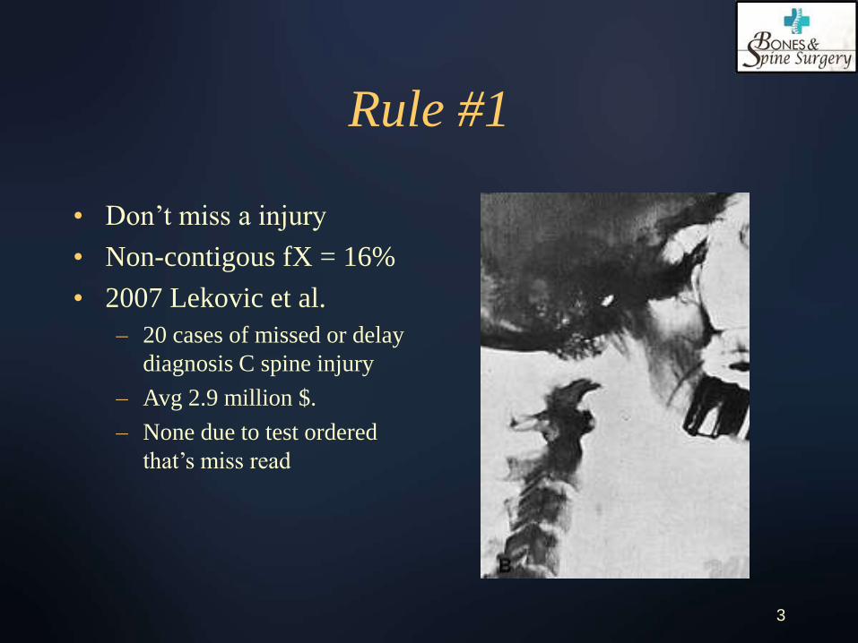
• Don’t miss a injury
• Non-contigous fX = 16%
• 2007 Lekovic et al.
– 20 cases of missed or delay
diagnosis C spine injury
– Avg 2.9 million $.
– None due to test ordered
that’s miss read
4
Anatomy
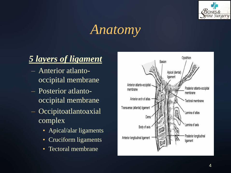
5 layers of ligament
– Anterior atlanto-
occipital membrane
– Posterior atlanto-
occipital membrane
– Occipitoatlantoaxial
complex
• Apical/alar ligaments
• Cruciform ligaments
• Tectoral membrane
5
Anatomy
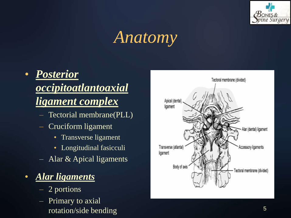
• Posterior
occipitoatlantoaxial
ligament complex– Tectorial membrane(PLL)
– Cruciform ligament
• Transverse ligament
• Longitudinal fasicculi
– Alar & Apical ligaments
• Alar ligaments
– 2 portions
– Primary to axial
rotation/side bending
6
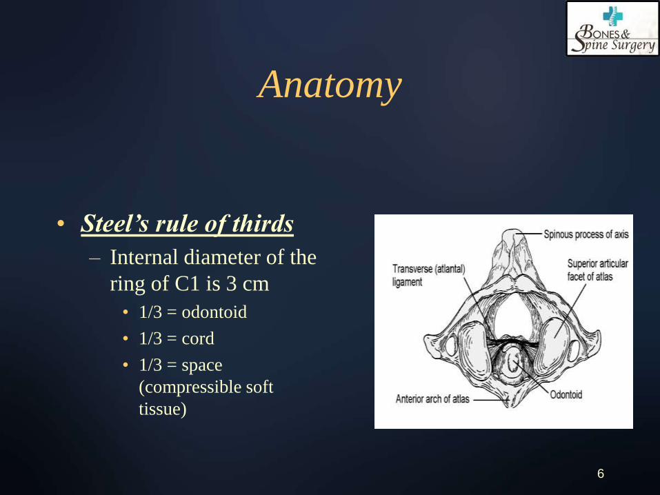
Anatomy
• Steel’s rule of thirds
– Internal diameter of the
ring of C1 is 3 cm
• 1/3 = odontoid
• 1/3 = cord
• 1/3 = space
(compressible soft
tissue)
7
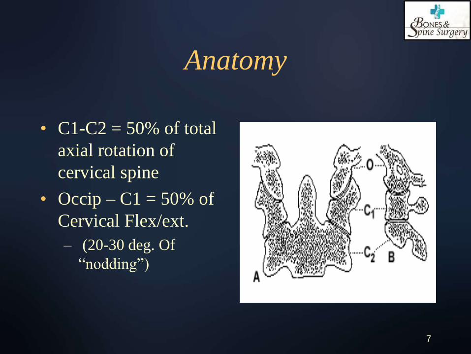
Anatomy
• C1-C2 = 50% of total
axial rotation of
cervical spine
• Occip – C1 = 50% of
Cervical Flex/ext.
– (20-30 deg. Of
“nodding”)
8
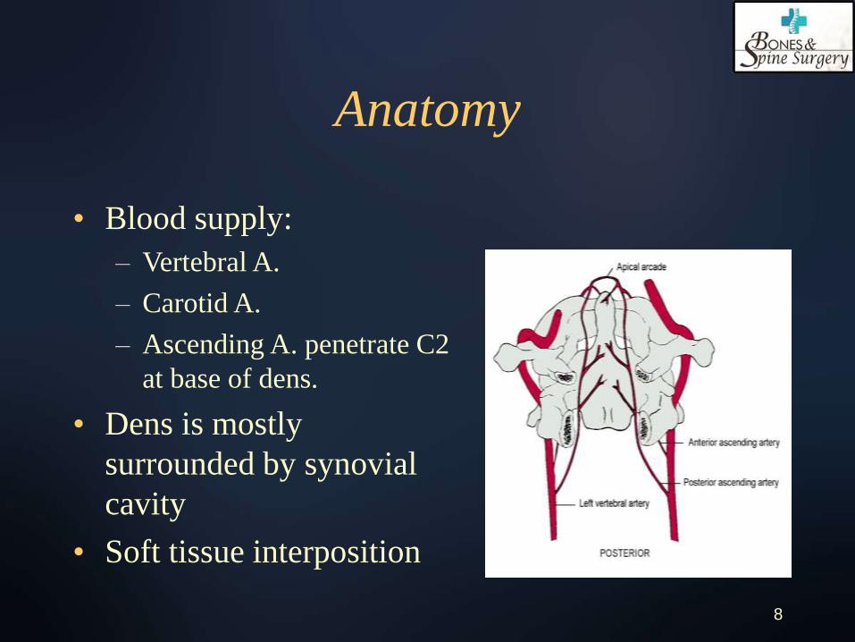
Anatomy
• Blood supply:
– Vertebral A.
– Carotid A.
– Ascending A. penetrate C2
at base of dens.
• Dens is mostly
surrounded by synovial
cavity
• Soft tissue interposition
9
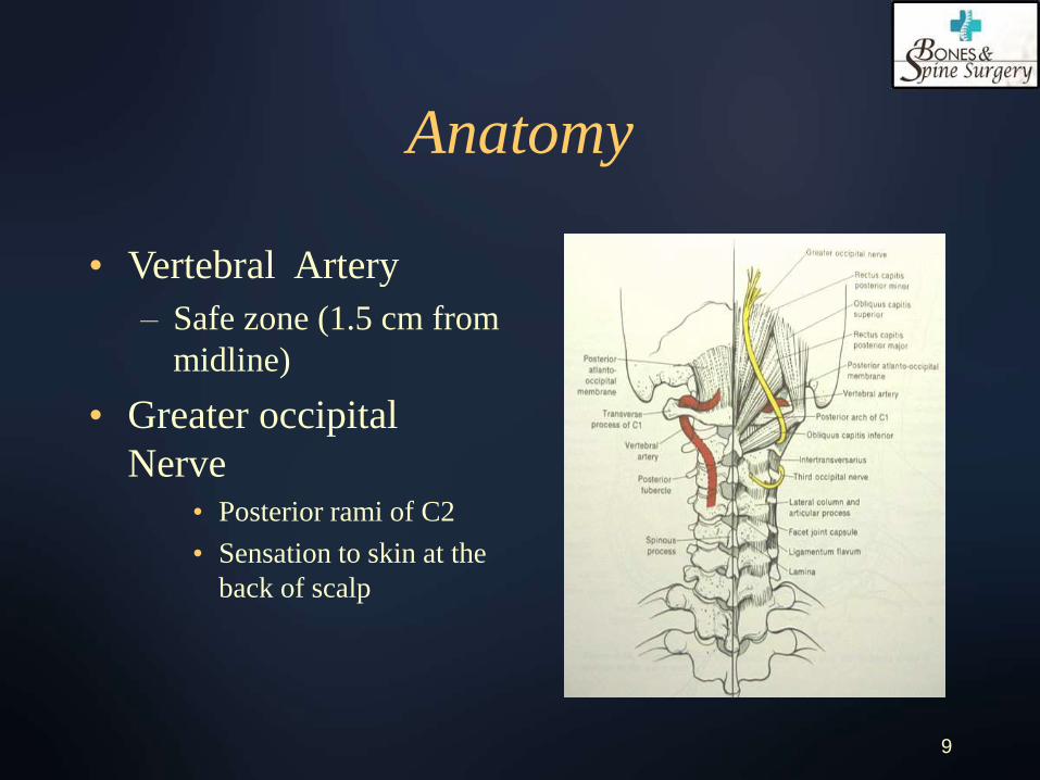
Anatomy
• Vertebral Artery
– Safe zone (1.5 cm from
midline)
• Greater occipital
Nerve• Posterior rami of C2
• Sensation to skin at the
back of scalp
10
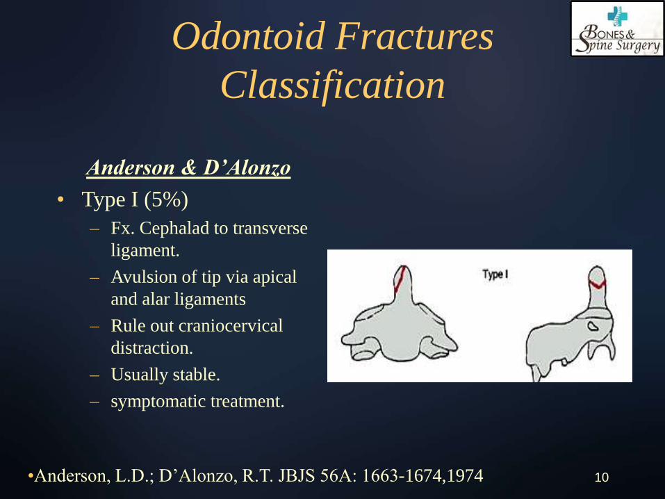
Odontoid Fractures
Classification
Anderson & D’Alonzo
• Type I (5%)
– Fx. Cephalad to transverse
ligament.
– Avulsion of tip via apical
and alar ligaments
– Rule out craniocervical
distraction.
– Usually stable.
– symptomatic treatment.
•Anderson, L.D.; D’Alonzo, R.T. JBJS 56A: 1663-1674,1974
11
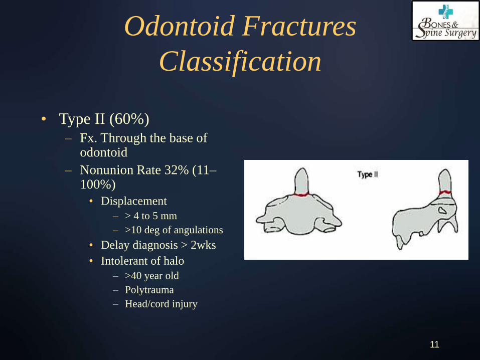
Odontoid Fractures
Classification
• Type II (60%)
– Fx. Through the base of odontoid
– Nonunion Rate 32% (11–100%)
• Displacement
– > 4 to 5 mm
– >10 deg of angulations
• Delay diagnosis > 2wks
• Intolerant of halo
– >40 year old
– Polytrauma
– Head/cord injury
12
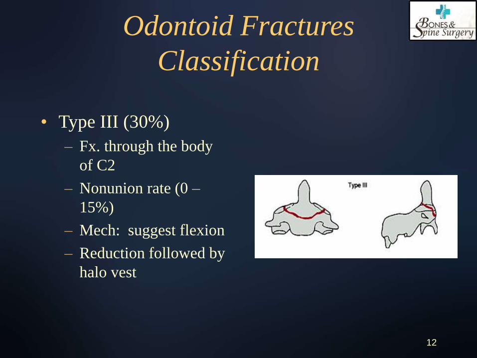
Odontoid Fractures
Classification
• Type III (30%)
– Fx. through the body
of C2
– Nonunion rate (0 –
15%)
– Mech: suggest flexion
– Reduction followed by
halo vest
13
Treatment of
Type II Odontoid Fractures
• Stable = Halo vest
– Displacement <
4mm,10 deg.
– Age <40 year old
– Injury recognition < 2
weeks.
• Unstable = primarysurgical stabilization
– Displacement > 4mm, 10 deg.
– Delay diagnosis > 2wks.
– Intolerant of halo
• Older, polytrauma, head/cord injury.
– Irreducible C1-2 fx. dislocation.
14
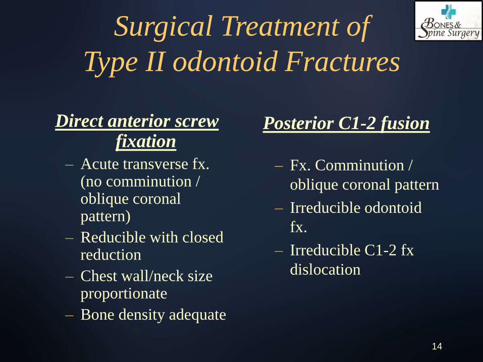
Surgical Treatment of
Type II odontoid Fractures
Direct anterior screw fixation
– Acute transverse fx. (no comminution / oblique coronal pattern)
– Reducible with closed reduction
– Chest wall/neck size proportionate
– Bone density adequate
Posterior C1-2 fusion
– Fx. Comminution /
oblique coronal pattern
– Irreducible odontoid
fx.
– Irreducible C1-2 fx
dislocation
15
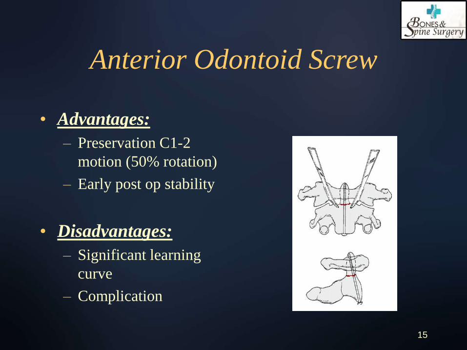
Anterior Odontoid Screw
• Advantages:
– Preservation C1-2
motion (50% rotation)
– Early post op stability
• Disadvantages:
– Significant learning
curve
– Complication
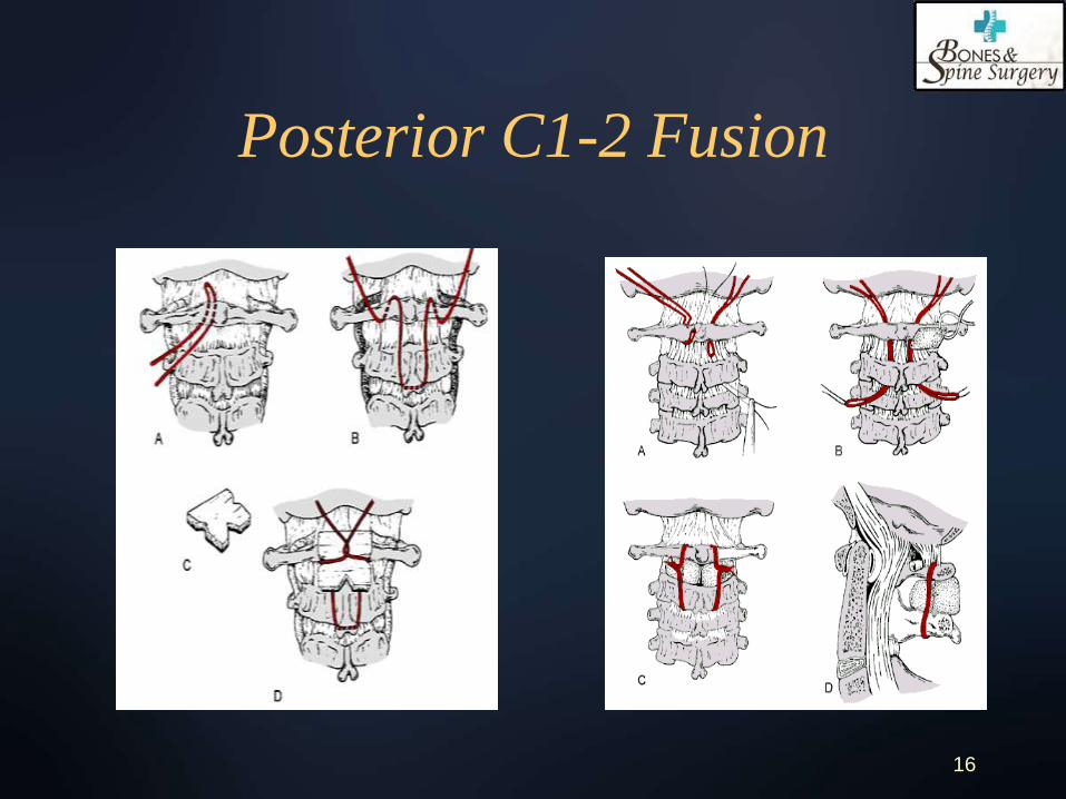
16
Posterior C1-2 Fusion
17
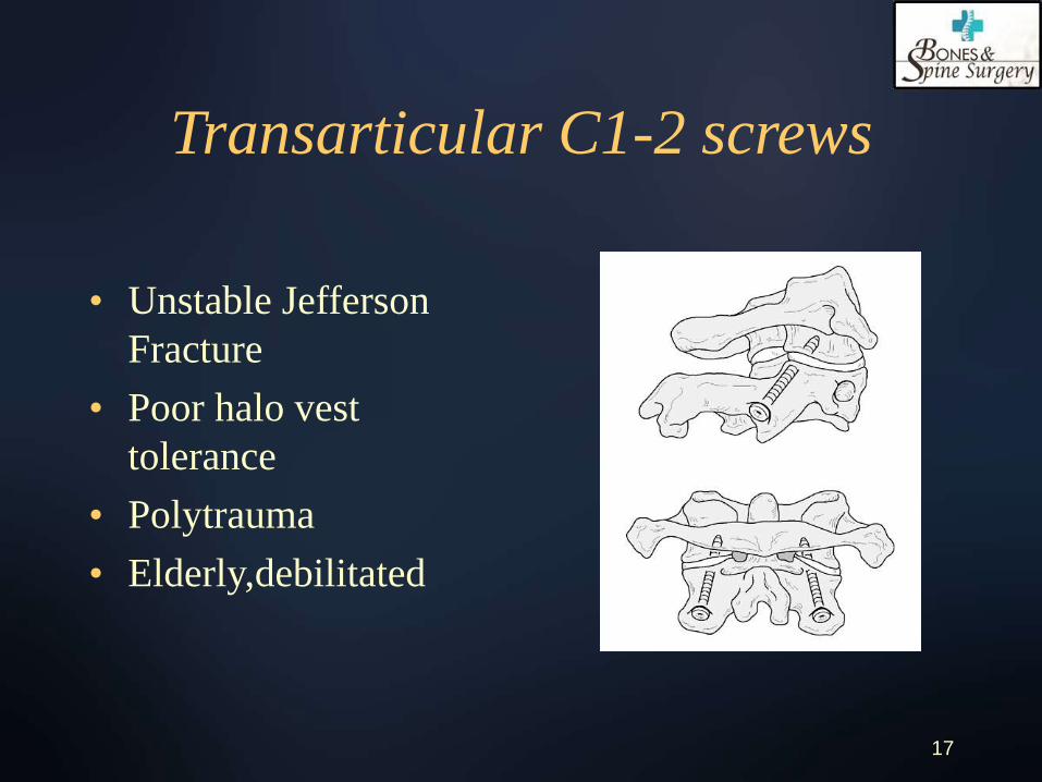
Transarticular C1-2 screws
• Unstable Jefferson
Fracture
• Poor halo vest
tolerance
• Polytrauma
• Elderly,debilitated
18
Atlas Fractures
• 10% of all cervical spine injuries.
• 48% has additional fractures in the C-spine
– #1 Dens fractures.
– #2 Traumatic spondylolisthesis C2
– #3 Lower cervical fractures.
• Mechanism – axial loading (MVA,diving)
19
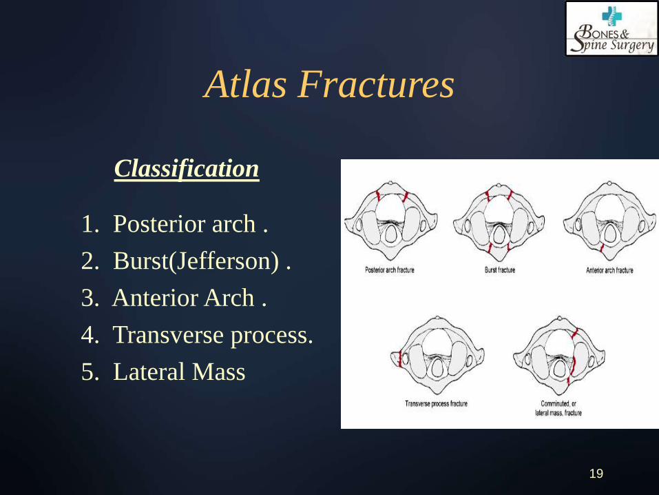
Atlas Fractures
Classification
1. Posterior arch .
2. Burst(Jefferson) .
3. Anterior Arch .
4. Transverse process.
5. Lateral Mass
20
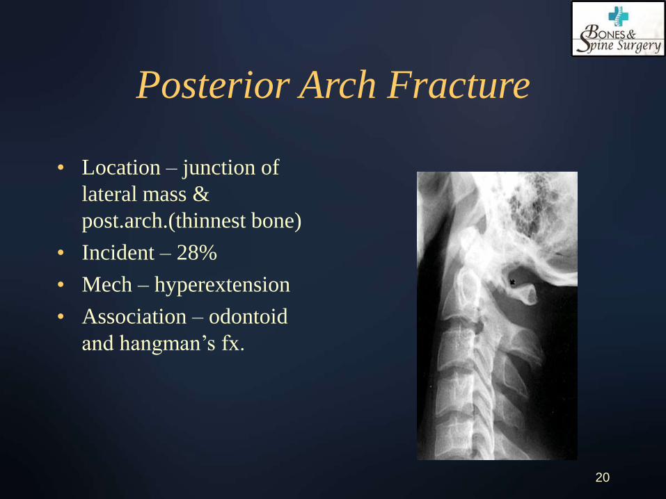
Posterior Arch Fracture
• Location – junction of
lateral mass &
post.arch.(thinnest bone)
• Incident – 28%
• Mech – hyperextension
• Association – odontoid
and hangman’s fx.
21
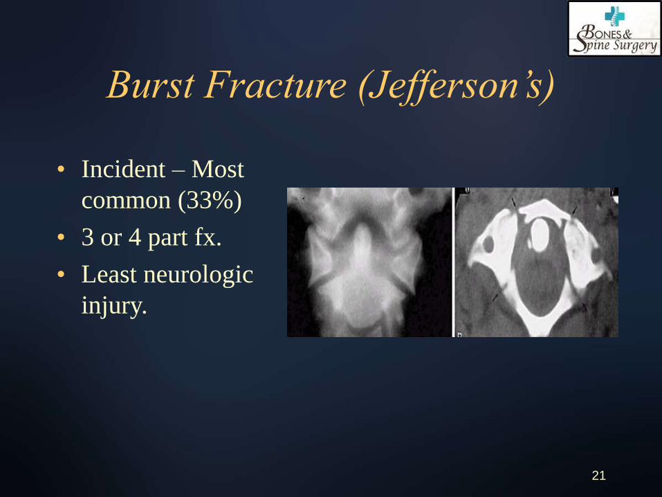
Burst Fracture (Jefferson’s)
• Incident – Most
common (33%)
• 3 or 4 part fx.
• Least neurologic
injury.
22
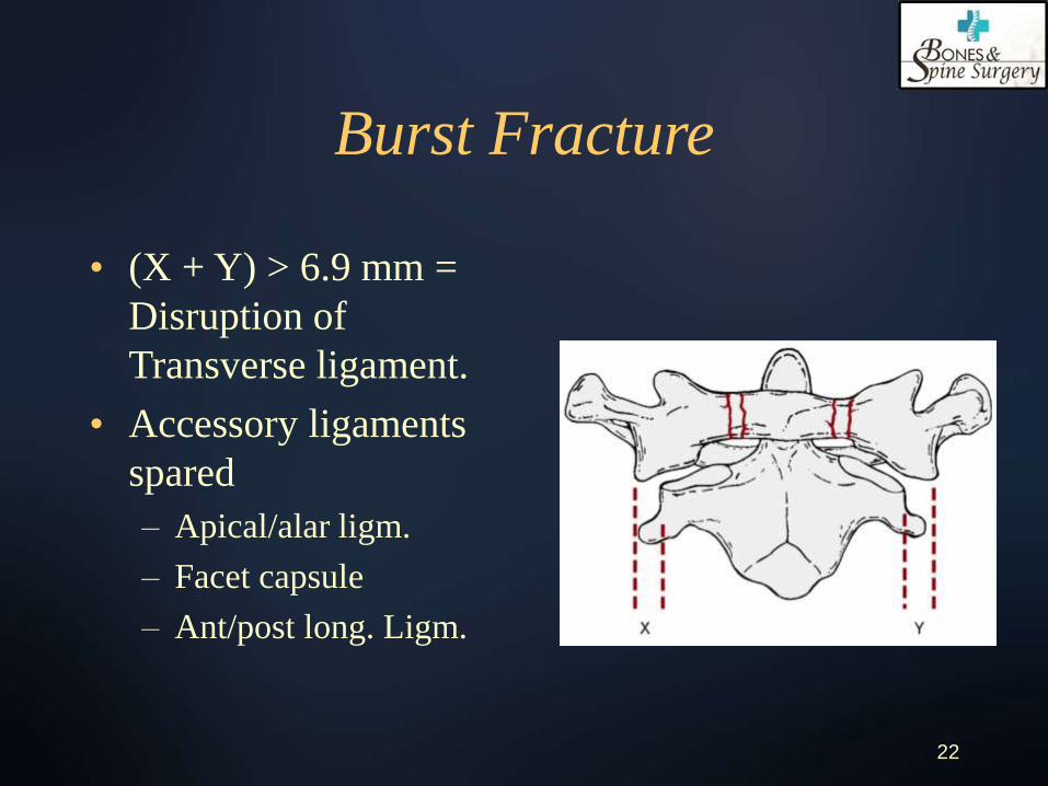
Burst Fracture
• (X + Y) > 6.9 mm =
Disruption of
Transverse ligament.
• Accessory ligaments
spared
– Apical/alar ligm.
– Facet capsule
– Ant/post long. Ligm.
23
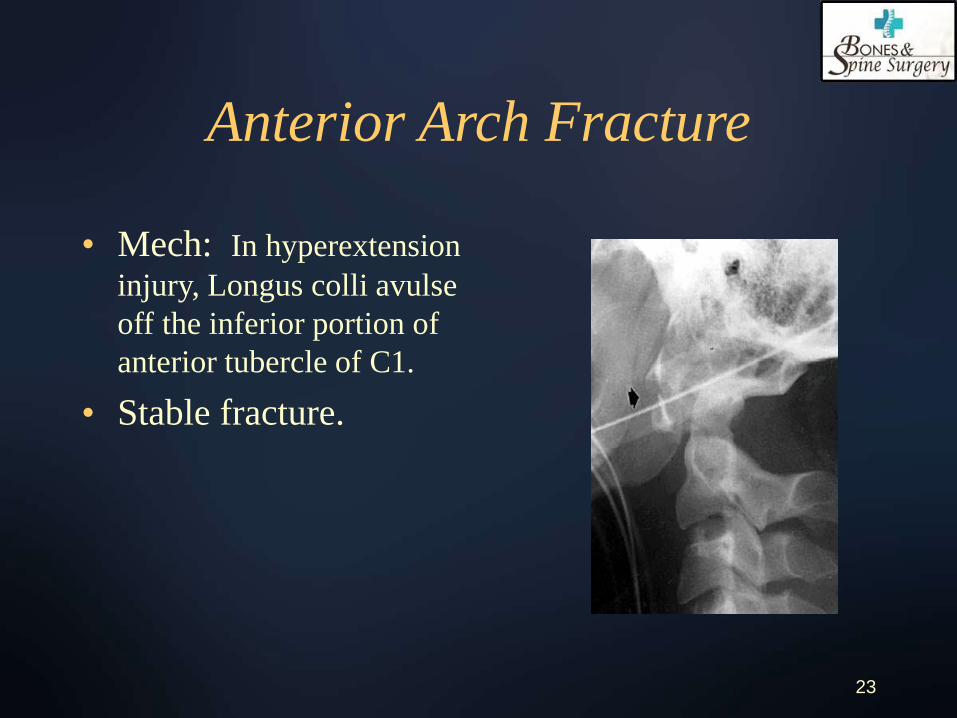
Anterior Arch Fracture
• Mech: In hyperextension
injury, Longus colli avulse
off the inferior portion of
anterior tubercle of C1.
• Stable fracture.
24
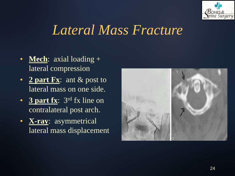
Lateral Mass Fracture
• Mech: axial loading +
lateral compression
• 2 part Fx: ant & post to
lateral mass on one side.
• 3 part fx: 3rd fx line on
contralateral post arch.
• X-ray: asymmetrical
lateral mass displacement
25
Treatment-Posterior arch fx.
• Isolated posterior arch fx. :
– collar
• Post arch fx. + type I “hangman’s fx”
– collar
• Post arch fx. + type II dens fx. :
– Reduction by traction then halo vest
– Anterior dens screw + collar
– C1-2 arthrodesis with Transarticular screw
– Halo then delayed standard C1-2 fusion
26
Treatment – Jefferson &
lateral Mass fractures
• Nondisplaced:
– Collar or halo
• Displacement < 7 mm:
– Halo
• Displacement > 7 mm
1. Axial traction (6 weeks),
reduction confirmed by
open mouth view.
2. Halo vest (6weeks)
3. Flex/Ext view end of 3
month
• Immediate C1-2 fusion
1. reduction via traction
2. Transarticular screws
27
Treatment: Combined Injuries
Jefferson/lateral mass + others
• Stable Jefferson + stable dens
– Halo vest
• Stable Jefferson + “unstable dens”
– Anterior dens screw with halo vest?
– Halo then delayed C12 fusion?
• Unstable Jefferson + “unstable dens”
– Halo traction?
– C1-2 fusion with Transarticular screws?
28
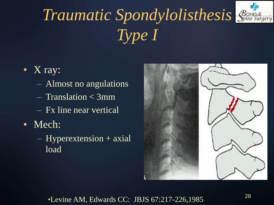
Traumatic Spondylolisthesis
Type I
• X ray:
– Almost no angulations
– Translation < 3mm
– Fx line near vertical
• Mech:
– Hyperextension + axial
load
•Levine AM, Edwards CC: JBJS 67:217-226,1985
29
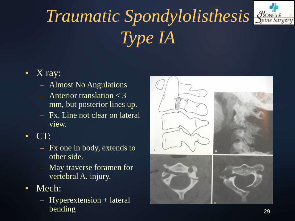
Traumatic Spondylolisthesis
Type IA
• X ray:
– Almost No Angulations
– Anterior translation < 3 mm, but posterior lines up.
– Fx. Line not clear on lateral view.
• CT:
– Fx one in body, extends to other side.
– May traverse foramen for vertebral A. injury.
• Mech:
– Hyperextension + lateral bending
30
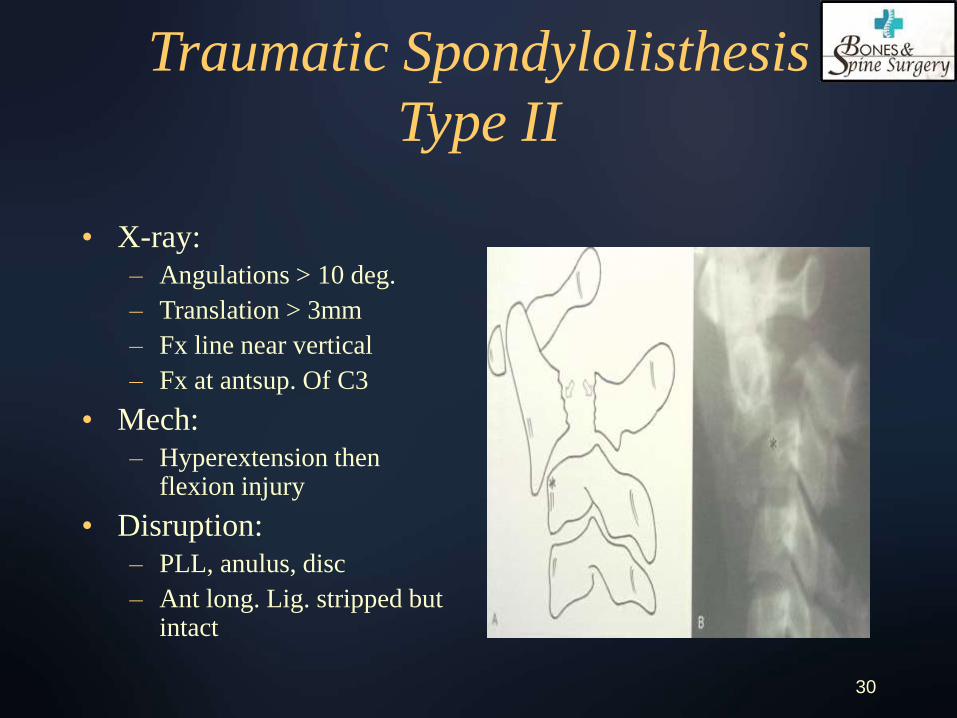
Traumatic Spondylolisthesis
Type II
• X-ray:
– Angulations > 10 deg.
– Translation > 3mm
– Fx line near vertical
– Fx at antsup. Of C3
• Mech:
– Hyperextension then flexion injury
• Disruption:
– PLL, anulus, disc
– Ant long. Lig. stripped but intact
31
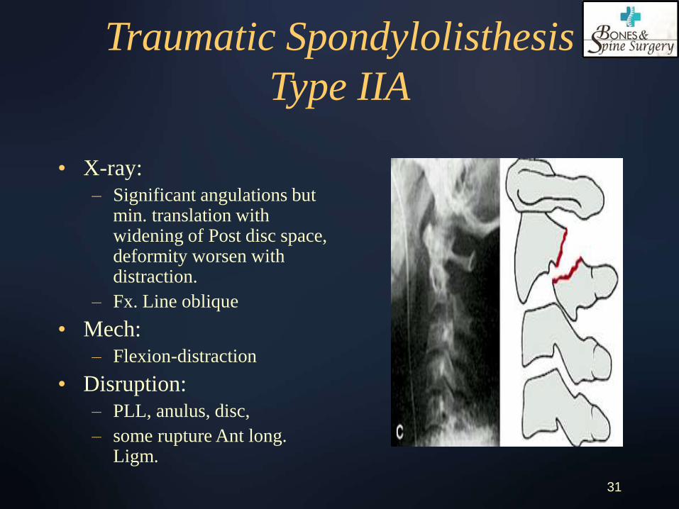
Traumatic Spondylolisthesis
Type IIA
• X-ray:
– Significant angulations but min. translation with widening of Post disc space, deformity worsen with distraction.
– Fx. Line oblique
• Mech:
– Flexion-distraction
• Disruption:
– PLL, anulus, disc,
– some rupture Ant long. Ligm.
32
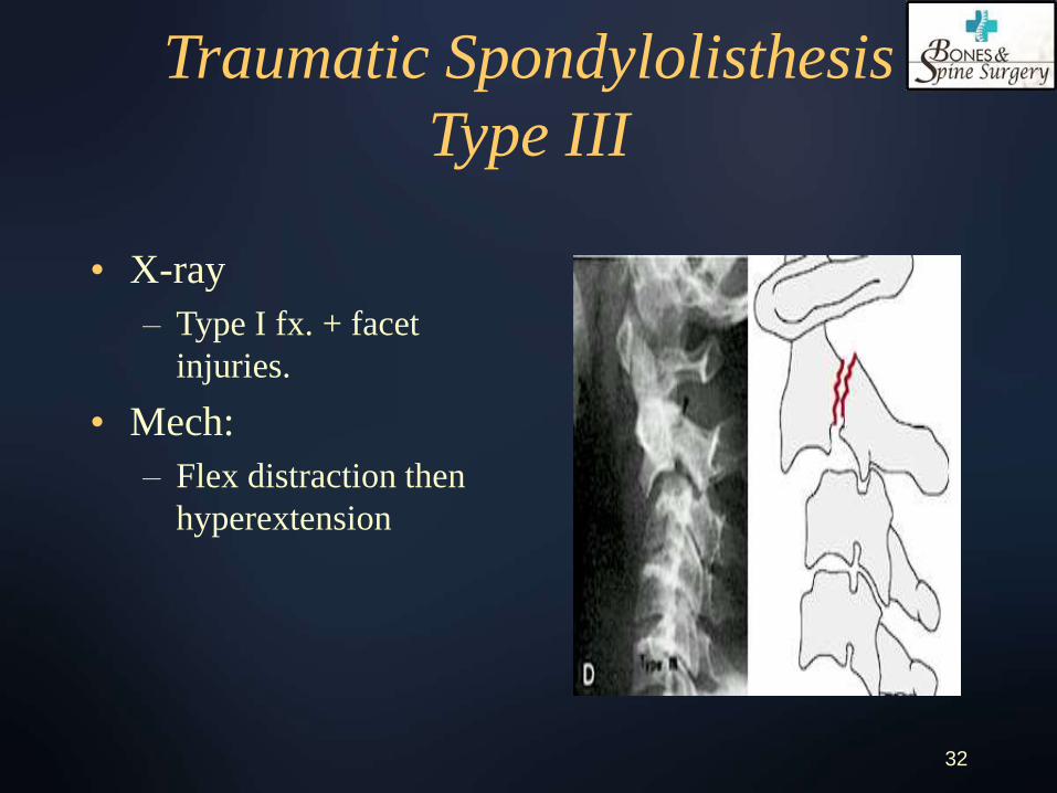
Traumatic Spondylolisthesis
Type III
• X-ray
– Type I fx. + facet
injuries.
• Mech:
– Flex distraction then
hyperextension
33
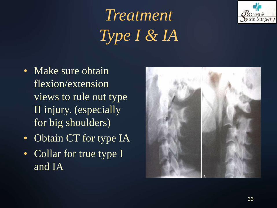
Treatment
Type I & IA
• Make sure obtain
flexion/extension
views to rule out type
II injury. (especially
for big shoulders)
• Obtain CT for type IA
• Collar for true type I
and IA
34
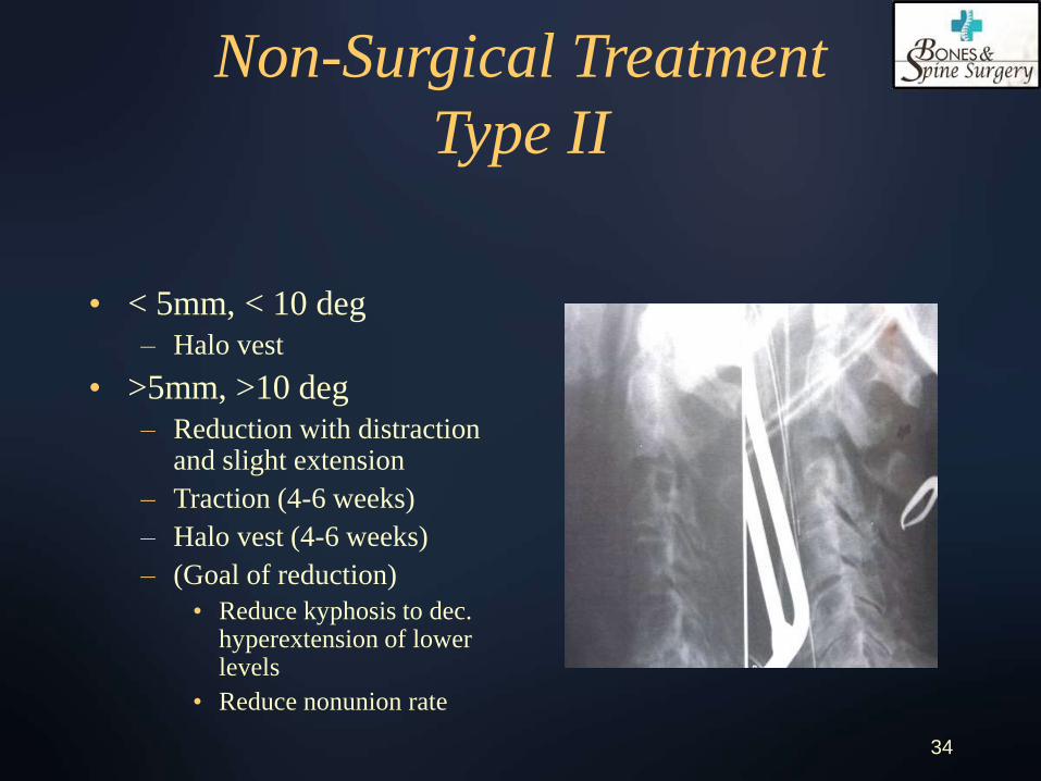
Non-Surgical Treatment
Type II
• < 5mm, < 10 deg
– Halo vest
• >5mm, >10 deg
– Reduction with distraction and slight extension
– Traction (4-6 weeks)
– Halo vest (4-6 weeks)
– (Goal of reduction)
• Reduce kyphosis to dec. hyperextension of lower levels
• Reduce nonunion rate
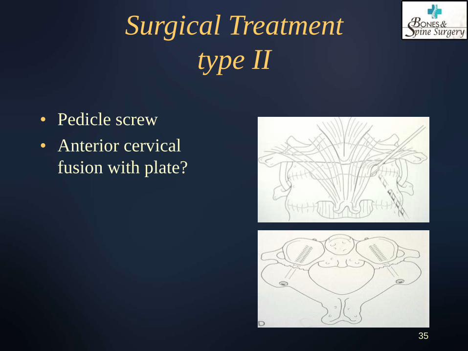
35
Surgical Treatment
type II
• Pedicle screw
• Anterior cervical
fusion with plate?
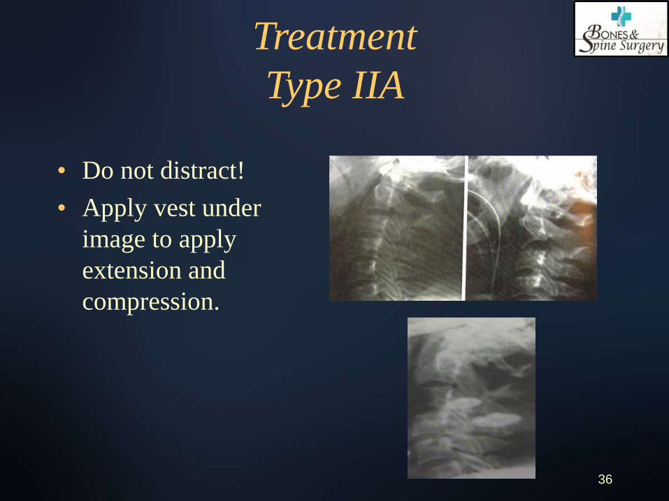
36
Treatment
Type IIA
• Do not distract!
• Apply vest under
image to apply
extension and
compression.
37
Treatment
Type III
• (Usually can not close reduce.)
1. Obtain MRI to r/o disc herniation
2. Posterior open reduction of facets
3. Fusion of C2-3 by wire/plates
38
Insufficiency of the
Transverse Ligament
• Incidence
– Fifth decade
• Mechanism
– Forced flexion of the neck
• Clinical Presentation
– Usually fatal
– Survivor have neurologic symptoms from normal to
transient quadriparesis.
– Symptoms worse with flexion of neck.
39
Insufficiency of the
Transverse Ligament
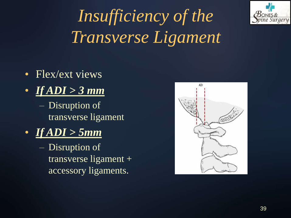
• Flex/ext views
• If ADI > 3 mm
– Disruption of
transverse ligament
• If ADI > 5mm
– Disruption of
transverse ligament +
accessory ligaments.
40
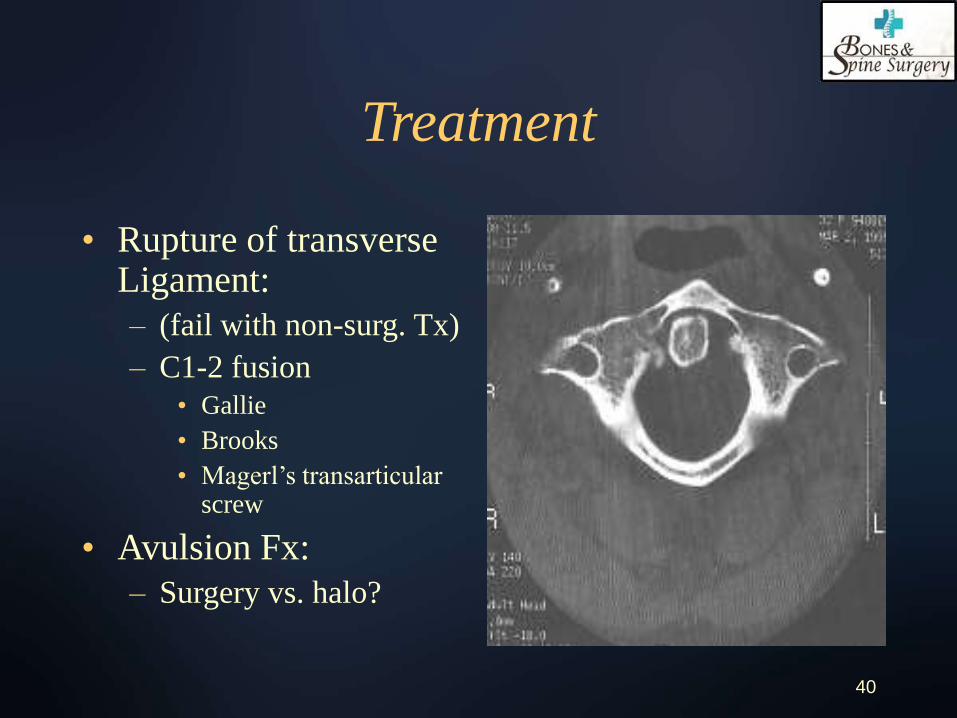
Treatment
• Rupture of transverse Ligament:
– (fail with non-surg. Tx)
– C1-2 fusion
• Gallie
• Brooks
• Magerl’s transarticular screw
• Avulsion Fx:
– Surgery vs. halo?
41
Atlantoaxial Rotatory Deformity
• Incidence
– Rare in adults
• Cause
– MVA
• Mechanism
– Flexion and rotation
• Max. rotation
– bilateral dislocation = 65 degree (intact transverse ligament)
– Unilateral dislocation = 45 deg. (deficiency of transverse ligament)
• Clinical Presentation
(wide spectrum)
– Neck pain
– torticollis (cock-robin)
– Neural deficit
– Vertebral Artery Injury
42
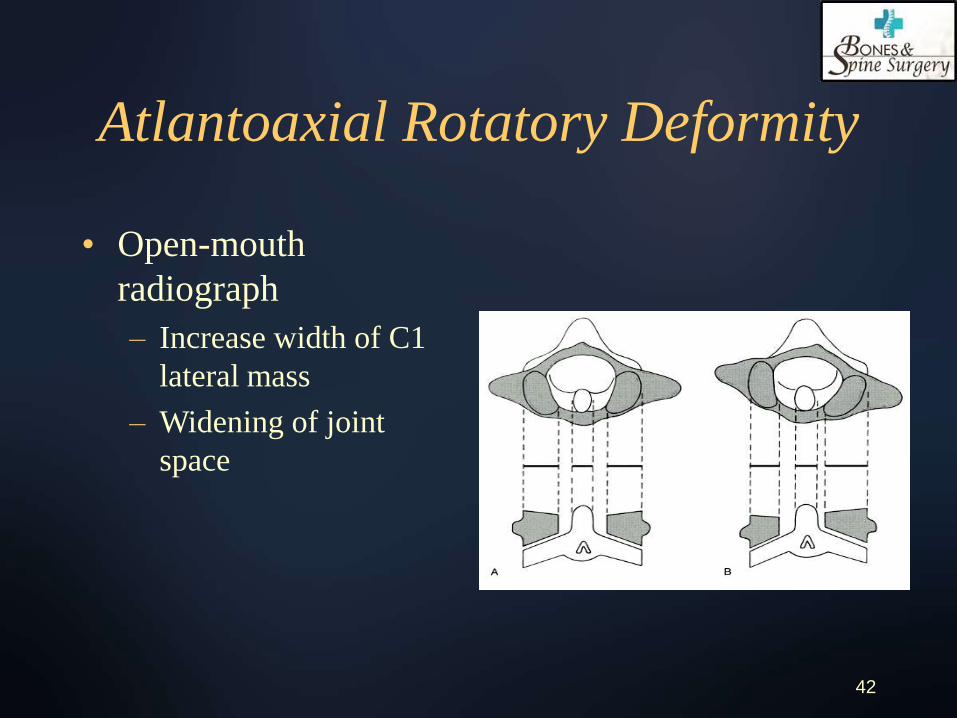
Atlantoaxial Rotatory Deformity
• Open-mouth
radiograph
– Increase width of C1
lateral mass
– Widening of joint
space
43
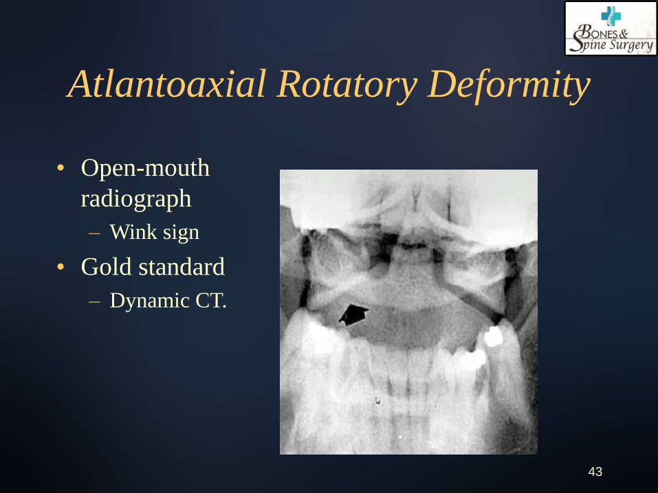
Atlantoaxial Rotatory Deformity
• Open-mouth
radiograph
– Wink sign
• Gold standard
– Dynamic CT.
44
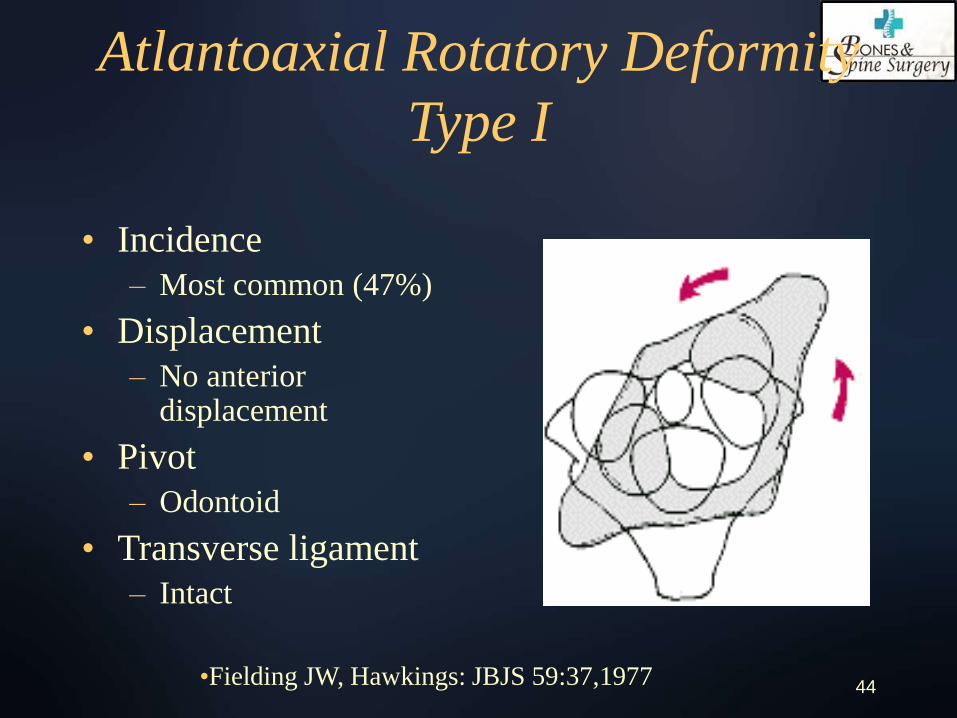
Atlantoaxial Rotatory Deformity
Type I
• Incidence
– Most common (47%)
• Displacement
– No anterior displacement
• Pivot
– Odontoid
• Transverse ligament
– Intact
•Fielding JW, Hawkings: JBJS 59:37,1977
45
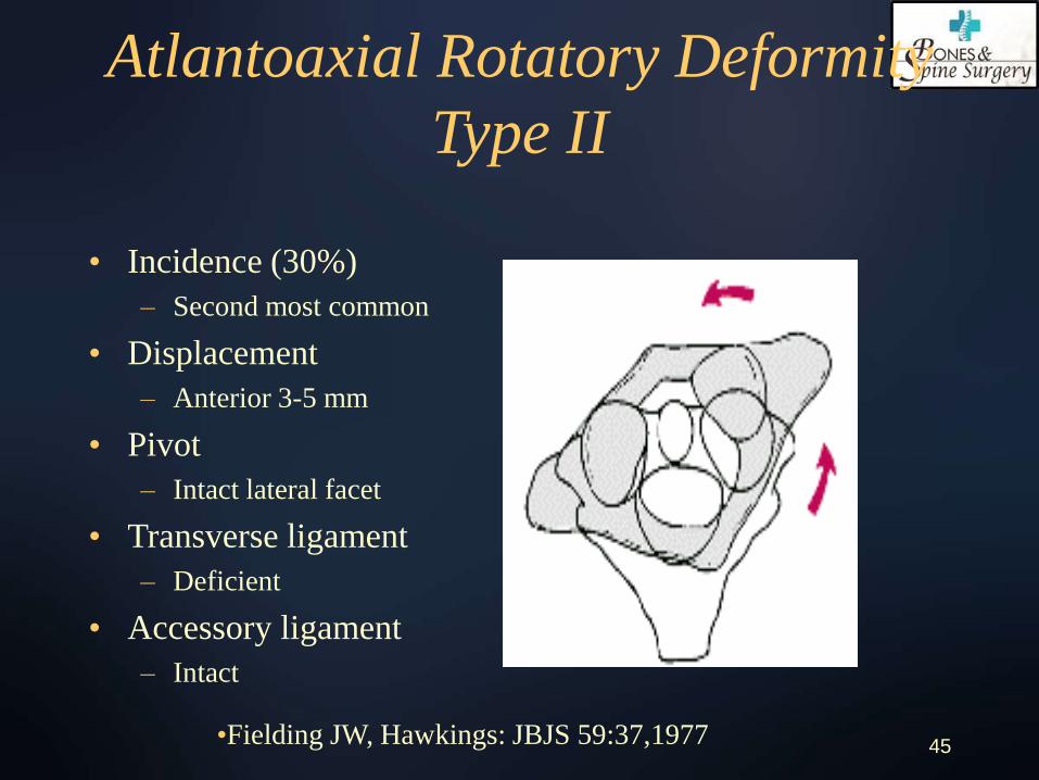
Atlantoaxial Rotatory Deformity
Type II
• Incidence (30%)
– Second most common
• Displacement
– Anterior 3-5 mm
• Pivot
– Intact lateral facet
• Transverse ligament
– Deficient
• Accessory ligament
– Intact
•Fielding JW, Hawkings: JBJS 59:37,1977
46
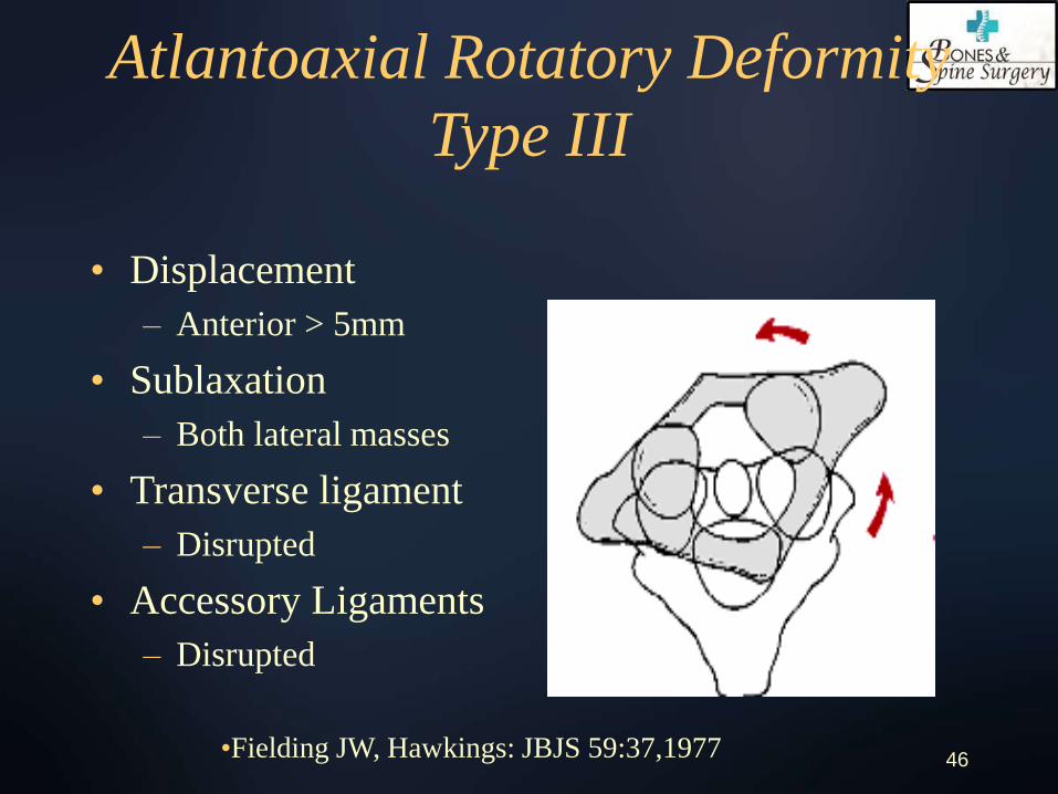
Atlantoaxial Rotatory Deformity
Type III
• Displacement
– Anterior > 5mm
• Sublaxation
– Both lateral masses
• Transverse ligament
– Disrupted
• Accessory Ligaments
– Disrupted
•Fielding JW, Hawkings: JBJS 59:37,1977
47
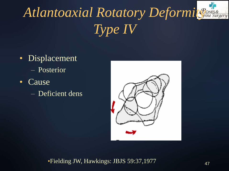
Atlantoaxial Rotatory Deformity
Type IV
• Displacement
– Posterior
• Cause
– Deficient dens
•Fielding JW, Hawkings: JBJS 59:37,1977
48
Atlantoaxial Rotatory
Deformities - Treatment
• Look for etiology
• Traction
– Start with 6.8 Kg.
– Increase 0.5 to 0.9 Kg every three days.
– Maximum 9.1KG
• Post reduction
– Immobilization for 2-3 months.
– Flex/ext. x-ray to check stability
49
Surgical Treatment
Indications
• Spinal instability
• Neural involvement
• Fail to achieve
reduction
• Fail to maintain
reduction
Methods
• Gallie
• Brooks-Jenkins
• Transarticular screws
50
Thank you