Embed Size (px)
Citation preview

1
Chapter 5 The Integumentary System

2
Introduction
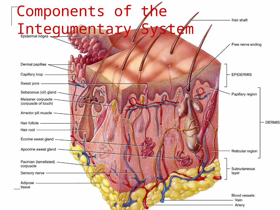
The organs of the integumentary system include the skin and its accessory structures including hair, nails, and glands, as well as blood vessels, muscles and nerves
Dermatology is the medical specialty for the diagnosis and treatment of disorders of the integumentary system.

3
Structure of the Skin
The skin (cutaneous membrane) covers the body and is the largest organ of the body by surface area and weight
Its area is about 2 square meters (22 square feet) and weighs 4.5-5kg (10-11 lb), about 7% of body weight
It is 0.5 – 4 mm thick, thinnest on the eyelids, thickest on the heels; the average thickness is 1 – 2 mm

4
Structure of the Skin
It consists of two major layers: outer, thinner layer called the epidermis,
consists of epithelial tissue (see video) inner, thicker layer called the dermis Beneath the dermis is a subcutaneous
(subQ) layer (also called hypodermis) which attaches the skin to the underlying tissues and organs.
5
Components of the Integumentary System

6
Structure of the Skin
The epidermis has a number of important characteristics:
the epidermis is composed of keratinized stratified squamous epithelium
it contains four major types of cells: Keratinocytes (90% of the cells) produce
keratin which is a tough fibrous protein that provides protection

7
Structure of the Skin Melanocytes: which produce the pigment
melanin that protects against damage by ultraviolet radiation
Langerhans cells: involved in immune responses, arise from red bone marrow
Merkel cells: which function in the sensation of touch along with the adjacent tactile discs

8
Types of Cells in the Epidermis

9
Epidermis The epidermis contains four major layers (thin
skin) or five major layers (thick skin) Stratum basale (deepest layer) or stratum
germinativum, where continuous cell division occurs which produces all the other layers
Stratum spinosum, 8-10 layers of keratinocytes
Stratum granulosum, which includes keratohyalin and lamellar granules

10
Epidermis Stratum lucidum is present only in thick skin (the
skin of the fingertips, palms, and soles) Stratum corneum: composed of many sublayers of
flat, dead keratinocytes called corneocytes or squames that are continuously shed and replaced by cells from deeper strata; constant friction can stimulate formation of a callus.
Keratinization, the accumulation of more and more protective keratin, occurs as cells move from the deepest layer to the surface layer
Dandruff - an excess of keratinized cells shed from the scalp

11
Layers of the Epidermis

12
Dermis The dermis has several important
characteristics: is composed of connective tissue containing
collagen and elastic fibers contains two layers the outer papillary region consists of areolar
connective tissue containing thin collagen and elastic fibers, dermal papillae (including capillary loops), corpuscles of touch and free nerve endings

13
Dermis
The deeper reticular region consists of dense irregular connective tissue containing collagen and elastic fibers adipose cells, hair follicles, nerves, sebaceous (oil) glands, and sudoriferous (sweat) glands Striae or stretch marks can appear if the skin is
stretched too much

14
Dermis
Lines of cleavage - “tension lines” in the skin indicate the predominant direction of underlying collagen fibers
Epidermal ridges reflect contours of the underlying dermal papillae and form the basis for fingerprints (and footprints); their function is to increase firmness of grip by increasing friction.
Dermatoglyphics - the study of the pattern of epidermal ridges

15
Cleavage (Tension) Lines and Striae Cleavage (tension) lines:
elastin and collagen fibers oriented in some directions more than in others
Important in surgery If incision parallel to lines,
there is less gapping, faster healing, less scar tissue
If skin is overstretched, striae (stretch marks) occur

16

17
Structural Basis of Skin Color Variations in skin color arise from variations in the amounts of three pigments: melanin, carotene, and hemoglobin
Melanin - a yellow-red or brown-black pigment produced by melanocytes (located mostly in the epidermis, where it absorbs UV radiation)
The amount of melanin causes the skin’s color to vary from pale yellow to red to tan to black
The number of melanocytes are about the same in all people; differences in skin color is due to the amount of pigment produced
18
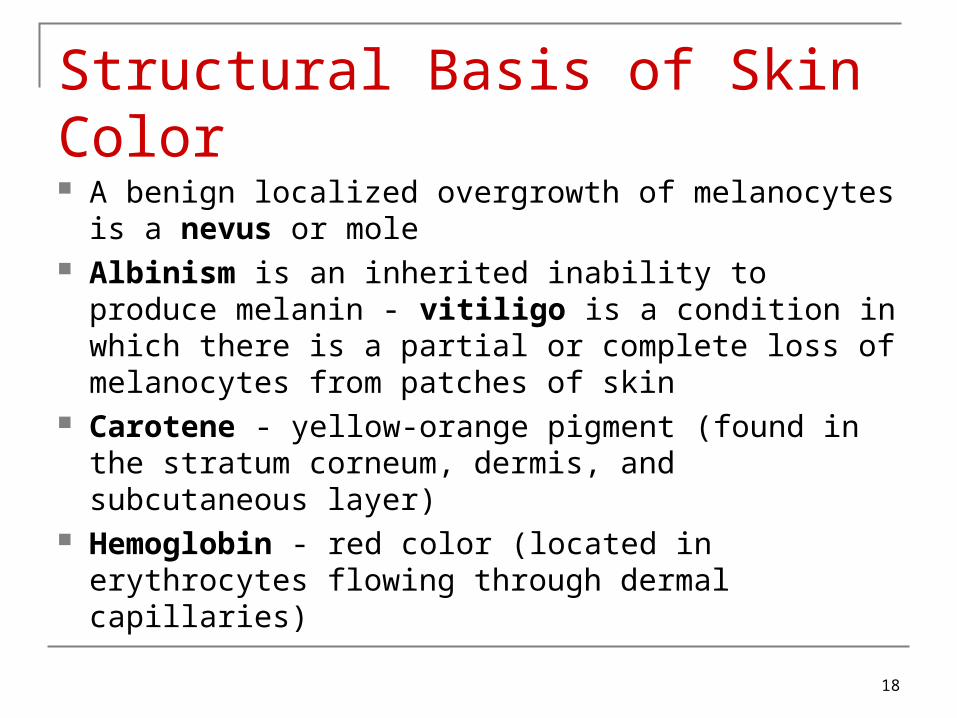
Structural Basis of Skin Color A benign localized overgrowth of melanocytes is a
nevus or mole Albinism is an inherited inability to produce melanin
- vitiligo is a condition in which there is a partial or complete loss of melanocytes from patches of skin
Carotene - yellow-orange pigment (found in the stratum corneum, dermis, and subcutaneous layer)
Hemoglobin - red color (located in erythrocytes flowing through dermal capillaries)

19
Subcutaneous Layer
Subcutaneous (subQ) layer (also called hypodermis) is not part of the skin but, among its functions, it attaches the skin to the underlying tissues and organs; this layer (and sometimes the dermis) contains lamellated (pacinian) corpuscles which detect external pressure applied to the skin.

20
Accessory Structures of the Skin include hair, skin glands, and nails Hairs (pili) have a number of important
functions: protection reduction of heat loss sensing light touch

21
Accessory Structures of the Skin - Hair Hair is composed of dead, keratinized
epidermal cells Hair consists of: shaft which mostly projects above the surface
of the skin root which penetrates into the dermis hair follicle epithelial root sheath – (downward
continuation of the epidermis) dermal root sheath -

22

23

24
Accessory Structures of the Skin There are different types of hairs including
lanugo, vellus hairs and terminal hairs Hair color is determined by the amount and
type of melanin Sebaceous (oil) glands are connected to
hair follicles

25
Skin Glands
Sebaceous glands secrete an oily substance called sebum which prevents dehydration of hair and skin, and inhibits growth of certain bacteria (Sebum=triglycerides, cholesterol, proteins, and inorganic salts)
Sudoriferous (sweat) glands-- 2 types: Eccrine sweat glands Apocrine sweat glands
26
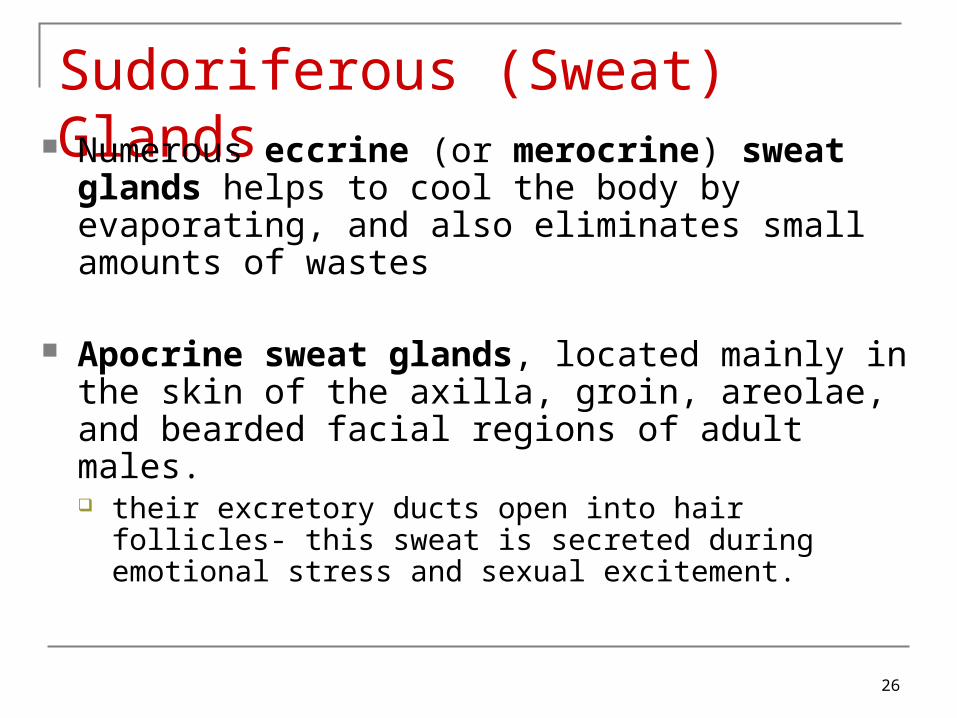
Sudoriferous (Sweat) Glands Numerous eccrine (or merocrine) sweat glands
helps to cool the body by evaporating, and also eliminates small amounts of wastes
Apocrine sweat glands, located mainly in the skin of the axilla, groin, areolae, and bearded facial regions of adult males. their excretory ducts open into hair follicles- this sweat is
secreted during emotional stress and sexual excitement.

27
Ceruminous Glands
Modified sweat glands located in the ear canal
Along with nearby sebaceous glands, they are involved in producing a waxy secretion called cerumen (earwax) which provides a sticky barrier that prevents entry of foreign bodies into the ear canal.

28
Nails
Nails are composed of hard, keratinized epidermal cells located over the dorsal surfaces of the ends of fingers and toes
Each nail consists of: free edge transparent nail body (plate) with a whitish
lunula at its base nail root embedded in a fold of skin

29
Nails

30

31
Types of Skin
There are two major types of skin: thin (hairy) skin covers all body regions
except the palms, palmar surfaces of digits, and soles
thick (hairless) skin covers the palms, palmar surfaces of digits, and soles

32
Epidermal Wound Healing

33
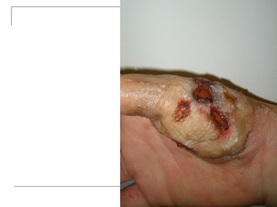
Deep Wound Healing Injury extends into dermis & hypodermis Scar tissue is formed
Some normal function lost Occurs in four phases
1. Inflammatory phase2. Migratory phase3. Proliferative phase4. Maturation phase

34
Inflammatory Phase Blood clot forms loosely uniting wound
edges Inflammation occurs
Eliminates microbes, foreign material, and dying tissue
Increases diameter of local blood vessles Enhancing delivery of nutrients, immune cells, and
fibroblasts

35
Migratory Phase Clot dries into scab Epithelial cells migrate beneath scab and
bridge wound Fibroblasts migrate and lay down
collagen fibers and glycoproteins in dermis
New blood vessels grow Tissue called granulation tissue
during this phase destined to become scar tissue
36
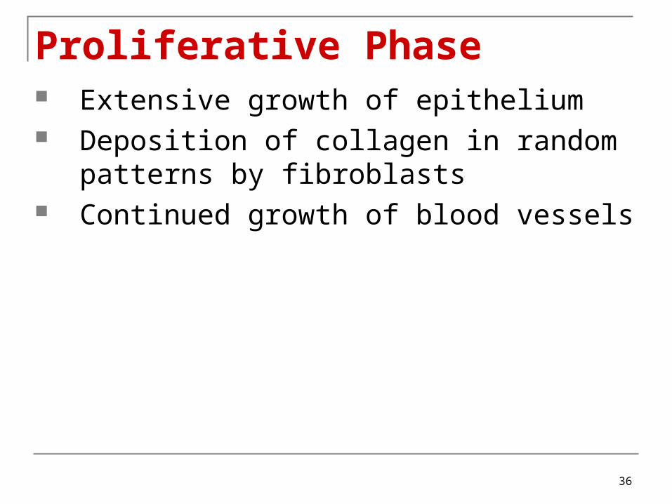
Proliferative Phase Extensive growth of epithelium Deposition of collagen in random
patterns by fibroblasts Continued growth of blood vessels

37
Maturation Phase Scab sloughs off once epidermis restored to
normal thickness Granulation tissue developing into scar tissue Fibroblasts decrease in number Blood vessels restored to normal Scar tissue formation called fibrosis
Elevated scars called Hypertrophic scars
If contained within sight of original wound Keloid scars
If extended beyond original wound

38
Deep Wound Healing

39

40
Aging and the Integumentary System Effects:
• wrinkling• decrease of skin’s immune responsiveness• dehydration and cracking of the skin• decreased sweat production• decreased numbers of functional melanocytes resulting
in gray hair and atypical skin pigmentation• loss of subcutaneous fat• a general decrease in skin thickness• an increased susceptibility to pathological conditions Growth of hair and nails decreases; nails may also
become more brittle with age.


05_09

43

44
45

46

05_11
49
Functions of the Skin
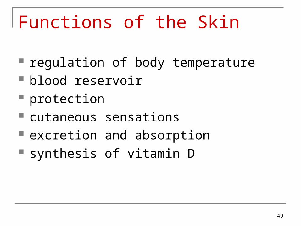
regulation of body temperature blood reservoir protection cutaneous sensations excretion and absorption synthesis of vitamin D


51

52

53