Embed Size (px)
Citation preview
Two transverse sections of the midbrain have been discussed:
1- At the level of the inferior colliculus.
2- At the level of the superior colliculus, characterized by the presence of the
red
nuclei.
Now, let’s start talking about the cranial nerves:
1-Oculomotor Nerve (III)
Two modalities:
a) Motor for skeletal muscles(voluntary): all the extraocular muscles except the lat. Rectus
and the sup. Rectus.
b) Motor for smooth muscles(parasympathetic, involuntary): ciliary and constrictor
pupillae muscles.
- It’s the first cranial nerve to leave the brainstem.
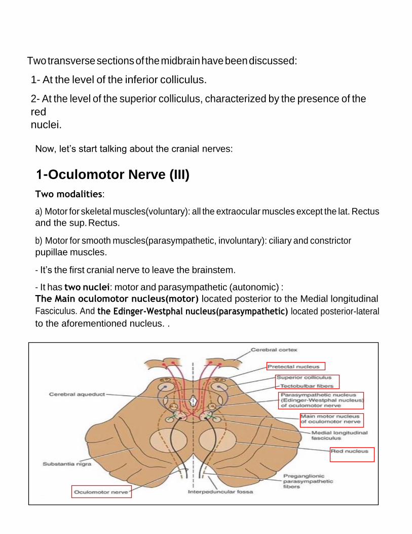
- It has two nuclei: motor and parasympathetic (autonomic) :
The Main oculomotor nucleus(motor) located posterior to the Medial longitudinal
Fasciculus. And the Edinger-Westphal nucleus(parasympathetic) located posterior-lateral
to the aforementioned nucleus. .
** Course of the nerve:
The fibers from the two nuclei will pass through red nucleus without synapse. From
the red nucleus fibers then pass via the substantia nigra exiting through the
interpeduncular fossa. Then the nerve enters the lateral wall of the cavernous sinus.
The nerve leaves the cranial cavity (middle cranial fossa) via the superior orbital
fissure where they divide to two branches: superior and inferior, which supply most
of the extraocular muscles .
NOTE: the parasympethatic fibers(preganglionic) synapse in the ciliary ganglion, and
come out as postganglionic fibers through short ciliary nerve.
** oculomotor nerve innervates:
1- Extrinsic muscles or extraoccular( Innervated by the main motor nucleus) :
The levator palpebrae superioris, superior rectus, medial rectus, inferior
rectus, and inferior oblique.
*all the muscles except superior oblique (innervated by the trochlear nerve and has
lateral rotation action and downword movement) and lateral rectus (supplied by the
abducent nerve and has abduction movement)
2- Intrinsic muscles( innervated by the parasympathetic nucleus ):
The constrictor pupillae of the iris and ciliary muscles (parasympathetic).
-Action: Lifting the upper eyelid; turning the eye upward, downward, and medially;
constricting the pupil; and accommodating the eye. ( all the movements except abduction
of the eye which is the lateral movement of the eyeball {lateral rectus} and turning the eye
downward and lateral {superior oblique} ).
**Oculomotor Nerve injury
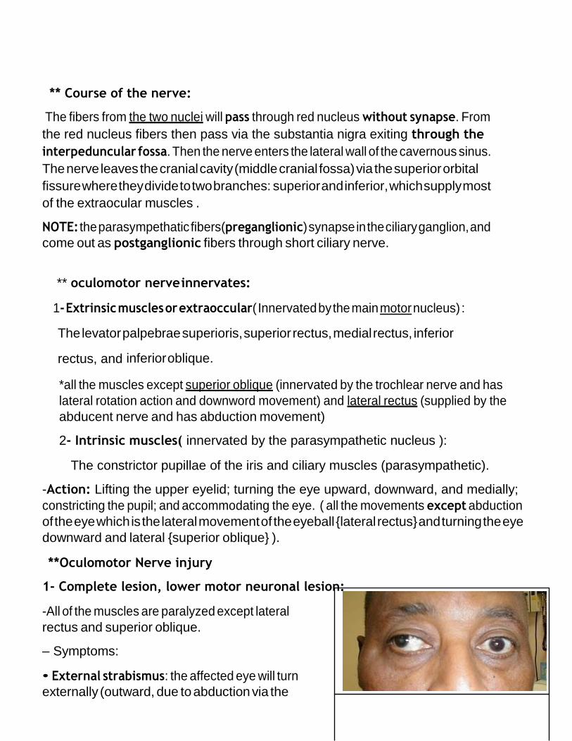
1- Complete lesion, lower motor neuronal lesion:
-All of the muscles are paralyzed except lateral
rectus and superior oblique.
– Symptoms:
• External strabismus: the affected eye will turn
externally (outward, due to abduction via the
unopposed lateral rectus, that is supplied by the abducent nerve) which causes diplopia.
• Diplopia (double vision )
• Ptosis: drooping of the upper eyelid because of the
paralysis of levator palpebrae superioris action.
• mydriasis: The pupil is widely dilated and nonreactive to light, dilation is overriding.
• Paralyzed accommodation
2-Incomplete lesions:
- Internal ophthalmoplegia: loss of the autonomic innervation of the sphincter pupillae
and ciliary muscle (Symptoms: the pupil is widely dilated and nonreactive to light only). the
parasympathetic fibers run superficial in the nerve, so if a pressure applied on the nerve,
the parasympathetic will be affected without motor (so it’s more susceptible to injury than
the motor).
- External ophthalmoplegia.: paralysis of the extraocular muscles, paralysis in motor
part.(Symptoms: External strabismus, diplopa and ptosis only)
Example: diabetic neuropathy that affects the motor fiber only.
TO SUM UP : you may have external ophthalmoplegia if the motor part is affected , OR
internal ophthalmoplegia if the autonomic part is affected , OR both if the whole nerve
is affected.
2-Trochlear nerve
- It has one nucleus, it’s a motor nucleus.
- Location of the nucleus: it’s found anterior to the cerebral aqueduct, at the level of the
inferior colliculi in the midbrain.
** Course of the nerve:
It goes posteriorly around the central gray matter then immediately decussates, it’s the
only nerve that emerging from the posterior aspect of the midbrain. The nerve then
moves along the lateral wall of the cavernous sinus (along with the oculomotor nerve)
before entering the orbit of the eye via the superior orbital fissure to innervate the
superior oblique muscle.
** Superior oblique muscle has a unique structure it’s a pulley-like structure (tackle),
this pulley system what gives superior oblique its actions, causing depression of the
eyeball despite being inserted on the superior surface and its turn the eye laterally
(downward & laterally movement of the eye ) .

9 | P a g e
**trochlear nerve injury 1- Diplopia 2- Difficulty in turning the eye downward and laterally, so at resting State the patient eye will go upward & medially.
3- Difficulty in descending stairs so patient will tilt his head to
the side opposite the paralyzed eye (compensatory adjustment).
3-Abducent Nerve
- has one motor nucleus found underneath the fourth ventricle, at the level of the facial
colliculus. Axons from the facial nerve loop around the abducent nucleus, creating a slight
bulge (the facial colliculus) that is visible on the dorsal surface of the floor of the fourth
ventricle; it’s close to the midline.
** Course of the nerve:
The abducent nerve leaves the brainstem anteriorly at the junction of the pons and the
medulla oblangata ( pontomedullary junction ) medial to the facial nerve. Then enter the
cavernous sinus below and lateral to the internal carotid artery,
then it enters the orbit through the superior orbital fissure and
innervates the lateral rectus muscle of the eye that turning the eye
laterally.
**Abducent Nerve injury: 1- Diplopia.
2- Internal strabismus: Difficulty in turning the eye laterally,
t
h
i
s
h
a
p
p
e
n
s
b

ecause the eye at rest is pulled medially by the overriding of
medial rectus that is supplied by the oculomotor
4-Trigeminal nerve
*the biggest cranial nerve
*It has 4 nuclei: 3 sensory and 1 motor.
The Sensory component:
Receives sensations from all the face except the angle
of the mandible, all the oral cavity, nasal cavity,
paranasal sinuses .
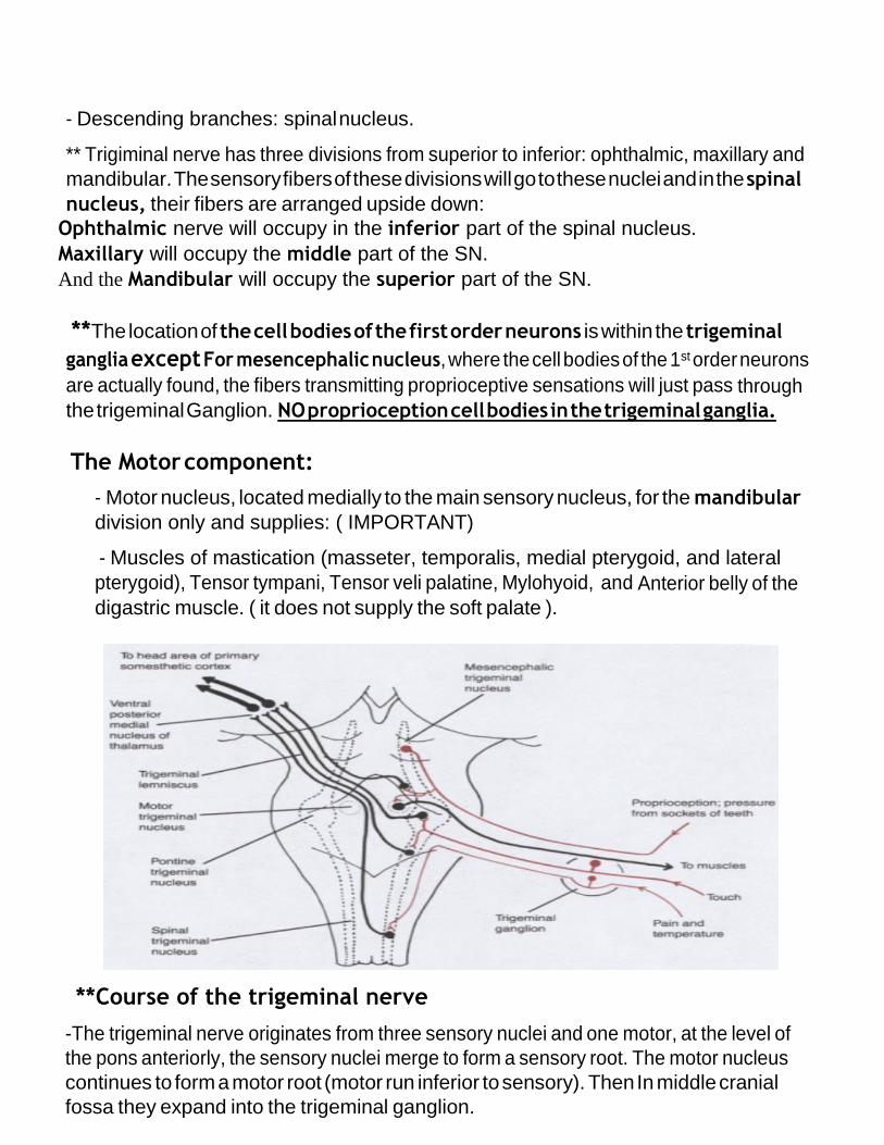
1- Main sensory nucleus:
- Found between spinal and mesencephalic nuclei in the pons. -receives crude touch and pressure sensations.
2- Spinal nucleus of trigeminal :
-Found in the medulla and the pons. And extends from the main sensory nucleus(superiorly
) to the C2 segment of the spinal cord(inferiorly).
-receives thermal and nociceptive sensations
3- Mesencephalic nucleus:
- Found in the Lateral part of the gray matter around the cerebral aqueduct in the mid brain
above the main sensory
nucleus.
-receives proprioceptive sensations
- Ascending branches: main sensory nucleus.
- Descending branches: spinal nucleus.
** Trigiminal nerve has three divisions from superior to inferior: ophthalmic, maxillary and
mandibular. The sensory fibers of these divisions will go to these nuclei and in the spinal
nucleus, their fibers are arranged upside down:
Ophthalmic nerve will occupy in the inferior part of the spinal nucleus.
Maxillary will occupy the middle part of the SN.
And the Mandibular will occupy the superior part of the SN.
**The location of the cell bodies of the first order neurons is within the trigeminal
ganglia except For mesencephalic nucleus, where the cell bodies of the 1st order neurons
are actually found, the fibers transmitting proprioceptive sensations will just pass through
the trigeminal Ganglion. NO proprioception cell bodies in the trigeminal ganglia.
The Motor component:
- Motor nucleus, located medially to the main sensory nucleus, for the mandibular
division only and supplies: ( IMPORTANT)
- Muscles of mastication (masseter, temporalis, medial pterygoid, and lateral
pterygoid), Tensor tympani, Tensor veli palatine, Mylohyoid, and Anterior belly of the
digastric muscle. ( it does not supply the soft palate ).
**Course of the trigeminal nerve
-The trigeminal nerve originates from three sensory nuclei and one motor, at the level of
the pons anteriorly, the sensory nuclei merge to form a sensory root. The motor nucleus
continues to form a motor root (motor run inferior to sensory). Then In middle cranial
fossa they expand into the trigeminal ganglion.
- Trigeminal ganglion located lateral to the cavernous sinus, Upper surface of the apex of
the petrous bone in a depression of the temporal bone, it has a pouch in the dura mater
known as Meckel cave.
The divisions of this nerve will go out through:
– Ophthalmic: through superior orbital fissure.
– Maxillary: through foramen rotundum to pterygopalatine fossa.
– Mandibular: through foramen ovale to infratemporal fossa .
5-Facial nerve
1- Main Motor Nucleus
• Found Deep in the reticular formation of the caudal part of the pons. And its fibers will
turn around the nucleus of the abducent nerve( facial colliculus)
• it supplies muscles of facial expressions.
2- Parasympathetic Nuclei(superior salivatory lacrimatory ):
• Location: Posterolateral to the main motor nucleus.
**It’s also named salivatory lacrimatory nucleus because it has:
- Superior salivatory:
supplies submandibular and sublingual glands.
- Lacrimal nucleus:
Receives from:
– Hypothalamus (Emotional), it’s the major output of limbic system that
appear as responses by autonomic nervous system like if you feel sad you will cry
(lacrimal gland will be stimulated to produce tears).
– Sensory nuclei of the trigeminal (reflex), like when a foreign body enter
the eye as a reflex the eye start tearing (lacrimation).
3- Sensory Nucleus:
** Remember: there are two types of sensation in the facial nerve one is general sensory and the other related to the taste.
**Course of facial nerve
→ Facial nerve will go out from the anterior surface between the pons and the medulla
oblongata, after coming out from the brainstem it will face a canal within the petrous
part of the temporal bone of the skull which is the internal acoustic meatus.
- Internal acoustic meatus gives passage to two cranial nerves the facial and
vestibulocochlear nerves, the facical nerve will run laterally through the cavity of inner ear
on the medial wall then it goes to the posterior wall of the tympanic cavity and finally
Emerges from the stylomastoid foramen and ends in the parotid gland (it doesn’t supply
this gland )and within the parotid gland, the nerve terminates by splitting into five
branches: Temporal branch, Zygomatic branch, Buccal branch, Marginal mandibular branch and
Cervical branch. They are the extracranial branches of the facial nerve.
-These branches are responsible for innervating the muscles of facial expression.
-When the facial nerve was in the petrous part of the temporal bone (in the cavity of
middle ear), it will give two intracranial branches:
1- Greater petrosal nerve 2- chorda tympani nerve
**Taste sensation/ the chorda tympani:
• Location: upper part of the nucleus of the tractus solitaries.
- The taste fibers of facial nerve will go to the nucleus of tractus solitaries.
* The Cell bodies found in geniculate ganglion.
- From the nucleus of the tractus solitaries the 2nd order neuron will make a cross and goes
up to the VPM, and then it will go from ventral posteriomedial to the Primary gustatory
cortex (area 43),this area found in the parital lobe just superior to the lateral fissure.
the chorda tympani, a branch from the facial nerve in the petrous part of the temporal
bone, has two fibers parasympathetic and taste.
It originates from the facial nerve, then it travels through the middle ear and go out
through the petrotympanic fissure (also called glaserian fissure) after which it emerges
from the skull into the infratemporal fossa, it soon combines with lingual nerve.
It combines the lingual nerve (a branch of the mandibular, trigeminal which lacks
parasympathetic and taste fibers) the taste fibers will continue with the lingual nerve that
give taste sensory to Anterior 2/3 of tongue.
The parasympathetic fibers : in the submandibular ganglion, the fibers(Preganglionic
parasympathetic fibers) that come from Superior salivatory nucleus will make synapse in
this ganglion and the postganglionic fibers will supply the submandibular and sublingual
glands.

**the geniculate ganglia
→ the ganglia where the facial nerve curve called geniculate ganglion (geniculate mean
curve)
* If you notice the facial fibers has two curves, the first where the motor fibers curve on
the floor of the 4th ventricle around the abducent nucleus and the other in the geniculate
ganglion.
Geniculate ganglion is sensory ganglia, has the cell body of the sensory mainly for taste
fibers Be careful! It’s not an autonomic ganglion there is no synapse.
**the greater petrosal
It carries preganglionic parasympethatic fibers that will reach the lacrimal gland. its found
in the cavity of the inner ear, then it will leave the cavity to the Middle cranial fossa through
the greater petrosal foramen (or hiatus), in the middle cranial fossa it Passes over Foramen
lacerum, where it joins the Postganglionic sympathetic fibers that found on the internal
carotid that go to the middle cranial fossa through the carotid canal. These Postganglionic
sympathetic fibers are the deep petrosal nerve (they are mainly originated in the superior
cervical sympathetic ganglion in the neck). Greater petrosal join the deep petrosal to form
the Vidian nerve (nerve to pterygoid canal). The Vidian will leave the middle cranial fossa
and inter the pterygopalatine fossa where a parasympathetic autonomic ganglia found
which is pterygopalatine ganglion
After they enter the pterygopalatine ganglion that is suspended by the maxillary nerve, the
fibers of the greater petrosal will synapse and give postganglionic parasympethatic fibers,
the deep petrosal will go through the pterygopalatine ganglion without synapse . these
fibers(parasympathetic and sympathetic) then run in the maxillary nerve to the zygomatic
nerve (branch from maxillary nerve), then they will go to zygomaticotemporal nerve
(branch from zygomatic nerve). Zygomaticotemporal nerve will join the lacrimal nerve
(remember lacrimal nerve is a general sensory branch from opthalmic nerve that is a branch
from trigeminal nerve). And that what we mean when we said that some autonomic fibers
Snoop in a branches from trigeminal which is lacrimal nerve. Then supply the lacrimal gland.
**General sensory
The fibers of this general sensation come from spinal trigeminal nucleus that its cell
body located in the geniculate ganglion which give fibers carried by facial nerve to the
skin of the external acoustic meatus for general sensation.
**Facial Nerve injury
* It’s important to know if it lower or upper motor lesion since they differ in their
symptoms.
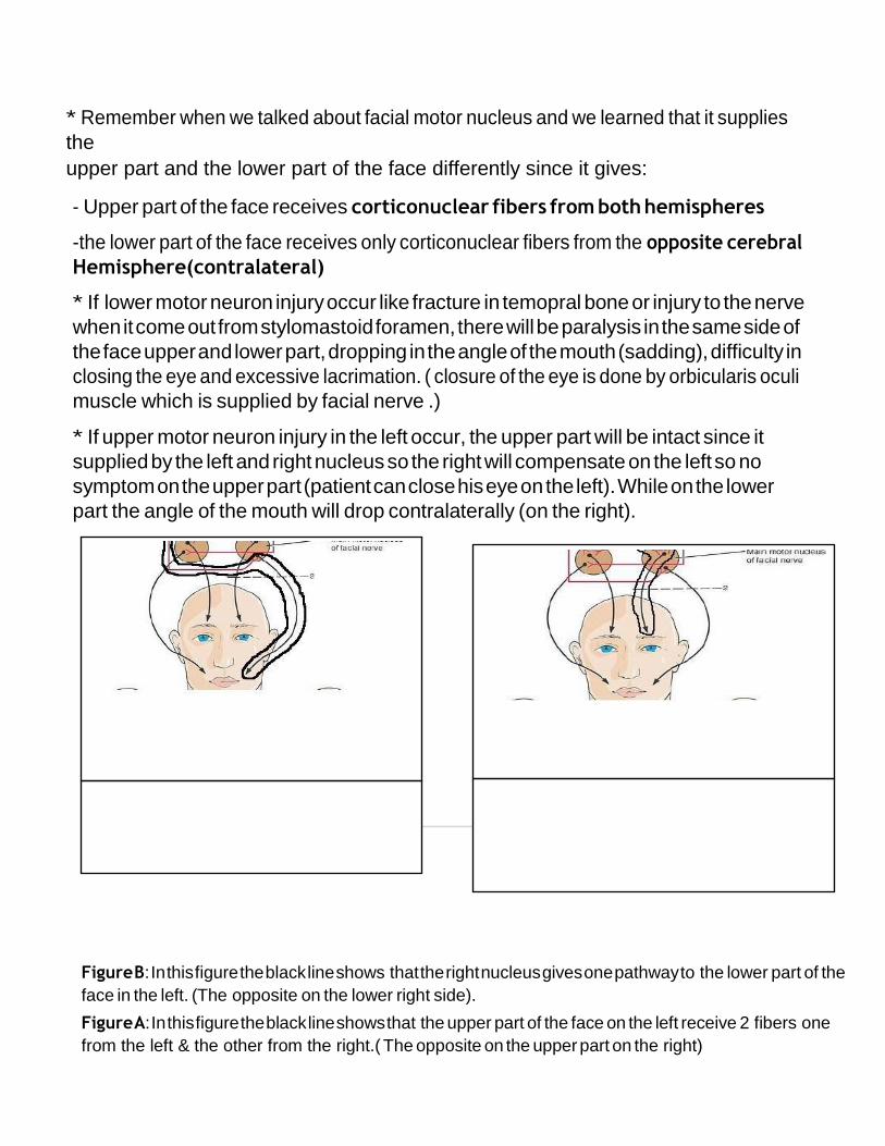
* Remember when we talked about facial motor nucleus and we learned that it supplies
the
upper part and the lower part of the face differently since it gives:
- Upper part of the face receives corticonuclear fibers from both hemispheres
-the lower part of the face receives only corticonuclear fibers from the opposite cerebral
Hemisphere(contralateral)
* If lower motor neuron injury occur like fracture in temopral bone or injury to the nerve
when it come out from stylomastoid foramen, there will be paralysis in the same side of
the face upper and lower part, dropping in the angle of the mouth (sadding), difficulty in
closing the eye and excessive lacrimation. ( closure of the eye is done by orbicularis oculi
muscle which is supplied by facial nerve .)
* If upper motor neuron injury in the left occur, the upper part will be intact since it
supplied by the left and right nucleus so the right will compensate on the left so no
symptom on the upper part (patient can close his eye on the left). While on the lower
part the angle of the mouth will drop contralaterally (on the right).
Figure B: In this figure the black line shows that the right nucleus gives one pathway to the lower part of the
face in the left. (The opposite on the lower right side).
Figure A: In this figure the black line shows that the upper part of the face on the left receive 2 fibers one
from the left & the other from the right.( The opposite on the upper part on the right)

This figure show:
If an injury happen here it will be upper motor
lesion, symptoms will appear as paralysis
of the lower contralateral part of the face
If an injury happens here it will be lower motor
lesion, symptoms will appear as paralysis of the
entire ipsilateral part of face .
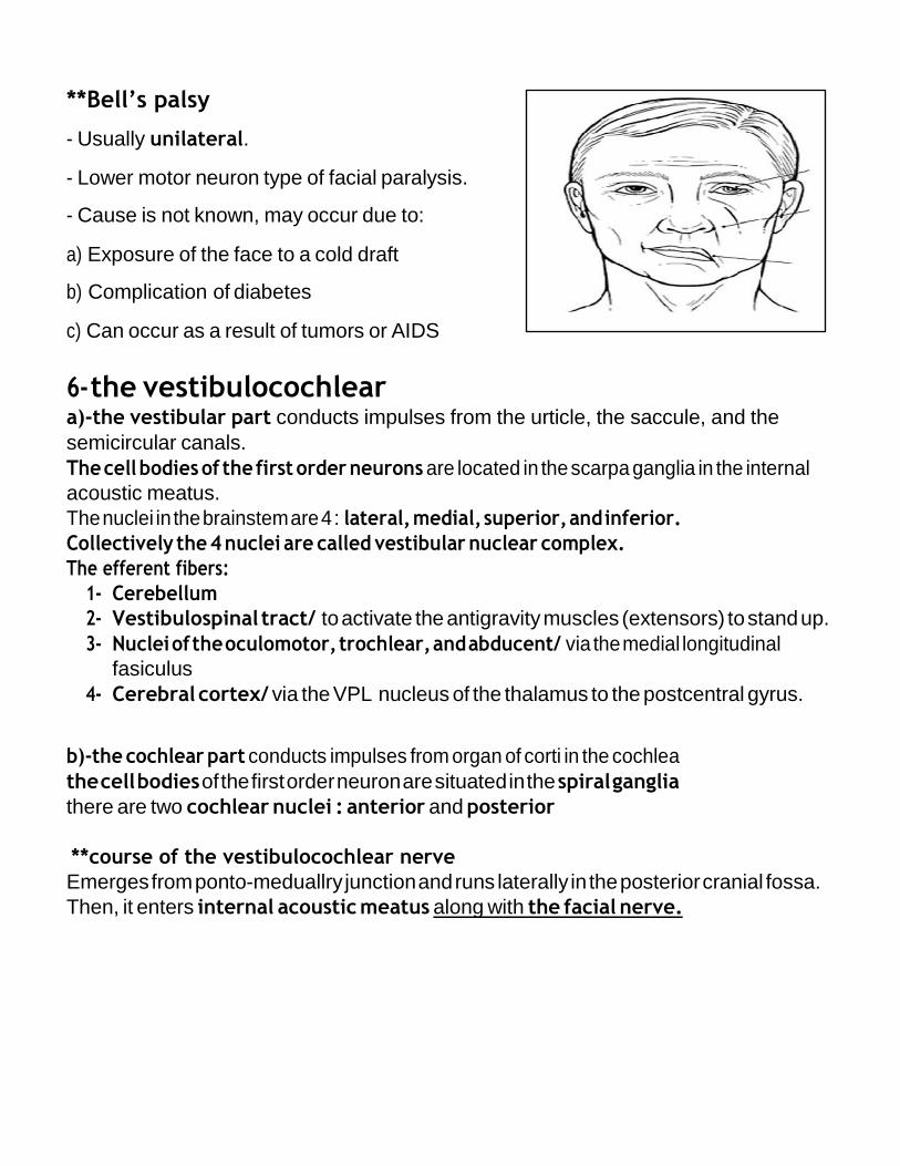
**Bell’s palsy
- Usually unilateral.
- Lower motor neuron type of facial paralysis.
- Cause is not known, may occur due to:
a) Exposure of the face to a cold draft
b) Complication of diabetes
c) Can occur as a result of tumors or AIDS
6- the vestibulocochlear a)-the vestibular part conducts impulses from the urticle, the saccule, and the
semicircular canals.
The cell bodies of the first order neurons are located in the scarpa ganglia in the internal
acoustic meatus.
The nuclei in the brainstem are 4 : lateral, medial, superior, and inferior.
Collectively the 4 nuclei are called vestibular nuclear complex.
The efferent fibers:
1- Cerebellum
2- Vestibulospinal tract/ to activate the antigravity muscles (extensors) to stand up.
3- Nuclei of the oculomotor, trochlear, and abducent/ via the medial longitudinal
fasiculus
4- Cerebral cortex/ via the VPL nucleus of the thalamus to the postcentral gyrus.
b)-the cochlear part conducts impulses from organ of corti in the cochlea
the cell bodies of the first order neuron are situated in the spiral ganglia
there are two cochlear nuclei : anterior and posterior
**course of the vestibulocochlear nerve
Emerges from ponto-meduallry junction and runs laterally in the posterior cranial fossa.
Then, it enters internal acoustic meatus along with the facial nerve.