Embed Size (px)
Citation preview
1
The metabolic functions of the liverCatabolism of haemoglobin, bilirubin
Metabolism of iron
Biochemistry VFULecture 15 2009 (J.S.)

2
Portal vein
A. hepatica
Hepatic veins
V. cava inf.
The major metabolic functions of the liver:
– uptake of most nutrients from the gastrointestinal tract,– intensive intermediary metabolism, conversion of nutrients,– controlled supply of essential compounds (glucose, VLD lipoproteins,
ketone bodies, plasma proteins, etc.),– ureosynthesis, biotransformation of xenobiotics (detoxification),– excretion (cholesterol, bilirubin, hydrophobic compounds, some metals).
3
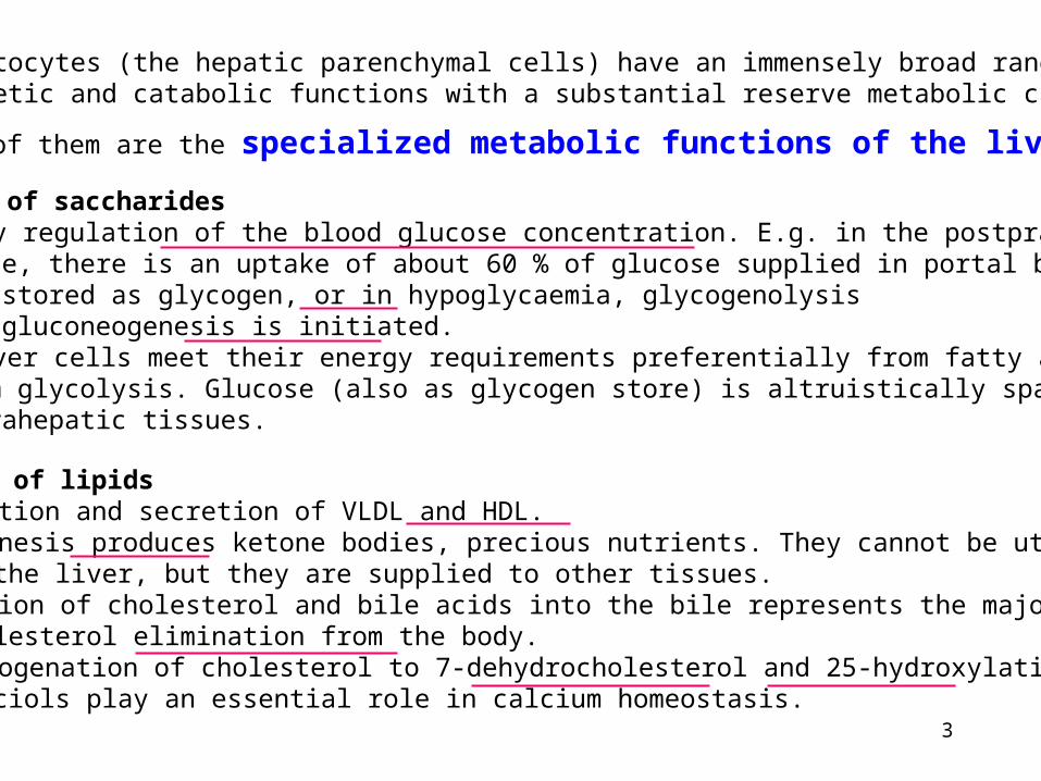
The hepatocytes (the hepatic parenchymal cells) have an immensely broad rangeof synthetic and catabolic functions with a substantial reserve metabolic capacity.
Many of them are the specialized metabolic functions of the liver:
Metabolism of saccharides – Primary regulation of the blood glucose concentration. E.g. in the postprandial
state, there is an uptake of about 60 % of glucose supplied in portal bloodand stored as glycogen, or in hypoglycaemia, glycogenolysisand gluconeogenesis is initiated.
– The liver cells meet their energy requirements preferentially from fatty acids, notfrom glycolysis. Glucose (also as glycogen store) is altruistically spared forextrahepatic tissues.
Metabolism of lipids – Completion and secretion of VLDL and HDL. – Ketogenesis produces ketone bodies, precious nutrients. They cannot be utilized
in the liver, but they are supplied to other tissues. – Secretion of cholesterol and bile acids into the bile represents the major way of
cholesterol elimination from the body. – Dehydrogenation of cholesterol to 7-dehydrocholesterol and 25-hydroxylation of
calciols play an essential role in calcium homeostasis.
4
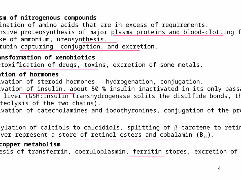
Metabolism of nitrogenous compounds – Deamination of amino acids that are in excess of requirements. – Intensive proteosynthesis of major plasma proteins and blood-clotting factors. – Uptake of ammonium, ureosynthesis. – Bilirubin capturing, conjugation, and excretion.
Biotransformation of xenobiotics – Detoxification of drugs, toxins, excretion of some metals.
Iron and copper metabolism – Synthesis of transferrin, coeruloplasmin, ferritin stores, excretion of copper.
Transformation of hormones – Inactivation of steroid hormones – hydrogenation, conjugation. – Inactivation of insulin, about 50 % insulin inactivated in its only passage through
the liver (GSH:insulin transhydrogenase splits the disulfide bonds, thenproteolysis of the two chains).
– Inactivation of catecholamines and iodothyronines, conjugation of the products.
Vitamins – Hydroxylation of calciols to calcidiols, splitting of -carotene to retinol. – The liver represent a store of retinol esters and cobalamin (B12).
5
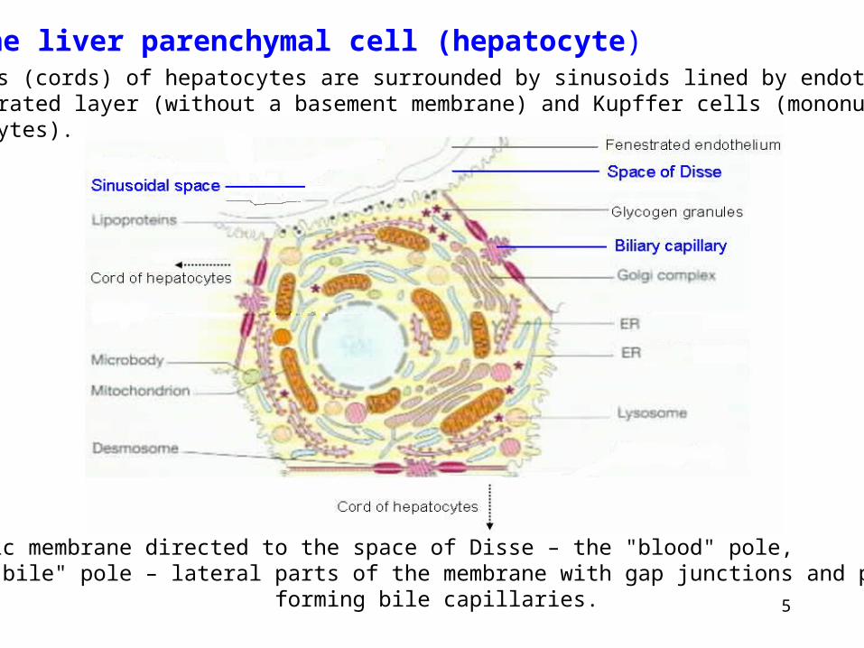
The liver parenchymal cell (hepatocyte)Columns (cords) of hepatocytes are surrounded by sinusoids lined by endothelialfenestrated layer (without a basement membrane) and Kupffer cells (mononuclearphagocytes).
Plasmatic membrane directed to the space of Disse – the "blood" pole, the "bile" pole – lateral parts of the membrane with gap junctions and parts forming bile capillaries.
6
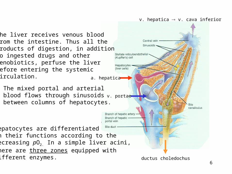
a. hepatica
v. portae
ductus choledochus
v. hepatica v. cava inferior
The mixed portal and arterialblood flows through sinusoidsbetween columns of hepatocytes.
The liver receives venous bloodfrom the intestine. Thus all theproducts of digestion, in additionto ingested drugs and otherxenobiotics, perfuse the liverbefore entering the systemiccirculation.
Hepatocytes are differentiatedin their functions according to thedecreasing pO2. In a simple liver acini,there are three zones equipped withdifferent enzymes.
7
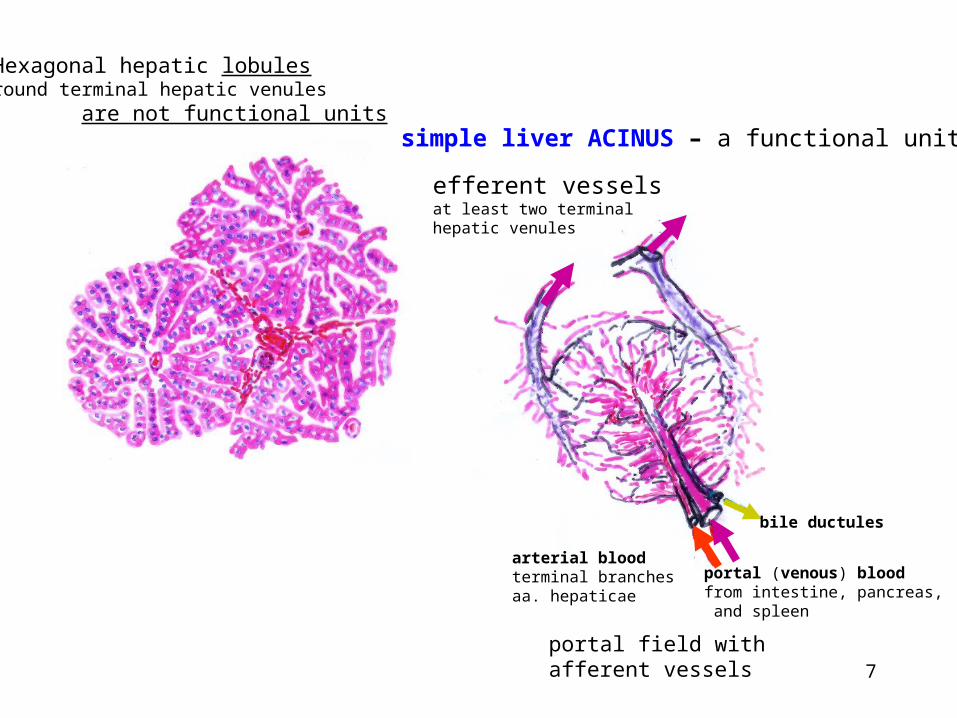
Hexagonal hepatic lobulesround terminal hepatic venules are not functional units
portal (venous) bloodfrom intestine, pancreas, and spleen
arterial bloodterminal branchesaa. hepaticae
bile ductules
simple liver ACINUS – a functional unit
portal field with afferent vessels
efferent vesselsat least two terminalhepatic venules
8
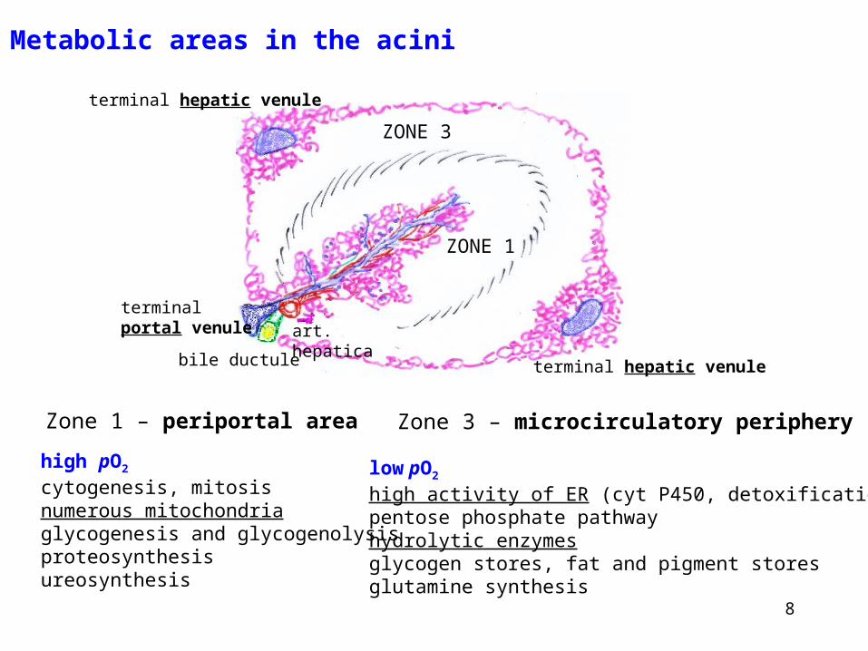
Zone 1 – periportal area Zone 3 – microcirculatory periphery
ZONE 1
ZONE 3
terminal hepatic venule
terminal hepatic venulebile ductule
art. hepaticaterminalportal venule
high pO2
cytogenesis, mitosisnumerous mitochondriaglycogenesis and glycogenolysisproteosynthesisureosynthesis
Metabolic areas in the acini
low pO2
high activity of ER (cyt P450, detoxification)pentose phosphate pathwayhydrolytic enzymesglycogen stores, fat and pigment storesglutamine synthesis
9
Liver of a patient who died in hepatic coma:.
Seastar-shaped necrotic lesion around the terminal hepatic venule. Thisshape is produced by necrosis creeping along zones 3 of the simple acini,intercalating between them and reaching portal spaces.
10
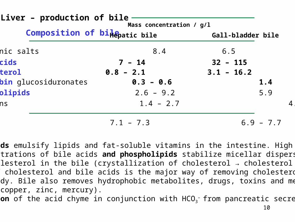
Liver – production of bile
Composition of bile Hepatic bile Gall-bladder bile
Mass concentration / g/l
Inorganic salts 8.4 6.5
Bile acids 7 – 14 32 – 115Cholesterol 0.8 – 2.1 3.1 – 16.2Bilirubin glucosiduronates 0.3 – 0.6 1.4
Phospholipids 2.6 – 9.2 5.9
Proteins 1.4 – 2.7 4.5
pH 7.1 – 7.3 6.9 – 7.7
FunctionsThe bile acids emulsify lipids and fat-soluble vitamins in the intestine. High concentrations of bile acids and phospholipids stabilize micellar dispersion of cholesterol in the bile (crystallization of cholesterol → cholesterol gall-stones).Excretion of cholesterol and bile acids is the major way of removing cholesterol from the body. Bile also removes hydrophobic metabolites, drugs, toxins and metals (e.g. copper, zinc, mercury).Neutralization of the acid chyme in conjunction with HCO3
– from pancreatic secretion.
11
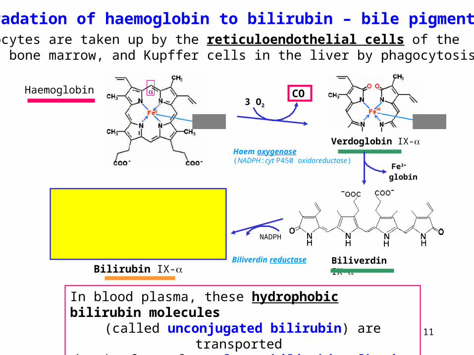
Degradation of haemoglobin to bilirubin – bile pigments
In blood plasma, these hydrophobic bilirubin molecules(called unconjugated bilirubin) are transported
in the form of complexes bilirubin-albumin.
Biliverdin IX-
Erythrocytes are taken up by the reticuloendothelial cells of thespleen, bone marrow, and Kupffer cells in the liver by phagocytosis.
Haemoglobin
Verdoglobin IX-
CO3 O2
Haem oxygenase(NADPH:cyt P450 oxidoreductase)
Fe3+
globin
Biliverdin reductase
NADPH
Bilirubin IX-
12
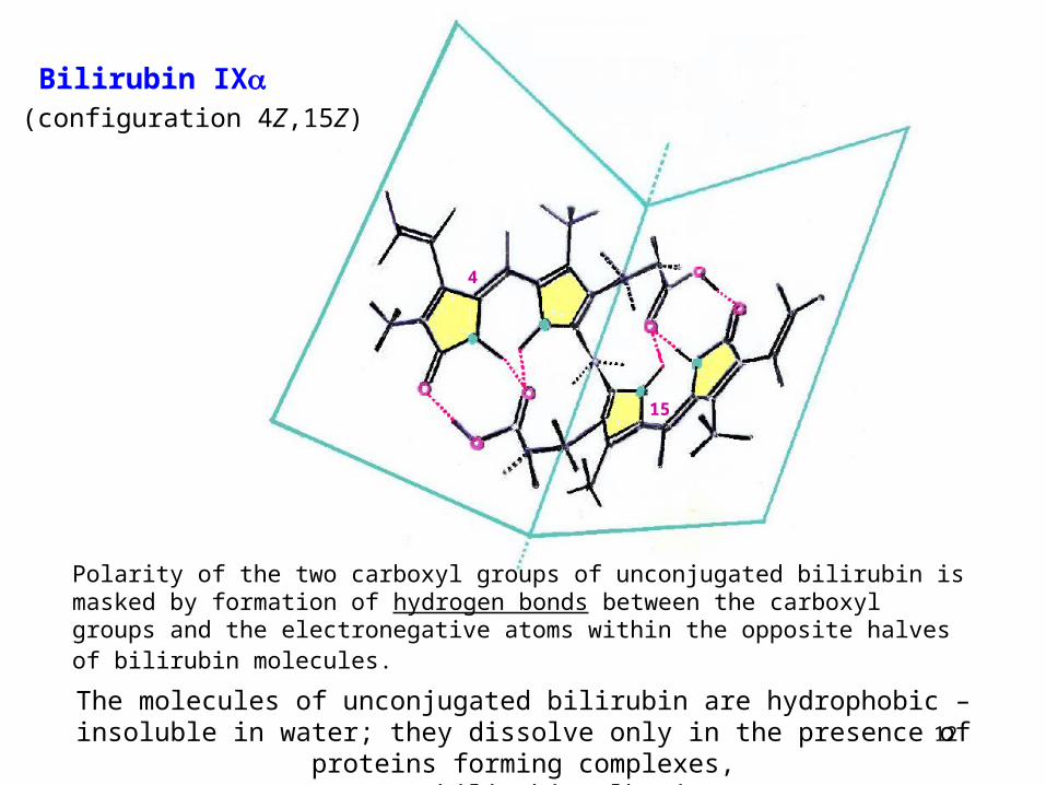
Bilirubin IX(configuration 4Z,15Z)
4
15
Polarity of the two carboxyl groups of unconjugated bilirubin is masked by formation of hydrogen bonds between the carboxyl groups and the electronegative atoms within the opposite halves of bilirubin molecules.
The molecules of unconjugated bilirubin are hydrophobic – insoluble in water; they dissolve only in the presence of proteins forming complexes,
e.g., bilirubin-albumin.
13
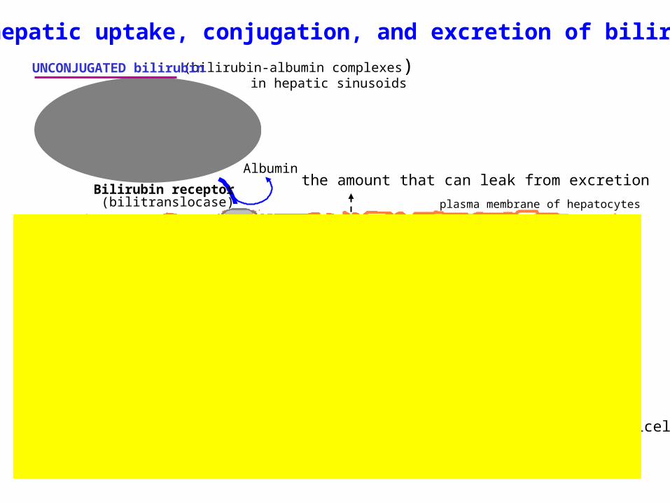
The hepatic uptake, conjugation, and excretion of bilirubin
(bilirubin-albumin complexes)in hepatic sinusoids
UNCONJUGATED bilirubin
bilirubin monoglucosiduronatebilirubin bisglucosiduronate
terminal bileductule
CONJUGATED bilirubin
is polar, water-soluble –active transport into bilecapillaries in the form of micellesdepends on the bile acids
plasma membrane of hepatocytes
Albumin
Ligandin(protein Y)
UDP-glucuronate
UDPglucosyluronate
transferaseon ER membranes
the amount that can leak from excretion Bilirubin receptor
(bilitranslocase)
14
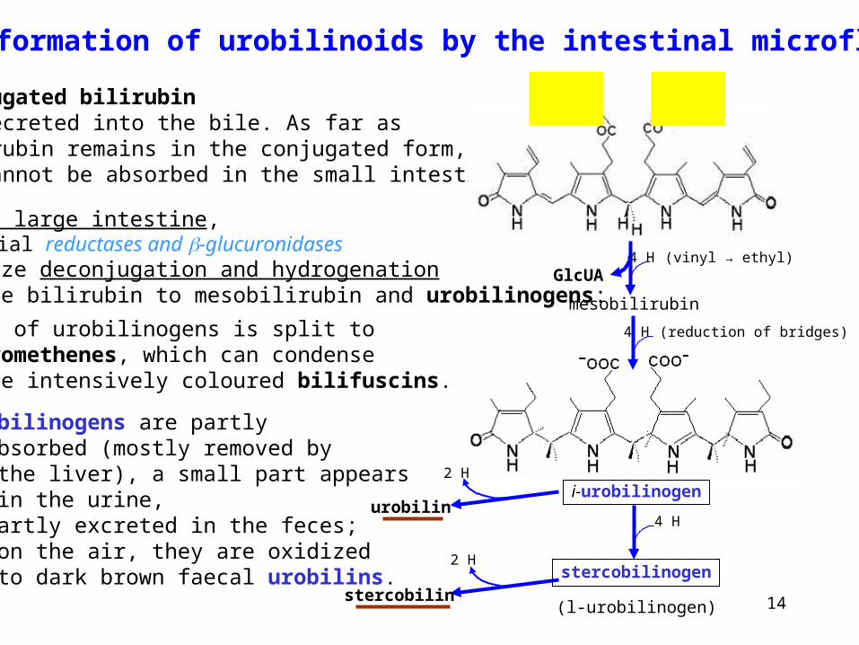
The formation of urobilinoids by the intestinal microflora
Conjugated bilirubin is secreted into the bile. As far asbilirubin remains in the conjugated form,it cannot be absorbed in the small intestine.
i-urobilinogen
In the large intestine,bacterial reductases and -glucuronidasescatalyze deconjugation and hydrogenationof free bilirubin to mesobilirubin and urobilinogens:
A part of urobilinogens is split todipyrromethenes, which can condenseto give intensively coloured bilifuscins.
4 H
4 H (reduction of bridges)
(l-urobilinogen)
4 H (vinyl → ethyl)
mesobilirubin
GlcUA
Urobilinogens are partly – absorbed (mostly removed by the liver), a small part appears in the urine, – partly excreted in the feces; on the air, they are oxidized to dark brown faecal urobilins.
urobilin
stercobilin
2 H
2 Hstercobilinogen
15
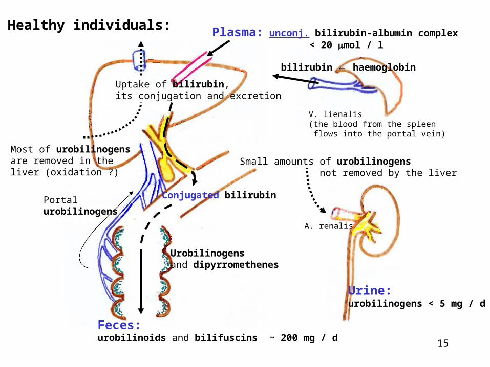
Healthy individuals:
Portalurobilinogens
Conjugated bilirubin
Urobilinogensand dipyrromethenes
Feces:urobilinoids and bilifuscins ~ 200 mg / d
V. lienalis(the blood from the spleen flows into the portal vein)
Most of urobilinogensare removed in theliver (oxidation ?)
Uptake of bilirubin,its conjugation and excretion
Urine:urobilinogens < 5 mg / d
Plasma: unconj. bilirubin-albumin complex < 20 mol / l
bilirubin ← haemoglobin
Small amounts of urobilinogens not removed by the liver
A. renalis
16
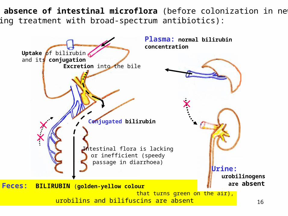
Conjugated bilirubin
Uptake of bilirubinand its conjugation
Excretion into the bile
Feces: BILIRUBIN (golden-yellow colour that turns green on the air), urobilins and bilifuscins are absent
Intestinal flora is lackingor inefficient (speedypassage in diarrhoea)
In the absence of intestinal microflora (before colonization in newbornsor during treatment with broad-spectrum antibiotics):
Plasma: normal bilirubin concentration
Urine: urobilinogens are absent
17
Major types of hyperbilirubinaemias
Hyperbilirubinaemia – serum bilirubin > 20 – 22 mol / l
Icterus (jaundice) – yellowish colouring of scleras and skin, serum bilirubin usually more than 30 – 35 mol / l
The causes of hyperbilirubinaemia are conventionally classified as
– prehepatic (haemolytic) – increased production of bilirubin,
– hepatocellular due to inflammatory disease (infectioushepatitis), hepatotoxic compounds (e.g. ethanol,acetaminophen), or autoimmune disease; chronichepatitis can result in liver cirrhosis – fibrosis ofhepatic lobules,
– posthepatic (obstructive) – insufficient drainage of intrahepaticor extrahepatic bile ducts (cholestasis).
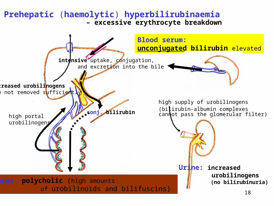
18
Prehepatic (haemolytic) hyperbilirubinaemia– excessive erythrocyte breakdown
Feces: polycholic (high amounts of urobilinoids and bilifuscins)
Blood serum:unconjugated bilirubin elevated
conj. bilirubinhigh portalurobilinogens
intensive uptake, conjugation, and excretion into the bile
Urine: increased urobilinogens (no bilirubinuria)
high supply of urobilinogens(bilirubin-albumin complexescannot pass the glomerular filter)
increased urobilinogensare not removed sufficiently
19
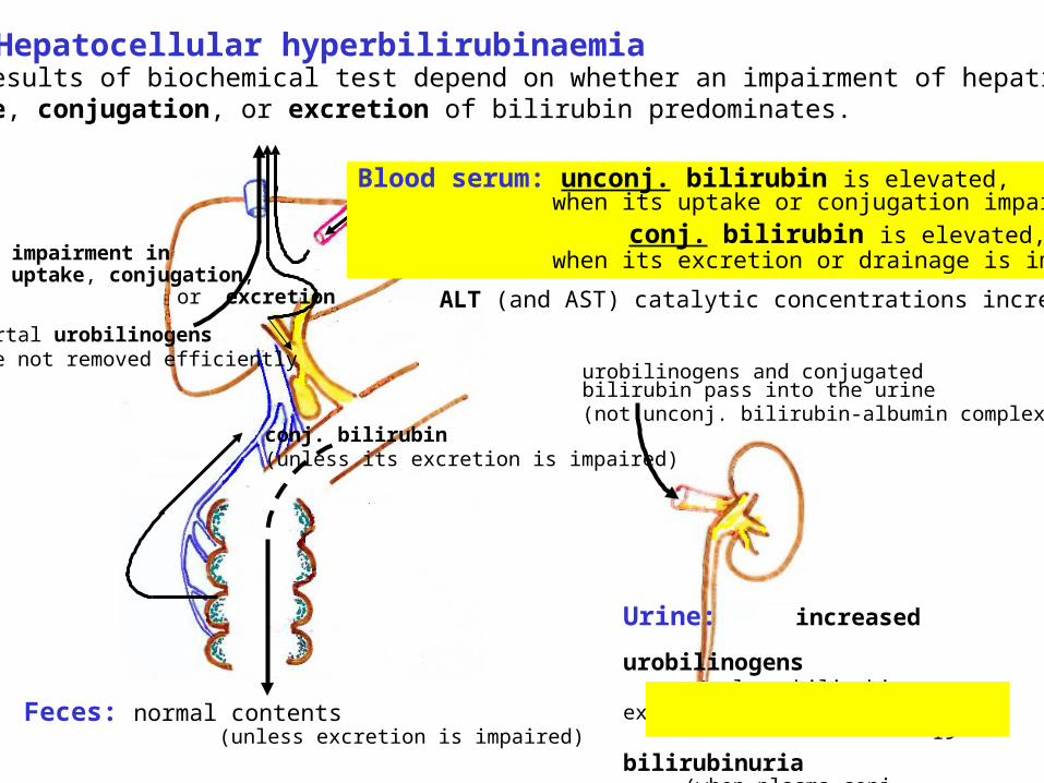
Hepatocellular hyperbilirubinaemiaThe results of biochemical test depend on whether an impairment of hepaticuptake, conjugation, or excretion of bilirubin predominates.
urobilinogens and conjugatedbilirubin pass into the urine(not unconj. bilirubin-albumin complexes)
Urine: increased urobilinogens (unless bilirubin excretion is impaired) bilirubinuria (when plasma conj. bilirubin increases)
Feces: normal contents (unless excretion is impaired)
conj. bilirubin(unless its excretion is impaired)
portal urobilinogensare not removed efficiently
impairment inuptake, conjugation,
or excretion
Blood serum: unconj. bilirubin is elevated, when its uptake or conjugation impaired
conj. bilirubin is elevated, when its excretion or drainage is impaired
ALT (and AST) catalytic concentrations increased
20
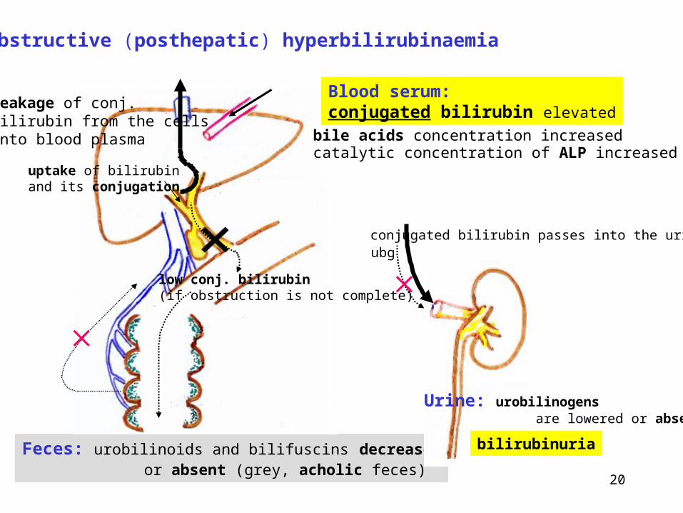
Obstructive (posthepatic) hyperbilirubinaemia
Feces: urobilinoids and bilifuscins decreased or absent (grey, acholic feces)
Blood serum:conjugated bilirubin elevated
bile acids concentration increasedcatalytic concentration of ALP increased
Urine: urobilinogens are lowered or absent
low conj. bilirubin(if obstruction is not complete)
conjugated bilirubin passes into the urineubg
bilirubinuria
uptake of bilirubinand its conjugation
leakage of conj.bilirubin from the cellsinto blood plasma
21
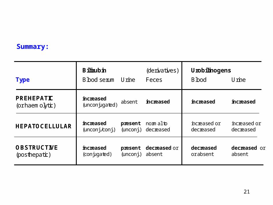
Bilirubin (derivatives) Urobilinogens
Type Blood serum Urine Feces Blood Urine
PREHEPATIC (or haemolytic)
increased (unconjugated)
absent increased increased increased
HEPATOCELLULAR increased (unconj./conj.)
present (unconj.)
normal to decreased
increased or decreased
increased or decreased
OBSTRUCTIVE (posthepatic)
increased (conjugated)
present (unconj.)
decreased or absent
decreased or absent
decreased or absent
Summary:
22
Laboratory testsfor detecting an impairment of liver functions ("liver tests")
• Plasma markers of hepatocyte membrane integrity
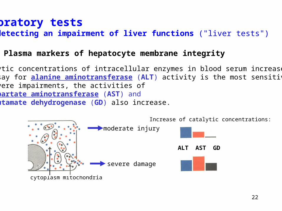
Catalytic concentrations of intracellular enzymes in blood serum increase:An assay for alanine aminotransferase (ALT) activity is the most sensitive one.In severe impairments, the activities of aspartate aminotransferase (AST) and glutamate dehydrogenase (GD) also increase.
cytoplasm mitochondria
ALT AST GD
moderate injury
severe damage
Increase of catalytic concentrations:
23
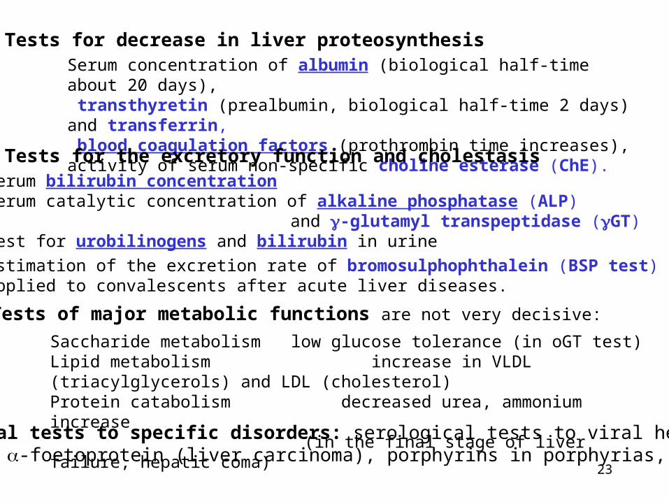
• Tests for decrease in liver proteosynthesisSerum concentration of albumin (biological half-time about 20 days), transthyretin (prealbumin, biological half-time 2 days) and transferrin, blood coagulation factors (prothrombin time increases),activity of serum non-specific choline esterase (ChE).
Saccharide metabolism low glucose tolerance (in oGT test)Lipid metabolism increase in VLDL (triacylglycerols) and LDL (cholesterol)Protein catabolism decreased urea, ammonium increase
(in the final stage of liver failure, hepatic coma)
• Tests of major metabolic functions are not very decisive:
• Tests for the excretory function and cholestasisSerum bilirubin concentrationSerum catalytic concentration of alkaline phosphatase (ALP)
and -glutamyl transpeptidase (GT)Test for urobilinogens and bilirubin in urine
Estimation of the excretion rate of bromosulphophthalein (BSP test) isapplied to convalescents after acute liver diseases.
• Special tests to specific disorders: serological tests to viral hepatitis, serum -foetoprotein (liver carcinoma), porphyrins in porphyrias, etc.
24
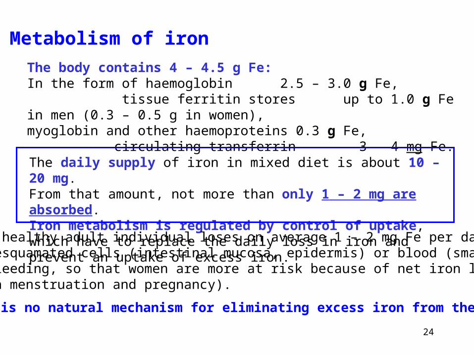
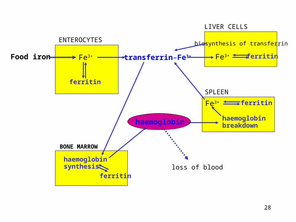
Metabolism of iron
The body contains 4 – 4.5 g Fe:In the form of haemoglobin 2.5 – 3.0 g Fe, tissue ferritin stores up to 1.0 g Fe in men (0.3 – 0.5 g in women),myoglobin and other haemoproteins 0.3 g Fe, circulating transferrin 3 – 4 mg Fe.
The daily supply of iron in mixed diet is about 10 – 20 mg.From that amount, not more than only 1 – 2 mg are absorbed.Iron metabolism is regulated by control of uptake, which have to replace the daily loss in iron and prevent an uptake of excess iron.
A healthy adult individual loses on average 1 – 2 mg Fe per day in desquamated cells (intestinal mucosa, epidermis) or blood (smallbleeding, so that women are more at risk because of net iron lossin menstruation and pregnancy).
There is no natural mechanism for eliminating excess iron from the body.
25
elimination of insolubleFe salts in feces
10 – 20 mf Fe
8 – 19 mg Fe
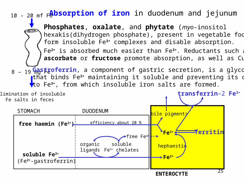
Absorption of iron in duodenum and jejunum
Gastroferrin, a component of gastric secretion, is a glycoproteinthat binds Fe2+ maintaining it soluble and preventing its oxidationto Fe3+, from which insoluble iron salts are formed.
Phosphates, oxalate, and phytate (myo-inositolhexakis(dihydrogen phosphate), present in vegetable food)form insoluble Fe3+ complexes and disable absorption.
Fe2+ is absorbed much easier than Fe3+. Reductants such asascorbate or fructose promote absorption, as well as Cu2+.
STOMACH DUODENUM
ENTEROCYTE
hephaestin
free haemin (Fe3+)
(Fe2+-gastroferrin)soluble Fe2+
ferritin
organicligands
free Fe3+
solubleFe3+ chelates
bile pigments
Fe3+
Fe2+
transferrin–2 Fe3+
efficiency about 20 %
26
Transferrin (Trf)
is a plasma glycoprotein (a major component of 1-globulin fraction), Mr 79 600.
Plasma (serum) transferrin concentration 2.5 – 4 g / l (30 – 50 mol / l)
Transferrin molecules have two binding sites for Fe ions,total iron binding capacity (TIBC) for Fe ions is higher than 60 mol / l.
Serum Fe3+ (i.e. transferrin-Fe3+) concentration is about 10 – 20 mol / l,14 – 26 mol / l in men,11 – 22 mol / l in women.
Circadian rhythm exists, the morning concentrations are higher by 10 - 30 % than those at night..
Saturation of transferrin with Fe3+ equals usually about 1/3.Because the biosynthesis of transferrin is stimulated during iron deficiency (andplasma iron concentration decreases), the decrease in saturation of transferrinis observed.
27
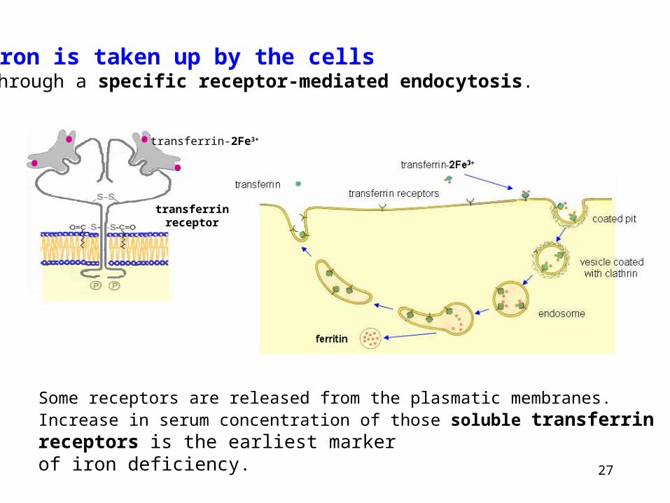
Iron is taken up by the cellsthrough a specific receptor-mediated endocytosis.
transferrin-2Fe3+
•••
•
transferrinreceptor
Some receptors are released from the plasmatic membranes. Increase in serum concentration of those soluble transferrin receptors is the earliest markerof iron deficiency.
28
ENTEROCYTES
Food iron Fe3+
ferritin
transferrin–Fe3+
LIVER CELLS
ferritinFe3+
biosynthesis of transferrin
SPLEEN
BONE MARROW
haemoglobinsynthesis
haemoglobin haemoglobinbreakdown
ferritinFe3+
ferritinloss of blood
29
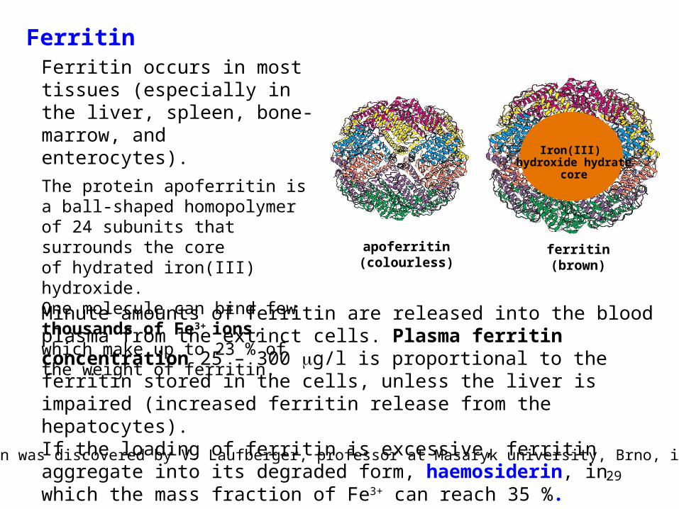
Ferritin
Iron(III) hydroxide hydrate
core
apoferritin(colourless)
ferritin(brown)
Minute amounts of ferritin are released into the blood plasma from the extinct cells. Plasma ferritin concentration 25 – 300 g/l is proportional to the ferritin stored in the cells, unless the liver is impaired (increased ferritin release from the hepatocytes).If the loading of ferritin is excessive, ferritin aggregate into its degraded form, haemosiderin, in which the mass fraction of Fe3+ can reach 35 %.
Ferritin occurs in most tissues (especially in the liver, spleen, bone-marrow, and enterocytes).
The protein apoferritin is a ball-shaped homopolymer of 24 subunits that surrounds the coreof hydrated iron(III) hydroxide.One molecule can bind few thousands of Fe3+ ions, which make up to 23 % of the weight of ferritin.
Ferritin was discovered by V. Laufberger, professor at Masaryk university, Brno, in 1934.
30
Hepcidin is a polypeptide (Mr ~ 2000, 25 amino acid residues, from which 8 are Cys), discovered as the liver-expressed antimicrobial peptide, LEAP-1, in 2000.
It is produced by the liver (to some extent in myocard and pancreas, too) as a hormone that limits the accessibility of iron and also exhibits certain antimicrobial and antifungal activity.
Effects of hepcidin: It – reduces Fe2+ absorption in the duodenum, – prevents the release of recyclable Fe from macrophages, – inhibits Fe transport across the placenta, – diminishes the accessibility of Fe for invading pathogens.
The biosynthesis of hepcidin is stimulated in iron overload and in inflammations (hepcidin belongs to acute phase proteins type 2),and is supressed during iron deficiency . Notice the fact that the same two factors stimulating hepcidin synthesis inhibit the biosynthesis of transferrin.
Hepcidin is filtered in renal glomeruli and not reabsorbed in the renal tubules. Sothe amount of hepcidin excreted into the urine corresponds with the amountsynthesized in the body. There is a positive correlation between this amount ofhepcidin and the concentration of ferritin in blood plasma.


![VFU SEM - PHP OOP [12.10.2013]](https://img.pdfslide.net/doc/110x75/554fb9c0b4c9053d018b4694/vfu-sem-php-oop-12102013.jpg)


![VFU SEM - PHP Intro [05.10.2013]](https://img.pdfslide.net/doc/110x75/554fb871b4c9050e7d8b470b/vfu-sem-php-intro-05102013.jpg)
