Embed Size (px)
Citation preview

Investigation
10Introduction
-
Purpose
Materials
Procedure A
Questions
Cell Structure and Organelles
A cell is a functional unit that carries on all metabolic activities associ¬ated with life. The outer limits of a cell are bounded by a continuousmembrane that serves both to enclose its contents and to regulate pas¬sage of molecules into and out of the cell. Within the cell are a number ofspecialized stuctures called organelles. Each organelle is speciallyadapted for certain functions. One current theory suggests that manyorganelles and the outer membrane have been formed from one singlemembrane structure, the unit membrane. Along with a large densestructure called the nucleus, which serves as the control center of thecell, the organelles are embedded in the cytoplasm. Prokaryotic cellssuch as bacteria and blue-green algae lack an organized nucleus andlack membrane-bound organelles. Eukaryotic cells have a membrane-bounded nucleus as well as membrane-bounded organelles within thecytoplasm.
Much of the cytoplasm in a cell is a colloidal mixture of protein macro-molecules and fat globules dispersed in water. Smaller molecules suchas simple sugars and inorganic ions are mixed with water to form a truesolution.
Before electron microscopy, some of the cell organelles could be viewedwith light microscopes, but these had many limitations. Since theirintroduction, transmission electron microscopes and scanning electronmicroscopes have enhanced the study of cell organelles greatly.
To study the structure and function of organelles in eukaryotic plantand animal cells.
Electron micrographs and drawings of eukaryotic plant and animalcells.
*-
Answer the following questions while making reference to the electronmicrographs and figures 1 to 15. Your text should also be used to helpyou answer the questions.
1. The cell membrane is made up of phospholipids and proteins (seefigure 1). What is the function of the cell membrane?
2. Figure 1 shows two adjoining animal cells. Does there appear to bea space between the two adjoining cell membranes? If so, whatwould be found in this space?
3. On observing the cell membranes of two animal cells in figure 1,each membrane appears as a light layer sandwiched between twodarker layers. Why is this so? Explain,
4. Microtubules, intermediate filaments, and microfilaments are com¬ponents of a cell's cytoskeleton (see figure 2). What roles does thecytoskeleton play in the cell?
5. Look at figures 2b and 2c. Describe the structural differences be¬tween microtubules and microfilaments.

M
Figure la. Electron micrograph
showing the junction between twoamphibian cardiac celLi. Note the
triiaminar appearance nfthe. adjoim
mg cell membranes (CM). The fila¬mentous rnasseH (FM) acsociaied
wLlk each membrane internally are
thought to be aHHociaied withmicrofilaments; intercellularSpace (IS). ( K 93 000)
b. A diagram showing a junctionheiWCPn tir.n rr!is nnci. the
trilaminar appearance of the cellmembrane nf each ceii
Figure 2a, Longitudinal sectionthrough an amphibian small intes¬tine cell displaying microtubules(MT) and intermediate filaments(IM). Intermediate filaments areintermediate in size between micro¬tubules and microfilaments. Unlikemicrofiilaments, they cannot con*
tract and as a result play an impor¬
tant supportive function in thecytoplasm. (x 35 500)
b, c. Drawings of a nlicroiubuleand a microfilament showing thearrangement of the globularproteins.
Figure 3a, b (inset). Electronmicrograph of an amphibian car¬diac muscle cell showing a portionof the nucleus. Note the nucleolus(NU), nuclear envelope (NE), con¬densed chromatin or heterochroma-tin (HE) along the inner surface ofthe nuclear envelope and dispersedchromatin or euchromatin (EU) scat¬tered throughout the nucleoplasm.Inset shows a pore (arrows) with adiaphragm (D) closing the pore.Heterochromatin is seen as clumps.fx 23 900, inset *34 900)
27

6. Look at figure 3. Does the nuclear envelope seem to be made up ofmore than a single membrane? How many membranes are visible?
7. In figure 3 (inset), a pore whose opening is covered by a diaphragmis clearly visible. What role do the pores serve? What would be therole of the diaphragm?
8. Look at figure 3 and describe the physical differences between het-erochromatin and euchromatin.
9. Note the nucleolus in figure 3. Describe the physical characteris¬tics of this organelle.
10. Often free-floating ribosomes are organized into functional groupscalled polysomes (see figure 4). What is the function of polysomes?
11. Are polysomes larger than ribosomes?12. Many ribosomes are attached to the membranes of the endoplasmic
reticulum (see figure 4). What is their function?
13. Glycogen granules are often associated with muscle cells (see fig¬ure 4). What role might they serve in muscle cell function?
14. About half a dozen saccules comprise the Golgi apparatus as shownin figure 5. What seems to be the major role of this structure?
15. Lysosomes are special vesicles which are most likely formed by theGolgi apparatus (see figure 6b). What role do lysosomes play withrespect to the metabolic activities within a cell?
16. List two other roles that lysosomes may play in the cell.
17. Two types of lysosomes may be observed in figure 6b. Vesicles thatappear darker in an electron micrograph are residual bodies con¬taining nondigested materials, while lighter coloured vacuoles areprobably autodigestive vacuoles. Look at figure 6b and explainwhich of the two lysosomes is shown.
18. As observed in figure 6a, what seems to be the relationship be¬tween vacuoles and vesicles?
19. How are vesicles and vacuoles formed? Explain.20. Note that in figure 7a, the inner membrane of a mitochondrion is
invaginated to form shelf-like structures called cristae. How doesthis inner structure of the mitochrondrion aid its function? Ex¬plain.
21. Flagella and cilia are composed of a special arrangement of mem¬brane-bounded microtubules (see figures 8b, c). List some functionsof flagella and cilia.
22. How are basal bodies structurally related to cilia and flagella? (seefigure 8b)
23. Look at figure 8c. Describe the microtubule arrangement of a fla-gellum.
24. Describe the structural differences between a desmosome and atight junction (see figures 9 and 10).
25. What are the roles of the above two junctions?26. Describe the structural differences between a gap junction and an
intermediate junction (see figures 11 and 12).27. Observe figures 9 to 12. Of all the junctions illustrated, which
junctions seem to allow for the movement of molecules from cell tocell? Explain.

6Z
(OOS 92 X)ipo dpsnui v m (j^j) saiuososfy
/CuviuudJo ydnutiojouu uoJio&lg 'Q
(001 SIX)•lusvjdotfo at/? m usas 3Jt>
(A) s&joisdn tfvvus iCuofli spripoudsnoiuvn ypm pd]jiJ (VA) sajonovn
dSuvj unoj Smmoys jpo ajosnw vJoydrugouojiu voupaj^ •ny aunBitf
jmr?. -
¦snjounddD iSjOQ v Jo dvujdpp JoyDvp oJo SuioTDip oifDiuayos y 'q
(000 60 x i T^pfS aavDUOD) aor/J ajo)-3J.09S 3ljJ lUOjJ pVV SdjnOJDS 32/? Jo
HzSpzayj mojJJJo poyomdsjo (as)SdlDtSdn AJOJdJOdB puv (AS) ssiuos
-osaj -pnixuvddv jBjof) aytjo (dpisxdcruajj BjpJSvinjjqJ ay] oj Binnotu
ujnpwuaj. ouusnidopua at/? tmojJsopwan psjfiJ-upiQjdaq o? lySnoyj
3JD ypiym (AX) saptsan J-aJs-uvpayj ajop ¦fyo) sainooos Jo pasod
-woo snpouvddv iSjOQ nSvA-moys
pw-jS anpaaSip podonpouq t>JoydvuSoujtui uuj.pajy -sc aunSij
¦anujajsTo wnpiotpj orw
-svidopTja ySnoj.Jo Butmvjp y 'q
(OOS l£"X) 'avitjpsio otiusvidopuaayj Bin/ fvuajvw uiajoud snopaw'Tl1M 1Pm — ivapma ait> np) sajn
-twj'B uaBoDAjS puo (Ad) satuos^odjo saiuosoqijjn sjajsniQ payoupD
(jS) satuoaoyij snodawnv ynm(US) 1-JJ7t]TlZr!ldJ onusvidopua Bm
Hnoya jiaD znosniu uoiqiyduw vnjo
ydujMojonu UQjjjdjs "nt ajtiSu
^ M s Nt3 % b- t

Figure 7a, Longitudinal sectionof mitochondria in a cardiac mun-cle cell Note the large calciumgranules (CG) in the matrix space.These granules are found in cellsnoted for calcium depositions,("x 55 900)
b. A schematic diagram of mito¬chondria depicting the double mem¬brane, the outer smooth membraneand the convoluted inner membranemaking shelf-like projections calledcristae.
Figure 8a. Scanning electronmicrograph offiagella found on acell lining the tubefoot of a starfish,fx 13 600)
b. Longitudinal section offiagellafound on a tunicate branchial bas¬ket Centriolar components of twobasal bodies (BA) are seen to becontinuous with microtubules (MT)extending into the ciliary shaft;microvillus (MS), glycocalyx (GC).(x 25 000)
c. A cross section in a plane indi¬cated at the arrow shown at ahigher magnification. Note the typ¬ical 9+2 ciliary arrangement ofmicrotubules, (x 53 500)
30

Figure fti. Electron micrographshowing dsSmosameH binding turn
adjacent cells. Note the fibrousplaque (FP) to the inside of each cellmembrane and the extracclluiar
plaques (EP) in the intercellularspace. These two dense mats of fila¬
ments give cells greater stability,fx43 500)
6. A schematic drawing ofdesmQ-somes connecting two cells.
Figure 10a. Electron micrographof two tunicate (sea squirt) epider¬mal cells bound together by- a tightjunction (TJ). fx 100 300)
b, c. Schematic drawings of a tightjunction.
Figure 11a. Electron micrographshowing a portion of two tunicatemuscle cells connected by a gapjunction (GJ). (x 78 400)
b, A drawing -showing a gapjunction,
c. A schematic drawing showingchannels inproteins embedded incell membranes which allowsexchange of molecules betweencells.
Figure 12a. Electron micrograph-showing an intermediate junction(IJ) between two tunicate musclecells. Microfilaments (MF) of theterminal web fTW) attach to the cellmembranes at the intermediate junc¬tion, f x 39 500); intercellular space(IS).
b, A drawing of an intermediatejunction.
31
t "*i
y... *>^ vit v-?* * S . \*-Yh,%
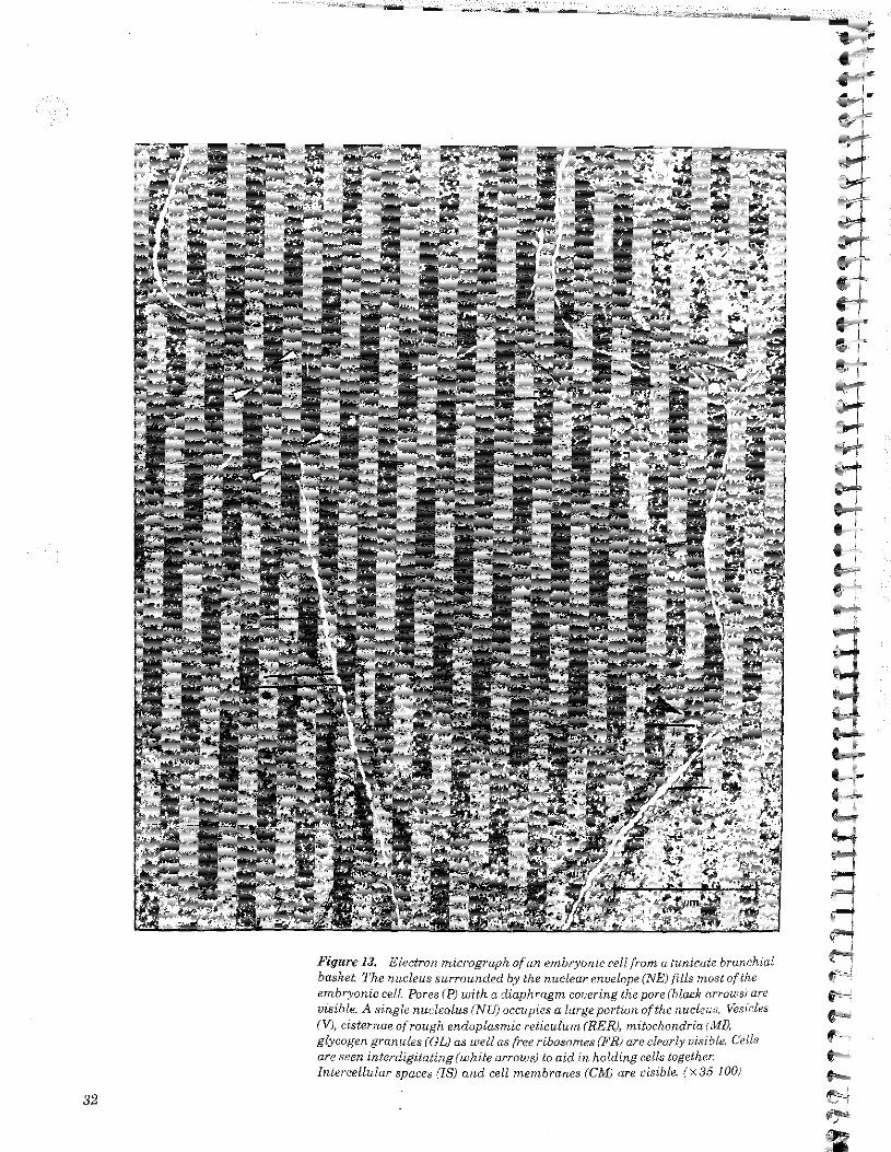
Figure 13, Electron micrograph of an embryonic cell from a tunicate branchialbasket The nucleus surrounded by the nuclear envelope (NE) fills most of theembryonic cell Pores (P) with a diaphragm covering the pore (black arrows) arevisible. A single nucleolus (NTJ) occupies a large portion of the nucleus. Vesicles(V), cistemae of rough endoplasmic reticulum (RER), mitochondria (MI),glycogen granules (GL) as well as free ribosomes (FR) are clearly visible. Cellsare seen interdigitating (white arrows) to aid in holding cells together.Intercellular spaces (IS) and cell membranes (CM) are visible. (x.3.5 100)
r

Figure 14a, l.^ongitudinal nection
of a ehlornpiast from a corn leaf ceilshowing stacks ofchlorophyli-con-
taining sacks which make up agranum, fx 40 000}
ft. A schematic drawing showinga chhropLaxt urniposed of organizedthyia'r membranes. Individualthylakoid sacks are interconnectedand they tend to stack to formgratia, (sing, granum). Surround¬
ing the grana is a colourless matrix,the stroma, The stroma and ikeinter-connecting thylakoid mem¬
branes are enclosed by a pair ofmembranes,
outermembrane
thylakoid soace
Figure 15a. Electron micrographof a plant leaf cell Note the largecentral vacuole which makes up thelargest part of the cell volume. Notethe nucleus (N), chloroplasts (CDand cell wall (CW). (x 6000;
b. Cellulose fibres of a primary cell wall of a green alga, fx 17 000)Higher plants are composed of large numbers of cells cemented together in fixedpositions by rigid cell walls that surround them.
33

28. In figure 13, note the slzy of the nucleus in comparisoii io the size ofthe cell- As cells mature, the size of the nucleus in relation to cellvolume decreases. How could you explain this/
29. Note in figure 13 that the cytoplasm appears to be granular due tonumerous free ribosomes suspended in the cytoplasm. Distinguishbetween the function of tree ribosomes and attached ribosomes.
30. Look at figure 13. What organelle seems to fill most of the nucleus?
31. Look at- figures 14b, d. The chloroplast envelope is a double mem¬brane enclosing a matrix called the stroma in which stacks of thyla-koid sacks (grana) are embedded. Where in the ehloroplast ischlorophyll found?
32. How many membranes enclose the contents of the ehloroplast? (seefigures 14b, c)
33. Are the outer membranes of a ehloroplast continuous with the Lhy-lakoid membranes? (see figures 14b, c)
34. How are the thylakoid sacks of one granum interconnected withthylakoid sacks of another granum? (see figures 14b, c)
35. Look at figure 15a. The cell wall lies externally to the cell mem¬brane and is much thicker than the cell membrane. "What is themain function of the cell wall?
36. Look at figures 15a, b. Describe the structure of the cell wall.
37. Look at figure 15b. Would the cell wall be permeable or imperme¬able to molecules? Explain.
38. In figure 15a, note the large central vacuole in the plant cell. Whatis the role of the central vacuole?
39. Observe figure 15a. Explain why the organelles located in the cellcytoplasm are near the cell wall.
40. Some smaller vacuoles (vesicles) arise from the infolding of the cellmembrane or by the pinching action of the Golgi apparatus. Howdoes the large central vacuole found in a plant cell arise?
Procedure B Figures 16a and 16b represent electron micrographs of plant and ani¬mal cells. Various organelles and cellular structures have been num¬bered. For each cell construct a table like the one shown below. Identifyby name each of the numbered structures and list its function(s). Esti¬mate the size of each numbered structure by referring to the scaleindicator in the lower right hand corner of each photomicrograph. Fig¬ures 1 to 15 may also be used to find the sizes of the various organelles.All values may also be expressed in nanometres, since
1 pm = 1000 nm
34

Figura ida. Electron micrograph,of a plant cell tciih numbered cellu¬lar structures-
.V
•» «
Figure 16b. Electron micrographof an animai cell with numberedceilu lar Hiru-ctures.
1
8 —
35