Embed Size (px)
Citation preview

CONTENT:
1. Examination of one or a couple of molecules
2. Examination of many molecules (genomics)
3. Complex techniques
Discussed in another lectures

CONTENT (1):
1. Molecular cloning
2. Polymerase chain reaction (PCR)
3. Gel electrophoresis
4. Detection of macromolecules
5. Gel retardation
6. Footprint analysis
7. Immunohistochemistry
8. In situ hybridization
9. FRET
10. Flow cytometry
11. FACS

1. Molecular cloning
a. Restriction endonucleases
b. DNA ligases
c. Plasmid vectors
d. Restriction/ligation cloning
e. Other enzymes

Restriction enzymes
Werner Arber Daniel Nathans Hamilton O. Smith
1978
1
EcoRI

2
5’-…GAATTC…-3’ 5’-…G-3’ 5’-AATTC…-3’
3’-…CTTAAG…-5’ 3’-…CTTAA-5’ 3’-G…-5’ 5’ protruding end: (e.g. EcoRI)
5’-…CCCGGG…-3’ 5’-…CCC-3’ 5’-GGG…-3’
3’-…GGGCCC…-5’ 3’-…GGG-5’ 3’-CCC…-5’ Blunt end: (e.g. SmaI)
5’-…GGTACC…-3’ 5’-…GGTAC-3’ 5’-C…-3’
3’-…CCATGG…-5’ 3’-…C-5’ 3’-CATGG…-5’ 3’ protruding end: (e.g. KpnI)
BEFORE CUTTING AFTER CUTTING
Restriction enzymes
5’-…GAATTC…-3’ 5’-…G-3’ 5’-AATTC…-3’
3’-…CTTAAG…-5’ 3’-…CTTAA-5’ 3’-G…-5’
CH3
CH3
X EcoRI methylase
RESTRICTION – MODIFICATION SYSTEM
EcoRI RE does not cut

One DNA molecule Two DNA molecules
DNA ligase DNA ligase
3
4
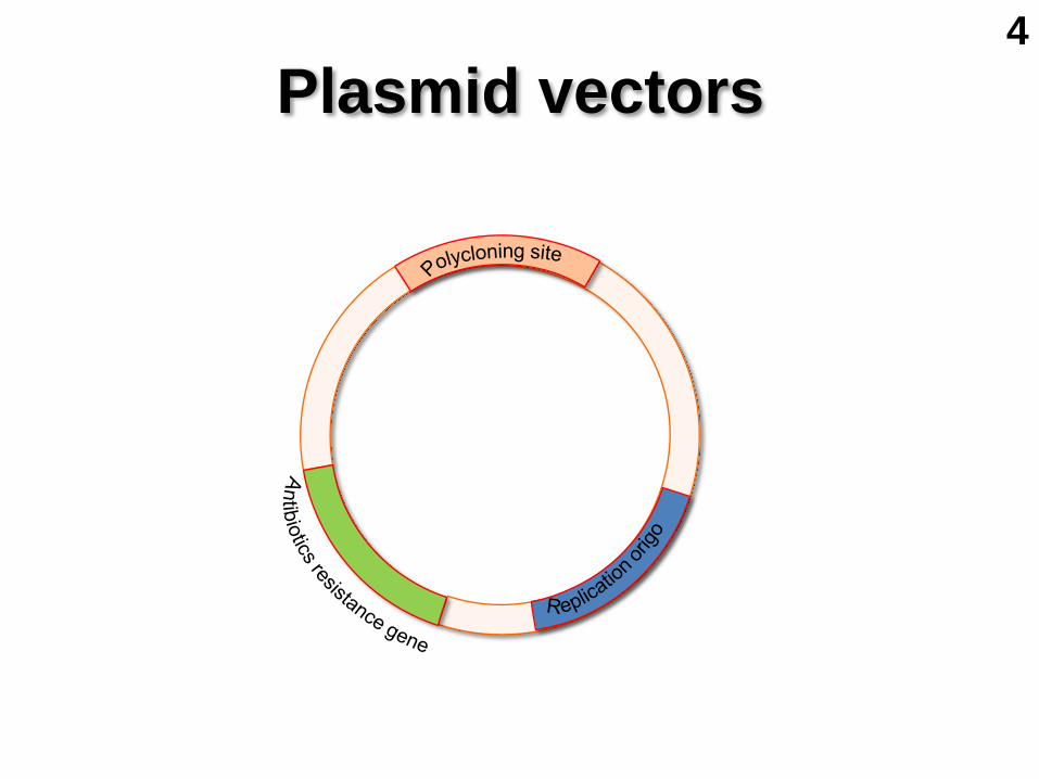
Plasmid vectors
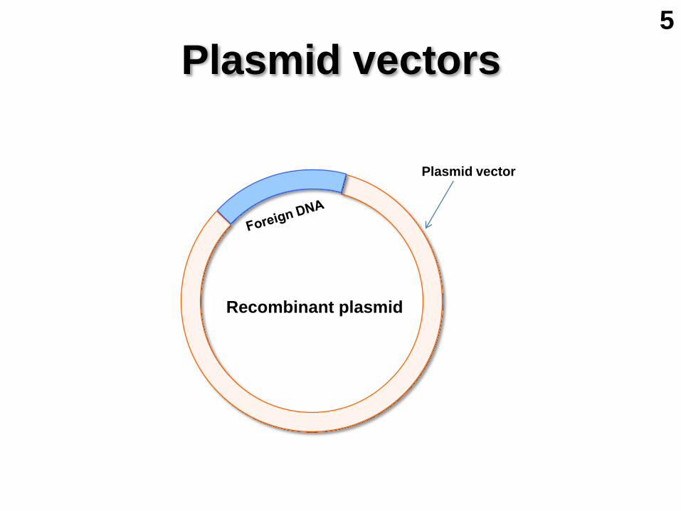
Plasmid vectors
Recombinant plasmid
Plasmid vector
5
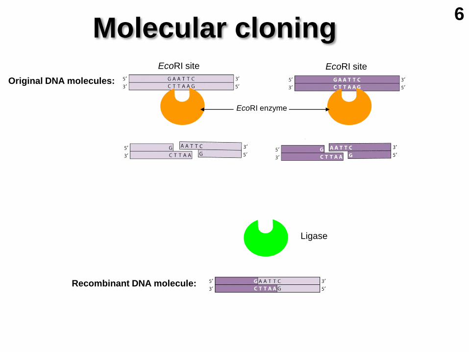
Molecular cloning
Original DNA molecules:
EcoRI site EcoRI site
EcoRI enzyme
Ligase
Recombinant DNA molecule:
6

Generation of recombinant
plasmid by DNA ligation
Transformation (delivery of DNA to cells)
Multiplication of plasmids
within the cells
Multiplication of
bacterial cells
A colony originates
from a single cell
Plasmid vector DNA fragment Recombinant plasmid
E. coli
Molecular cloning 7

DNA DNA lacZ lacI O lacY lacA C E P
rep
T
repressor CAP site pol-binding site operator structural genes terminator
lac operon
lactose
8
DNA DNA lacZ lacI O lacY lacA C E P rep
galactose
indoxyl
T
repressor CAP site pol-binding site operator structural genes terminator
IPTG
X-Gal
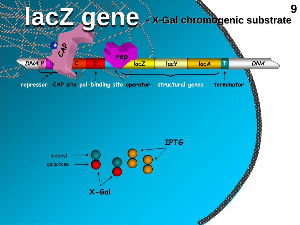
- X-Gal chromogenic substrate lacZ gene 9

DNS DNA lacZ lacI O lacY lacA C E P
trans- acetylase
permease
rep
-gal
T
pol
repressor CAP site pol-binding site operator structural genes terminator
lacZ gene - X-Gal chromogenic substrate 9

DNA DNA
lacZ lacI O lacY lacA C E P T
lacZ gene in cloning
lacZ -fragment
-fragment
lacZ- lacZ-
Foreign DNA
repressor CAP site pol-binding site operator structural genes terminator
10
5’- P P - 5’
- 5’
OH-3’
3’-HO
P
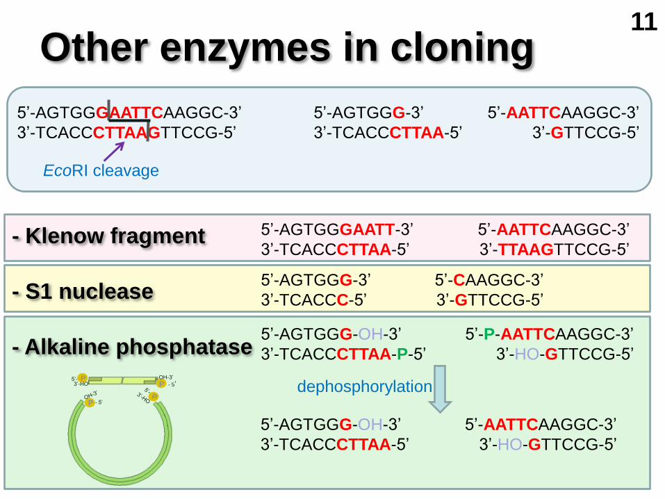
Other enzymes in cloning
- Klenow fragment
- S1 nuclease
- Alkaline phosphatase
5’-AGTGGGAATTCAAGGC-3’
3’-TCACCCTTAAGTTCCG-5’
5’-AGTGGG-3’ 5’-AATTCAAGGC-3’
3’-TCACCCTTAA-5’ 3’-GTTCCG-5’
5’-AGTGGGAATT-3’ 5’-AATTCAAGGC-3’
3’-TCACCCTTAA-5’ 3’-TTAAGTTCCG-5’
5’-AGTGGG-3’ 5’-CAAGGC-3’
3’-TCACCC-5’ 3’-GTTCCG-5’
5’-AGTGGG-OH-3’ 5’-P-AATTCAAGGC-3’
3’-TCACCCTTAA-P-5’ 3’-HO-GTTCCG-5’
EcoRI cleavage
5’-AGTGGG-OH-3’ 5’-AATTCAAGGC-3’
3’-TCACCCTTAA-5’ 3’-HO-GTTCCG-5’
dephosphorylation
11

1993
2. PCR: polymerase chain reaction
1. cycle
2. cycle
3. cycle
denaturation annealing synthesis
Forward primer
Reverse primer
12
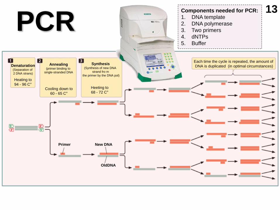
PCR
Denaturation (Separation of
2 DNA strans)
Heating to
94 - 96 C°
Annealing (primer binding to
single-stranded DNA
Cooling down to
60 - 65 C°
Synthesis (Synthesis of new DNA
strand fro m
the primer by the DNA pol)
Heeting to
68 - 72 C°
Each time the cycle is repeated, the amount of
DNA is duplicated (in optimal circumstances)
Primer New DNA
OldDNA
13 Components needed for PCR:
1. DNA template
2. DNA polymerase
3. Two primers
4. dNTPs
5. Buffer

3. Gel electrophoresis
a. Agarose gel electrophoresis
b. Polyacrilamide gel electrophoresis (PAGE)
14
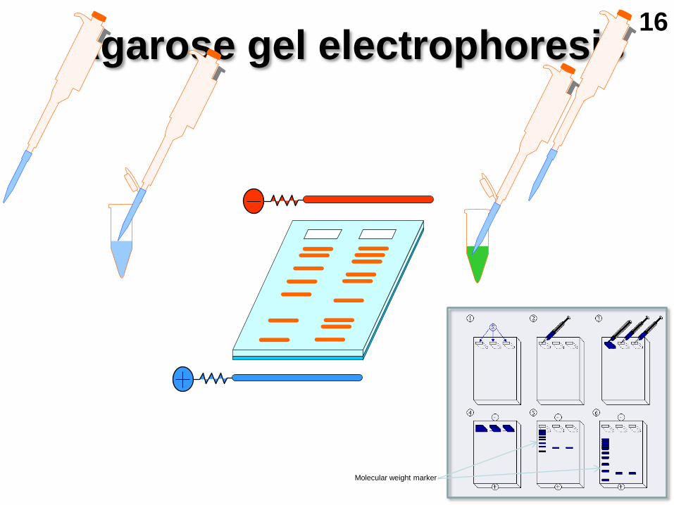
Agarose gel electrophoresis
Molecular weight marker
16

Fluorescence of DNA bands
UV UV UV
Soaking in Ethidium bromide
Agarose gel electrophoresis 16
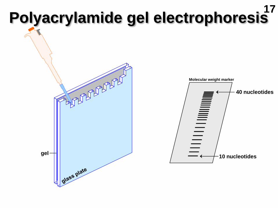
10 nucleotides
40 nucleotides
gel
Molecular weight marker
Polyacrylamide gel electrophoresis 17

Protein with two subunits
joined by a disulfide bridge Single subunit protein C
Heated with SDS and mercaptoethanol
C
B
A
SH
HS Negatively charged
SDS molecules
-S-S-
A B C
SDS-PAGE 18

4. Detection of macromolecules 19
a. Southern blot
b. Northern blot
c. Western blot
d. Eastern blot
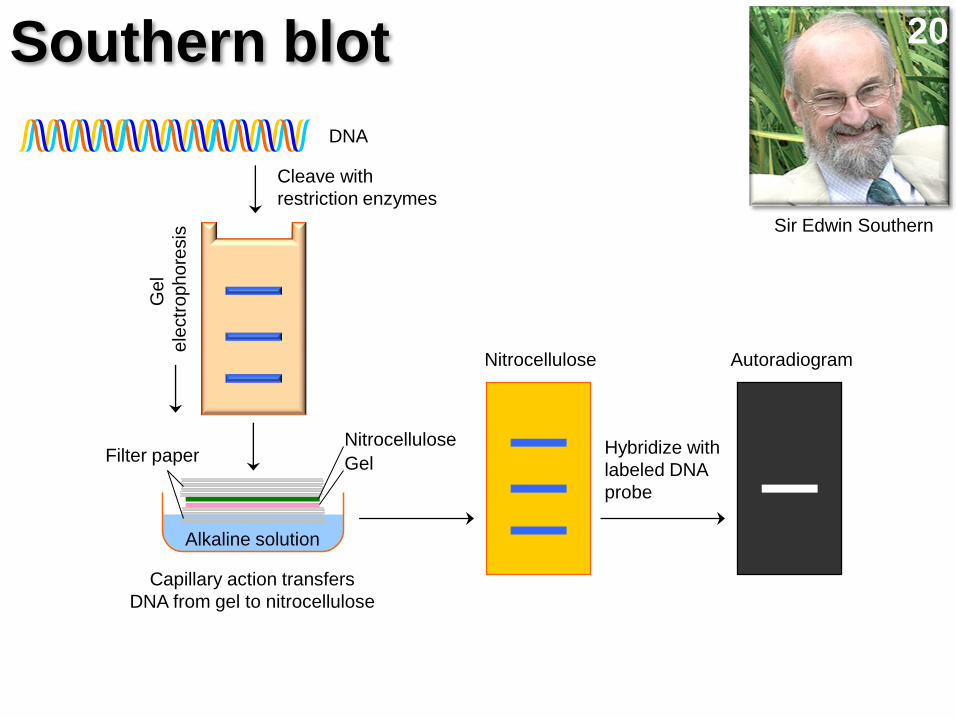
Cleave with
restriction enzymes
DNA G
el
ele
ctr
op
ho
resis
Alkaline solution
Capillary action transfers
DNA from gel to nitrocellulose
Nitrocellulose Autoradiogram
Hybridize with
labeled DNA
probe
Filter paper Nitrocellulose
Gel
Southern blot
Sir Edwin Southern
20

(A) Transfer of DNA from gel to membrane
(B) Hybridization analysis
Nylon membrane
Nylon membrane Autoradiograph
Hybridizing
bands
Labeled DNA probe
DNA
markers
Restricted
DNA
Agarose gel Support
Gel
Nylon membrane
Paper tower
Buffer
Southern blot 21
P
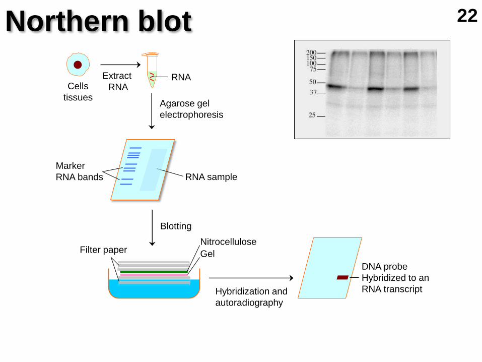
Extract
RNA Cells
tissues
Filter paper Nitrocellulose
Gel
RNA
Agarose gel
electrophoresis
Blotting
Hybridization and
autoradiography
DNA probe
Hybridized to an
RNA transcript
RNA sample
Marker
RNA bands
Northern blot 22

Cells
Extract RNA
Denaturing agarose gel electrophoresis
Blotting, northern hybridization,
autoradiography
RNA bands
DNA probe hybridizes to
a single RNA transcript
Northern blot 23
Electroblot
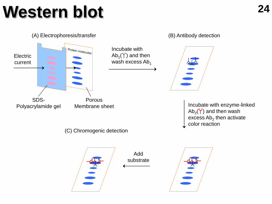
Incubate with
Ab1( ) and then
wash excess Ab1
Incubate with enzyme-linked
Ab2( ) and then wash
excess Ab2 then activate
color reaction
(A) Electrophoresis/transfer (B) Antibody detection
(C) Chromogenic detection
Add
substrate
Electric
current
SDS-
Polyacrylamide gel
Porous
Membrane sheet
Western blot 24

Retarded band
DNA markers
Restriction fragments Restriction fragments
+ nuclear protein
5. Gel retardation analyis Detection of DNA/protein interaction
25
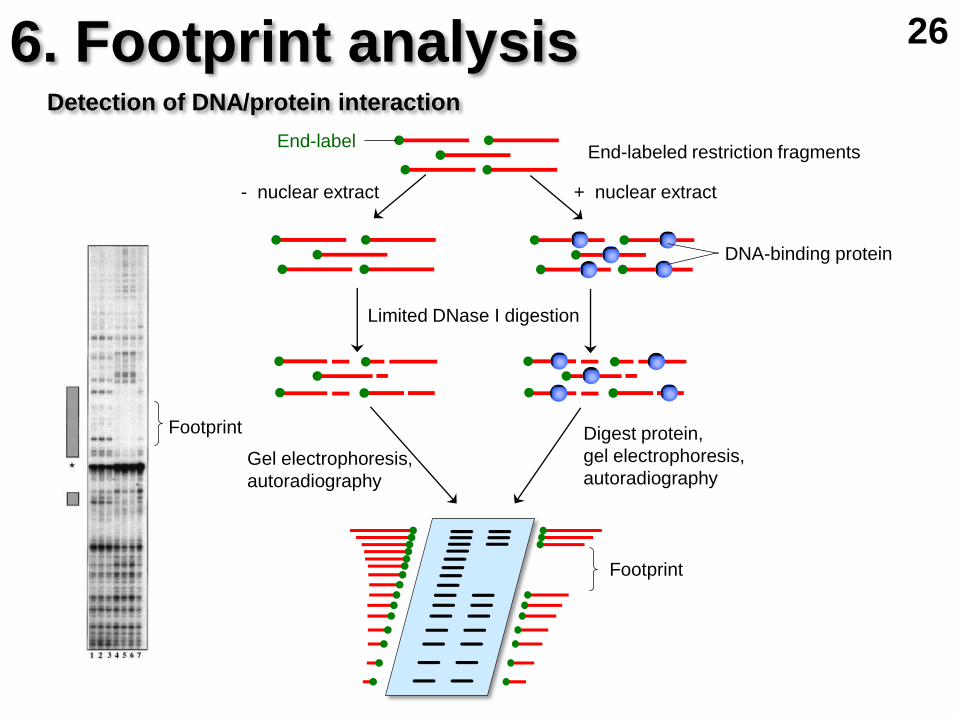
Footprint
End-label End-labeled restriction fragments
+ nuclear extract - nuclear extract
Limited DNase I digestion
Digest protein,
gel electrophoresis,
autoradiography Gel electrophoresis,
autoradiography
DNA-binding protein
6. Footprint analysis Detection of DNA/protein interaction
26
Footprint
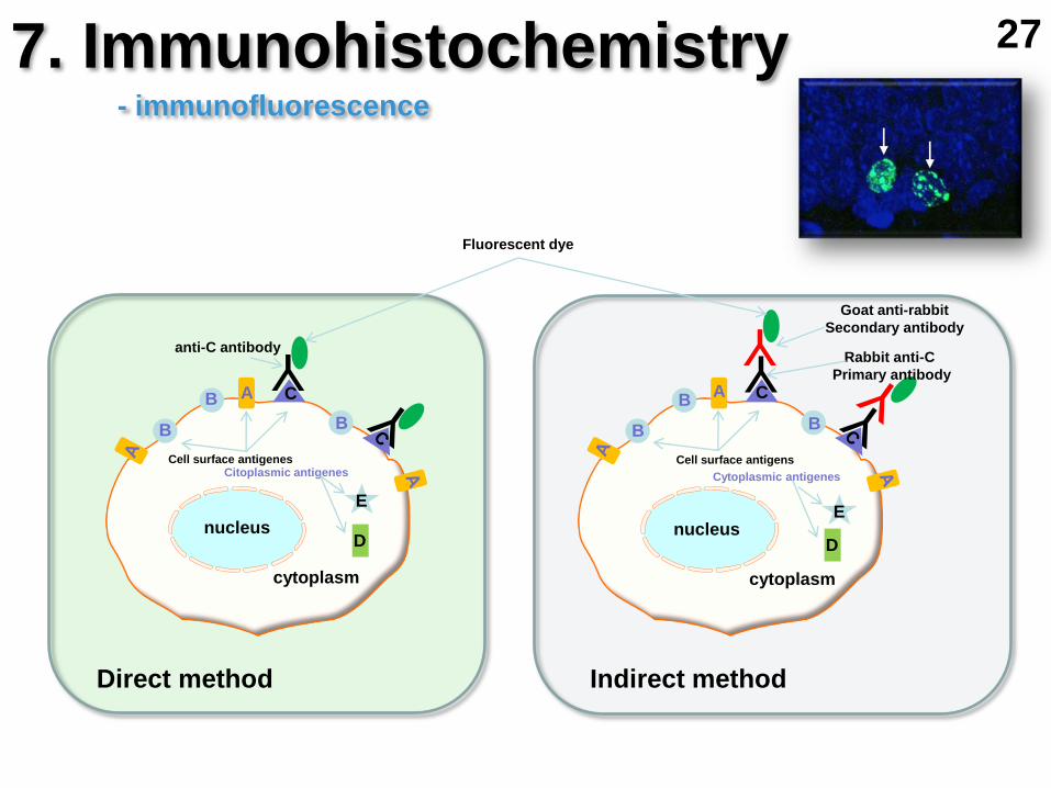
7. Immunohistochemistry
Y Y Y
nucleus nucleus
cytoplasm
Cell surface antigenes
anti-C antibody
Fluorescent dye
Rabbit anti-C
Primary antibody
Goat anti-rabbit
Secondary antibody
Direct method Indirect method
B
A B
B B
B
B
A C C
- immunofluorescence
Citoplasmic antigenes
cytoplasm
D
E
Cytoplasmic antigenes
D
E
Cell surface antigens
27

peroxidase
Y Y
nucleus
cytoplasm
Cell surfice antigens
peroxidase Rabbit anti-C
primary antibody
Goat anti-rabbit
secondary antibody
Indirect methods
B
B
B
A C
Y peroxidase
peroxidase
peroxidase
Y
avidin
- peroxidase
- biotin – avidin – peroxidase
Anti-rabbit antibody (secondary antibody)
- immunoperoxidase-based methods
DAB + H2O2 Ni-DAB + H2O2
cytoplazmic antigens
anti-rabbit antibody (secondary antibody)
D
E
Y anti-C antibody
Y
anti-C antibody
7. Immunohistochemistry 28
biotin

biotin
avidin
ABC method (ABC = avidin-biotin complex)
anti-rabbit antibody (secondary antibody)
Y
peroxidase
Y
primary antibody
antigen
7. Immunohistochemistry - immunoperoxidase-based methods
29
DAB + H2O2 Ni-DAB + H2O2
Y Y

Y Y
nucleus
cytoplasm
Alkaline phosphatase Rabbit anti-C
primary antibody
Goat anti-rabbit
secondary antibody
Indirect method
B
B
B
A C
- alkaline phosphatase
- digoxigenin – anti-digoxigenin – alkaline phosphatase
alk. phosph. Y
Sheep anti-rabbit antibody
alk. phosph
Y
DIG
anti-C antibody
Cell surfice antigens
cytoplasmic antigens
D
E
Y
Rabbit anti-C antibody
30
Y
alk. phosph
DIG
anti-C antitbody
Y
anti-rabbit antibody
7. Immunohistochemistry
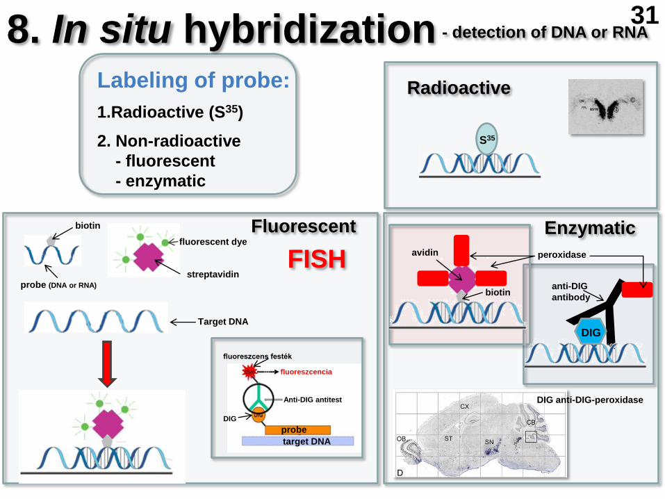
8. In situ hybridization
DIG anti-DIG-peroxidase
Radioactive
Enzymatic Fluorescent
Labeling of probe:
1.Radioactive (S35)
2. Non-radioactive
- fluorescent
- enzymatic
- detection of DNA or RNA
streptavidin
fluorescent dye
probe (DNA or RNA)
biotin
Target DNA
target DNA
probe
DIG
Anti-DIG antitest
fluoreszcens festék
fluoreszcencia
FISH peroxidase
DIG
biotin
avidin
anti-DIG
antibody
S35
31

slide
Dividing cells
Denaturation of DNA
Fluorescent
labeling
Fluorescent in situ hybridization
Metaphase chromosome
32

Fluorescence resonance energy transfer
Protein X Protein Y
No protein interaction Protein interaction
UV light
excitation
Blue light
emission Blue light
excitation
Yellow light
emission
Yellow light UV light UV light Blue light
Blue fluorescent
protein
Yellow fluorescent
protein
9. FRET 33

Sample
Sorter
Laser Flow cell
Scatter detectors
Laser
Band pass filters
Dichroic
filters
Photomultipliers
PMT4
PMT3
PMT1
PMT2
10. Flow cytometry 34

Fluorescence activated cell sorting
Waste
Sample
Collection tubes
Sheath fluid
Vibrating
flow cell
Deflection plates
Charging
collar
Laser
Beam
splitters
Red
fluorescence
Green
fluorescence
900 light
scatter
(granularity) Forward
light
scatter
(size)
11. FACS 35