Embed Size (px)
Citation preview
23 Amino Acids, Peptides, and Proteins
959
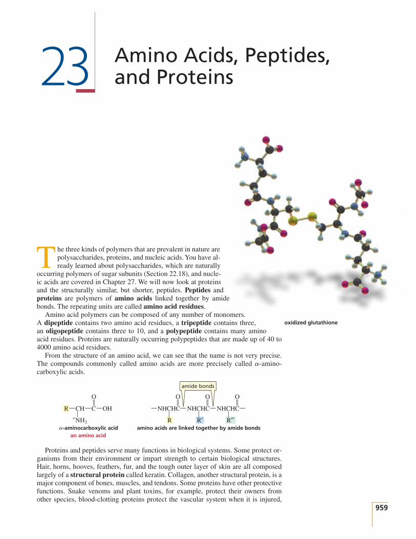
oxidized glutathione
The three kinds of polymers that are prevalent in nature arepolysaccharides, proteins, and nucleic acids. You have al-ready learned about polysaccharides, which are naturally
occurring polymers of sugar subunits (Section 22.18), and nucle-ic acids are covered in Chapter 27. We will now look at proteinsand the structurally similar, but shorter, peptides. Peptides andproteins are polymers of amino acids linked together by amidebonds. The repeating units are called amino acid residues.
Amino acid polymers can be composed of any number of monomers.A dipeptide contains two amino acid residues, a tripeptide contains three,an oligopeptide contains three to 10, and a polypeptide contains many aminoacid residues. Proteins are naturally occurring polypeptides that are made up of 40 to4000 amino acid residues.
From the structure of an amino acid, we can see that the name is not very precise.The compounds commonly called amino acids are more precisely called amino-carboxylic acids.
Proteins and peptides serve many functions in biological systems. Some protect or-ganisms from their environment or impart strength to certain biological structures.Hair, horns, hooves, feathers, fur, and the tough outer layer of skin are all composedlargely of a structural protein called keratin. Collagen, another structural protein, is amajor component of bones, muscles, and tendons. Some proteins have other protectivefunctions. Snake venoms and plant toxins, for example, protect their owners fromother species, blood-clotting proteins protect the vascular system when it is injured,
O O O O
CH
-aminocarboxylic acidan amino acid
R C OH NHCHC
R
NHCHC
R′
NHCHC
R′′amino acids are linked together by amide bonds
amide bonds
+NH3
a-
960 C H A P T E R 2 3 Amino Acids, Peptides, and Proteins
and antibodies and protein antibiotics protect us from disease. A group of proteinscalled enzymes catalyzes the chemical reactions that occur in living systems, andsome of the hormones that regulate these reactions are peptides. Proteins are also re-sponsible for many physiological functions, such as the transport and storage of oxy-gen in the body and the contraction of muscles.
23.1 Classification and Nomenclature of Amino Acids
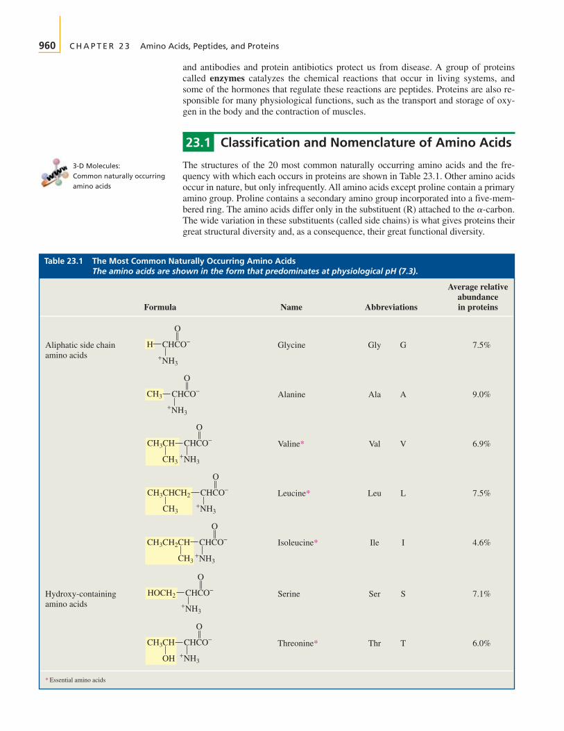
The structures of the 20 most common naturally occurring amino acids and the fre-quency with which each occurs in proteins are shown in Table 23.1. Other amino acidsoccur in nature, but only infrequently. All amino acids except proline contain a primaryamino group. Proline contains a secondary amino group incorporated into a five-mem-bered ring. The amino acids differ only in the substituent (R) attached to the The wide variation in these substituents (called side chains) is what gives proteins theirgreat structural diversity and, as a consequence, their great functional diversity.
a-carbon.
Table 23.1 The Most Common Naturally Occurring Amino Acids The amino acids are shown in the form that predominates at physiological pH (7.3).
Average relativeabundance
Formula Name Abbreviations in proteins
Aliphatic side chainamino acids
Glycine Gly G 7.5%
Alanine Ala A 9.0%
Valine* Val V 6.9%
Leucine* Leu L 7.5%
Isoleucine* Ile I 4.6%
Hydroxy-containingamino acids
Serine Ser S 7.1%
Threonine* Thr T 6.0%
* Essential amino acids
O
+NH3
CHCO−CH3CH
OH
O
+NH3
CHCO−HOCH2
O
+NH3
CHCO−CH3CH2CH
CH3
O
+NH3
CHCO−CH3CHCH2
CH3
O
+NH3
CHCO−CH3CH
CH3
O
+NH3
CHCO−CH3
H CHCO−
O
+NH3
3-D Molecules:Common naturally occurringamino acids

Section 23.1 Classification and Nomenclature of Amino Acids 961
Sulfur-containingamino acids
Cysteine Cys C 2.8%
Methionine* Met M 1.7%
Acidic amino acids Aspartate Asp D 5.5%(aspartic acid)
Glutamate Glu E 6.2%(glutamic acid)
Amides of acidicamino acids
Asparagine Asn N 4.4%
Glutamine Gln Q 3.9%
Basic amino acids Lysine* Lys K 7.0%
Arginine* Arg R 4.7%
Benzene-containingamino acids
Phenylalanine* Phe F 3.5%
Tyrosine Tyr Y 3.5%
Heterocylicamino acids
Proline Pro P 4.6%
* Essential amino acids
O
CO−
N
Η Η+
+NH3
O
CHCO−CH2HO
+NH3
O
CHCO−CH2
+NH3
O
CHCO−
NH2
H2NCNHCH2CH2CH2
+
+NH3
O
CHCO−H3NCH2CH2CH2CH2+
+NH3
OO
CHCO−H2NCCH2CH2
+NH3
OO
CHCO−H2NCCH2
+NH3
CHCO−−OCCH2CH2
OO
OO
+NH3
CHCO−−OCCH2
O
+NH3
CHCO−CH3SCH2CH2
O
+NH3
CHCO−HSCH2
Average relativeabundance
Formula Name Abbreviations in proteins
Table 23.1 (continued)

Heterocyclicamino acids(continued)
Histidine* His H 2.1%
Tryptophan* Trp W 1.1%
* Essential amino acids
O
+NH3
CHCO−CH2
NH
O
CHCO−CH2
N NH+NH3
962 C H A P T E R 2 3 Amino Acids, Peptides, and Proteins
The amino acids are almost always called by their common names. Often, the nametells you something about the amino acid. For example, glycine got its name becauseof its sweet taste (glykos is Greek for “sweet”), and valine, like valeric acid, has fivecarbon atoms. Asparagine was first found in asparagus, and tyrosine was isolated fromcheese (tyros is Greek for “cheese”).
Dividing the amino acids into classes makes them easier to learn. The aliphatic sidechain amino acids include glycine, the amino acid in which and four amino acidswith alkyl side chains. Alanine is the amino acid with a methyl side chain, and valine hasan isopropyl side chain. Can you guess which amino acid—leucine or isoleucine—has anisobutyl side chain? If you gave the obvious answer, you guessed incorrectly. Isoleucinedoes not have an “iso” group; it is leucine that has an isobutyl substituent—isoleucine hasa sec-butyl substituent. Each of the amino acids has both a three-letter abbreviation (thefirst three letters of the name in most cases) and a single-letter abbreviation.
Two amino acid side chains—serine and threonine—contain alcohol groups. Serineis an HO-substituted alanine and threonine has a branched ethanol substituent. Thereare also two sulfur-containing amino acids: Cysteine is an HS-substituted alanine andmethionine has a 2-methylthioethyl substituent.
There are two acidic amino acids (amino acids with two carboxylic acid groups):aspartate and glutamate. Aspartate is a carboxy-substituted alanine and glutamate hasone more methylene group than aspartate. (If their carboxyl groups are protonated,they are called aspartic acid and glutamic acid, respectively.) Two amino acids—asparagine and glutamine—are amides of the acidic amino acids; asparagine is theamide of aspartate and glutamine is the amide of glutamate. Notice that the obviousone-letter abbreviations cannot be used for these four amino acids because A and G areused for alanine and glycine. Aspartic acid and glutamic acid are abbreviated D and E,and asparagine and glutamine are abbreviated N and Q.
There are two basic amino acids (amino acids with two basic nitrogen-containinggroups): lysine and arginine. Lysine has an group and arginine has a -guanidinogroup. At physiological pH, these groups are protonated. The and can remind you howmany methylene groups each amino acid has.
O
H3N+NH3
+NH3
CH2CH2CH2CH2CHCO− H2N C CH2CH2CH2CHCO−NH
O+NH2+
lysine
an -amino group
arginine
a -guanidino group
dPdP-amino
R = H,
glycine
leucine
Average relativeabundance
Formula Name Abbreviations in proteins
Table 23.1 (continued)

NH
indole
Section 23.1 Classification and Nomenclature of Amino Acids 963
Two amino acids—phenylalanine and tyrosine—contain benzene rings. As its nameindicates, phenylalanine is phenyl-substituted alanine. Tyrosine is phenylalanine witha para-hydroxy substituent.
Proline, histidine, and tryptophan are heterocyclic amino acids. Proline has its nitro-gen incorporated into a five-membered ring—it is the only amino acid that contains asecondary amino group. Histidine is an imidazole-substituted alanine. Imidazole is anaromatic compound because it is cyclic and planar and has three pairs of delocalized electrons (Section 21.11). The of a protonated imidazole ring is 6.0, so the ring willbe protonated in acidic solutions and nonprotonated in basic solutions (Section 23.3).
Tryptophan is an indole-substituted alanine (Section 21.11). Like imidazole, indole isan aromatic compound. Because the lone pair on the nitrogen atom of indole is need-ed for the compound’s aromaticity, indole is a very weak base. (The of protonatedindole is ) Therefore, the ring nitrogen in tryptophan is never protonated underphysiological conditions.
Ten amino acids are essential amino acids. We humans must obtain these 10essential amino acids from our diets because we either cannot synthesize them at allor cannot synthesize them in adequate amounts. For example, we must have a dietarysource of phenylalanine because we cannot synthesize benzene rings. However, we donot need tyrosine in our diets, because we can synthesize the necessary amounts fromphenylalanine. The essential amino acids are denoted by red asterisks (*) inTable 23.1. Although humans can synthesize arginine, it is needed for growth ingreater amounts than can be synthesized. So arginine is an essential amino acid forchildren, but a nonessential amino acid for adults. Not all proteins contain the sameamino acids. Bean protein is deficient in methionine, for example, and wheat protein isdeficient in lysine. They are incomplete proteins: They contain too little of one or moreessential amino acids to support growth. Therefore, a balanced diet must contain pro-teins from different sources.
Dietary protein is hydrolyzed in the body to individual amino acids. Some of theseamino acids are used to synthesize proteins needed by the body, some are broken downfurther to supply energy to the body, and some are used as starting materials for thesynthesis of nonprotein compounds the body needs, such as adrenaline, thyroxine, andmelanin (Section 25.6).
PROBLEM 1
a. Explain why, when the imidazole ring of histidine is protonated, the double-bondednitrogen is the nitrogen that accepts the proton.
b. Explain why, when the guanidino group of arginine is protonated, the double-bondednitrogen is the nitrogen that accepts the proton.
2 H++
NH2
H2NCNHCH2CH2CH2CHCO−
ONH
+NH3
H2NCNHCH2CH2CH2CHCO−
O+NH2
2 H++ HN NH+
NHNNH2
CH2CHCOO−
+NH3
CH2CHCOO−
-2.4.pKa
HN NH H+++
NHN
protonated imidazole imidazole
pKa
p
aspartate
lysine
Tutorial:Basic nitrogens in histidineand arginine

964 C H A P T E R 2 3 Amino Acids, Peptides, and Proteins
23.2 Configuration of Amino Acids
The of all the naturally occurring amino acids except glycine is an asymmet-ric carbon. Therefore, 19 of the 20 amino acids listed in Table 23.1 can exist as enan-tiomers. The D and L notation used for monosaccharides (Section 22.2) is also used foramino acids. The D and L isomers of monosaccharides and amino acids are defined thesame way. Thus, an amino acid drawn in a Fischer projection with the carboxyl groupon the top and the R group on the bottom of the vertical axis is a D-amino acid if theamino group is on the right and an L-amino acid if the amino group is on the left. Un-like monosaccharides, where the D isomer is the one found in nature, most amino acidsfound in nature have the L configuration. To date, D-amino acid residues have beenfound only in a few peptide antibiotics and in some small peptides attached to the cellwalls of bacteria.
Why D-sugars and L-amino acids? While it makes no difference which isomer nature“selected” to be synthesized, it is important that the same isomer be synthesized by allorganisms. For example, if mammals ended up having L-amino acids, then L-aminoacids would need to be the isomers synthesized by the organisms upon which mammalsdepend for food.
C H
CH2OH
O
OHH
C O−
R
O
NH3+H
C H
CH2OH
O
HHO
C O−
R
O
HH3N+
D-glyceraldehyde
D-amino acid
L-glyceraldehyde
L-amino acid
a-carbon
AMINO ACIDS AND DISEASE
The Chamorro people of Guam have a high inci-dence of a syndrome that resembles amyotrophic
lateral sclerosis (ALS) with elements of Parkinson’s diseaseand dementia. This syndrome developed during World War IIwhen, as a result of food shortages, the tribe ate large quantities
of Cycas circinalis seeds. These seeds contain -methylamino-L-alanine, an amino acid that binds to glutamate receptors.When monkeys are given -methylamino-L-alanine, they de-velop some of the features of this syndrome. There is hope that,by studying the mechanism of action of -methylamino-L-alanine, we may gain an understanding of how ALS andParkinson’s disease arise.
b
b
b
alaninean amino acid
PROBLEM 2◆
a. Which isomer—(R)-alanine or (S)-alanine—is D-alanine?b. Which isomer—(R)-aspartate or (S)-aspartate—is D-aspartate?c. Can a general statement be made relating R and S to D and L?
PROBLEM 3◆
Which amino acids in Table 23.1 have more than one asymmetric carbon?

Section 23.3 Acid–Base Properties of Amino Acids 965
Recall from the Henderson–Hasselbalchequation (Section 1.20) that the acidicform predominates if the pH of the solu-tion is less than the of the com-pound and the basic form predominatesif the pH of the solution is greater thanthe of the compound.pKa
pKa
Table 23.2 The Values of Amino AcidspKa
Amino acid -COOH - side chain
Alanine 2.34 9.69 —
Arginine 2.17 9.04 12.48
Asparagine 2.02 8.84 —
Aspartic acid 2.09 9.82 3.86
Cysteine 1.92 10.46 8.35
Glutamic acid 2.19 9.67 4.25
Glutamine 2.17 9.13 —
Glycine 2.34 9.60 —
Histidine 1.82 9.17 6.04
Isoleucine 2.36 9.68 —
Leucine 2.36 9.60 —
Lysine 2.18 8.95 10.79
Methionine 2.28 9.21 —
Phenylalanine 2.16 9.18 —
Proline 1.99 10.60 —
Serine 2.21 9.15 —
Threonine 2.63 9.10 —
Tryptophan 2.38 9.39 —
Tyrosine 2.20 9.11 10.07
Valine 2.32 9.62 —
NH3�AA
pKapKapKa
23.3 Acid–Base Properties of Amino Acids
Every amino acid has a carboxyl group and an amino group, and each group can existin an acidic form or a basic form, depending on the pH of the solution in which theamino acid is dissolved. The carboxyl groups of the amino acids have values ofapproximately 2, and the protonated amino groups have values near 9(Table 23.2). Both groups, therefore, will be in their acidic forms in a very acidic solu-tion At the pH of the solution is greater than the of the car-boxyl group, but less than the of the protonated amino group. The carboxyl group,therefore, will be in its basic form and the amino group will be in its acidic form. In astrongly basic solution both groups will be in their basic forms.
Notice that an amino acid can never exist as an uncharged compound, regardless ofthe pH of the solution. To be uncharged, an amino acid would have to lose a protonfrom an group with a of about 9 before it would lose a proton from a COOHgroup with a of about 2. This clearly is impossible: A weak acid cannot be moreacidic than a strong acid. Therefore, at physiological pH (7.3) an amino acid exists as adipolar ion, called a zwitterion. A zwitterion is a compound that has a negative charge
pKa
pKa+NH3
H++
R CH C OH R
a zwitterionpH = 7
CH C O−
H++
R
OOO
NH2
CH C O−
pH = 0 pH = 11
+NH3+NH3
(pH ' 11),
pKa
pKapH = 7,(pH ' 0).
pKa
pKa

966 C H A P T E R 2 3 Amino Acids, Peptides, and Proteins
on one atom and a positive charge on a nonadjacent atom. (The name comes fromzwitter, German for “hermaphrodite” or “hybrid.”)
A few amino acids have side chains with ionizable hydrogens (Table 23.2). Theprotonated imidazole side chain of histidine, for example, has a of 6.04. Histidine,therefore, can exist in four different forms, and the form that predominates depends onthe pH of the solution.
PROBLEM 4◆
Why are the carboxylic acid groups of the amino acids so much more acidic than a carboxylic acid such as acetic acid
PROBLEM 5 SOLVED
Draw the form in which each of the following amino acids predominantly exists at physio-logical pH (7.3):
a. aspartic acid c. glutamine e. arginine
b. histidine d. lysine f. tyrosine
SOLUTION TO 5a Both carboxyl groups are in their basic forms because the pH isgreater than their . The protonated amino group is in its acidic form because the pHis less than its
PROBLEM 6◆
Draw the form in which glutamic acid predominantly exists in a solution with the follow-ing pH:
a. b. c. d.
PROBLEM 7
a. Why is the of the glutamic acid side chain greater than the of the aspartic acidside chain?
b. Why is the of the arginine side chain greater than the of the lysine side chain?
23.4 The Isoelectric Point
The isoelectric point (pI) of an amino acid is the pH at which it has no net charge. Inother words, it is the pH at which the amount of positive charge on an amino acidexactly balances the amount of negative charge:
pI (isoelectric point) � pH at which there is no net charge
pKapKa
pKapKa
pH = 11pH = 6pH = 3pH = 0
+NH3
O O−OCCH2CHCO−
pKa .pKa’s
(pKa = 4.76)?(pKa
' 2)
pKa
+NH3+NH3
+NH3HN NH
CH2CHCOH
+HN NH
CH2CHCO−
+N NH
CH2CHCO−
N NH
CH2CHCO−
NH2
O O O O
pH = 0 pH = 4 pH = 8 pH = 12
histidine
Section 23.4 The Isoelectric Point 967
Recall from the Henderson–Hasselbalchequation that when , half thegroup is in its acidic form and half is inits basic form (Section 1.20).
pH � pKa
An amino acid will be positively chargedif the pH of the solution is less than thepI of the amino acid and will be nega-tively charged if the pH of the solutionis greater than the pI of the amino acid.
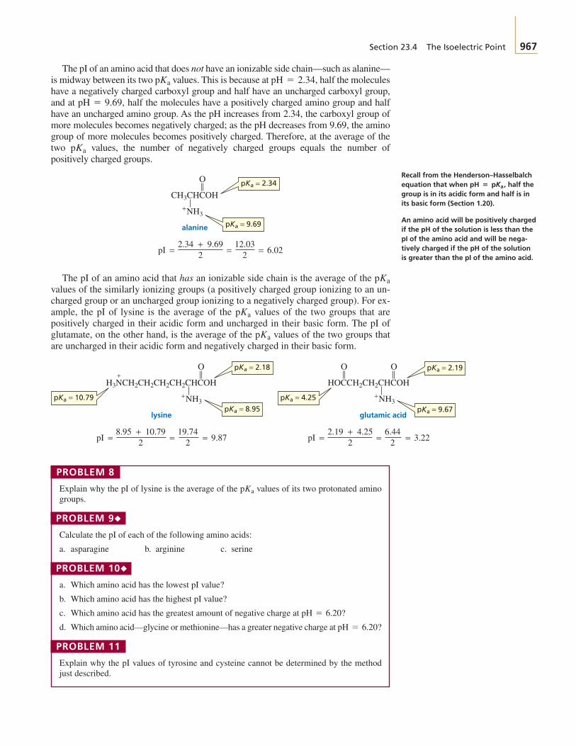
The pI of an amino acid that does not have an ionizable side chain—such as alanine—is midway between its two values. This is because at half the moleculeshave a negatively charged carboxyl group and half have an uncharged carboxyl group,and at half the molecules have a positively charged amino group and halfhave an uncharged amino group. As the pH increases from 2.34, the carboxyl group ofmore molecules becomes negatively charged; as the pH decreases from 9.69, the aminogroup of more molecules becomes positively charged. Therefore, at the average of thetwo values, the number of negatively charged groups equals the number ofpositively charged groups.
The pI of an amino acid that has an ionizable side chain is the average of the values of the similarly ionizing groups (a positively charged group ionizing to an un-charged group or an uncharged group ionizing to a negatively charged group). For ex-ample, the pI of lysine is the average of the values of the two groups that arepositively charged in their acidic form and uncharged in their basic form. The pI ofglutamate, on the other hand, is the average of the values of the two groups thatare uncharged in their acidic form and negatively charged in their basic form.
PROBLEM 8
Explain why the pI of lysine is the average of the values of its two protonated aminogroups.
PROBLEM 9◆
Calculate the pI of each of the following amino acids:
a. asparagine b. arginine c. serine
PROBLEM 10◆
a. Which amino acid has the lowest pI value?
b. Which amino acid has the highest pI value?
c. Which amino acid has the greatest amount of negative charge at
d. Which amino acid—glycine or methionine—has a greater negative charge at
PROBLEM 11
Explain why the pI values of tyrosine and cysteine cannot be determined by the methodjust described.
pH = 6.20?
pH = 6.20?
pKa
pKa
pKa
pKa
+NH3
O
= = 6.02=pI2.34 + 9.69
2
CH3CHCOH
12.032
pKa = 9.69
pKa = 2.34
alanine
pKa
pH = 9.69,
pH = 2.34,pKa
+NH3+NH3
pKa = 9.67pKa = 8.95
pKa = 2.19pKa = 2.18
pKa = 4.25pKa = 10.79
+O
= = 9.87=pI8.95 + 10.79
2
H3NCH2CH2CH2CH2CHCOH
19.742
OO
= = 3.22=pI2.19 + 4.25
2
HOCCH2CH2CHCOH
6.442
lysine glutamic acid
968 C H A P T E R 2 3 Amino Acids, Peptides, and Proteins
23.5 Separation of Amino Acids
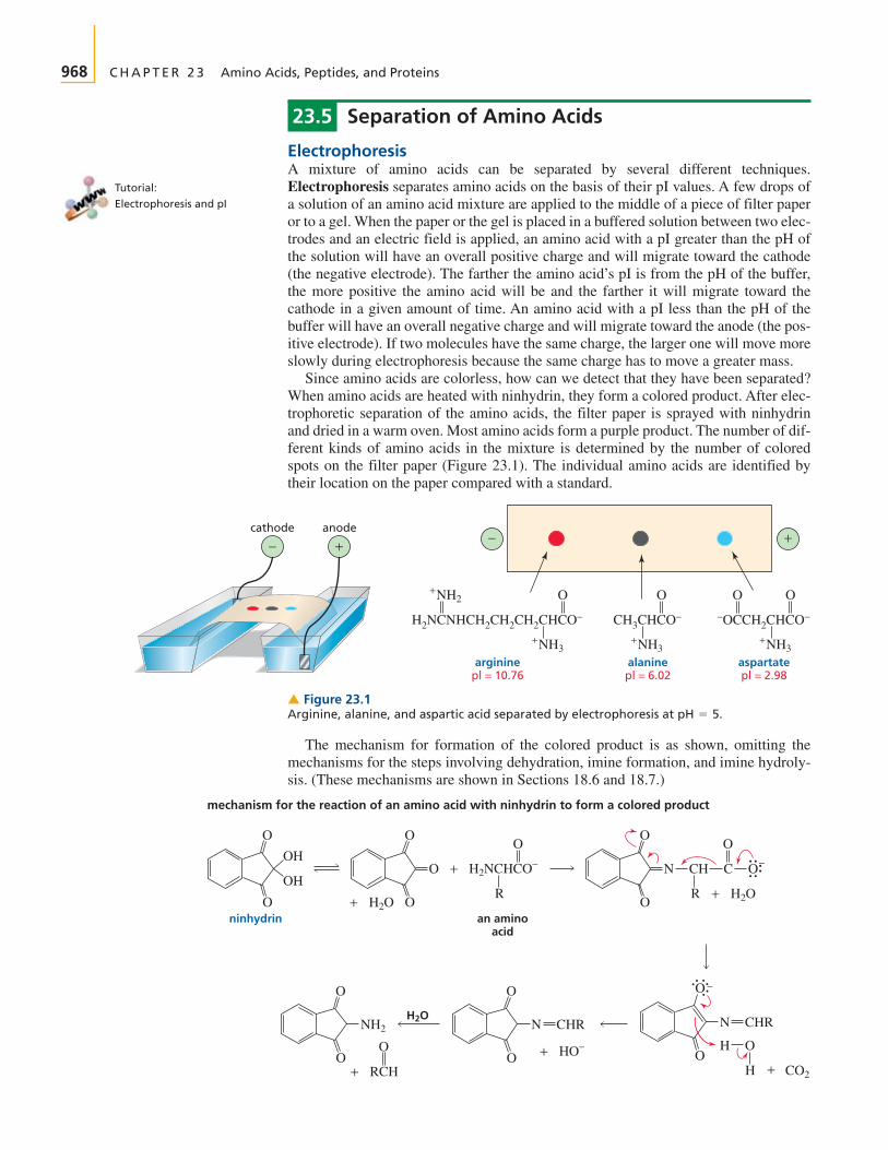
ElectrophoresisA mixture of amino acids can be separated by several different techniques.Electrophoresis separates amino acids on the basis of their pI values. A few drops ofa solution of an amino acid mixture are applied to the middle of a piece of filter paperor to a gel. When the paper or the gel is placed in a buffered solution between two elec-trodes and an electric field is applied, an amino acid with a pI greater than the pH ofthe solution will have an overall positive charge and will migrate toward the cathode(the negative electrode). The farther the amino acid’s pI is from the pH of the buffer,the more positive the amino acid will be and the farther it will migrate toward thecathode in a given amount of time. An amino acid with a pI less than the pH of thebuffer will have an overall negative charge and will migrate toward the anode (the pos-itive electrode). If two molecules have the same charge, the larger one will move moreslowly during electrophoresis because the same charge has to move a greater mass.
Since amino acids are colorless, how can we detect that they have been separated?When amino acids are heated with ninhydrin, they form a colored product. After elec-trophoretic separation of the amino acids, the filter paper is sprayed with ninhydrinand dried in a warm oven. Most amino acids form a purple product. The number of dif-ferent kinds of amino acids in the mixture is determined by the number of coloredspots on the filter paper (Figure 23.1). The individual amino acids are identified bytheir location on the paper compared with a standard.
+NH3+NH3
+NH3
+NH2
−OCCH2CHCO−CH3CHCO−H2NCNHCH2CH2CH2CHCO−
−− ++
O O O O
cathode anode
aspartatepl = 2.98
alaninepl = 6.02
argininepl = 10.76
▲ Figure 23.1Arginine, alanine, and aspartic acid separated by electrophoresis at pH = 5.
Tutorial:Electrophoresis and pI
O O
R
an aminoacid
R
H2NCHCO−+O N CH
O
OH
OH
N CHR
O −
HO
O
O
O
O
O
O
O
O
O
H CO2+
H2O+H2O+
O
C O−
mechanism for the reaction of an amino acid with ninhydrin to form a colored product
O
H2ON CHR
HO−+RCH+
NH2
ninhydrin
The mechanism for formation of the colored product is as shown, omitting themechanisms for the steps involving dehydration, imine formation, and imine hydroly-sis. (These mechanisms are shown in Sections 18.6 and 18.7.)

Section 23.5 Separation of Amino Acids 969
Ch
rom
ato
gra
ph
y Leu
Ala
Glumost polar amino acid
least polar amino acid
origin
> Figure 23.2Separation of glutamate, alanine,and leucine by paperchromatography.
Movie:Column chromatography
Paper Chromatography and Thin-Layer ChromatographyPaper chromatography once played an important role in biochemical analysis be-cause it provided a method for separating amino acids using very simple equipment.Although more modern techniques are now more commonly used, we will describethe principles behind paper chromatography because many of the same principles areemployed in modern separation techniques.
The technique of paper chromatography separates amino acids on the basis of po-larity. A few drops of a solution of an amino acid mixture are applied to the bottom ofa strip of filter paper. The edge of the paper is then placed in a solvent (typically a mix-ture of water, acetic acid, and butanol). The solvent moves up the paper by capillaryaction, carrying the amino acids with it. Depending on their polarities, the amino acidshave different affinities for the mobile (solvent) and stationary (paper) phases andtherefore travel up the paper at different rates. The more polar the amino acid, themore strongly it is adsorbed onto the relatively polar paper. The less polar amino acidstravel up the paper more rapidly, since they have a greater affinity for the mobilephase. Therefore, when the paper is developed with ninhydrin, the colored spot closestto the origin is the most polar amino acid and the spot farthest away from the origin isthe least polar amino acid (Figure 23.2).
O
O
O
O
O
O
O
N
O
O
O
H2O+
O−
N
purple-colored product
H2O+
H
O−H
The most polar amino acids are those with charged side chains, the next most polarare those with side chains that can form hydrogen bonds, and the least polar are thosewith hydrocarbon side chains. For amino acids with hydrocarbon side chains, the larg-er the alkyl group, the less polar the amino acid. In other words, leucine is less polarthan valine.
Paper chromatography has largely been replaced by thin-layer chromatography(TLC). Similar to paper chromatography, TLC differs from it in that TLC uses a platewith a coating of solid material instead of filter paper. The physical property on whichthe separation is based depends on the solid material and the solvent chosen for themobile phase.
PROBLEM 12◆
A mixture of seven amino acids (glycine, glutamate, leucine, lysine, alanine, isoleucine,and aspartate) is separated by TLC. Explain why only six spots show up when the chro-matographic plate is sprayed with ninhydrin and heated.

970 C H A P T E R 2 3 Amino Acids, Peptides, and Proteins
WATER SOFTENERS:EXAMPLES OF CATION-EXCHANGE CHROMATOGRAPHY
Water softeners contain a column with a cation-exchange resinthat has been flushed with concentrated sodium chloride. InSection 17.13, we saw that the presence of calcium and magne-
sium ions in water is what causes the water to be “hard.” Whenwater passes through the column, the resin binds magnesiumand calcium ions more tightly than it binds sodium ions. In thisway, the water softener removes magnesium and calcium ionsfrom water, replacing them with sodium ions. The resin must berecharged from time to time by flushing it with concentratedsodium chloride to replace the bound magnesium and calciumions with sodium ions.
SO3 Na+ SO3 Na+ SO3 Na+
SO3 Na+SO3 Na+
CH CH2
CH CH2CH CH2
CHCH2 CH CH2CHCH2
CH2
CHCH2
CH CH2 CH CH2 CH CH2 CH
CH CH2 CH
Figure 23.3 NA section of a cation-exchangeresin. This particular resin is calledDowex 50.®
Cations bind most stronglyto cation-exchange resins.
Anions bind most stronglyto anion-exchange resins.
Ion-Exchange ChromatographyElectrophoresis and thin-layer chromatography are analytical separations—smallamounts of amino acids are separated for analysis. Preparative separation, in whichlarger amounts of amino acids are separated for use in subsequent processes, can beachieved using ion-exchange chromatography. This technique employs a columnpacked with an insoluble resin. A solution of a mixture of amino acids is loaded ontothe top of the column and eluted with a buffer. The amino acids separate because theyflow through the column at different rates, as explained below.
The resin is a chemically inert material with charged side chains. One commonlyused resin is a copolymer of styrene and divinylbenzene with negatively charged sul-fonic acid groups on some of the benzene rings (Figure 23.3). If a mixture of lysineand glutamate in a solution with a pH of 6 were loaded onto the column, glutamatewould travel down the column rapidly because its negatively charged side chainwould be repelled by the negatively charged sulfonic acid groups of the resin. Thepositively charged side chain of lysine, on the other hand, would cause that aminoacid to be retained on the column. This kind of resin is called a cation-exchangeresin because it exchanges the counterions of the groups for the positivelycharged species that are added to the column. In addition, the relatively nonpolarnature of the column causes it to retain nonpolar amino acids longer than polar aminoacids. Resins with positively charged groups are called anion-exchange resinsbecause they impede the flow of anions by exchanging their negatively charged coun-terions for negatively charged species that are added to the column. A common anion-exchange resin (Dowex®1) has groups in place of the groups in Figure 23.3.
SO3-Na+CH2N+(CH3)3Cl-
SO3-Na+
An amino acid analyzer is an instrument that automates ion-exchange chromatog-raphy. When a solution of an amino acid mixture passes through the column of anamino acid analyzer containing a cation-exchange resin, the amino acids movethrough the column at different rates, depending on their overall charge. The solutionleaving the column is collected in fractions, which are collected often enough that adifferent amino acid ends up in each fraction (Figure 23.4). If ninhydrin is added toeach of the fractions, the concentration of the amino acid in each fraction can be

Section 23.5 Separation of Amino Acids 971
Fractions sequentially collected
> Figure 23.4Separation of amino acids byion-exchange chromatography.
MetAsp
Thr
Val
Tyr
Lys
Ser Glu AlaIle
Leu Phe
Arg
40 80 120 160
Gly
Pro
HisNH3
200 240 280 320
Effluent (mL)
Ab
sorb
ance
330 370 410 450 490 9050 130
pH 3.3 buffer pH 4.3 bufferpH 5.3 buffer > Figure 23.5
A typical chromatogram obtainedfrom the separation of a mixture ofamino acids using an automatedamino acid analyzer.
determined by the amount of absorption at 570 nm—because the colored compoundformed by the reaction of an amino acid with ninhydrin has a of 570(Section 8.11). In this way, the identity and the relative amount of each amino acid canbe determined (Figure 23.5).
lmax
PROBLEM 13
Why are buffer solutions of increasingly higher pH used to elute the column that generatesthe chromatogram shown in Figure 23.5?
PROBLEM 14
Explain the order of elution (with a buffer of pH 4) of each of the following pairs of aminoacids on a column packed with Dowex 50 (Figure 23.3):
a. aspartate before serine c. valine before leucineb. glycine before alanine d. tyrosine before phenylalanine
PROBLEM 15◆
In what order would the following amino acids be eluted with a buffer of pH 4 from a col-umn containing an anion-exchange resin?
histidine, serine, aspartate, valine
®

972 C H A P T E R 2 3 Amino Acids, Peptides, and Proteins
23.6 Resolution of Racemic Mixtures of Amino Acids
Chemists do not have to rely on nature to produce amino acids; they can synthesizethem in the laboratory, using a variety of methods. One of the oldest methods replacesan of a carboxylic acid with a bromine in a Hell–Volhard–Zelinski reac-tion (Section 19.5). The resulting acid then undergoes an re-action with ammonia to form the amino acid (Section 10.4).
PROBLEM 16
Why is excess ammonia used in the preceding reaction?
When amino acids are synthesized in nature, only the L-enantiomer is formed(Section 5.20). However, when amino acids are synthesized in the laboratory, theproduct is usually a racemic mixture of D and L enantiomers. If only one isomer is de-sired, the enantiomers must be separated. They can be separated by means of an en-zyme-catalyzed reaction. Because an enzyme is chiral, it will react at a different ratewith each of the enantiomers (Section 5.20). For example, pig kidney aminoacylase isan enzyme that catalyzes the hydrolysis of N-acetyl-L-amino acids, but not N-acetyl-D-amino acids. Therefore, if the racemic amino acid is converted into a pair ofN-acetylamino acids and the N-acetylated mixture is hydrolyzed with pig kidneyaminoacylase, the products will be the L-amino acid and N-acetyl-D-amino acid, whichare easily separated. Because the resolution (separation) of the enantiomers dependson the difference in the rates of reaction of the enzyme with the two N-acetylated com-pounds, this technique is known as a kinetic resolution.
PROBLEM 17
Pig liver esterase is an enzyme that catalyzes the hydrolysis of esters. It hydrolyzes estersof L-amino acids more rapidly than esters of D-amino acids. How can this enzyme be usedto separate a racemic mixture of amino acids?
PROBLEM 18◆
Amino acids can be synthesized by reductive amination of acids (Section 21.8).
+NH3
O
O
excess ammoniaH2/Raney NiRC C OH
O
RCH C O−
a-keto
O
Bra carboxylic
acid
1. Br2, PBr3
2. H3O+RCH2COH
O
RCHCOH
O
RCHCO− NH4Br−++
excessNH3
an amino acid
+NH3
SN2a-bromocarboxylica-hydrogen
O
D-amino acid+
L-amino acid
CH3COCCH3H2NCHCO−
R
O O
N-acetyl-D-amino acid+
N-acetyl-L-amino acid
CH3C NHCHCO−
O
CH3CO−
R
pig kidneyaminoacylase
H2O
O O
CH3C+ NHCHCO−
R
R
CO−
H +H2N
OO O
L-amino acid
N-acetyl-D-amino acid

Section 23.7 Peptide Bonds and Disulfide Bonds 973
Biological organisms can also convert acids into amino acids, but because and metal catalysts are not available to the cell, they do so by a different mechanism(Section 25.6.)
a. What amino acid is obtained from the reductive amination of each of the followingmetabolic intermediates in the cell?
b. What amino acids are obtained from the same metabolic intermediates when they aresynthesized in the laboratory?
23.7 Peptide Bonds and Disulfide Bonds
Peptide bonds and disulfide bonds are the only covalent bonds that hold amino acidresidues together in a peptide or a protein.
Peptide BondsThe amide bonds that link amino acid residues are called peptide bonds. By conven-tion, peptides and proteins are written with the free amino group (the N-terminalamino acid) on the left and the free carboxyl group (the C-terminal amino acid) onthe right.
When the identities of the amino acids in a peptide are known but their sequence isnot known, the amino acids are written separated by commas. When the sequence ofamino acids is known, the amino acids are written separated by hyphens. In the fol-lowing pentapeptide shown on the right, valine is the N-terminal amino acid and histi-dine is the C-terminal amino acid. The amino acids are numbered starting with theN-terminal end. The glutamate residue is referred to as Glu 4 because it is the fourthamino acid from the N-terminal end. In naming the peptide, adjective names (endingin “yl”) are used for all the amino acids except the C-terminal amino acid. Thus, thispentapeptide is named valylcysteylalanylglutamylhistidine.
A peptide bond has about 40% double-bond character because of electron delo-calization. Steric hindrance causes the trans configuration to be more stable than the
Glu, Cys, His, Val, Ala Val-Cys-Ala-Glu-Histhe pentapeptide contains the indicatedamino acids, but their sequence is not known
the amino acids in the pentapeptidehave the indicated sequence
O
H3NCHC
R
O
NHCHC
R′
O
NHCHCO− ++
2 H2O
R′′
O
H3NCHCO− + +
R R′
+O
H3NCHCO−+O
H3NCHCO−+
R′′
a tripeptide
peptide bondsthe C-terminal amino acidthe N-terminal amino acid
OO
CH3C C OH
OO O
HOCCH2 C COH
OO O
HOCCH2CH2 C COHpyruvic acid oxaloacetic acid -ketoglutaric acid
H2a-keto
974 C H A P T E R 2 3 Amino Acids, Peptides, and Proteins
cis configuration, so the of adjacent amino acids are trans to each other(Section 4.11).
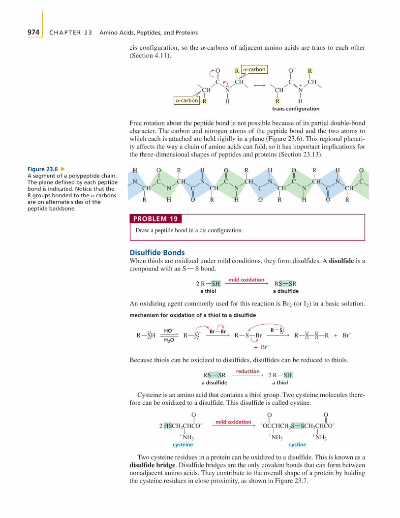
Free rotation about the peptide bond is not possible because of its partial double-bondcharacter. The carbon and nitrogen atoms of the peptide bond and the two atoms towhich each is attached are held rigidly in a plane (Figure 23.6). This regional planari-ty affects the way a chain of amino acids can fold, so it has important implications forthe three-dimensional shapes of peptides and proteins (Section 23.13).
CH NC
R H
R
CH
O
CH N+C
R H
R
CH
O−
trans configuration
-carbon
-carbon
a-carbons
O
CCH
H
NN
HR
R
CHC
O
O
CCH
H
NN
HR
R
CHC
O
O
CCH
H
NN
HR
R
CHC
O
CCH
H
N
RO
Figure 23.6 NA segment of a polypeptide chain.The plane defined by each peptidebond is indicated. Notice that theR groups bonded to the are on alternate sides of thepeptide backbone.
a-carbons
PROBLEM 19
Draw a peptide bond in a cis configuration.
Disulfide BondsWhen thiols are oxidized under mild conditions, they form disulfides. A disulfide is acompound with an bond.
An oxidizing agent commonly used for this reaction is (or ) in a basic solution.
Because thiols can be oxidized to disulfides, disulfides can be reduced to thiols.
Cysteine is an amino acid that contains a thiol group. Two cysteine molecules there-fore can be oxidized to a disulfide. This disulfide is called cystine.
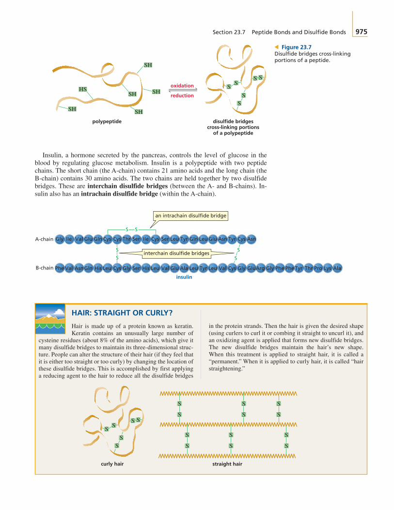
Two cysteine residues in a protein can be oxidized to a disulfide. This is known as adisulfide bridge. Disulfide bridges are the only covalent bonds that can form betweennonadjacent amino acids. They contribute to the overall shape of a protein by holdingthe cysteine residues in close proximity, as shown in Figure 23.7.
cysteine cystine
mild oxidation2 HSCH2CHCO−
O−OCCHCH2S
O
SCH2CHCO−
O
+NH3+NH3
+NH3
reduction
a disulfideRS SR
a thiol2 R SH
HO−
H2OR SH
Br BrR S S R Br−+R S− R S Br
+ Br−
R S−
mechanism for oxidation of a thiol to a disulfide
I2Br2
mild oxidation
a disulfideRS SR
a thiol2 R SH
S¬S
Section 23.7 Peptide Bonds and Disulfide Bonds 975
oxidation
reduction
polypeptide disulfide bridgescross-linking portions
of a polypeptide
SH
SH SH
SH
SH
HS SS
SS
S S
> Figure 23.7Disulfide bridges cross-linkingportions of a peptide.
HAIR: STRAIGHT OR CURLY?
Hair is made up of a protein known as keratin.Keratin contains an unusually large number of
cysteine residues (about 8% of the amino acids), which give itmany disulfide bridges to maintain its three-dimensional struc-ture. People can alter the structure of their hair (if they feel thatit is either too straight or too curly) by changing the location ofthese disulfide bridges. This is accomplished by first applyinga reducing agent to the hair to reduce all the disulfide bridges
straight hair
S
S
S
S
S
S
S
S
S
S
S
S
SS
SS
S S
curly hair
Insulin, a hormone secreted by the pancreas, controls the level of glucose in theblood by regulating glucose metabolism. Insulin is a polypeptide with two peptidechains. The short chain (the A-chain) contains 21 amino acids and the long chain (theB-chain) contains 30 amino acids. The two chains are held together by two disulfidebridges. These are interchain disulfide bridges (between the A- and B-chains). In-sulin also has an intrachain disulfide bridge (within the A-chain).
S S
SS
Gly Ile Val Glu Asn AsnGluGln GlnCys Cys Cys CysThr Tyr TyrLeu LeuSer SerIleA-chain
Phe Val Asn Gln Val Glu PheArg AlaPheLeuHis LeuLeu Cys Leu GlyGly Gly Tyr Thr Pro LysAla CysGlu TyrSer ValHisB-chain
an intrachain disulfide bridge
S S
interchain disulfide bridges
insulin
in the protein strands. Then the hair is given the desired shape(using curlers to curl it or combing it straight to uncurl it), andan oxidizing agent is applied that forms new disulfide bridges.The new disulfide bridges maintain the hair’s new shape.When this treatment is applied to straight hair, it is called a“permanent.” When it is applied to curly hair, it is called “hairstraightening.”

ornithine
H3NCH2CH2CH2CHCO−
O+
+NH3
L-Val
L-Val
L-OrnL-Pro
L-LeuL-Phe
D-PheL-Leu
L-ProD-Orn
gramicidin S
976 C H A P T E R 2 3 Amino Acids, Peptides, and Proteins
PROBLEM 20◆
a. How many different octapeptides can be made from the 20 naturally occurring aminoacids?
b. How many different proteins containing 100 amino acids can be made from the 20 nat-urally occurring amino acids?
PROBLEM 21◆
Which bonds in the backbone of a peptide can rotate freely?
23.8 Some Interesting Peptides
Enkephalins are pentapeptides that are synthesized by the body to control pain. Theydecrease the body’s sensitivity to pain by binding to receptors in certain brain cells.Part of the three-dimensional structures of enkephalins must be similar to those ofmorphine and painkillers such as Demerol because they bind to the same receptors(Sections 30.3 and 30.6).
Bradykinin, vasopressin, and oxytocin are peptide hormones. They are all nona-peptides. Bradykinin inhibits the inflammation of tissues. Vasopressin controlsblood pressure by regulating the contraction of smooth muscle. It is also an antidi-uretic. Oxytocin induces labor in pregnant women and stimulates milk production innursing mothers. Vasopressin and oxytocin both have an intrachain disulfide bond,and their C-terminal amino acids contain amide rather than carboxyl groups. Noticethat the C-terminal amide group is indicated by writing after the name of theC-terminal amino acid. In spite of their very different physiological effects, vaso-pressin and oxytocin differ only by two amino acids.
Gramicidin S is an antibiotic produced by a strain of bacteria. It is a cyclic dec-apeptide. Notice that it contains the amino acids L-ornithine (L-Orn), D-ornithine(D-Orn), and also D-phenylalanine. Ornithine is not listed in Table 23.1 because it oc-curs rarely in nature. Ornithine resembles lysine, but has one less methylene group inits side chain.
The synthetic sweetener aspartame, or NutraSweet (Section 22.21), is the methylester of a dipeptide of L-aspartate and L-phenylalanine. Aspartame is about 200 timessweeter than sucrose. The ethyl ester of the same dipeptide is not sweet. If a D-aminoacid is substituted for either of the L-amino acids of aspartame, the resulting dipeptideis bitter rather than sweet.
COO−
CH2 CH2
+O
H3NCHC NHCHCOCH3
O
aspartameNutraSweet
®
bradykinin Arg-Pro-Pro-Gly-Phe-Ser-Pro-Phe-Arg
vasopressin Cys-Tyr-Phe-Gln-Asn-Cys-Pro-Arg-Gly-NH2
S S
oxytocin Cys-Tyr-Ile-Gln-Asn-Cys-Pro-Leu-Gly-NH2
S S
“NH2”
leucine enkephalinTyr-Gly-Gly-Phe-Leu
methionine enkephalinTyr-Gly-Gly-Phe-Met
®
Oxytocin was the first small peptideto be synthesized. Its synthesis wasachieved in 1953 by Vincent duVigneaud (1901–1978), who latersynthesized vasopressin. Du Vi-gneaud was born in Chicago and wasa professor at George WashingtonUniversity Medical School and laterat Cornell University MedicalCollege. For synthesizing thesenonapeptides, he received the NobelPrize in chemistry in 1955.

Section 23.9 Strategy of Peptide Bond Synthesis: N-Protection and C-Activation 977
3-D Molecules:Glutathione;Oxidized glutathione
Glutathione is a tripeptide of glutamate, cysteine, and glycine. Its function is to de-stroy harmful oxidizing agents in the body. Oxidizing agents are thought to be respon-sible for some of the effects of aging and are believed to play a role in cancer(Section 9.8). Glutathione removes oxidizing agents by reducing them. Consequently,glutathione is oxidized, forming a disulfide bond between two glutathione molecules.An enzyme subsequently reduces the disulfide bond, allowing glutathione to reactwith more oxidizing agents.
PROBLEM 22
What is unusual about glutathione’s structure? (If you can’t answer this question, draw thestructure you would expect for a tripeptide of glutamate, cysteine, and glycine, and com-pare your structure with the structure of glutathione.)
23.9 Strategy of Peptide Bond Synthesis: N-Protection and C-Activation
Because amino acids have two functional groups, a problem arises when one attemptsto make a particular peptide bond. For example, suppose you wanted to make thedipeptide Gly-Ala. That dipeptide is only one of four possible dipeptides that could beformed from alanine and glycine.
If the amino group of the amino acid that is to be on the N-terminal end (in thiscase, Gly) is protected, it will not be available to form a peptide bond. If the carboxylgroup of this same amino acid is activated before the second amino acid is added, theamino group of the added amino acid (in this case, Ala) will react with the activated
oxidizing agentreducing agent
SH
COO−
CH2
+O O O
H3NCHCH2CH2C NHCHC NHCH2CO−
S
S
COO−
CH2
CH2
+O O O
H3NCHCH2CH2C NHCHC NHCH2CO−
COO−
+
OO O
H3NCHCH2CH2C NHCHC NHCH2CO−
2
glutathione
oxidized glutathione
NHCHCO−
CH3
O O
H3NCH2C NHCHCO−
CH3CH3
O O
H3NCHC NHCH2CO−
O O
H3NCH2C NHCH2CO−
CH3
O O
H3NCHC+ + + +
Gly-Ala Ala-Ala Gly-Gly Ala-Gly
978 C H A P T E R 2 3 Amino Acids, Peptides, and Proteins
carboxyl group of glycine in preference to reacting with a nonactivated carboxyl groupof another alanine molecule.
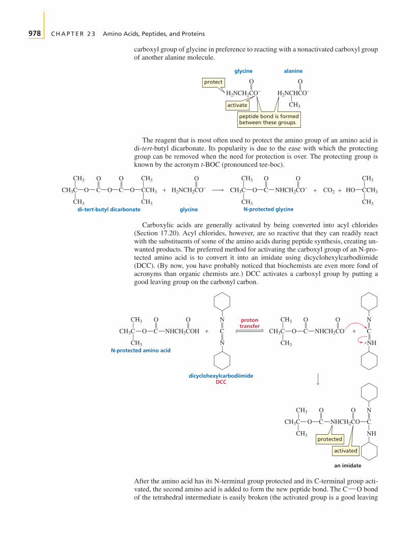
The reagent that is most often used to protect the amino group of an amino acid isdi-tert-butyl dicarbonate. Its popularity is due to the ease with which the protectinggroup can be removed when the need for protection is over. The protecting group isknown by the acronym t-BOC (pronounced tee-boc).
Carboxylic acids are generally activated by being converted into acyl chlorides(Section 17.20). Acyl chlorides, however, are so reactive that they can readily reactwith the substituents of some of the amino acids during peptide synthesis, creating un-wanted products. The preferred method for activating the carboxyl group of an N-pro-tected amino acid is to convert it into an imidate using dicyclohexylcarbodiimide(DCC). (By now, you have probably noticed that biochemists are even more fond ofacronyms than organic chemists are.) DCC activates a carboxyl group by putting agood leaving group on the carbonyl carbon.
After the amino acid has its N-terminal group protected and its C-terminal group acti-vated, the second amino acid is added to form the new peptide bond. The bondof the tetrahedral intermediate is easily broken (the activated group is a good leaving
C¬O
CH3
O O
H2NCH2CO− H2NCHCO−
glycine
protect
activate
peptide bond is formedbetween these groups
alanine
O
H2NCH2CO−CCH3CH3C
CH3
CH3
+
O
OCO
CH3
CH3
O O
CO2NHCH2CO−CH3C + HO CCH3+CO
CH3
CH3
CH3
CH3
O
OC
glycinedi-tert-butyl dicarbonate N-protected glycine
protontransfer
an imidate
CH3
CH3
CH3C
OO N
CO C
N
NHCH2COH
CH3
CH3
CH3C
N
C
+NH
+
OO
O C NHCH2CO− +
CH3
CH3
CH3C
N
C
NH
OO
O C NHCH2CO
N-protected amino acid
dicyclohexylcarbodiimideDCC
protected
activated

Section 23.9 Strategy of Peptide Bond Synthesis: N-Protection and C-Activation 979
group) because the bonding electrons are delocalized, forming dicyclohexylurea, astable diamide. [Recall that the weaker (more stable) the base, the better it is as a leav-ing group; see Section 17.5.]
Amino acids can be added to the growing C-terminal end by repeating these two steps:activating the carboxyl group of the C-terminal amino acid of the peptide by treating itwith DCC and then adding a new amino acid.
When the desired number of amino acids has been added to the chain, the protect-ing group on the N-terminal amino acid is removed. t-BOC is an ideal protectinggroup because it can be removed by washing with trifluoroacetic acid and methylenechloride, reagents that will not break any other covalent bonds. The protecting group isremoved by an elimination reaction, forming isobutylene and carbon dioxide. Becausethese products are gases, they escape, driving the reaction to completion.
amino acid
CH3
CH3
CH3C O
O O N
C
NH
OCNHCH2C
H B+
HB+
CH3
O
H2NCHCO−
tetrahedral intermediate
CH3 NH
CH
CH3
CH3C O
O O H N
CH3
C
NH
OCNHCH2C
−OC
CH3 CH3
CH3
CH3C O+NHCHCO−
O O O NH
C
NH
OCNHCH2C
O
dicyclohexylureaa diamide
new peptide bond
1. DCC
2. H2NCHCO−
N-protected dipeptide N-protected tripeptide
O
R
CH3
NHCHCO−
O O
NHCH2C
CH3
CH3
CH3C
O
OC
CH3
CH3
CH3C
O
OC
CH3
NHCHC
O O
R
NHCHCO−
O
NHCH2C
CF3COOHCH2Cl2
O
O NHCH2CC
CH3
NHCHCOH
O O
NHCHC
N-protected tripeptide
tripeptide
CH2
H
CH3
CH3C
CH2
CH3
CH3C
OHCF3COO
O
CO
R
R
CH3
NHCHCOH
O O
NHCHC
−
+
CO2 +
O
NHCH2C
+O
H3NCH2C
CH3
NHCHCOH
O O
NHCHC
R
H B+
H B+

980 C H A P T E R 2 3 Amino Acids, Peptides, and Proteins
Theoretically, one should be able to make as long a peptide as desired with this tech-nique. Reactions do not produce 100% yields, however, and the yields are further de-creased during the purification process. After each step of the synthesis, the peptidemust be purified to prevent subsequent unwanted reactions with leftover reagents. As-suming that each amino acid can be added to the growing end of the peptide chain in an80% yield (a relatively high yield, as you can probably appreciate from your own expe-rience in the laboratory), the overall yield of a nonapeptide such as bradykinin would beonly 17%. It is clear that large polypeptides could never be synthesized in this way.
Number of amino acids 2 3 4 5 6 7 8 9Overall yield 80% 64% 51% 41% 33% 26% 21% 17%
PROBLEM 23
What dipeptides would be formed by heating a mixture of valine and N-protected leucine?
PROBLEM 24
Suppose you are trying to synthesize the dipeptide Val-Ser. Compare the product that wouldbe obtained if the carboxyl group of N-protected valine were activated with thionyl chloridewith the product that would be obtained if the carboxyl group were activated with DCC.
PROBLEM 25
Show the steps in the synthesis of the tetrapeptide Leu-Phe-Lys-Val.
PROBLEM 26◆
a. Calculate the overall yield of bradykinin if the yield for the addition of each amino acidto the chain is 70%.
b. What would be the overall yield of a peptide containing 15 amino acid residues if theyield for the incorporation of each is 80%?
23.10 Automated Peptide Synthesis
In addition to producing low overall yields, the method of peptide synthesis describedin Section 23.9 is extremely time-consuming because the product must be purified ateach step of the synthesis. In 1969, Bruce Merrifield described a method that revolu-tionized the synthesis of peptides because it provided a much faster way to producepeptides in much higher yields. Furthermore, because it is automated, the synthesis re-quires fewer hours of direct attention. Using this technique, bradykinin was synthesizedwith an 85% yield in 27 hours. Subsequent refinements in the technique now allow areasonable yield of a peptide containing 100 amino acids to be synthesized in four days.
In the Merrifield method, the C-terminal amino acid is covalently attached to asolid support contained in a column. Each N-terminal blocked amino acid is added oneat a time, along with other needed reagents, so the protein is synthesized from theC-terminal end to the N-terminal end. Notice that this is opposite to the way proteinsare synthesized in nature (from the N-terminal end to the C-terminal end;Section 27.13). Because it uses a solid support and is automated, Merrifield’s methodof protein synthesis is called automated solid-phase peptide synthesis.
N-protected amino acid
Merrifield automated solid-phase synthesis of a tripeptide
CH3
CH3
CH3C ClCH2
O
CO
R
NHCHCO−
O
+
resin
R. Bruce Merrifield was born in1921 and received a B.S. and a Ph.D.from the University of California, LosAngeles. He is a professor of chem-istry at Rockefeller University. Merri-field received the 1984 Nobel Prize inchemistry for developing automatedsolid-phase peptide synthesis.

Section 23.10 Automated Peptide Synthesis 981
HF
CH2
CH3
CH3C CO2
RRR
RR
H2NCHC NHCHC NHCHCO CH2
O O O
+
+
+
HOCH2
R
H3NCHC NHCHC NHCHCOH
O O O
+
CF3COOHCH2Cl2
CH3
CH3
CH3C
O
CO
RR
NHCHC NHCHCO CH2
O
CH2
CH3
CH3C CO2
R R
H2NCHC NHCHCO CH2
O O
+ +
O
CF3COOHCH2Cl2
CH3
CH3
CH3C
O
CO NHCHCO CH2
O
N-protected amino acid N-protected andC-activated amino acid
DCC
CH3
CH3
CH3C
O
CO NHCHCOH
O
CH3
CH3
CH3C
O
CO
RR
R
R
NHCHCO DCC
O
CH2
CH3
CH3C CO2 H2NCHCO CH2
O
+ +
Tutorial:Merrifield automated solid-phase synthesis
N-protected amino acid N-protected andC-activated amino acid
DCC
CH3
CH3
CH3C
O
CO
R
NHCHCOH
O
CH3
CH3
CH3C
O
CO
R
NHCHCO DCC
O
CF3COOHCH2Cl2
CH3
CH3
CH3C
O
CO
RR
NHCHC
R
NHCHC NHCHCO CH2
OOO

982 C H A P T E R 2 3 Amino Acids, Peptides, and Proteins
The solid support to which the C-terminal amino acid is attached is a polystyreneresin similar to the one used in ion-exchange chromatography (Section 23.5), exceptthat the benzene rings have chloromethyl substituents instead of sulfonic acid sub-stituents. Before the C-terminal amino acid is attached to the resin, its amino groupis protected with t-BOC to prevent the amino group from reacting with the resin.The C-terminal amino acid is attached to the resin by means of an reaction—itscarboxyl group attacks a benzyl carbon of the resin, displacing a chloride ion(Section 10.4).
After the C-terminal amino acid is attached to the resin, the t-BOC protecting groupis removed (Section 23.9). The next amino acid, with its amino group protected witht-BOC and its carboxyl group activated with DCC, is added to the column.
A huge advantage of the Merrifield method of peptide synthesis is that thegrowing peptide can be purified by washing the column with an appropriate solventafter each step of the procedure. The impurities are washed out of the column be-cause they are not attached to the solid support. Since the peptide is covalentlyattached to the resin, none of it is lost in the purification step, leading to high yieldsof purified product.
After the required amino acids have been added one by one, the peptide can be re-moved from the resin by treatment with HF under mild conditions that do not breakthe peptide bonds.
Merrifield’s technique is constantly being improved so that peptides can bemade more rapidly and more efficiently. However, it still cannot begin to comparewith nature: A bacterial cell is able to synthesize a protein thousands of aminoacids long in seconds and can simultaneously synthesize thousands of differentproteins with no mistakes.
Since the early 1980s, it has been possible to synthesize proteins by genetic engi-neering techniques. Strands of DNA can be introduced into bacterial cells, causing thecells to produce large amounts of a desired protein (Section 27.13). For example, massquantities of human insulin are produced from genetically modified E. coli. Geneticengineering techniques also have been useful in synthesizing proteins that differ in oneor a few amino acids from the natural protein. Such synthetic proteins have been used,for example, to learn how a change in a single amino acid affects the properties of aprotein (Section 24.9).
PROBLEM 27
Show the steps in the synthesis of the peptide in Problem 25, using Merrifield’s method.
23.11 Protein Structure
Protein molecules are described by several levels of structure. The primary struc-ture of a protein is the sequence of amino acids in the chain and the location of allthe disulfide bridges. The secondary structure describes the regular conformationassumed by segments of the protein’s backbone. In other words, the secondarystructure describes how local regions of the backbone fold. The tertiary structuredescribes the three-dimensional structure of the entire polypeptide. If a protein hasmore than one polypeptide chain, it has quaternary structure. The quaternarystructure of a protein is the way the individual protein chains are arranged with re-spect to each other.
Proteins can be divided roughly into two classes. Fibrous proteins containlong chains of polypeptides that occur in bundles. These proteins are insoluble inwater. All the structural proteins described at the beginning of this chapter, such askeratin and collagen, are fibrous proteins. Globular proteins are soluble in waterand tend to have roughly spherical shapes. Essentially all enzymes are globularproteins.
SN2

Section 23.12 Determining the Primary Structure of a Protein 983
PRIMARY STRUCTURE AND EVOLUTIONWhen we examine the primary structures of pro-
teins that carry out the same function in different organisms,we can relate the number of amino acid differences betweenthe proteins to the taxonomic differences between thespecies. For example, cytochrome c, a protein that transfers
electrons in biological oxidations, has about 100 amino acidresidues. Yeast cytochrome c differs by 48 amino acids fromhorse cytochrome c, while duck cytochrome c differs by onlytwo amino acids from chicken cytochrome c. Chickens andturkeys have cytochrome c’s with identical primary struc-tures. Humans and chimpanzees also have identical cyto-chrome c’s, differing by one amino acid from the cytochromec of the rhesus monkey.
23.12 Determining the Primary Structure of a Protein
The first step in determining the sequence of amino acids in a peptide or a protein is toreduce any disulfide bridges in the peptide or protein. A commonly used reducingagent is 2-mercaptoethanol, which is oxidized to a disulfide. Reaction of the proteinthiol groups with iodoacetic acid prevents the disulfide bridges from reforming as a re-sult of oxidation by
PROBLEM 28
Write the mechanism for the reaction of a cysteine residue with iodoacetic acid.
The next step is to determine the number and kinds of amino acids in the peptide orprotein. To do this, a sample of the peptide or protein is dissolved in 6 N HCl and heat-ed at for 24 hours. This treatment hydrolyzes all the amide bonds in the pro-tein, including the amide bonds of asparagine and glutamine.
6 N HCI100 °C24 h
protein amino acids
100 °C
ICH2COH
cleaving disulfide bridges
O
CH2
CH2
S
S
C
O
NHCH
C
O
NHCH
+ 2 HSCH2CH2OH
CH2
CH2
SH
SH
SCH2CH2OH
SCH2CH2OH
C
O
NHCH
C
O
NHCH
+
CH2
CH2
SCH2COH
SCH2COH
C
O
O
O
NHCH
C
O
2 HI
NHCH
+
iodoacetic acid
2-mercaptoethanol
O2.
Insulin was the first protein for whichthe primary sequence was deter-mined. This was done in 1953 byFrederick Sanger, who received the1958 Nobel Prize in chemistry for hiswork. Sanger was born in England in1918 and received a Ph.D. fromCambridge University, where he hasworked for his entire career. He alsoreceived a share of the 1980 NobelPrize in chemistry (Section 27.15) forbeing the first to sequence a DNAmolecule (with 5375 nucleotidepairs).

984 C H A P T E R 2 3 Amino Acids, Peptides, and Proteins
The mixture of amino acids is then passed through an amino acid analyzer to deter-mine the number and kind of each amino acid in the peptide or protein (Section 23.5).
Because all the asparagine and glutamine residues have been hydrolyzed to aspar-tate and glutamate residues, the number of aspartate or glutamate residues in theamino acid mixture tells us the number of aspartate plus asparagine—or glutamateplus glutamine—residues in the original protein. A separate technique must be used todistinguish between aspartate and asparagine or between glutamate and glutamine inthe original protein.
The strongly acidic conditions used for hydrolysis destroy all the tryptophanresidues because the indole ring is unstable in acid (Section 21.9). The tryptophancontent of the protein can be determined by hydroxide-ion-promoted hydrolysis of theprotein. This is not a general method for peptide bond hydrolysis because the stronglybasic conditions destroy several other amino acid residues.
There are several ways to identify the N-terminal amino acid of a peptide or pro-tein. One of the most widely used methods is to treat the protein with phenyl isothio-cyanate (PITC), more commonly known as Edman’s reagent. This reagent reactswith the N-terminal amino group, and the resulting thiazolinone derivative is cleavedfrom the protein under mildly acidic conditions. The thiazolinone derivative is extract-ed into an organic solvent and in the presence of acid, rearranges to a more stablephenylthiohydantoin (PTH).
Because each amino acid has a different substituent (R), each amino acid forms adifferent PTH. The particular PTH can be identified by chromatography using knownstandards. Several successive Edman degradations can be carried out on a protein.The entire primary sequence cannot be determined in this way, however, because side
OHC
HN
HN
R
S
HCl
thiazolinonederivative
O
H3NCHC
R′
O
NHCHC
R′′
OHC
HN
S
R
N
++
+
PTH–amino acid
peptide without the originalN-terminal amino acid
phenyl isothiocyanatePITC
Edman's reagent
N H2NCHC NHCHC NHCHCC
O O O
R′′R′R
S
NH
NHCHC NHCHC NHCHCC
O O O
NHCHC
O
R′′R′RS
NH
HRN
C
S
CH H F
C
O− R′
NHCHC
O
R′′
HF
+

Section 23.12 Determining the Primary Structure of a Protein 985
products accumulate that interfere with the results. An automated instrument knownas a sequenator allows about 50 successive Edman degradations to be carried out ona protein.
The C-terminal amino acid of the peptide or protein can be identified by treating theprotein with carboxypeptidase A. Carboxypeptidase A cleaves off the C-terminalamino acid as long as it is not arginine or lysine (Section 24.9). On the other hand, car-boxypeptidase B cleaves off the C-terminal amino acid only if it is arginine or lysine.Carboxypeptidases are exopeptidases. An exopeptidase is an enzyme that catalyzesthe hydrolysis of a peptide bond at the end of a peptide chain.
Once the N-terminal and C-terminal amino acids have been identified, a sample ofthe protein is hydrolyzed with dilute acid. This treatment, called partial hydrolysis,hydrolyzes only some of the peptide bonds. The resulting fragments are separated, andthe amino acid composition of each is determined. The N-terminal and C-terminalamino acids of each fragment can also be identified. The sequence of the originalprotein can then be determined by lining up the peptides and looking for pointsof overlap.
PROBLEM-SOLVING STRATEGY
A nonapeptide undergoes partial hydrolysis to give peptides whose amino acid composi-tions are shown. Reaction of the intact nonapeptide with Edman’s reagent releasesPTH-Leu. What is the sequence of the nonapeptide?
a. Pro, Ser c. Met, Ala, Leu e. Glu, Ser, Val, Pro g. Met, Leu
b. Gly, Glu d. Gly, Ala f. Glu, Pro, Gly h. His, Val
Let’s start with the N-terminal amino acid. We know that it is Leu. Now we need to lookfor a fragment that contains Leu. Fragment (g) tells us that Met is next to Leu and fragment(c) tells us that Ala is next to Met. Now we look for a fragment that contains Ala. Fragment(d) contains Ala and tells us that Gly is next to Ala. From fragment (b), we know that Glucomes next. Glu is in both fragments (e) and (f). Fragment (e) has two amino acids we haveyet to place in the growing peptide, but fragment (f) has only one, so from fragment (f), weknow that Pro is the next amino acid. Now we can use fragment (e). Fragment (e) tells usthat the next amino acid is Val, and fragment (h) tells us that His is the last (C-terminal)amino acid. Thus, the amino acid sequence of the nonapeptide is
Leu-Met-Ala-Gly-Glu-Pro-Ser-Val-His
Now continue on to Problem 29.
PROBLEM 29◆
A decapeptide undergoes partial hydrolysis to give peptides whose amino acid composi-tions are shown. Reaction of the intact decapeptide with Edman’s reagent releasesPTH-Gly. What is the sequence of the decapeptide?
a. Ala, Trp c. Pro, Val e. Trp, Ala, Arg g. Glu, Ala, Leu
b. Val, Pro, Asp d. Ala, Glu f. Arg, Gly h. Met, Pro, Leu, Glu
The peptide or protein can also be partially hydrolyzed using endopeptidases. Anendopeptidase is an enzyme that catalyzes the hydrolysis of a peptide bond that is notat the end of a peptide chain. Trypsin, chymotrypsin, and elastase are endopeptidases
R′′R′R
NHCHCO−
OO O
NHCHCNHCHC
site where carboxypeptidase cleaves

986 C H A P T E R 2 3 Amino Acids, Peptides, and Proteins
Table 23.3 Specificity of Peptide or Protein Cleavage
Reagent Specificity
Chemical reagentsEdman’s reagent removes the N-terminal amino acidCyanogen bromide hydrolyzes on the C-side of Met
Exopeptidases*Carboxypeptidase A removes the C-terminal amino acid (not Arg or Lys)Carboxypeptidase B removes the C-terminal amino acid (only Arg or Lys)
Endopeptidases*Trypsin hydrolyzes on the C-side of Arg and LysChymotrypsin hydrolyzes on the C-side of amino acids that contain
aromatic six-membered rings (Phe, Tyr, Trp)
Elastase hydrolyzes on the C-side of small amino acids(Gly and Ala)
*Cleavage will not occur if Pro is on either side of the bond to be hydrolyzed.
that catalyze the hydrolysis of only the specific peptide bonds listed in Table 23.3.Trypsin, for example, catalyzes the hydrolysis of the peptide bond on the C-side ofonly arginine or lysine residues.
Thus, trypsin will catalyze the hydrolysis of three peptide bonds in the followingpeptide, creating a hexapeptide, a dipeptide, and two tripeptides.
Chymotrypsin catalyzes the hydrolysis of the peptide bond on the C-side of aminoacids that contain aromatic six-membered rings (Phe, Tyr, Trp).
Elastase catalyzes the hydrolysis of peptide bonds on the C-side of small amino acids(Gly, Ala). Chymotrypsin and elastase are much less specific than trypsin. (An expla-nation for the specificity of these enzymes is given in Section 24.9.)
cleavage by elastase
Ala-Lys-Phe-Gly-Asp-Trp-Ser-Arg-Met-Val-Arg-Tyr-Leu-His
cleavage by chymotrypsin
Ala-Lys-Phe-Gly-Asp-Trp-Ser-Arg-Met-Val-Arg-Tyr-Leu-His
cleavage by trypsin
Ala-Lys-Phe-Gly-Asp-Trp-Ser-Arg-Met-Val-Arg-Tyr-Leu-His
R′CH2
CH2
CH2
CH2
+NH3
CH2
CH2
NH
C
NH2
NH2
R
NHCHC
OO O
R′′
NHCHC
O
CH2
NHCHC
O
R′′′
NHCHC
O
NHCHCNHCHC
+
C-side of arginineC-side of lysine
3-D Molecules:Carboxypeptidase A; Chymotrypsin

Section 23.12 Determining the Primary Structure of a Protein 987
None of the exopeptidases or endopeptidases that we have mentioned will catalyze thehydrolysis of an amide bond if proline is at the hydrolysis site. These enzymes recog-nize the appropriate hydrolysis site by its shape and charge, and proline’s structurecauses the hydrolysis site to have an unrecognizable three-dimensional shape.
Cyanogen bromide causes the hydrolysis of the amide bond on theC-side of a methionine residue. Cyanogen bromide is more specific than the en-dopeptidases about what peptide bonds it cleaves, so it provides more reliable infor-mation about the primary structure (the sequence of amino acids). Because cyanogenbromide is not a protein and therefore does not recognize the substrate by its shape,cyanogen bromide will still cleave the peptide bond if proline is at the cleavage site.
The first step in the mechanism for cleavage of a peptide bond by cyanogen bro-mide is attack by the highly nucleophilic sulfur of methionine on cyanogen bromide.Formation of a five-membered ring with departure of the weakly basic leaving groupis followed by acid-catalyzed hydrolysis, which cleaves the protein (Section 18.6).Further hydrolysis can cause the lactone (a cyclic ester) to open to a carboxyl groupand an alcohol group (Section 17.11).
H2O
H2O
N
R R′
NHCHCNHCH H3NCHC
O O
NHCHCNHCH
R
CH2
CH2
OH
CH2
CH2
O
O
OO
COH C O
R R′
NHCHCNHCH
O OCH2
CH2
C NHCHC+
++
CH3SC+
HCl
HCl
R R′R′
NHCHCNHCH
O O O
NHCHCNHCHCNHCH
R
CH2
CH2
S
CH3
CH2
CH2
S
CH3
OO
C
C N
C N
Br
O
NHCHCC
+
mechanism for the cleavage of a peptide bond by cyanogen bromide
+ Br−
Ala-Lys-Phe-Gly-Lys-Trp-Ser-Arg-Met-Val-Arg-Tyr-Leu-His
cleavage by cyanogen bromide
(BrC‚N)
Ala-Lys-Pro Leu-Phe-Pro Pro-Phe-Val
chymotrypsin will cleavechymotrypsin will not cleavetrypsin will not cleave

988 C H A P T E R 2 3 Amino Acids, Peptides, and Proteins
The last step in determining the primary structure of a protein is to figure out the lo-cation of any disulfide bonds. This is done by hydrolyzing a sample of the protein thathas intact disulfide bonds. From a determination of the amino acids in the cysteine-containing fragments, the locations of the disulfide bonds in the protein can be estab-lished (Problem 47).
PROBLEM 30
Why won’t cyanogen bromide cleave at cysteine residues?
PROBLEM 31◆
In determining the primary structure of insulin, what would lead you to conclude that ithad more than one polypeptide chain?
PROBLEM 32 SOLVED
Determine the amino acid sequence of a polypeptide from the following results:
Acid hydrolysis gives Ala, Arg, His, 2 Lys, Leu, 2 Met, Pro, 2 Ser, Thr, Val.Carboxypeptidase A releases Val.Edman’s reagent releases PTH-Leu.Cleavage with cyanogen bromide gives three peptides with the following amino acidcompositions:
1. His, Lys, Met, Pro, Ser 3. Ala, Arg, Leu, Lys, Met, Ser2. Thr, Val
Trypsin-catalyzed hydrolysis gives three peptides and a single amino acid:1. Arg, Leu, Ser 3. Lys2. Met, Pro, Ser, Thr, Val 4. Ala, His, Lys, Met
SOLUTION Acid hydrolysis shows that the polypeptide has 13 amino acids. The N-ter-minal amino acid is Leu (Edman’s reagent), and the C-terminal amino acid is Val (car-boxypeptidase A).
Because cyanogen bromide cleaves on the C-side of Met, any peptide containing Metmust have Met as its C-terminal amino acid. The peptide that does not contain Met must bethe C-terminal peptide. We know that peptide 3 is the N-terminal peptide because it con-tains Leu. Since it is a hexapeptide, we know that the 6th amino acid in the 13-amino acidpeptide is Met. We also know that the eleventh amino acid is Met because cyanogen bro-mide cleavage gave the dipeptide Thr, Val.
Because trypsin cleaves on the C-side of Arg and Lys, any peptide containing Arg or Lysmust have that amino acid as its C-terminal amino acid. Therefore, Arg is the C-terminalamino acid of peptide 1, so we now know that the first three amino acids are Leu-Ser-Arg.We also know that the next two are Lys-Ala because if they were Ala-Lys, trypsin cleav-age would give an Ala, Lys dipeptide. The trypsin data also identify the positions of Hisand Lys.
Finally, because trypsin successfully cleaves on the C-side of Lys, Pro cannot be adja-cent to Lys. Thus, the amino acid sequence of the given polypeptide is
Leu Ser Arg Lys Ala Met His Lys Ser Pro Met Thr Val
Leu Ser Arg Lys Ala Met His Lys
Pro, Ser
Met Thr Val
Leu Met
Ala, Arg, Lys, Ser His, Lys, Pro, Ser
Met Thr Val
Leu Val
Section 23.13 Secondary Structure of Proteins 989
PROBLEM 33◆
Determine the primary structure of an octapeptide from the following data:
Acid hydrolysis gives 2 Arg, Leu, Lys, Met, Phe, Ser, Tyr.Carboxypeptidase A releases Ser.Edman’s reagent releases Leu.Cyanogen bromide forms two peptides with the following amino acid compositions:
1. Arg, Phe, Ser 2. Arg, Leu, Lys, Met, TyrTrypsin forms the following two peptides and two amino acids:
1. Arg 3. Arg, Met, Phe2. Ser 4. Leu, Lys, Tyr
23.13 Secondary Structure of Proteins
Secondary structure describes the conformation of segments of the backbone chain ofa peptide or protein. To minimize energy, a polypeptide chain tends to fold in a repeat-ing geometric structure such as an or a sheet. Three factors deter-mine the choice of secondary structure:
• the regional planarity about each peptide bond (as a result of the partial double-bond character of the amide bond), which limits the possible conformations ofthe peptide chain (Section 23.7)
• maximization of the number of peptide groups that engage in hydrogen bonding(i.e., hydrogen bonding between the carbonyl oxygen of one amino acid residueand the amide hydrogen of another)
• adequate separation between nearby R groups to avoid steric hindrance andrepulsion of like charges
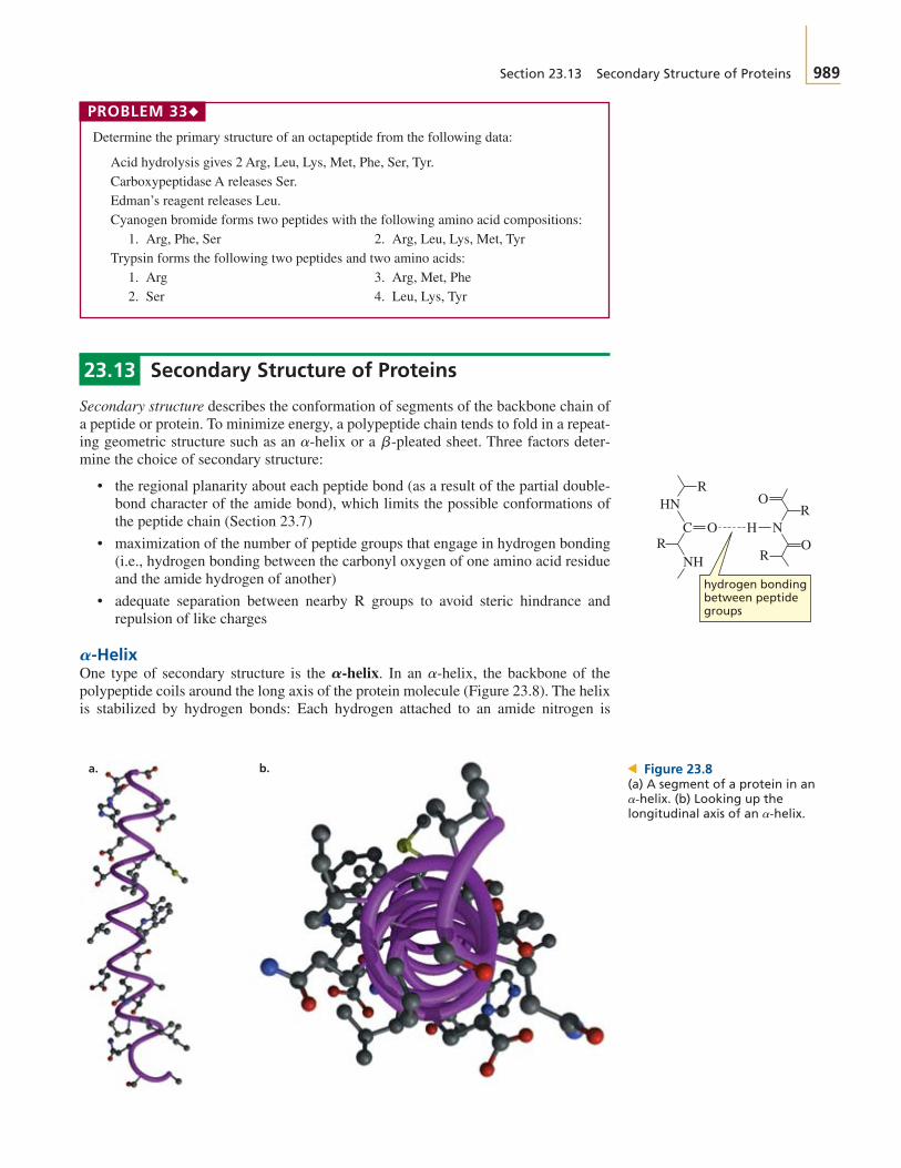
-HelixOne type of secondary structure is the In an the backbone of thepolypeptide coils around the long axis of the protein molecule (Figure 23.8). The helixis stabilized by hydrogen bonds: Each hydrogen attached to an amide nitrogen is
a-helix,A-helix.A
b-pleateda-helix
C
HN
NH
R
RR
RH NO
O
O
hydrogen bondingbetween peptidegroups
a. b. > Figure 23.8(a) A segment of a protein in an
(b) Looking up thelongitudinal axis of an a-helix.a-helix.

990 C H A P T E R 2 3 Amino Acids, Peptides, and Proteins
3-D Molecule:An a-helix
hydrogen bonded to a carbonyl oxygen of an amino acid four residues away. The sub-stituents on the of the amino acids protrude outward from the helix, therebyminimizing steric hindrance. Because the amino acids have the L-configuration, the
is a right-handed helix; that is, it rotates in a clockwise direction as it spiralsdown. Each turn of the helix contains 3.6 amino acid residues, and the repeat distanceof the helix is 5.4 Å.
Not all amino acids are able to fit into an A proline residue, for exam-ple, forces a bend in a helix because the bond between the proline nitrogen and the
cannot rotate to enable it to fit readily into a helix. Similarly, two adja-cent amino acids that have more than one substituent on a (valine,isoleucine, or threonine) cannot fit into a helix because of steric crowding betweenthe R groups. Finally, two adjacent amino acids with like-charged substituentscannot fit into a helix because of electrostatic repulsion between the R groups. Thepercentage of amino acid residues coiled into an varies from protein toprotein, but, on average, about 25% of the residues in globular proteins are in
-Pleated SheetThe second type of secondary structure is the In a sheet,the polypeptide backbone is extended in a zigzag structure resembling a series ofpleats. A sheet is almost fully extended—the average two-residue repeatdistance is 7.0 Å. The hydrogen bonding in a sheet occurs between neigh-boring peptide chains. The adjacent hydrogen-bonded peptide chains can run in thesame direction or in opposite directions. In a parallel sheet, the adjacentchains run in the same direction. In an antiparallel sheet, the adjacentchains run in opposite directions (Figure 23.9).
B-pleatedB-pleated
b-pleatedb-pleated
b-pleatedB-pleated sheet.B
a-helices.
a-helix
b-carbona-carbon
a-helix.
a-helix
a-carbons
3-D Molecule:Antiparallel sheetb-pleated
CHR
NH
C
CHR
N
NH
C
R
H
R
HC RHC
CO
HC
CHR
NH
C
CHR
N
NH
C
CO
HC
O
O
H
R
O
O
Parallel
N-terminalN-terminal
C-terminalC-terminal
CHR
NH
C
CHR
N
NH
C
R
H
R
HC R CH
R CH
CO
HC
RHC
NH
R
C
CH
N
NH
C
CO
O
O
H
O
O
Antiparallel
N-terminal
C-terminalN-terminal
C-terminal
Figure 23.9 NSegment of a sheetdrawn to illustrate its pleatedcharacter.
b-pleated
Because the substituents (R) on the of the amino acids on adjacent chainsare close to each other, the chains can nestle closely together to maximize hydrogen-bonding interactions only if the substituents are small. Silk, for example, a proteinwith a large number of relatively small amino acids (glycine and alanine), has largesegments of sheets. The number of side-by-side strands in a sheetranges from 2 to 15 in a globular protein. The average strand in a sheet sec-tion of a globular protein contains six amino acid residues.
Wool and the fibrous protein of muscle are examples of proteins with secondarystructures that are almost all Consequently, these proteins can be stretched.In contrast, the secondary structures of silk and spider webs are predominantly
sheets. Because the sheet is a fully extended structure, these pro-teins cannot be stretched.
b-pleatedb-pleated
a-helices.
b-pleatedb-pleatedb-pleated
a-carbons

Section 23.14 Tertiary Structure of Proteins 991
Coil ConformationGenerally, less than half of a globular protein is in an or sheet(Figure 23.10). Most of the rest of the protein is still highly ordered but is difficult todescribe. These polypeptide fragments are said to be in a coil conformation or a loopconformation.
b-pleateda-helix
> Figure 23.10The backbone structure ofcarboxypeptidase A:
segments are purple;sheets are indicated
by flat green arrows pointing inthe direction.N ¡ C
b-pleateda-helical
23.14 Tertiary Structure of Proteins
The tertiary structure of a protein is the three-dimensional arrangement of all theatoms in the protein. Proteins fold spontaneously in solution in order to maximize theirstability. Every time there is a stabilizing interaction between two atoms, free energy isreleased. The more free energy released (the more negative the ), the more stablethe protein. So a protein tends to fold in a way that maximizes the number of stabiliz-ing interactions (Figure 23.11).
¢G°
AN ATTEMPT TO IMPROVE ON NATUREChemists are currently studying
which are polymers of acids. These peptides havebackbones one carbon longer than the peptides nature synthe-sizes using acids. Therefore, each acidresidue has two carbons to which side chains can be attached.
Like fold into relatively sta-ble helical and pleated sheet conformations, causing scientiststo wonder whether biological activity might be possible withsuch peptides. Recently, a with biological activityhas been synthesized—one that mimics the activity of the hor-
b-peptide
b-polypeptidesa-polypeptides,
b-aminoa-amino
b-aminob-peptides,
B-PEPTIDES: mone somatostatin. There is hope that will pro-vide a source of new drugs and catalysts. Surprisingly, the pep-tide bonds in are resistant to the enzymes thatcatalyze the hydrolysis of peptide bonds in This resistance to hydrolysis means that a drugwill have a longer duration of action in the bloodstream.
H3N
R
O−
O
CH C
-amino acid
+H3N
R R′
O−
O
CH CH C
-amino acid
+
b-polypeptidea-polypeptides.
b-polypeptides
b-polypeptides
PROBLEM 34◆
How long is an that contains 74 amino acids? Compare the length of this with the length of a fully extended peptide chain containing the same number of aminoacids. (The distance between consecutive amino acids in a fully extended chain is 3.5 Å.)
a-helixa-helix

992 C H A P T E R 2 3 Amino Acids, Peptides, and Proteins
The stabilizing interactions include covalent bonds, hydrogen bonds, electrostaticattractions (attractions between opposite charges), and hydrophobic (van derWaals) interactions. Stabilizing interactions can occur between peptide groups(atoms in the backbone of the protein), between side-chain groups and between peptide and side-chain groups. Because the side-chain groups help de-termine how a protein folds, the tertiary structure of a protein is determined by itsprimary structure.
Disulfide bonds are the only covalent bonds that can form when a protein folds. Theother bonding interactions that occur in folding are much weaker, but because thereare so many of them (Figure 23.12), they are the important interactions in determininghow a protein folds.
Most proteins exist in aqueous environments. Therefore, they tend to fold in away that exposes the maximum number of polar groups to the aqueous environ-ment and that buries the nonpolar groups in the interior of the protein, awayfrom water.
The interactions between nonpolar groups are known as hydrophobic interac-tions. These interactions increase the stability of a protein by increasing the entropy ofwater molecules. Water molecules that surround nonpolar groups are highlystructured. When two nonpolar groups come together, the surface area in contact withwater decreases, decreasing the amount of structured water. Decreasing structure in-creases entropy, which in turn decreases the free energy, which increases the stabilityof the protein. (Recall that )¢G° = ¢H° - T¢S°.
(a-substituents),
Helical Structure
Pleated SheetStructure
COO−
H3N+
HN
C O
CH2 OH O C
CH
CH
2 CH
3
CH
3C
H3
CH
3
CH
CH2C NHH
OCH2H
(CH
2 )4 NH
3+
−OC
CH
2
O
CH
SS
CH
2
O
▲ Figure 23.11Stabilizing interactions responsible for the tertiary structure of a protein.
Max Perutz was born in Austria in1914. In 1936, because of the rise ofNazism, he moved to England. Hereceived a Ph.D. from, and became aprofessor at, Cambridge University.He worked on the three-dimensionalstructure of hemoglobin and assignedthe work on myoglobin (a smaller pro-tein) to John Kendrew (1917–1997).Kendrew was born in England and was educated at CambridgeUniversity.
Max Ferdinand Perutz and JohnCowdery Kendrew were the first todetermine the tertiary structure of aprotein. Using X-ray diffraction, theydetermined the tertiary structure ofmyoglobin (1957) and hemoglobin(1959). For this work, they shared the1962 Nobel Prize in chemistry.

▲ Figure 23.13Computer graphic representation ofthe quaternary structure ofhemoglobin. The orange and pinksubunits are identical, as are thegreen and purple subunits. Thecylindrical tubes represent thepolypeptide chains, while the beadsrepresent the iron-containingporphyrin rings (Section 21.11).
Section 23.15 Quaternary Structure of Proteins 993
> Figure 23.12The three-dimensional structure ofcarboxypeptidase A.
PROBLEM 35◆
How would a protein that resides in the interior of a membrane fold, compared with thewater-soluble protein just discussed?
23.15 Quaternary Structure of Proteins
Proteins that have more than one peptide chain are called oligomers. The individualchains are called subunits. A protein with a single subunit is called a monomer, onewith two subunits is called a dimer; one with three subunits is called a trimer, andone with four subunits is called a tetramer. Hemoglobin is an example of a tetramer.It has two different kinds of subunits and two of each kind. The quaternary structureof hemoglobin is shown in Figure 23.13.
The subunits are held together by the same kinds of interactions that hold the indi-vidual protein chains in a particular three-dimensional conformation: hydrophobic in-teractions, hydrogen bonding, and electrostatic attractions. The quaternary structure ofa protein describes the way the subunits are arranged in space. Some of the possiblearrangements of the six subunits of a hexamer are shown here:
PROBLEM 36◆
a. Which of the following water-soluble proteins would have the greatest percentage ofpolar amino acids—a spherical protein, a cigar-shaped protein, or a subunit of ahexamer?
b. Which of these would have the smallest percentage of polar amino acids?
possible quaternary structures for a hexamer

994 C H A P T E R 2 3 Amino Acids, Peptides, and Proteins
23.16 Protein Denaturation
Destroying the highly organized tertiary structure of a protein is called denaturation.Anything that breaks the bonds responsible for maintaining the three-dimensional shapeof the protein will cause the protein to denature (unfold). Because these bonds are weak,proteins are easily denatured. The totally random conformation of a denatured protein iscalled a random coil. The following are some of the ways that proteins can be denatured:
• Changing the pH denatures proteins because it changes the charges on many ofthe side chains. This disrupts electrostatic attractions and hydrogen bonds.
• Certain reagents such as urea and guanidine hydrochloride denature proteins byforming hydrogen bonds to the protein groups that are stronger than the hydro-gen bonds formed between the groups.
• Detergents such as sodium dodecyl sulfate denature proteins by associating withthe nonpolar groups of the protein, thus interfering with the normal hydrophobicinteractions.
• Organic solvents denature proteins by disrupting hydrophobic interactions.• Proteins can also be denatured by heat or by agitation. Both increase molecular
motion, which can disrupt the attractive forces. A well-known example is thechange that occurs to the white of an egg when it is heated or whipped.
Summary
Peptides and proteins are polymers of amino acids linkedtogether by peptide (amide) bonds. A dipeptide containstwo amino acid residues, a tripeptide contains three, anoligopeptide contains three to 10, and a polypeptide con-tains many amino acid residues. Proteins have 40 to 4000amino acid residues. The amino acids differ only in thesubstituent attached to the Most amino acidsfound in nature have the L configuration.
The carboxyl groups of the amino acids have valuesof and the protonated amino groups have values of
At physiological pH, an amino acid exists as azwitterion. A few amino acids have side chains with ioniz-able hydrogens. The isoelectric point (pI) of an amino acidis the pH at which the amino acid has no net charge. A mix-ture of amino acids can be separated based on their pI’s byelectrophoresis or based on their polarities by paperchromatography or thin-layer chromatography. Prepar-ative separation can be achieved using ion-exchangechromatography employing a cation-exchange resin. Anamino acid analyzer is an instrument that automatesion-exchange chromatography. A racemic mixture of aminoacids can be separated by a kinetic resolution.
The amide bonds that link amino acid residues are calledpeptide bonds. A peptide bond has about 40% double-bond character. Two cysteine residues can be oxidized to adisulfide bridge. Disulfide bridges are the only covalentbonds that can form between nonadjacent amino acids. Byconvention, peptides and proteins are written with the freeamino group (the N-terminal amino acid) on the left andthe free carboxyl group (the C-terminal amino acid) onthe right.
'9.pKa
'2pKa
a-carbon.
To synthesize a peptide bond, the amino group of thefirst amino acid must be protected (by t-BOC) and its car-boxyl group activated (with DCC). The second amino acidis added to form a dipeptide. Amino acids can be added tothe growing C-terminal end by repeating these two steps:activating the carboxyl group of the C-terminal amino acidwith DCC and adding a new amino acid. Automated solid-phase peptide synthesis allows peptides to be made morerapidly and in higher yields.
The primary structure of a protein is the sequence of itsamino acids and the location of all its disulfide bridges. TheN-terminal amino acid of a peptide or protein can be deter-mined with Edman’s reagent. The C-terminal amino acidcan be identified with carboxypeptidase. Partial hydrolysishydrolyzes only some of the peptide bonds. An exopeptid-ase catalyzes the hydrolysis of a peptide bond at the end of apeptide chain. An endopeptidase catalyzes the hydrolysis ofa peptide bond that is not at the end of a peptide chain.
The secondary structure of a protein describes how localsegments of the protein’s backbone folds. A protein foldsso as to maximize the number of stabilizing interactions:covalent bonds, hydrogen bonds, electrostatic attractions(attraction between opposite charges), and hydrophobic in-teractions (interactions between nonpolar groups). An
a and a coil conformation aretypes of secondary structure. The tertiary structure of aprotein is the three-dimensional arrangement of all the atomsin the protein. Proteins with more than one peptide chain arecalled oligomers. The individual chains are called subunits.The quaternary structure of a protein describes the way thesubunits are arranged with respect to each other in space.
B-pleated sheet,A-helix,
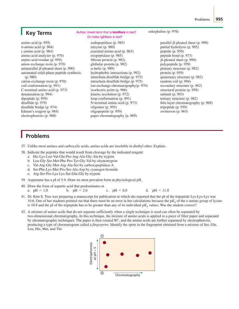
Problems 995
Key Terms
amino acid (p. 959)D-amino acid (p. 964)L-amino acid (p. 964)amino acid analyzer (p. 970)amino acid residue (p. 959)anion-exchange resin (p. 970)antiparallel sheet (p. 990)automated solid-phase peptide synthesis
(p. 980)cation-exchange resin (p. 970)coil conformation (p. 991)C-terminal amino acid (p. 973)denaturation (p. 994)dipeptide (p. 959)disulfide (p. 979)disulfide bridge (p. 974)Edman’s reagent (p. 984)electrophoresis (p. 968)
b-pleated
endopeptidase (p. 985)enzyme (p. 960)essential amino acid (p. 963)exopeptidase (p. 985)fibrous protein (p. 982)globular protein (p. 982)
(p. 989)hydrophobic interactions (p. 992)interchain disulfide bridge (p. 975)intrachain disulfide bridge (p. 975)ion-exchange chromatography(p. 970)isoelectric point (p. 966)kinetic resolution (p. 972)loop conformation (p. 991)N-terminal amino acid (p. 973)oligomer (p. 993)oligopeptide (p. 959)paper chromatography (p. 969)
a-helix
parallel sheet (p. 990)partial hydrolysis (p. 985)peptide (p. 959)peptide bond (p. 973)
sheet (p. 990)polypeptide (p. 959)primary structure (p. 982)protein (p. 959)quaternary structure (p. 982)random coil (p. 994)secondary structure (p. 982)structural protein (p. 959)subunit (p. 993)tertiary structure (p. 982)thin-layer chromatography (p. 969)tripeptide (p. 959)zwitterion (p. 965)
b-pleated
b-pleated
Problems
37. Unlike most amines and carboxylic acids, amino acids are insoluble in diethyl ether. Explain.
38. Indicate the peptides that would result from cleavage by the indicated reagent:a. His-Lys-Leu-Val-Glu-Pro-Arg-Ala-Gly-Ala by trypsinb. Leu-Gly-Ser-Met-Phe-Pro-Tyr-Gly-Val by chymotrypsinc. Val-Arg-Gly-Met-Arg-Ala-Ser by carboxypeptidase Ad. Ser-Phe-Lys-Met-Pro-Ser-Ala-Asp by cyanogen bromidee. Arg-Ser-Pro-Lys-Lys-Ser-Glu-Gly by trypsin
39. Aspartame has a pI of 5.9. Draw its most prevalent form at physiological pH.
40. Draw the form of aspartic acid that predominates ata. b. c. d.
41. Dr. Kim S. Tree was preparing a manuscript for publication in which she reported that the pI of the tripeptide Lys-Lys-Lys was10.6. One of her students pointed out that there must be an error in her calculations because the of the group of lysineis 10.8 and the pI of the tripeptide has to be greater than any of its individual values. Was the student correct?
42. A mixture of amino acids that do not separate sufficiently when a single technique is used can often be separated bytwo-dimensional chromatography. In this technique, the mixture of amino acids is applied to a piece of filter paper and separatedby chromatographic techniques. The paper is then rotated 90°, and the amino acids are further separated by electrophoresis,producing a type of chromatogram called a fingerprint. Identify the spots in the fingerprint obtained from a mixture of Ser, Glu,Leu, His, Met, and Thr.
Elec
tro
ph
ore
sis
at p
H =
5
Chromatography
−
+
pKa
e-aminopKa
pH = 11.0pH = 6.0pH = 2.6pH = 1.0
Author: Insert term that is boldface in text?
Or make lightface in text?
enkephalins (p. 976)

996 C H A P T E R 2 3 Amino Acids, Peptides, and Proteins
43. Explain the difference in the values of the carboxyl groups of alanine, serine, and cysteine.
44. Which would be a more effective buffer at physiological pH, a solution of 0.1 M glycylglycylglycylglycine or a solution of 0.2 Mglycine?
45. Identify the location and type of charge on the hexapeptide Lys-Ser-Asp-Cys-His-Tyr ata. b. c.
46. The following polypeptide was treated with 2-mercaptoethanol and then with iodoacetic acid. After reacting with maleicanhydride, the peptide was hydrolyzed by trypsin. (Treatment with maleic anhydride causes trypsin to cleave a peptide only atarginine residues.)
Gly-Ser-Asp-Ala-Leu-Pro-Gly-Ile-Thr-Ser-Arg-Asp-Val-Ser-Lys-Val-Glu-Tyr-Phe-Glu-Ala-Gly-Arg-Ser-Glu-Phe-Lys-Glu-Pro-Arg-Leu-Tyr-Met-Lys-Val-Glu-Gly-Arg-Pro-Val-Ser-Ala-Gly-Leu-Trpa. Why, after a peptide is treated with maleic anhydride, does trypsin no longer cleave it at lysine residues?b. How many fragments are obtained from the peptide?c. In what order would the fragments be eluted from an anion-exchange column using a buffer of
47. Treatment of a polypeptide with 2-mercaptoethanol yields two polypeptides with the following primary sequences:
Val-Met-Tyr-Ala-Cys-Ser-Phe-Ala-Glu-Ser
Ser-Cys-Phe-Lys-Cys-Trp-Lys-Tyr-Cys-Phe-Arg-Cys-Ser
Treatment of the original intact polypeptide with chymotrypsin yields the following peptides:a. Ala, Glu, Ser c. Tyr, Val, Met e. Ser, Phe, 2 Cys, Lys, Ala, Trpb. 2 Phe, 2 Cys, Ser d. Arg, Ser, Cys f. Tyr, LysDetermine the positions of the disulfide bridges in the original polypeptide.
48. Show how aspartame can be synthesized using DCC.
49. Reaction of a polypeptide with carboxypeptidase A releases Met. The polypeptide undergoes partial hydrolysis to give thefollowing peptides. What is the sequence of the polypeptide?a. Ser, Lys, Trp e. Met, Ala, Gly i. Lys, Serb. Gly, His, Ala f. Ser, Lys, Val j. Glu, His, Valc. Glu, Val, Ser g. Glu, His k. Trp, Leu, Glud. Leu, Glu, Ser h. Leu, Lys, Trp l. Ala, Met
50. Glycine has values of 2.3 and 9.6. Would you expect the values of glycylglycine to be higher or lower than these values?
51. A mixture of 15 amino acids gave the fingerprint shown in the below (see also Problem 42). Identify the spots. (Hint 1: Pro reactswith ninhydrin to form a yellow color; Phe and Tyr form a yellow-green color. Hint 2: Count the number of spots before you start.)
52. Dithiothreitol reacts with disulfide bridges in the same way that 2-mercaptoethanol does. With dithiothreitol, however, theequilibrium lies much more to the right. Explain.
HO HOSH RSSRSH
S
SHO HO
+ 2 RSH+
dithiothreitol
Elec
tro
ph
ore
sis
at p
H =
5
Chromatography
Origin
Starting mixture:
AlaArgAspGluGly
IleLeuMetPhePro
SerThrTrpTyrVal
−
+
pKapKa
pH = 5?
pH = 9pH = 5pH = 7
pKa

Problems 997
53. acids can be prepared by treating an aldehyde with ammonia and hydrogen cyanide, followed by acid-catalyzedhydrolysis.a. Give the structures of the two intermediates formed in this reaction.b. What amino acid is formed when the aldehyde that is used is 3-methylbutanal?c. What aldehyde would be needed to prepare valine?
54. The UV spectra of tryptophan, tyrosine, and phenylalanine are shown here. Each spectrum is that of a solution of theamino acid, buffered at Calculate the approximate molar absorptivity of each of the three amino acids at 280 nm.
55. A normal polypeptide and a mutant of the polypeptide were hydrolyzed by an endopeptidase under the same conditions. Thenormal and mutant differ by one amino acid residue. The fingerprints of the peptides obtained from the normal and mutantpolypeptides are as shown. What kind of amino acid substitution occurred as a result of the mutation (That is, the substituted aminoacid more or less polar than the original amino acid? Is its pI lower or higher?)
Elec
tro
ph
ore
sis
at p
H 6
.5
Paper chromatography
Elec
tro
ph
ore
sis
at p
H 6
.5
Paper chromatography
Normal Mutant
−
+
−
+
0
Ab
sorb
ance
.6
.5
.4
.3
.2
.1
230 300290280240
Wavelength (nm)
260 310270
tryptophan
tyrosine
phenylalanine
250
pH = 6.0.1 * 10-3 M
a-Amino
998 C H A P T E R 2 3 Amino Acids, Peptides, and Proteins
56. Determine the amino acid sequence of a polypeptide from the following results:a. Complete hydrolysis of the peptide yields the following amino acids: Ala, Arg, Gly, 2 Lys, Met, Phe, Pro, 2 Ser, Tyr, Val.b. Treatment with Edman’s reagent gives PTH-Val.c. Carboxypeptidase A releases Ala.d. Treatment with cyanogen bromide yields the following two peptides:
1. Ala, 2 Lys, Phe, Pro, Ser, Tyr2. Arg, Gly, Met, Ser, Val
e. Treatment with chymotrypsin yields the following three peptides:1. 2 Lys, Phe, Pro2. Arg, Gly, Met, Ser, Tyr, Val3. Ala, Ser
f. Treatment with trypsin yields the following three peptides:1. Gly, Lys, Met, Tyr2. Ala, Lys, Phe, Pro, Ser3. Arg, Ser, Val
57. The C-terminal end of a protein extends into the aqueous environment surrounding the protein. The C-terminal amino acids areGln, Asp, 2 Ser, and three nonpolar amino acids. Assuming that the for formation of a hydrogen bond is and the
for removal of a hydrophobic group from water is calculate the for folding the C-terminal end of theprotein into the interior of the protein under the following conditions:a. All the polar groups form one intramolecular hydrogen bond.b. All but two of the polar groups form intramolecular hydrogen bonds.
58. Professor Mary Gold wanted to test her hypothesis that the disulfide bridges that form in many proteins do so after the minimumenergy conformation of the protein has been achieved. She treated a sample of lysozyme, an enzyme containing four disulfidebridges, with 2-mercaptoethanol and then added urea to denature the enzyme. She slowly removed these reagents so that theenzyme could refold and reform the disulfide bridges. The lysozyme she recovered had 80% of its original activity. What would bethe percent activity in the recovered enzyme if disulfide bridge formation were entirely random rather than determined by thetertiary structure? Does this experiment support Professor Gold’s hypothesis?
¢G°-4 kcal>mol,¢G°-3 kcal>mol¢G°