Embed Size (px)
Citation preview

Chapter 2.4 — Identification and management of freshwater algae • 98
2.4 Identification and management of freshwater algaeDr Simon Mitrovic and Dr Lee Bowling Algal Management Team, NSW Office of Water Australia
Abstract
Freshwater algae are important components of water systems as they form the base of the aquatic food chain. They are a diverse group of autotrophs that can be indicative of the health of aquatic systems as they are susceptible to impacts within the catchment and water body. Nutrient enrichment and resultant eutrophication can lead to algal blooms and loss of algal diversity. The cyanobacteria are a group of photosynthesising bacteria that can form large blooms and some species can produce potent toxins. This can render waters unsafe for drinking supplies, recreational use and livestock watering. This chapter discusses information about freshwater algae, their identification and enumeration, algal blooms, toxin production and potential strategies to control algae blooms in wetlands and other small waterways.

Introduction to Freshwater Algae
Definition of an algaIt is very difficult to provide an adequate definition of an alga, because the organisms commonly called algae constitute a group of very diverse and different organisms with few taxonomic relationships between them. For example, the greatest divide is between the organisms commonly called “blue-green algae” or more technically cyanobacteria or cyanoprokaryota. These are actually prokaryotic organisms, a type of bacteria, lacking the internal organelles that separate different cellular functions into different structures in higher organisms. In comparison, all other algae are eukaryotic organisms that possess these organelles, such as a nucleus which contains most of the genetic material (DNA), chloroplasts where photosynthesis occurs, and mitrochondria where cellular respiration occurs. While this feature makes these other algae more alike to plants, there are still large differences between the many different sorts of algae.
Some biologists have tried to define algae based on their reproductive structure, in that they lack a row of sterile cells around their reproductive cells, but this has little meaning in real terms, especially to a non-biologist.
Depending on the taxonomic authority consulted, up to 16 different groups of algae can be recognised, each with its own very distinctive features. The main groups and their particular features are discussed later in this chapter. In addition, algae live in a wide range of habitats, from hypersaline ponds to the marine environment to freshwaters. Although they are often considered to be aquatic organisms, algae are found in a wide range of habitats, including in the soil, growing on rocks and on snow, and growing in association with plants, fungi (as lichens) and with animals (such as corals, and even in the fur of giant sloths). They have a range of morphologies, ranging from single celled organisms, colonial organisms and filamentous organisms through to having large plant bodies with specialised cells for different functions. They range in size from very tiny of less then 1 micron (1/1000 of a millimetre) diameter through to some large sub-Antarctic seaweeds greater than 50 metres in length.
Common to higher plants, algae (including cyanobacteria) usually contain a green pigment known as chlorophyll-a that they use to capture light energy to manufacture their own food (phototrophic), although some lack any pigment at
all and can live off dissolved organic substances in the water (saprotrophic) and some can even engulf and digest other algae and bacteria (heterotrophic). Most algae also have a range of accessory pigments for light capture, some of which are specific to only one or two different groups of algae. Unlike higher plants, all algae lack roots, stems and leaves, although some larger seaweeds have structures analogous to these.
Why is it important that we know about freshwater algae?As freshwater is required for life, the quality and health of algal communities in waters is very important. Algae are key players in the food webs of freshwater lakes and rivers. These food webs may be based on phytoplankton, the free floating algae within the water, or the benthic algae, which are algae that are attached to a substrate within the water such as cobbles in a river, or on sediment in a lake. Algae are one of the base food resources that higher trophic levels rely on. For instance, free floating algae (phytoplankton) are consumed by herbivorous zooplankton, which are consumed by predatory zooplankton, which may then become a food source for fish. As the different trophic levels rely on algae as the base resource, any changes in this community may influence energy pathways of the food web within a lake or river. The composition of the phytoplankton assemblage is important as it dictates which species are dominating the algae. Algae communities are made up of a variety of different groups and the proportions of these may influence the trophic web. Some groups are more prone to consumption by grazers and some may have different nutritional value than others. If one group dominates most of the algae community then there is less diversity in the available base resources.
One group within the freshwater algae, the cyanobacteria, are particularly important. The cyanobacteria produce a range of secondary metabolites (chemicals) some of which can act against competitors or grazers. Some species of cyanobacteria produce metabolites which are potent toxins to mammals or act as contact irritants. This can make the water a risk to drink for humans, for primary recreation such as swimming, and to a lesser extent for secondary recreation such as boating or fishing if the person is likely to come into frequent contact with the water. It is also a risk to animal life such as fish, water birds and other wildlife, pets and stock animals. Although algal cells do not produce a large quantity of toxin, planktonic (free floating) cyanobacteria have the ability to
Chapter 2.4 — Identification and management of freshwater algae • 99

grow to large biomass, and sometimes form visually striking green blooms. Cyanobacteria can form these blooms due to an ability to regulate buoyancy and hence receive a greater amount of light than other algae, particularly in lake environments. At high biomass a substantial amount of toxin is produced and this can be enough to cause serious illness and even death in humans and animals. So it is important that we know about the types of algae in freshwaters to determine if toxin producing cyanobacteria are occurring. It is also important to know about the concentration of algae in water as this may relate to the levels of toxin in the water. If a small amount of toxic algae is present, this may not be a problem. However, if a large amount of toxic algae occurs, then it may be a risk and require the closing of waterways or treatment of drinking water. Some cyanobacteria and other algae also produce taste and odour compounds that impart foul tastes to water. These chemicals are often removed before the water is supplied for human consumption and this can be costly to drinking water providers.
Are freshwater algae different from estuarine or marine algae?The algae found in freshwater environments can be similar to those found in estuarine and marine environments. Green algae, diatoms and dinoflagellates are common in both environments although they are usually represented by different species. Some algae such as Chrysophytes are mainly found in freshwaters while others like the red and brown algae are mainly marine with only a few freshwater representatives. Some species can tolerate a wide range of salt concentrations (salinity) and may be found in both freshwater and estuarine or marine environments. However, many freshwater algae are not tolerant of high salinity and will die if exposed to high salt concentrations. Within freshwaters, cyanobacteria are the main nuisance algal species occurring that produce toxins. Other algal groups such as diatoms and dinoflagellates are not known to produce toxins in freshwaters in Australia. However, in the saline estuarine and marine environment, several different diatom and dinoflagellate species are known to produce a variety of different toxins. Some of these produce the visually spectacular “red tides” that cause major community concern. Others do not form visible blooms but toxins can accumulate in shellfish causing risks to humans through food consumption which is a problem for aquaculture.
Freshwater algal blooms in AustraliaFreshwater algal blooms are a common occurrence in Australia. Blooms generally occur when a range of environmental factors favourable for algal growth occur in coincidence with each other. Blooms are more common in the warmer months from late spring to late autumn when the water warms, water bodies that are deep enough become thermally stratified (a layer of warm water on the surface, cold on the bottom – the density difference between the layers prevents them from mixing and creates a stable environment for algal growth), and hours of sunlight are longer. Excessive amounts of nutrients within the water also markedly increase the risk of algal blooms. However excessive nutrients or warm water alone may not lead to a bloom if other factors are unfavourable, for example in turbid waters where there may be insufficient light to support a bloom despite high nutrient concentrations and warm water. Blooms are also less common in shallow wetlands where there is plentiful submerged and emergent aquatic vegetation, as this vegetation competes with the algae for light and nutrients.
Blooms occur commonly in NSW each summer, especially in some of the larger reservoirs in the headwaters of the Murray Darling Basin, but also in many smaller reservoirs used for town water supply, and also in smaller urban wetlands in the Hunter Valley and in the Sydney metropolitan area. For example, a bloom occurred in Warragamba Reservoir, the main water supply reservoir for Sydney, in August 2007. Major blooms can also occur in slow-flowing lowland rivers, especially in the Murray Darling Basin. Cyanobacterial blooms impacted over 1000 kilometres of the Murray River during the autumns of 2009 and 2010, enhanced by drought conditions at the time and lower than normal amounts of water in the headwaters reservoirs and in the river itself (Al-Tebrineh et al. 2012; Bowling et al. 2013).
Cyanobacterial blooms are also a common occurrence in other parts of Australia, examples over the past 10 to 15 years including in Lake Burley Griffin in Canberra (ACT), in reservoirs supplying water to Adelaide in South Australia, in the Gippsland Lakes in Victoria, in water supply reservoirs in south-east and central Queensland, in several reservoirs in Tasmania, and in the Swan and Canning Rivers in Western Australia. Only in the Northern Territory, where there has been low anthropogenic impact on surface waters, are cyanobacterial blooms unusual.
Chapter 2.4 — Identification and management of freshwater algae • 100

Freshwater algae: types and identification
TypesA range of different types of algae occur in freshwaters. Most of the more commonly represented algal divisions are microalgae, occurring as phytoplankton floating within the water column, but many microalgae are benthic or grow attached to aquatic plants. Some algae grow as large visible accumulations of filaments attached to plants or a substrate and obtain their nutrients and other needs from the water flowing past. There are also some benthic macroalgae growing from the bottom in shallow water bodies. The most commonly occurring types of freshwater algae include:
• Blue-green algae (cyanobacteria). These are generally microscopic, occurring as single cells, colonies (of variable cell numbers) or filaments. Large colonies or aggregations can form clumps 1 to 2 millimetres across. Freshwater cyanobacteria are typically but not always green to blue-green in colour due to their accessory pigment called phycocyanin, although others have a dark reddish pigment called phycoerythyn. Being prokaryotic organisms cyanobacteria lack any internal structure to the cell although aggregations of gas vesicles occur in many planktonic species, which appear as black speckles within the cell. Some filamentous species have specialized cells for the fixation of atmospheric nitrogen (heterocysts) and as resting spores (akinetes).
• Green algae (Chlorophyceae). There is a great diversity in green algae. In freshwater they most commonly occur as components of the phytoplankton or as large filamentous mats attached to a substrate in shallow wetlands, streams and ditches. Planktonic species can be single celled, filamentous or colonial, often with a fixed number of cells (4, 8, 16, 32, etc.) per colony. Green algae often have a large chloroplast within the cell that may also contain pyrenoids, where starch is stored. Green algae contain both chlorophyll-a and b, and may have accessory pigments such as carotenes and xanthophylls (red and brown pigments). The cell walls are generally (but not always) composed of cellulose. Some species of green algae are motile, and can swim with the aid of 1, 2 or sometimes more flagella. Most however are non-motile, and have flattened cells shaped with protuberances and spines,
which create friction with the water and reduce the sinking rate of the cells. This occurs most typically in a group known as desmids.
• Diatoms (Bacillariophyceae). They are typified by their silica cell walls that are often decorated with small holes or pores. The cell walls themselves come in two halves (called valves), joined around the middle of the cell by a girdle band. This can make diatom cells look completely different when viewed from the top (valve view) or from the side (girdle view). Diatoms come in two basic shapes – centric diatoms that appear circular and have radial symmetry in valve view; and pennate diatoms that are longer than wide in valve view and have bilateral symmetry. The silica cell wall is a disadvantage as it makes the cells heavy and larger species can therefore sink rapidly. Many species have either flattened cells, cells with long spines, or long needle-like cells to provide resistance with the water and reduce sinking rates. Most of the large species of diatoms however live attached to a substrate rather than have a planktonic life style. Diatoms can be either single celled, filamentous or colonial, they can have many chloroplast that typically appear brown due to their accessory brown coloured pigment fucoxanthin, although they also contain chlorophylls-a, c1 and c2. Diatoms are non-flagellate.
• Dinoflagellates (Dinophyceae or Pyrrhophyceae). In freshwater they are typically single celled organisms that swim with the aid of two flagella. The cell is divided into two halves, or hemispheres, with a central groove (cingulum) around the equator of the cell. A second groove (the sulcus) runs longitudionally down the lower hemisphere from the cingulum to the pole. One flagella emerges backwards from the sulcus, while the second encircles the cell within the cingulum. Many freshwater dinoflagellates are ‘armoured’ having cellulose plates that cover the entire cell. However a few species lack these plates and are ‘naked’. Dinoflagellates contain several to many chloroplasts that usually radiate out from the centre of the cell, and which, in addition to chlorophyll-a, also contain chlorophyll-c2 and several unique carotenoides, one of which is peridinium. These give the dinoflagellates a reddish to brown colouration. Dinoflagellates also have specialised organelles called ejectosomes that fire out projectiles if the cell is irritated. They also display a wide range of
Chapter 2.4 — Identification and management of freshwater algae • 101

trophic strategies, from phototrophic, saprotrophic and heterotrophic.
• Euglenas (Euglenaphyceae). They are another group of flagellated single-celled algae. Euglenoids often appear as bright green due to the presence of both chlorophyll-a and b, but at other times they can appear brick red due to the presence of accessory pigments such as carotene and xanthophylls. Others lack any pigmentation and are colourless, and are heterotrophic. Euglenoid cells are naked, lacking a cell wall, but they do contain a structure just within their exterior cell membrane comprised of overlapping proteinaceous strips that wind helically around the cell. This imparts considerable flexibility to the cell, allowing it to change shape from long cigar shape to spherical. Photosynthetic species often have numerous disc-shaped chloroplasts. Eyespots may be present at the anterior end of the cell, along with a contractile vacuole for osmoregulation. There are usually two flagella, although only one is emergent, extending from a canal at the anterior end of the cell. Some species of euglenoids live within a non-living outer layer (lorica) surrounding the cell, with a pore through which the flagella emerges. The loricas are often ornamented with spines.
• Golden-brown algae (Chrysophyceae). Such algae are less common than the other groups described above, but can still be routinely found in some phytoplankton samples, especially from slightly acidic brown-water environments. These algae are motile and swim using two flagella, although one is often of reduced size. An eyespot may also be present in some cells. Chrysophytes contain chlorophyll-a and also chlorophyll-c1 and c2, and also accessory pigments such as fucoxanthin that give them their distinctive golden-brown colour. The exterior of the cell varies widely between species, with some genera being naked, others covered with a layer of silaceous scales or spines, and yet others with the cells contained
within urn-shaped loricas secreted by the cell itself. Chrysophytes are usually single celled or colonial organisms.
• Cryptomonads (Cryptophyceae). These are a common component of most freshwater phytoplankton communities, but they are rarely the dominant alga present. Their cells have a flattened bean shaped appearance that occur singly. They possess two flagella, one slightly shorter than the other, that emerge from a ventral depression which opens towards the anterior end of the cell. This depression with the flagella is usually lined with ejectosomes. Cryptomonad cells lack a cell wall, and they usually contain one or two chloroplasts, or in some species none at all. Pigments include both chlorophyll-a and c2, and some carotenes, xanthophylls, phycocyanin and phycoerythyn, giving them a range of colouration. However they often appear black under a microscope.
• Charophyes (Charophyceae). These algae are a group of freshwater macroalgae that grow in the bottom sediments of shallow water bodies to which sufficient light can penetrate for photosynthesis. They are generally green in colour due to the presence of chlorophyll-a and b, but may become encrusted externally with calcium carbonate. They have cell walls of cellulose. They may grow to greater than 30 centimetres in height in still waters. The plants are anchored to their substrate by a mat of rhizoids, while the plant body within the water consists of long internodal cells (in some species this can be a single cell per node several centimetres long) with whorls of branching cells at each node.
Figure 2.4.1. Colony of the potentially toxic cyanobacteria Microcystis aeruginosa (right) and a Anabaena circinalis (left) cell.
Chapter 2.4 — Identification and management of freshwater algae • 102

Identification techniquesApart from the Charophyceae and the green algae that form large clumps or filamentous mats, most phytoplankton are very small and require a microscope to identify them. Even the identification of macroscopic filamentous green algae requires a microscope to look at the structure of the cells, shape of the chloroplasts, and other microscopic features of these algae to help identify them.
The key steps involved in the identification of algae include the collection of a representative sample, its adequate preservation, its preparation for viewing under a microscope, and the actual observation of the organisms under a microscope. Appropriate taxonomic keys may be needed to obtain a correct identification based on the features of the alga as seen under the microscope.
Representative algal samples can be collected by a number of means for both qualitative and quantitative assessment of algal presence. Qualitative samples can be collected by dip samples of algal scums from around the shoreline, if a scum forming bloom is present, to enable identification of the main species causing the bloom. Concentrated samples of the phytoplankton community present in a water body can also be obtained using a plankton net. This is a small mesh sized (frequently 20 micron or 35 micron) net that is thrown out into the water a number of times and pulled back in. It concentrates the phytoplankton present in a bottle at the end of the net.
Quantitative sampling requires the collection of samples that are representative of the phytoplankton within the water column. Therefore accumulations such as scums need to be avoided. If sampling from off the shoreline, use a 250 mL plastic bottle, preferably new, and if possible a 3 metre long sampling pole. This allows collection of a dip sample approximately 3 metres offshore without the need to wade into the water. Immerse the bottle neck down to a depth of approximately 25 centimetres, then rotate it so that the neck is upwards and the bottle fills. If a boat, jetty or bridge is available, this enables offshore samples to be collected. Several samples should be collected from a water body as the phytoplankton will not be evenly dispersed across the entire water body. To save analytical costs, samples of the same size from different locations around the shoreline can be mixed together in a bucket to provide a composite sample that is then sent to the laboratory.
A boat enables the collection of depth specific samples using a closing bottle sampler that is lowered to the required depth on a rope and then closed by a weight that slides down the rope to trigger a closing device on the bottle. Boats also allow the collection of depth integrated samples, sometimes called ‘hosepipe samples’, as these are collected using a hosepipe over a specified depth of the water column. A hosepipe with a weight and rope at the lower end is lowered down the water column to the required depth, the end of the pipe still at the surface is then stoppered and the bottom end pulled up with the rope to the surface. The sampler is then retrieved, the water it contains emptied into a bucket and well mixed, and an aliquot collected in a sample bottle. Rigid depth integrated samplers made of PVC pipe with a closing valve at the bottom end are also available for the collection of these samples. The depth sampled is usually from the surface to several metres deep, depending on the depth of the water body, or to the euphotic depth (the depth over which 99% of the surface irradiance is attenuated).
Lugols iodine solution is recommended as the best preservative for algal samples. Several drops (about 1 mL in a 250 mL phytoplankton sample) is usually sufficient. The preserved samples should have a colour of weak tea. Other preservatives include formalin, but as this is carcinogenic, many laboratories will no longer analyse samples preserved with formalin. If the laboratory is nearby and the analyses can be performed immediately, it may not be necessary to preserve a sample at all. However preservatives kill motile algae and can assist the collapse of gas vesicles in cyanobacteria so that they do not float during analysis, making counting of these algae easier. Preservative can however stain algal cells so that their original colour is no longer apparent, and can cause some more fragile algal cells to disintegrate.
If there is a high density of phytoplankton cells within a sample, it can be analysed without concentration. However in samples with fewer cells present, a concentration step may be required. This is generally achieved by pouring 100 mL of the preserved sample into a 100 mL measuring cylinder, and waiting at least 24 hours (smaller sized cells take longer to sediment, so require a longer time) for the algal cells to sink to the bottom of the cylinder. Lugols iodine preservative assists sedimentation as it makes the cells heavier. Once the sample has sedimented, the top 90 mL of the sample volume is carefully drawn off using a pipette, leaving the phytoplankton cells in the
Chapter 2.4 — Identification and management of freshwater algae • 103

remaining 10 mL at the bottom of the measuring cylinder. This gives a 10 times concentration. The concentrated cells can then be mixed back into the remaining water prior to microscopic analysis.
Phytoplankton samples are usually analysed using a counting chamber and either an inverted or normal compound microscope. A range of taxonomic texts are also required, so that any unknown taxa within the sample can be keyed out and identified. Counting should be undertaken at a magnification of between 100 and 400 times, depending on the size of the phytoplankton species present. A lower magnification means that the field of view down the microscope covers a greater area and allows more cells to be counted for each field of view, but it has the disadvantage in that small sized cells may be missed or are difficult to identify. A range of counting chambers, ranging from Utermöhl chambers, Lund cells and Sedgwick-Rafter cells are available for counting, depending on the type of microscope used. A well mixed aliquot of a known volume of the sample to be analysed is placed within the counting chamber, which is then left to stand for a minimum of 15 minutes to let the algae settle to the bottom. This is then placed on the microscope for the analysis to commence. To obtain a counting precision of at least ±20%, at least 100 units of each dominant species need to be counted, a unit being either a single cell, a colony or a filament, depending on the morphology of the algal species. A minimum of 30 fields of view also need to be counted. These can be selected at random, or easier is to do randomly selected traverses of the counting cell (Lund cell of Sedgwick-Rafter cell). Colonies may be 3 dimensional, so counting over several focal planes may be necessary. For colonial or filamentous algae, it will be necessary to convert your count from units to cells. To do this, you need to estimate an average number of cells per unit and then multiply the unit count by this average. To obtain an average, count the number of cells in at least 10 units, or in 20% of the total unit count, whichever is the larger value, and divide the total cell count by the number of units counted. When very large colonies or aggregations of filaments are present, it may be necessary to count the number of cells in an estimated 10% or 20% of the colony, and the count is then “scaled up” to the entire colony. Colonies may also be broken up with mixing or sonification. Although this increases counting error, in these situations there is usually so much algae present that they already represent an obvious management problem, so counting error becomes less relevant.
Once the counting is complete, the number of cells per mL can be calculated as follows:
No of cells/mL = (cells counted x 1000 mm3) / (number of fields of view counted x the volume within each field of view in mm3 x concentration factor).
Cyanobacteria, including toxic groups and speciesThe identification and counting of cyanobacteria is often the main purpose of freshwater phytoplankton analysis, as some of these organisms can produce potent toxins that present a health risk to humans who may drink or recreate within the water, and to livestock and other animals. Most cyanobacteria also produce substances which are part of their cell walls that act as contact irritants to people who come into contact with them through body contact recreation, and cause skin rashes, eye irritations, gastric upsets and other health problems.
When undertaking the analysis of cyanobacterial samples, identification should preferably be to species level where species known to produce toxins occur within the sample, and to genus level otherwise. The following is a list of known toxin producers identified so far in Australia:
• Microcystis aeruginosa and Microcystis flos-aquae – may be different size classes of the same species, along with other Microcystis sp.
• Anabaena circinalis (recently renamed Dolichospermum circinalis)
• Anabaena bergii
• Cylindrospermopsis raciborskii
• Aphanizomenon ovalisporum
• Nodularia spumigena
• Nostoc cf. linkia
• Lyngbya wollei
• Raphidiopsis mediterranea
• Limnothrix species.
In addition, a toxic episode possibly involving the benthic cyanobacterium Phormidium sp. has been described from a reservoir in South Australia. The possibility of other potentially toxic species of cyanobacteria has also been indicated (Humpage et al. 2013).
A number of excellent Australian guides and keys to cyanobacterial identification are available, including Baker (1991, 1992), Baker and Fabbro (1999), McGregor (2007) and McGregor and Fabbro (2001).
Chapter 2.4 — Identification and management of freshwater algae • 104
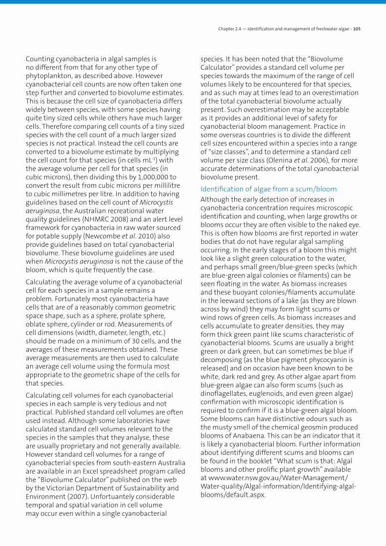
Counting cyanobacteria in algal samples is no different from that for any other type of phytoplankton, as described above. However cyanobacterial cell counts are now often taken one step further and converted to biovolume estimates. This is because the cell size of cyanobacteria differs widely between species, with some species having quite tiny sized cells while others have much larger cells. Therefore comparing cell counts of a tiny sized species with the cell count of a much larger sized species is not practical. Instead the cell counts are converted to a biovolume estimate by multiplying the cell count for that species (in cells mL-1) with the average volume per cell for that species (in cubic microns), then dividing this by 1,000,000 to convert the result from cubic microns per millilitre to cubic millimetres per litre. In addition to having guidelines based on the cell count of Microcystis aeruginosa, the Australian recreational water quality guidelines (NHMRC 2008) and an alert level framework for cyanobacteria in raw water sourced for potable supply (Newcombe et al. 2010) also provide guidelines based on total cyanobacterial biovolume. These biovolume guidelines are used when Microcystis aeruginosa is not the cause of the bloom, which is quite frequently the case.
Calculating the average volume of a cyanobacterial cell for each species in a sample remains a problem. Fortunately most cyanobacteria have cells that are of a reasonably common geometric space shape, such as a sphere, prolate sphere, oblate sphere, cylinder or rod. Measurements of cell dimensions (width, diameter, length, etc.) should be made on a minimum of 30 cells, and the averages of these measurements obtained. These average measurements are then used to calculate an average cell volume using the formula most appropriate to the geometric shape of the cells for that species.
Calculating cell volumes for each cyanobacterial species in each sample is very tedious and not practical. Published standard cell volumes are often used instead. Although some laboratories have calculated standard cell volumes relevant to the species in the samples that they analyse, these are usually proprietary and not generally available. However standard cell volumes for a range of cyanobacterial species from south-eastern Australia are available in an Excel spreadsheet program called the “Biovolume Calculator” published on the web by the Victorian Department of Sustainability and Environment (2007). Unfortuantely considerable temporal and spatial variation in cell volume may occur even within a single cyanobacterial
species. It has been noted that the “Biovolume Calculator” provides a standard cell volume per species towards the maximum of the range of cell volumes likely to be encountered for that species, and as such may at times lead to an overestimation of the total cyanobacterial biovolume actually present. Such overestimation may be acceptable as it provides an additional level of safety for cyanobacterial bloom management. Practice in some overseas countries is to divide the different cell sizes encountered within a species into a range of “size classes”, and to determine a standard cell volume per size class (Olenina et al. 2006), for more accurate determinations of the total cyanobacterial biovolume present.
Identification of algae from a scum/bloomAlthough the early detection of increases in cyanobacteria concentration requires microscopic identification and counting, when large growths or blooms occur they are often visible to the naked eye. This is often how blooms are first reported in water bodies that do not have regular algal sampling occurring. In the early stages of a bloom this might look like a slight green colouration to the water, and perhaps small green/blue-green specks (which are blue-green algal colonies or filaments) can be seen floating in the water. As biomass increases and these buoyant colonies/filaments accumulate in the leeward sections of a lake (as they are blown across by wind) they may form light scums or wind rows of green cells. As biomass increases and cells accumulate to greater densities, they may form thick green paint like scums characteristic of cyanobacterial blooms. Scums are usually a bright green or dark green, but can sometimes be blue if decomposing (as the blue pigment phycocyanin is released) and on occasion have been known to be white, dark red and grey. As other algae apart from blue-green algae can also form scums (such as dinoflagellates, euglenoids, and even green algae) confirmation with microscopic identification is required to confirm if it is a blue-green algal bloom. Some blooms can have distinctive odours such as the musty smell of the chemical geosmin produced blooms of Anabaena. This can be an indicator that it is likely a cyanobacterial bloom. Further information about identifying different scums and blooms can be found in the booklet “What scum is that: Algal blooms and other prolific plant growth” available at www.water.nsw.gov.au/Water-Management/Water-quality/Algal-information/Identifying-algal-blooms/default.aspx.
Chapter 2.4 — Identification and management of freshwater algae • 105

Toxicity of cyanobacteria and health risksCyanobacteria like many other algae and macrophytes produce a range of chemicals known as secondary metabolites which can act allelopathically against competitors, may be used to source various nutrients or may just be the resultant by-products of growth processes. The main categories of toxins (from Codd et al. 2005) are the:
• Neurotoxins - fast acting toxins that effect the nervous system;
• Hepatotoxins - damage the liver and promote tumour development;
• Cytotoxins – cause necrotic activity in mammals; and
• Irritants and gastrointestinal toxins - skin irritants, may cause inflammatory and gastrointestinal incidents.
Although all occur frequently, the hepatotoxins have been the most often implicated in cyanobacterial toxicoses.
Neurotoxins
There are many structural variants of saxitoxins known in cyanobacteria (particularly Anabaena circinalis in Australia) and marine dinoflagellates. These are carbamate alkaloids which block sodium channels and cause neurotoxicity. Saxitoxins (also known as paralytic shellfish poisons or PSPs) can have a LD50 in mice of 10-30 µg/kg body weight. Other neurotoxins include anatoxin-a and homoanatoxin-a which are postsynaptic, cholinergic neuromuscular blocking agents and are also alkaloids. They can have a LD50 in mice of 250 µg/kg body weight. Anatoxin-a(s)-like activity has recently been detected in samples from a range of Australian reservoirs (Humpage et al. 2013), but further work is required to confirm the presence of this or other anatoxins and homoanatoxins in Australia.
Beta-N-methylamine-L-alanine (BMAA) and its conger 2,4 diaminobutyric acid (DAB) are believed to be slow-acting neurotoxins that have been implicated with atypical motor neuron disease, Parkinsonism and Alzheimer’s-like dementia following gradual accumulation in brain tissues over many years. BMAA has been shown to be produced by a wide variety of (possibly all) cyanobacteria (Cox et al. 2009).
Hepatotoxins
These include the cyclic heptapeptide microcystins of which many structural variants are known. The most common are microcystin-LR, RR and YR. In this same category are the nodularins of which about 6 variants are known. These peptides inhibit protein phosphatases, cause liver damage and are tumour promoters.
Figure 2.4.2a. Cyanobacterial bloom (Anabaena circinalis) on a reservoir.
Figure 2.4.2b. Initial stages of an algal bloom with cells accumulating in wind rows.
Figure 2.4.2c. Bloom of the flagellated euglenoid Euglena sp.
Chapter 2.4 — Identification and management of freshwater algae • 106

Cytotoxins
These include the toxin cylindrospermopsin which is a guanidine alkaloid inhibitor of protein sysnthesis and causes necrotic (cell death) in mammals often in the liver, kidney, lungs, spleen and intestine.
A putative novel new cyanotoxin, limnothrixin, produced by Limnothrix spp. has also been identified recently (Humpage et al. 2013). It has been shown to destroy mammalian cells and to be toxic to mice. Its structure has yet to be determined.
Irritants and gastrointestinal toxins
Lipopolysaccharide endotoxins (LPS) are widely produced by cyanobacteria and can cause inflammatory and gastrointestinal incidents. Other irritants can cause skin irritation and potentially tumours.
Health effects for humans
The health effects of cyanobacterial blooms on humans are varied and there are both acute (short term) effects and chronic (long term) effects. As many symptoms are similar to other illnesses it can often be difficult to assign causality to cyanotoxins. However, there have been some serious acute health effects from cyanobacteria including the death of 88 people (mainly children) through drinking water supplies attributed to a massive toxic bloom of Microcystis and Anabaena in Brazil. In another case, 76 patients in a dialysis clinic that used water contaminated with hepatoxins died. Toxic effects of cyanobacteria on human health have been implicated for Palm Island, Queensland where 139 children and 10 adults were affected by drinking water with a Cylindrospermopsis bloom, and also for Malpas Dam, New South Wales. In South Australia, eight suspected cases of illness were associated with cyanobacteria, and documented for the summer of 1990/91. Conditions associated with cyanobacterial toxins exposure can be the development of a skin rash, conjunctivitis, abdominal pain, diarrhoea, vomiting-pneumonia like symptoms, headaches and breathing difficulty (Chorus and Bartram 1999).
There is also evidence for chronic effects from exposure to cyanobacterial toxins. There have been high incidences of primary liver cancer in China where people used pond and ditch water contaminated with microcystins as drinking water supplies. Communities that used well water (without toxins) had much lower rates of liver cancer. The lack of knowledge regarding the effects of cyanobacterial toxins on human health
and the nature of symptoms may allow affected individuals to go unreported. Symptoms can be easily attributed to other causes.
Toxic effects on aquatic organisms
Within the freshwater environment some of these secondary metabolites can cause toxic effects on other competing autotrophs such as algae and macrophytes. Zooplankton such as rotifers, cladocerans and copepods can be killed by the toxins and fecundity and growth can also be lowered, although not all genera and species within the same group have the same sensitivity (see De Mott et al. 1991). The death of fish during cyanobacterial blooms has been reported, but the links to cyanobacterial toxins are unclear. Hepatotoxin injection into fish has resulted in effects similar to those in mammals and fish killed during blooms have been shown to have liver damage. There have been many cases where water birds have also been killed as a result of toxins as well as other wild animals that rely on contaminated water sources.
There is also a risk for domestic animals with cyanobacterial blooms. There have been several recorded cases where dogs have been swimming in waters with toxic blooms and have died shortly afterwards from either neurotoxic or hepatoxic symptoms. Dogs seem to be particularly susceptible to toxins possibly exacerbated through the licking of their fur coats. Farm animals such as sheep and cattle are also prone to illness and death as they often rely on river water or farm dams for water supply and these can be contaminated with toxins.
Guidelines for cyanobacteria
Management guidelines targeted at protecting the health of humans using water sources contaminated with cyanobacterial blooms for potable water supply and for recreation have been produced internationally by the World Health Organisation (WHO) (Chorus and Bartram 1999), while similar guidelines taking account of more recent research are available for Australia
The Australian Drinking Water Guidelines (NHMRC and NRMMC 2011) provide information on several species of toxic cyanobacteria found in Australia and guidelines for long-term exposure to the toxins they produce. In addition, an Alerts Level Framework has been produced by Newcombe et al. (2010) which provides guidance on management actions required when cyanobacterial presence exceeds certain thresholds in raw waters that are sourced to supply potable water to urban communities.
Chapter 2.4 — Identification and management of freshwater algae • 107

For primary contact recreational activities, recreational guidelines have been provided by the National Health and Medical Research Council of Australia (NHMRC 2008). These provide for 3 levels of management and suggest actions that should be applied at each level – a Green level Surveillance mode when cyanobacteria are first detected at low numbers, an Amber level Alert mode that is implemented as cyanobacterial presence increases, and a Red level Action mode once cyanobacteria have reached bloom proportions and the water is no longer considered suitable for recreation without a potential adverse health risk to the users. The guidelines are based on cell counts of Microcystis aeruginosa and on concentrations of the toxin microcystin that may be present, but they also provide alternative guidelines based on total cyanobacterial biovolume present where blooms are comprised of species other than Microcystis aeruginosa.
Guidelines are also available for livestock watering, based mainly on cell counts of either Microcystis aeruginosa or Anabaena circinalis. In NSW, most management activities for cyanobacterial blooms in lakes, reservoirs and rivers are based on the recreational guidelines, as once these are exceeded, the guidelines for drinking water and livestock watering are also exceeded. Utilities and landholders with direct responsibility for potable supply and livestock watering should implement their management actions at the lower cyanobacterial concentrations relevant to those particular guidelines.
Management of freshwater algae
Causes of cyanobacterial bloomsThere are many causes to cyanobacterial blooms in freshwaters. In many cases a set of individual conditions can lead to a situation that allows a particular cyanobacteria to dominate and bloom. So in some ways each individual bloom situation needs to be studied to understand the environmental conditions that triggered the bloom. However, there are several factors that commonly contribute to the formation and the longevity / intensity of blooms.
Nutrients
One of the major factors that has led to the increased incidence and intensity of cyanobacterial blooms is increased nutrient loading of phosphorus and nitrogen to freshwaters through the process of eutrophication. This is largely an anthropogenic process whereby nutrients have entered freshwaters from a range of external and internal
sources. External sources include sewage inputs rich in P and N that have for many decades been discharged into rivers and lake systems as well as diffuse sources such as runoff from fertiliser application (such as super P) in agricultural areas. Erosion, soil carried in runoff and the loss of riparian vegetation have all contributed to the nutrient loads in freshwaters. In many deep reservoirs significant sources of P are also stored within the sediments and are released when bottom waters become anoxic during periods of thermal stratification. In many instances there are significant sources of P stored within the sediments meaning that even if all external inputs were stopped there would still be ample phosphorus available within the sediments.
Phosphorus is usually touted as the most important nutrient or the nutrient most limiting to algal growth in freshwaters. As such, phosphorus is often focused on more in both the causes and control of blooms. The relative nutrient levels can influence competition between different algal groups, as some smaller species with higher surface area to volume ratios are faster growers than larger celled algae such as cyanobacteria. So under low nutrient conditions, cyanobacterial dominance is often reduced. Nutrients can also influence algal growth as the biomass of algae and cyanobacteria is controlled by the nutrient availability (and other factors such as light). So in nutrient rich waters cyanobacteria (and other algae) can develop large biomass when conditions are right for blooms, with the potential of high toxin concentrations.
Light and water column stability
Sufficient light is required for phytoplankton to grow and to form high bloom densities. The availability of light is largely influenced by the mixing dynamics in lakes (i.e. is the lake stratified or not) and the light penetration within the water body. The depth to which algae are mixed is referred to as the mixing depth (Zmix) and is the depth of the water body when fully mixed, or is the depth of the thermocline (area of steep drop in temperature with depth) when a system is stratified. The depth to which enough light penetrates for algal growth is referred to as the euphotic depth (Zeu) and is the depth where only 1% of light at the water surface remains. The Zeu to Zmix ratio then dictates how much light is available for phytoplankton growth. When this ratio is above 1 phytoplankton are always within the light zone with plentiful light. When this ratio gets below 0.2, there is generally not enough light to enable positive growth of phytoplankton. So the greater the ratio, the more
Chapter 2.4 — Identification and management of freshwater algae • 108

light that is available for growth. Most algae are then reliant on the mixing energy within the water column to move into and out of the light region. Under these conditions (as all algae receive similar light levels) the faster growers tend to dominate (that is usually diatoms and small green algae).
Cyanobacteria have ways to overcome the limitations of slower growth and being out-competed by faster growers, at least during periods of reduced water column mixing. They have buoyancy regulation mechanisms (gas vacuoles) which some cyanobacteria use as a method of controlling their position in the water column. This is achieved through the presence of intracellular gas-filled spaces, which can change overall density relative to water, allowing the algae to be positively and negatively buoyant (Paerl 1988). The formation and buoyancy of gas vacuoles depends on an interaction between light and limiting nutrients and the accumulation of carbohydrates within the cell. In deeper waters where light intensity is low, production of gas vacuoles is most active, enabling the algae to return to the photic zone. Higher temperatures may also promote positive buoyancy and bloom formation among some cyanobacterial species.
Cyanobacteria using buoyancy mechanisms can form scum accumulations on water surfaces when numbers are sufficiently high. The formation of scums may be a mechanism by which cyanobacteria can dominate surface waters. Cyanobacteria have at least five Carbon Concentrating Mechanisms (CCMs) that enable them to sequester CO2 at low concentrations and also HCO3 and CO3 at high pH (Badger and Price 2003). At the water surface, cyanobacteria can utilise CO2 rather than HCO3 and CO3, as a photosynthesis carbon source (Paerl 1988). Under these conditions (often thermally stratified), supplies of free CO2 are depleted in the lower waters (where non-motile phytoplankton are trapped), and cyanobacteria use CO2 from the air -water interface. The scum may also shade other phytoplankton groups below the water surface reducing their light availability. Colonies of cyanobacteria avoid increased self shading by decreasing internal pigment concentration with increasing colony size, thereby allowing light to penetrate further down the water column. Above 10 mg chlorophyll-a per cubic metre, this effect becomes significant at inhibiting competitors (Paerl and Ustach 1982). If surface cyanobacteria
cannot accumulate enough carbohydrate to overcome buoyancy, persistent surface scums can be witnessed (Walsby et al. 1991).
As access to light by cyanobacteria using buoyancy is linked to water column stability, areas that stratify strongly for long periods are generally more prone to cyanobacterial blooms. This leads to lakes and reservoirs, ponds, wetlands and weir pools being particularly susceptible to blooms during the hotter periods (often October to April) when thermal energy is enough to maintain stratification. Often under stratified and stable water column conditions flagellates may also become dominant and may form surface accumulations. Flagellated phytoplankton can counteract weak mixing forces using motility, which allows them to be positioned at a preferred depth and to counteract sinking.
Conditions that reduce water column stability may act to reduce blooms such as high winds, lower temperatures and cloud cover. Increased water movement through increased flow velocities or mixing devices will also reduce water column stability and induce mixing of the water column.
Management of cyanobacterial blooms in wetlands and other small lakesMonitoring of cyanobacterial blooms
Due to the public and livestock/wildlife health issues associated with cyanobacterial blooms, it is important that regular monitoring of freshwaters be undertaken. Proactive sampling for algae and/or toxins is a way of ensuring that waters remain safe for drinking water and recreational uses. Ideally samples would be taken at various areas around the water body at weekly or fortnightly intervals. The number of samples would depend on the size and morphology of the water body, as well as including any areas that are frequently used for primary recreation or where water is extracted for other uses. When cyanobacterial numbers increase or toxins start to increase or are detected above guideline values the sampling frequency and spatial extent should be increased. Methods for sampling phytoplankton are covered in ‘Identification techniques’. For information on samples for toxin analysis and methods for determining toxins see Humpage et al. (2013), although it needs to be noted that there is no one ideal method of toxin analysis, with each method having its own strengths and weaknesses.
When cyanobacterial concentration or biomass exceeds guidelines then appropriate actions must be taken. These may include;
Chapter 2.4 — Identification and management of freshwater algae • 109

• Placement of warning signs advising of the risks associated with use of the water;
• Notification of the relevant authorities such as Regional Algal Coordinating Committees (RACC) in NSW;
• Media announcements either through the RACC or by the authority responsible for the water body; and
• Restriction of access and policing of the water body to ensure people are adhering to warnings.
Nutrient control
The control of phytoplankton by manipulating the nutrient availability in water bodies is termed ‘bottom up’ control. Methods of nutrient removal may be through reduction of nutrient inputs (i.e. sewage, better farming practices), regular removal of plant material that builds up (nutrients incorporated in plants are removed), increasing plant biomass in a system to take up nutrients or chemically treating the nutrients to make them less available (this may be achieved in the water column using alum or gypsum, or may be done at the sediments using a product such as Phoslock).
Caitcheon et al. (1994) predicted a reduction of 90% in sediment entering Chaffey Dam (Peel River, NSW) would be required to limit excess phytoplankton growth, so catchment management may not always be successful. The largest problem in using nutrient removal as a control method for cyanobacterial blooms is the ability to effectively reduce phosphorus to levels low enough to suppress bloom formation and stop replenishment from sediment bound sources. After a 50% reduction in phosphorus loading to Lake Ontario, no significant changes in phytoplankton biomass or species composition were found (Nicholls and Hurley 1989). Regardless of some of these findings, any reductions in nutrients entering a waterway will in the long run be a useful measure to combat eutrophication and potentially algal blooms.
Mixing of the water column
One frequently employed technique to reduce extensive periods of thermal stratification in a water body is to mix the water column using various methods. These can be quite expensive to install and run but can be effective at reducing cyanobacterial blooms that are related to thermal stratification i.e. summer blooms in deeper lakes and ponds. The water column can be mixed using a compressor that pumps air into a pipe with holes
in it that is placed low in the water column. The bubbles that are generated then rise to the surface and in the process mix the water column and stop thermal stratification from occurring. Alternatively fans that mix the water column may be utilised and pumps that either circulate water in small ponds or move bottom waters to the surface may be useful.
Biomanipulation
Some studies have shown a “top down” approach, often referred to as biomanipulation, where the food web is manipulated to allow organisms to feed on and remove cyanobacteria has been successful, particularly in the northern hemisphere. The basis of biomanipulation is that within an ecosystem, the survival and proliferation of one organism is often dependent on other organisms. The trophic interactions, which influence cyanobacterial biomass and composition, are predation by zooplankton and planktivorous fish and the abundance of these predators and their grazing habits.
Some Australian experiences have shown that biomanipulation may be possible. Kobayashi (1993) found the filtering rate of Daphnia carinata was sufficient to have some control over the growth of the blue-green algae Microcystis aeruginosa and Anabaena cylindrica. This was found to be true only if the cyanobacteria remained small enough and their morphology is suitable. Most Australian studies have shown problems with the application of biomanipulation with zooplankton, especially the ability of zooplankton to consume cyanobacteria. In overseas studies, the zooplankton responsible for most cyanobacterial consumption have been cladocerans such as Daphnia. Calanoid copepods and rotifers make up the majority of zooplankton in Australian waters. The ability of these zooplankton to effectively consume cyanobacteria has not been established. Cyanobacteria are also considered low food value for zooplankton and in the presence of toxic algae, preferential feeding by zooplankton on other more nutritionally beneficial algae may occur. Cyanobacteria may also clog the filter feeding apparatus of zooplankton and larger filaments and colonies of cyanobacteria are also less easily grazed by zooplankton. Toxins have been shown to be active against zooplankton.
Competition with macrophytes
There is evidence that competition of macrophytes with cyanobacteria can be successful in reducing cyanobacterial growth, though little is known about the interactions. In terms of freshwater management, one model of interest is the lakes
Chapter 2.4 — Identification and management of freshwater algae • 110
states model where one state is dominated by cyanobacteria (known as the phytoplankton or turbid water state) and the other state is dominated by macrophytes (known as the clear water state) (Scheffer et al. 2001). In lakes, allelopathy likely contributes to the overall strength of positive feedbacks that maintain alternative states. For example, many macrophyte species suppress phytoplankton through exudation of algicidal chemicals which can induce modifications in phytoplankton community structure away from cyanobacteria (Jasser 1995). These include diverse chemical classes such as oxygenated fatty acids, polyacetylenes, polyphenols and sulphur compounds. Similarly, many cyanobacteria suppress growth of macrophytes through exudation of deleterious chemicals such as the toxins. The effectiveness of exuded allelochemicals by macrophytes and cyanobacteria is dependent on environmental factors, such as light and nutrients. Although this area needs further research it is likely increased macrophyte density may help in reducing cyanobacteria by the above mechanisms but also by competing with algae for nutrients. Macrophytes also provide a supporting substrate for epiphytic algae (often diatoms), which may also compete with the cyanobacteria for available nutrients. Floating macrophytes and to a lesser extent the emergent and submerged macrophytes will shade water systems and light-limit phytoplankton growth.
Summary
Freshwater algae are important components of water systems as they form the base of the aquatic food chain. Up to 8 groups of algae, all very different from each other, occur commonly in freshwater ecosystems. Of these, blue-green algae, actually a type of bacteria, are of major management concern. Nutrient enrichment and resultant eutrophication can lead to algal blooms and loss of algal diversity causing ecosystem stress. The cyanobacteria in particular can form large blooms and some species can produce potent toxins. This can render waters unsafe for drinking supplies, recreational use and livestock watering. Monitoring of freshwaters for algae involves sample collection and subsequent microscopic analysis of these samples, and can involve considerable effort when done adequately. However monitoring of waterways is very important to ensure waters stay within safe guidelines for drinking, recreational or stock watering uses. If guidelines are exceeded actions need to be taken to protect humans and animals from the toxins. Causes of blooms are usually
water body specific but some common elements are high phosphorus levels and high temperatures. Also, a stable water column such as during thermal stratification in lakes and weir pools can allow cyanobacteria to utilise buoyancy mechanisms to access a greater amount of light. Management of algal blooms can involve reduction in nutrient loading, promoting competition by introducing macrophytes and reducing water column stability through mixing of the water column such as with bubble plume aerators. Management of algal blooms requires a coordinated approach that combines short term actions such as monitoring and public safety responses to toxic blooms with longer term actions such as nutrient control and research into the causes of blooms.
References
Al-Tebrineh, J., Merrick, C., Humpage, A., Bowling L., and Neilan, B. A. (2012). Community composition, toxigenicity and environmental conditions during a cyanobacterial bloom occurring along 1,100 kilometers of the Murray River. Applied and Environmental Microbiology 78, 263–272.
Badger, M. R., and Price G. D. (2003) CO2 concentrating mechanisms in cyanobacteria: molecular components, their diversity and evolution. Journal of Experimental Botany 54, 609–622.
Baker, P. D. (1991). Identification of common noxious cyanobacteria. Part I – Nostocales. Research Report No. 29, Urban Water Research Association of Australia, Melbourne, Australia.
Baker, P. D. (1992). Identification of common noxious cyanobacteria. Part II – Chroococcales, Oscillatoriales. Research Report No. 46, Urban Water Research Association of Australia, Melbourne, Australia.
Baker, P. D., and Fabbro, L. D. (1999). A guide to the identification of common blue-green algae (Cyanoprokaryotes) in Australian freshwaters. Identification Guide No. 2, Cooperative Research Centre for Freshwater Ecology, Albury, Australia.
Bowling, L. C., Merrick, C., Swann, J., Green, D., Smith, G., et al. (2013). Effects of hydrology and river management on the distribution, abundance and persistence of cyanobacterial blooms in the Murray River, Australia. Harmful Algae (in press).
Chapter 2.4 — Identification and management of freshwater algae • 111
Caitcheon, G. G., Donnelly, T. H., Wallbrink, P. J., and Murray, A. S. (1994) Sources of phosphorus and sediment in the catchment of Chaffey Reservoir, New South Wales, Technical Report, CSIRO Division of Water Resources, Canberra ACT.
Carmichael, W. W., Mahmood, N. A., and Hyde, E. G. (1990) Natural toxins from cyanobacteria (blue-green algae). In ‘Marine toxins: origin, structure, and molecular pharmacology American Chemical Society’.
Chorus, I., and Bartram, J. (1999) Toxic cyanobacteria in water, a guide to their public health consequences, monitoring and management, World Health Organisation Publication, E and F Spon Publishers, London.
Cox, B. A., Richer, R., Metcalf, J. S., Banack, S. A., Codd, G. A., et al. (2009). Cyanobacteria and BMAA exposure from desert dust: A possible link to sporadic ALS among Gulf War veterans. Aymotrophic Lateral Sclerosis 10, 109–117.
De Mott, W. R., Zhang, Q. X., and Carmichael, W. W. (1991) Effects of toxic cyanobacteria and purified toxins on the survival and feeding of a copepod and three species of Daphnia. Limnology and Oceanography 36, 1346–1357.
Humpage, A., Gaget, V., Lau, M., Froscio, S., and Laingam, S. (2013). ‘CyanoSurvey: A national update on toxic cyanobacteria and their distribution. Research Project 1022.’ (Water Research Australia: Adelaide, Australia.)
Jasser, I. (1995) The influence of macrophytes on a phytoplankton community in experimental conditions. Hydrobiologia 306, 21–32.
Kobayashi, T. (1993) Filtering rates of Daphnia carinata King (Crustacea: Cladocera) on the blue-green algae Microcystis aeruginosa (Kutz.) and Anabaena cylindrica (Lemm.). Australian Journal of Ecology 18, 231–234.
McGregor, G. B. (2007). Freshwater cyanoprokaryota of North-Eastern Australia. In ‘Oscillatoriales. Flora of Australia Supplementary Series Number 24’. (Australian Biological Resources Study: Canberra, Australia.)
McGregor, G. B., and Farrro, L. D. (2001). A guide to the identification of Australian freshwater planktonic Chroococcales (Cyanoprokaryota/Cyanobacteria) Identification Guide No. 39. Cooperative Research centre for Freshwater Ecology, Albury, Australia.
Newcombe, G., House, J., Ho, L., Baker, P. and Burch, M. (2010). Management strategies for cyanobacteria (blue-green algae): A guide for water utilities. Research Report No. 74, Water Quality Research Australia, Adelaide, Auistralia.
NHMRC (2008). ‘Guidelines for managing risks in recreational waters.’ (National Health and Medical Research Council: Canberra, Australia.)
NHMRC and NRMMC (2011). National Water Quality Management Strategy. Australian Drinking Water Guidelines 6. 2011. National Health and Medical Research Council and Natural Resource Management Ministerial Council, Canberra, Australia.
Nicholls, K. H., and Hurley, D. A. (1989) Recent changes in the phytoplankton of the Bay of Quinte, Lake Ontario: the relative importance of fish, nutrients and other factors. Canadian Journal of Fisheries and Aquatic Sciences 46, 179–186.
Olenina, I., Hajdu, S., Edler, L., Andersson, A., Wasmund, N., et al. (2006). Biovolumes and size-classes of phytoplankton in the Baltic Sea. Baltic Marine Environment Protection Commission – Helsinki Commission. Helsinki, Finland.
Paerl, H. W. (1988) Freshwater blue-green algal ecology. In ‘Growth and Reproductive strategies of freshwater phytoplankton’. (Ed C. Sandgren.) (Cambridge University Press.)
Paerl, H. W., and Ustach, J. F. (1982) Blue-green algal scums: An explanation for their occurrence during Freshwater blooms. Limnology and Oceanography 27, 212–217.
Scheffer, M., Carpenter, S., Foley, J. A., Folke, C., and Walker, B. (2001). Catastrophic shifts in ecosystems. Nature 413, 591–596.
Victorian Department of Sustainability and Environment (2007). ‘Biovolume Calculator.’ Available at http://www.dse.vic.gov.au/dse/index.htm.
Walsby, A. E., Kinsman, R., Ibelings, B. W., Reynolds, C. S. (1991) Highly buoyant colonies of the cyanobacterium Anabaena lemmermanni form persistent water blooms. Archiv Fur Hydrobiologie 121, 261–280.
Chapter 2.4 — Identification and management of freshwater algae • 112