Embed Size (px)
Citation preview
Genetics
Tyler Reimschisel, MD, MHPEAssociate Professor of Pediatrics and Neurology
Vanderbilt University Medical Center, Nashville, TNMarch 4, 2017
Conflict of Interest
• Neither I nor any member of my immediate family has a financial relationship or interest with any entity producing, marketing, re-selling, or distributing health care goods or services consumed by, or used on, patients.
• I do not intend to discuss an unapproved/investigative use of a commercial product/device.
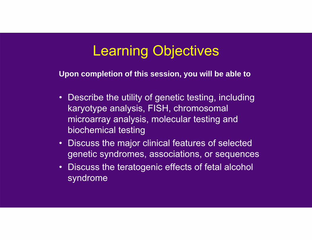
Learning ObjectivesUpon completion of this session, you will be able to
• Describe the utility of genetic testing, including karyotype analysis, FISH, chromosomal microarray analysis, molecular testing and biochemical testing
• Discuss the major clinical features of selected genetic syndromes, associations, or sequences
• Discuss the teratogenic effects of fetal alcohol syndrome
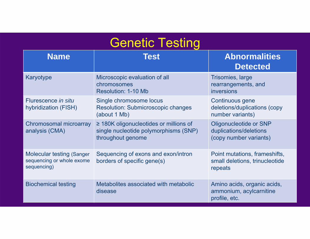
Genetic TestingName Test Abnormalities
DetectedKaryotype Microscopic evaluation of all
chromosomes Resolution: 1-10 Mb
Trisomies, large rearrangements, and inversions
Flurescence in situhybridization (FISH)
Single chromosome locusResolution: Submicroscopic changes (about 1 Mb)
Continuous gene deletions/duplications (copy number variants)
Chromosomal microarray analysis (CMA)
≥ 180K oligonucleotides or millions of single nucleotide polymorphisms (SNP) throughout genome
Oligonucleotide or SNP duplications/deletions(copy number variants)
Molecular testing (Sanger sequencing or whole exome sequencing)
Sequencing of exons and exon/intron borders of specific gene(s)
Point mutations, frameshifts, small deletions, trinucleotide repeats
Biochemical testing Metabolites associated with metabolic disease
Amino acids, organic acids, ammonium, acylcarnitineprofile, etc.
Genetic Syndromes & Teratogens
• HIPAA and copyright guidelines limit use of pictures in handouts
• Pictures do not reproduce well in handouts
• Handout does not include most pictures• Google Images is good resource for
representative images of syndromes
Trisomy 13• Associated with AMA• Microcephaly• Microophthalmia• Heart malformations• Ear anomalies• Severe growth and developmental impairment• Cutis aplasia• Holoprosencephaly• Orofacial clefting• Polydactyly (postaxial)• 45% die in first month, 85% in first year
Trisomy 18• Associated with AMA• Microcephaly• Microophthalmia• Heart malformations• Ear anomalies• Severe growth and developmental impairment• Prominent occiput• Micrognathia• Overlapping fingers• Hypoplastic nails• Clubfoot• Seizures• 50% die in first week, 95% in first yr

Klinefelter Syndrome• 80%: XXY• Remainder mosaic or variable sex chromosome
number (eg. XXYY, XXXY, XXXYY)
• Average or above average adult height• 50% with delayed speech• Average IQ with mild learning disability• 30% with delayed emotional development• Adults may have behavior problems
Klinefelter Syndrome• Hypergonadotropic hypogonadism• Most are sterile• Accounts for 5-15% of sterility in males• Sparse facial hair• Gynecomastia• Increased risk of carcinoma of breast and mediastinal
tumors• 20% with major malformations (no pattern of
anomalies)• Treat with androgen replacement
Turner Syndrome• 1/ 4,000 newborns• Causes:
– 50%: 45,X – 25%: RingX or isoX– 25%: Mosaic (45,X/46,XX or XY)
• Short, broad, webbed neck• Broad chest, widely spaced nipples• May only cause growth restriction• Gonadal dysgenesis common• May have hypothyroidism• Ovarian hormone replacement required• Growth hormone may be provided
Turner Syndrome• Heart anomalies (coarctation)• Can have renal anomalies• Autoimmune disease more common
(diabetes, IBD)• IQ is normal, but may have cognitive
impairment• Delayed speech• LD• Visual-spatial problems
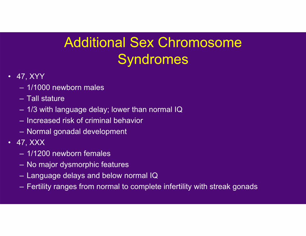
Additional Sex Chromosome Syndromes
• 47, XYY– 1/1000 newborn males– Tall stature– 1/3 with language delay; lower than normal IQ– Increased risk of criminal behavior– Normal gonadal development
• 47, XXX– 1/1200 newborn females– No major dysmorphic features– Language delays and below normal IQ– Fertility ranges from normal to complete infertility with streak gonads
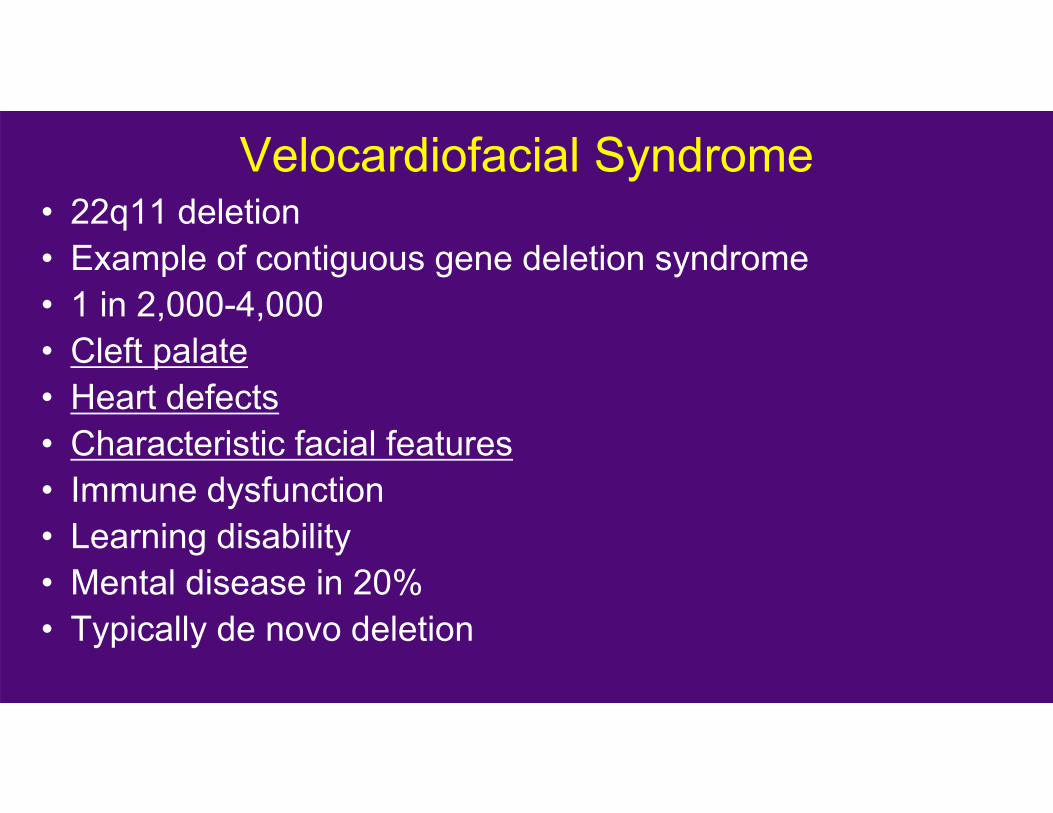
Velocardiofacial Syndrome• 22q11 deletion• Example of contiguous gene deletion syndrome• 1 in 2,000-4,000• Cleft palate• Heart defects• Characteristic facial features• Immune dysfunction• Learning disability• Mental disease in 20%• Typically de novo deletion
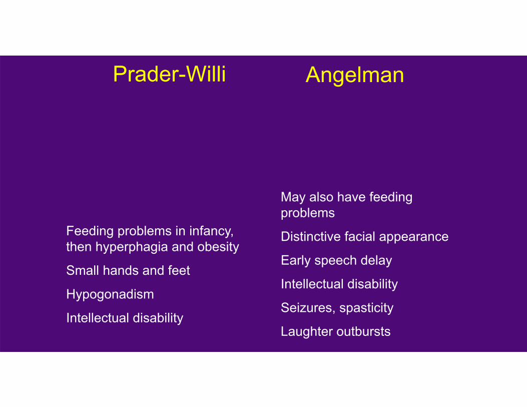
Prader-Willi
Feeding problems in infancy, then hyperphagia and obesity
Small hands and feet
Hypogonadism
Intellectual disability
Angelman
May also have feeding problems
Distinctive facial appearance
Early speech delay
Intellectual disability
Seizures, spasticity
Laughter outbursts
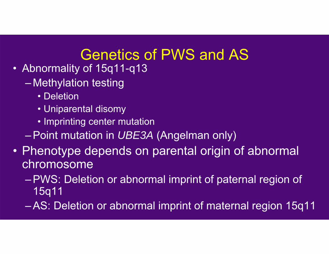
Genetics of PWS and AS• Abnormality of 15q11-q13
– Methylation testing• Deletion• Uniparental disomy• Imprinting center mutation
– Point mutation in UBE3A (Angelman only)• Phenotype depends on parental origin of abnormal
chromosome– PWS: Deletion or abnormal imprint of paternal region of
15q11– AS: Deletion or abnormal imprint of maternal region 15q11
Beckwith-Wiedemann syndrome• Overgrowth syndrome
– Macrosomia, macroglossia– Hemihyperplasia– Visceromegaly
• Ear creases/pits• Omphalocele• Renal anomalies• Neonatal hypoglycemia• Embryonal tumors
– Wilms, hepatoblastoma, neuroblastoma, rhabdomyosarcoma– AFP every 6 weeks, abd U/S every 3 months until 8yo
• Genetics: CDKN1C mutation, 11p15 methylation abnormality
Neurofibromatosis Type I• Autosomal dominant• NIH Criteria (2 or more)
– ≥ 6 café au lait macules• > 5mm in prepubertal• >15 mm in postpubertal
– > 2 neurofibromas or > 1 plexiform neurofibroma
– Axillary freckling– ≥ 2 Lisch nodules– Tibial pseudoarthrosis– Optic glioma– Family history Photos from GeneTests.org
Neurofibromatosis Type I• Café au lait macules can increase in number during first few
years of life• Optic gliomas typically develop in first 6 years of life• 50% with learning disability• Progressive scoliosis usually only seen at 6-10 years of age• Hypertension is common in adults• Malignant peripheral nerve sheath tumors in 10%• Increased risk for cerebral vasculopathy• Somewhat increased risk for leukemia and other tumors (GI
tumors)
Tuberous Sclerosis• Autosomal dominant• Mutation in TSC1 or TSC2• Skin
– Facial angiofibromas– Hypomelanotic macule– Shagreen patch– Ungual fibromas
Tuberous Sclerosis• Brain
– Cortical dysplasias– Subependymal nodules– Subependymal giant cell astrocytomas (SEGAs)– Epilepsy, autism, ADHD, LD, ID
• Kidney: angiomyolipomas, cysts, renal cell carcinoma
• Heart: rhabdomyomas, dysrhythmias• Lungs: lymphangioleiomyomatosis (LAM) in
women• Increased risk for neuroendocrine tumors
Ehlers-Danlos• Autosomal dominant• Hetergeneous group of connective tissue
disorders• Classic type (Type I)
– Skin hyperextensibility– Atrophic scars– Joint hypermobility– Smooth, velvety skin– Easy bruising– Hernia, rectal prolapse
Ehlers-Danlos Type IV
• Vascular type– Autosomal dominant– Mutation in COL3A1– Arterial, intestinal or uterine rupture– Translucent skin– Joint hypermobility with subluxations– Easy bruising
Marfan Syndrome• Autosomal dominant• Mutation in Fibrillin-1• Diagnostic criteria (> 2)
– Family history– Skeletal system– Ocular system– Cardiovascular– Lumbosacral dural ectasia– Pulmonary (minor criteria)
• Pneumothorax• Apical blebs
Marfan Syndrome– Skeletal system
– Arachnodactyly– Pectus carinatum or excavatum– Wrist and thumb signs– Typical facial features– Pes planus
– Ocular system– Ectopia lentis– Miosis
– Cardiovascular– Progressive aortic root dilatation– Treat with beta-blocker
Osteogenesis Imperfecta• “Brittle bone disease”• Autosomal dominant• Mutations in COL1A1 or COL1A2 cause
defect in a major structural protein of bone and fibrous tissue
• Bone fragility leads to fractures• Progressive bone deformities• Short stature• Blue sclera• Poor dentition• Hearing impairment• Spectrum of severity (Types I-IV)• In differential diagnosis for NAT
Achondroplasia• Autosomal dominant• Mutation in FGFR3
– 98%: G to A at 1138– 2%: G to C at 1138
• Glycine to arginine at codon 380• Rhizomelic short stature• Enlarged head• Flat nasal bridge
Achondroplasia• Short fingers and trident hand• Short vertebral bodies• Interpediculate distance narrowing in lumbar spine• Anterior wedging of vertebrae – kyphosis• Normal cognition• Delayed motor development• Increased rate of sudden death in infancy and early childhood• Narrow spinal cord can cause compression• Otitis media and hearing loss
Bardet-Biedl• Autosomal recessive (may be oligogenic)• Truncal obesity• Rod-cone dystrophy (night, then legal, blindness)• Postaxial polydactyly• Hypogonadism (males)• Genital abnormalities (females)• Renal abnormalities lead to ESRD• Global developmental delay, learning disability, ataxia• Anosmia
CHARGE Syndrome• Autosomal dominant• 60-70% with mutations in CHD7• Coloboma• Heart defect• Atresia choanae• Retarded growth/ development • (Cranial neuropathies)• Genital anomalies/ hypogonadism• Ear anomalies/deaf
CHARGE Syndrome
• Other associated features– Developmental delays and hypotonia– May have behavior problems– Feeding difficulties– Growth deficiency (70-80%)– Orofacial clefts (15-20%)– Tracheoesophageal fistula (15-20%)– Other life-threatening conditions in infancy
Treacher Collins Syndrome
• Mandibulofacial dysostosis• Treacle or TCOF1• Downslanting palpebral fissures with coloboma
of outer third of lower lid• Malformed pinnae and middle ear malformations• Nose appears large due to malar and
supraorbital ridge hypoplasia• Cleft palate, rarely CL/P
Apert/Crouzon• Mutation in FGFR2• Coronal craniosynostosis• Flat faces• Midface hypoplasia• Proptosis• Hearing loss• Syndactyly• Heart anomalies• Variable IQ
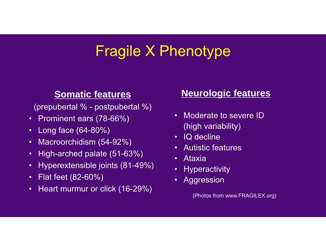
Fragile X Phenotype
Neurologic features
• Moderate to severe ID (high variability)
• IQ decline• Autistic features • Ataxia• Hyperactivity• Aggression
(Photos from www.FRAGILEX.org)
Somatic features(prepubertal % - postpubertal %)
• Prominent ears (78-66%)• Long face (64-80%)• Macroorchidism (54-92%)• High-arched palate (51-63%)• Hyperextensible joints (81-49%)• Flat feet (82-60%)• Heart murmur or click (16-29%)
Fragile X Phenotype
• Females usually less affected than males due to X-inactivation• Phenotype depends on the number of cells that contain normal X as active chromosome (activation ratio) • Phenotype ranges from normal to severely affected • Severity of intellectual disability correlates with prominence of dysmorphic features
Fragile X testing is indicated for girls with GDD/ID(Photos from www.FRAGILEX.org)
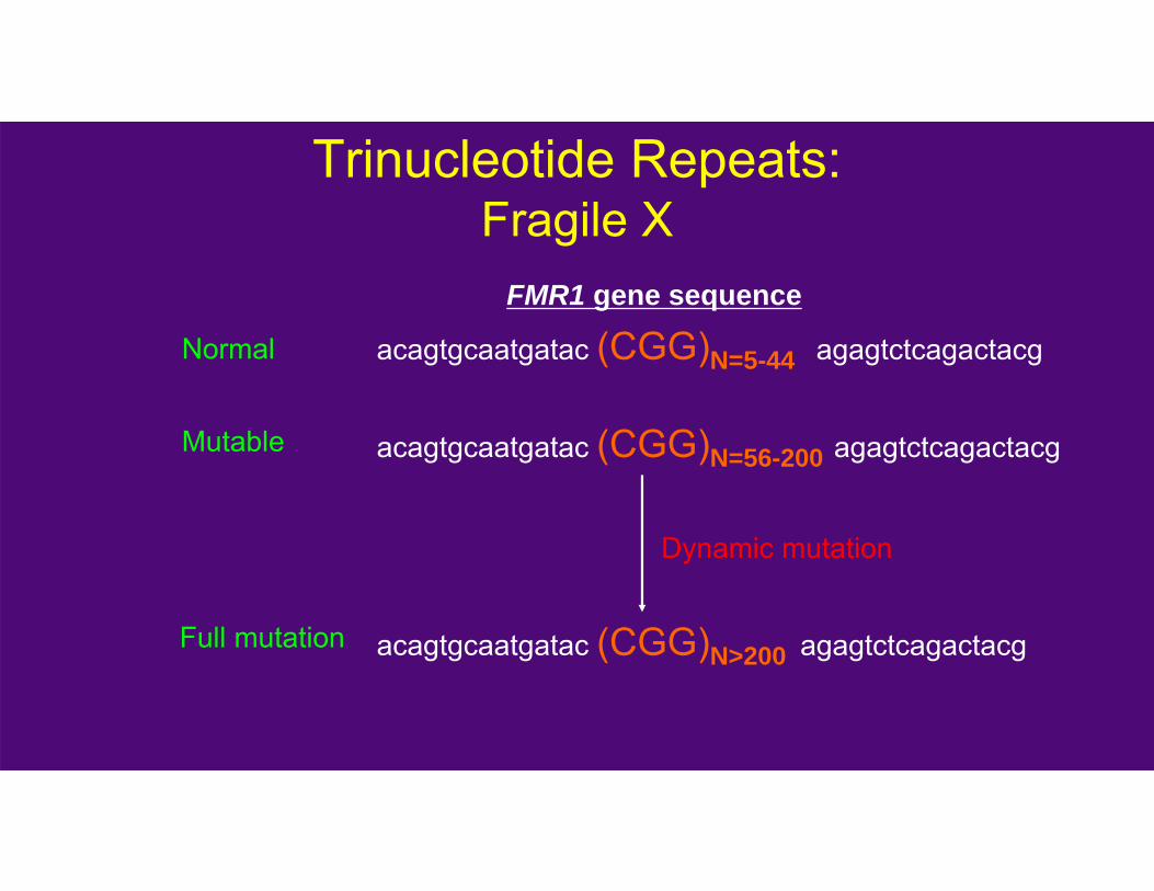
Trinucleotide Repeats:Fragile X
acagtgcaatgatac (CGG)N=5-44 agagtctcagactacg
acagtgcaatgatac (CGG)N=56-200 agagtctcagactacg
acagtgcaatgatac (CGG)N>200 agagtctcagactacg
FMR1 gene sequence
Normal
Mutable
Full mutation
Dynamic mutation
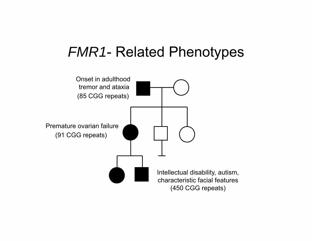
FMR1- Related Phenotypes
Intellectual disability, autism, characteristic facial features
(450 CGG repeats)
Onset in adulthood tremor and ataxia(85 CGG repeats)
Premature ovarian failure(91 CGG repeats)
Muscular DystrophiesDuchenne and Becker
• Childhood onset of progressive myopathy• X-linked (mutations in dystrophin gene)• Cognitive impairment is not rare• Pseudohypertrophy and Gowers sign• Massively elevated CPK (diagnostic)• Genetic testing available• Treatment
– Physical therapy and nutrition– Steroids seem to improve strength and maintain ambulation
• Prognosis death/ventilator dependence by adulthood for Duchenne, survival into adulthood for Becker type.
Rett Syndrome• Neurodegenerative disease in girls• Mutations in MECP2 on X chromosome• 1-3/10,000 live births• Acquired microcephaly• GDD, then regression• Autistic-like behavior • Seizures, ataxia, hyperventilation episodes • Stereotypic hand movements
(From website [email protected])
VACTERL Association• Cause is usually unknown (not a syndrome)• Diagnostic criteria - 3/7 following features:• Vertebral anomalies• Anal atresia• Cardiac anomalies (TOF, transposition, VSD)• TracheoEsophageal fistula or esophageal atresia• Renal anomalies (aplasia, hydronephrosis)• Limb anomalies (radial ray anomalies, polydactyly, syndactyly,
triphalangeal thumb)• Growth deficiency• Tethered cord, occipital encephlocele• Increased incidence in infants of diabetic mothers
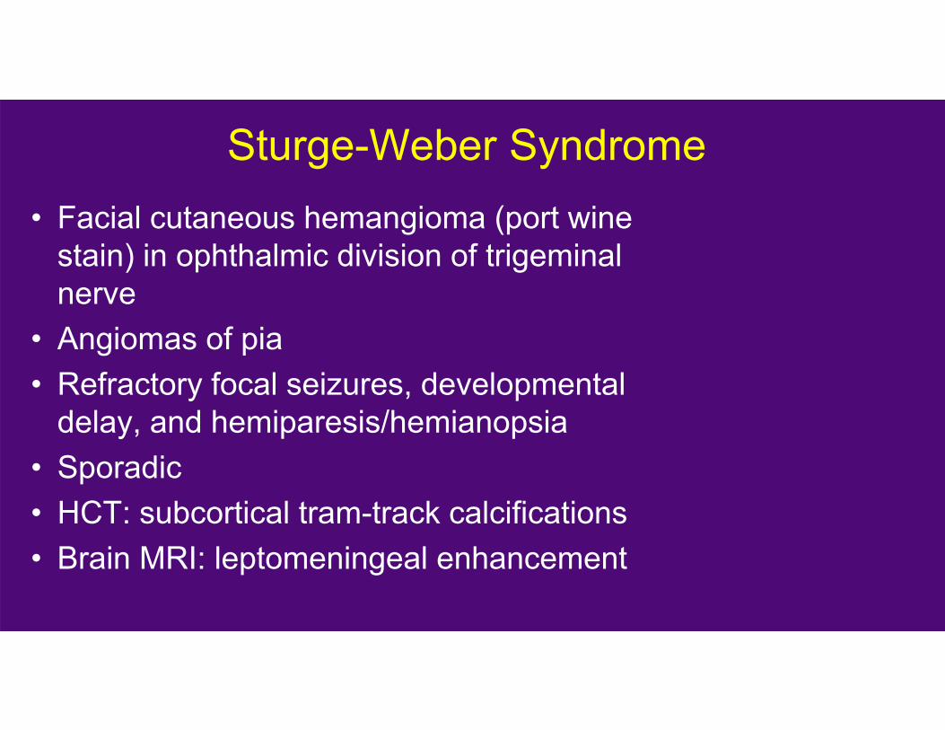
Sturge-Weber Syndrome• Facial cutaneous hemangioma (port wine
stain) in ophthalmic division of trigeminal nerve
• Angiomas of pia• Refractory focal seizures, developmental
delay, and hemiparesis/hemianopsia• Sporadic• HCT: subcortical tram-track calcifications• Brain MRI: leptomeningeal enhancement
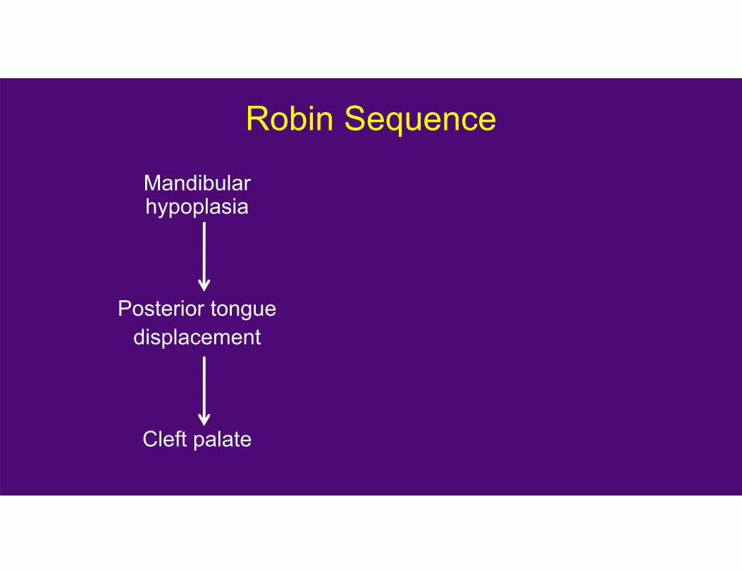
Robin Sequence
Mandibular hypoplasia
Posterior tonguedisplacement
Cleft palate
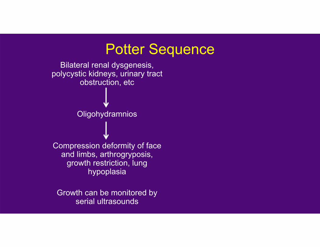
Potter SequenceBilateral renal dysgenesis,
polycystic kidneys, urinary tract obstruction, etc
Oligohydramnios
Compression deformity of face and limbs, arthrogryposis,
growth restriction, lung hypoplasia
Growth can be monitored by serial ultrasounds
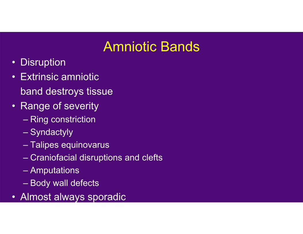
Amniotic Bands• Disruption• Extrinsic amniotic
band destroys tissue• Range of severity
– Ring constriction– Syndactyly– Talipes equinovarus– Craniofacial disruptions and clefts– Amputations– Body wall defects
• Almost always sporadic
Teratology• Adverse effects of environmental toxin (teratogens) on
growth and development of embryo• Drugs, infection, radiation, maternal disease• Time of exposure is usually important (except for alcohol)
– Critical time is 14-60 days – Effect can differ based on timing
• Recognizable pattern of abnormalities• Threshold and dose-response• Species-specific• Pathogenesis largely unknown
Fetal Alcohol Syndrome• In utero alcohol exposure• 1/30 women abuse alcohol• 6% of their children have FAS• Mild to moderate microcephaly• Dysmorphic face• Heart defects• Pre- and postnatal growth restriction• CNS malformations• Intellectual disability and learning disorders• Behavior problems
References• Gene Reviews: www.genetests.org• Gorlin’s Syndromes of the Head and Neck, 2nd
Edition• Online Mendelian Inheritance in Man
www.ncbi.nlm.nih.gov/omim• Smith’s Recognizable Patterns of Human
Malformations, 6th Edition• Thompson and Thompson’s Genetics in Medicine,
6th Edition
Change in Practice Opportunities
• Review the key features of common genetic syndromes• Confirm all patients with intellectual disability have a
chromosomal microarray analysis and Fragile X testing• Correlate specific genetic conditions with the genetic
testing that would be performed to diagnose conditions• Utilize genetic references to help diagnose genetic
conditions