Embed Size (px)
Citation preview

© Copyright 2013 Elsevier, Ltd. All rights reserved.
4 Clinical diagnosis of soft tissue lesions
CHAPTER CONTENTS
Introduction . . . . . . . . . . . . . . . . . . . . . . . . . 53
Clinical .evaluation . . . . . . . . . . . . . . . . . . . . . 57
History . . . . . . . . . . . . . . . . . . . . . . . . . 57Inspection . . . . . . . . . . . . . . . . . . . . . . . 62Preliminary examination . . . . . . . . . . . . . . . . 63Functional examination . . . . . . . . . . . . . . . . 63Accessory tests . . . . . . . . . . . . . . . . . . . . 68Palpation . . . . . . . . . . . . . . . . . . . . . . . 69Diagnostic infiltration or aspiration . . . . . . . . . . 72Technical investigations . . . . . . . . . . . . . . . . 72
Interpretation . . . . . . . . . . . . . . . . . . . . . . . . 72
Impairment of active movements . . . . . . . . . . . 73Impairment of passive movements . . . . . . . . . . 75Impairment of resisted movements . . . . . . . . . . 79Absence of pain on functional testing. . . . . . . . . 80Summary . . . . . . . . . . . . . . . . . . . . . . . 80Diagnostic difficulties . . . . . . . . . . . . . . . . . 80
Introduction
The major part of this book is about making a clinical diagnosis: a system of clinical reasoning leading to a proper diagnosis. The final stage of the diagnostic procedure is the precise anatomical description of the lesion, for example: supraspinatus tendinitis at the superficial aspect of the tenoperiosteal junction, chronic subdeltoid bursitis, lesion at the origin of the extensor carpi radialis brevis muscle, periostitis at the anteroinferior surface of the fibula, annular disc protrusion at the L4–L5 level irritat-ing the fifth lumbar nerve root.
During the last decades, new technology has revolutionized diagnosis and decision making in orthopaedic medicine. Previ-ously, soft tissue lesions were characterised by a lack of objec-tive findings. This has changed dramatically since the arrival of sonography, computed tomography and magnetic resonance imaging (MRI). These new techniques can demonstrate ana-tomical changes in soft tissues and therefore contribute signifi-cantly to the understanding of non-osseous orthopaedic lesions. However, they do not make clinical assessment redundant. Contrary to popular belief, diagnosis is not made by only looking at the result of a technical investigation. In the best case, an anatomical picture may be the ultimate confirmation of the clinical diagnosis. If imaging is undertaken too early during the diagnostic process it will create more problems and questions than it resolves, and often puts the examiner on the wrong track and leads to wrong therapeutic decisions.
First of all, not every detected anatomical lesion causes pain or dysfunction. Asymptomatic lesions do exist and are present in numbers that are much larger than previously assumed: asymptomatic herniations in cervical, thoracic and lumbar spine are present in up to 50% of the population.1,2,3 Also the high prevalence of rotator cuff tears in elderly asymptomatic individuals is very well known.4,5 It is estimated that in the general population, approximately two-thirds of all rotator cuff tears are asymptomatic.6,7 Large numbers of asymptomatic lesions have also been demonstrated in the knee. A recent MRI study on asymptomatic soccer players demonstrated one or more MRI abnormalities in no less than 64%. Another study with MRI scans performed on the knees of asymptomatic male professional basketball players demonstrated an overall prevalence of articular cartilage lesions of 47.5%8 and meniscal lesions of 20%.9
Another shortcoming of technical investigations is that they only detect an anatomical lesion (defect, swelling or other structural changes) and not the functional deficiency (weak-ness, limitation, laxity). In other words, the behaviour of the tissue during activity is not assessed.
General Principles
54
2 . Look .for .objective .physical .signs
Examination of the moving parts is an exercise in applied anatomyThe examination should include questioning (history taking) and testing which provokes or elicits symptoms and/or signs that can be assessed, judged and interpreted as objectively as possible.
The soft tissues of the locomotor system have the advantage that their functional anatomy is well known. It is clear how joints behave, how capsules and ligaments guide and limit movements, how muscles function and what movements they provoke.
Therefore, examination of the moving parts is an exercise in applied anatomy. Each tissue or group of tissues in turn must be tested and the answer interpreted in the light of the ana-tomical possibilities.
3 . Avoid .palpation .as .much .as .possible
The function of the different tissues is knownAlthough palpation is very often used as a diagnostic proce-dure, it is unreliable for several reasons:
• Some regions in the body are always tender to touch (e.g. lesser tuberosity at the shoulder, lateral epicondyle at the elbow, border of the trapezius muscle).
• Some structures lie too deeply and cannot be reached by the palpating finger (e.g. capsule of the hip joint, cruciate ligaments at the knee).
• The painful area does not always correspond to the site of the lesion (referred pain) and referred dural tenderness is sometimes present.
• Some patients with altered perception or desire to deceive the examiner may produce misleading responses.
It is easy to understand that, in these circumstances, palpation offers no help at all or, even worse, may misdirect the examiner.
Diagnosis, therefore, rests largely on the correlation of a series of semi-subjective data, obtained from a proper func-tional examination – an indirect approach. By assessing the function of each tissue in turn and interpreting the signs in the light of the anatomical knowledge, the examiner should be able to come to a correct description of the lesion.
The patient is asked to answer some very precise questions. A patient with an organic lesion exactly describes what is felt and gives the examiner a fairly precise clinical picture. The neurotic or malingering patient will feel the need to embellish so as to give a colourful description of suffering rather than of the symptoms.
4 . Functional .testing: .the .principle .of . .‘selective .tension’
The soft tissues can be put under tensionThe different tissues of the moving parts can be subjected to strain which may increase the pain and tests are used to elicit or influence the patient’s symptoms.
For all these reasons, there is no place for high-tech visuali-zation techniques in the beginning of the diagnostic process and they should never be used as screening tests. A biased examiner who is looking for a particular lesion will often find that lesion, whether it is responsible for the complaints or not. Too many and too early technical investigations substantially increase the cost of medical care but do not give a better outcome. On the contrary, in the hands of an unprofessional doctor, high-tech investigations are potentially dangerous as they may lead to major, unwanted and unnecessary surgery.
Principles of diagnostic procedure in orthopaedic medicine
Clinical examination is all about behaviour of the tissues involved. The examiner must have a very good knowledge of the behaviour of the lesions that he is dealing with and of the behaviour of the normal tissue. Tissue behaviour is described by the patient during the inquiry and checked by the examiner during the functional examination. Looking for tissue behav-iour, the following general principles are important.
1 . Look .for .‘inherent .likelihoods’
Some things are likely to happenSoft tissue lesions behave in a very typical way and the exam-iner will therefore regularly be faced with the same history and the same response to functional testing. The symptoms and signs are closely related to the lesion present. The examiner should therefore try to recognize ‘inherent likelihoods’, a term defined as the sequence of symptoms and/or signs that belong to the clinical picture of a certain pathological disorder and that are likely to be found, more or less in a sequence which is typical for that disorder.
For example: in the history, a patient with lumbar pain may mention that on some days the pain spreads down the lower limb; tennis elbow is characterized by sudden twinges when objects are picked up; and in lumbar disc lesions pain may shift from one side to the other.
Functional examination can also show some inherent likeli-hoods. When resisted extension of the wrist hurts at the elbow, a tennis elbow is suspect and can be confirmed by positive responses to resisted extension of the wrist with the fingers held actively flexed and to resisted radial deviation of the wrist. In tendinitis at the radial insertion of the brachial biceps, apart from pain on resisted flexion and supination of the elbow, full passive pronation is also painful. In L5 sciatica, pins and needles in the medial three toes may be accompanied by numbness in the same area and by weakness of the extensor hallucis longus and peroneal muscles.
The examiner who has a knowledge of what is likely to happen should recognize this and compare the pattern to the ‘unlikelihoods’ presented by some patients, which indicate either a non-organic lesion, a somatic but non-orthopaedic problem or an unusual lesion. These inherent probabilities can of course only be recognized if the clinical examination is per-formed thoroughly.
C H A P T E R 4 Clinical diagnosis of soft tissue lesions
55
The possibility of making a diagnosis by selective tension depends largely on the characteristics of each tissue and on its capacity either to contract or to become stretched.
Muscles and tendons may be stressed by isometric contrac-tion of the muscle or by passive stretching in the opposite direction. By contrast, ligaments and joint capsules can be put under tension by passive stretch.
If a certain test is positive, in that it provokes the symptom for which the patient consults, it establishes the relationship between the structure that becomes stretched, squeezed or contracted and the lesion.
It is important to try to use movements that put tension on one structure only, so that interpretation is as simple as pos-sible. If a movement tests more than one tissue, accessory tests or palpation may be required to obtain further information that can differentiate between potential causes. For example, when testing the lateral ligaments at the ankle, a combined move-ment of passive plantar flexion and inversion is performed. If this is positive and, later in the examination, passive internal rotation at the mid-tarsal joints is negative, involvement of the calcaneocuboid ligament is excluded. In examination of the shoulder, painful resisted flexion of the elbow incriminates either the biceps or brachialis muscle; if resisted supination of the elbow is also positive, the lesion lies in the biceps muscle.
5 . Use .physiological .movements .as .much .as .possible
Normal movements may become disturbedThis approach has some advantages:
• The structures that participate in the movement are well known (applied anatomy).
• The movements are easily controllable and reproducible. Pain may be provoked, but also limitation can be seen and weakness is not difficult to detect. The inter- and intra-tester reliability is quite high.10–15
• Patterns can be found: pain patterns, patterns of limitation and patterns of weakness. The recognition of a known pattern confirms the symptoms and signs presented.16
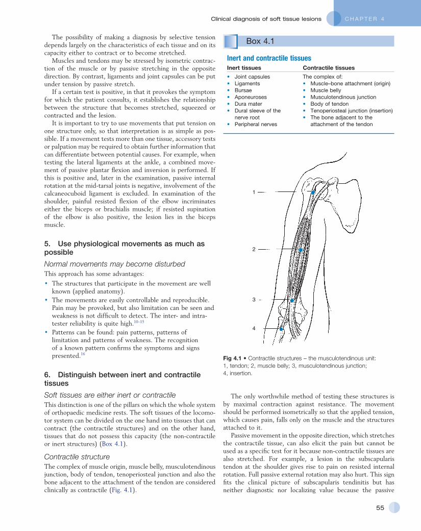
6 . Distinguish .between .inert .and .contractile .tissues
Soft tissues are either inert or contractileThis distinction is one of the pillars on which the whole system of orthopaedic medicine rests. The soft tissues of the locomo-tor system can be divided on the one hand into tissues that can contract (the contractile structures) and on the other hand, tissues that do not possess this capacity (the non-contractile or inert structures) (Box 4.1).
Contractile structureThe complex of muscle origin, muscle belly, musculotendinous junction, body of tendon, tenoperiosteal junction and also the bone adjacent to the attachment of the tendon are considered clinically as contractile (Fig. 4.1).
Box 4.1
Inert and contractile tissuesInert tissues Contractile tissues
• Joint capsules• Ligaments• Bursae• Aponeuroses• Dura mater• Dural sleeve of the
nerve root• Peripheral nerves
The complex of:• Muscle–bone attachment (origin)• Muscle belly• Musculotendinous junction• Body of tendon• Tenoperiosteal junction (insertion)• The bone adjacent to the
attachment of the tendon
Fig 4.1 • Contractile structures – the musculotendinous unit: 1, tendon; 2, muscle belly; 3, musculotendinous junction; 4, insertion.
1
2
3
4
The only worthwhile method of testing these structures is by maximal contraction against resistance. The movement should be performed isometrically so that the applied tension, which causes pain, falls only on the muscle and the structures attached to it.
Passive movement in the opposite direction, which stretches the contractile tissue, can also elicit the pain but cannot be used as a specific test for it because non-contractile tissues are also stretched. For example, a lesion in the subscapularis tendon at the shoulder gives rise to pain on resisted internal rotation. Full passive external rotation may also hurt. This sign fits the clinical picture of subscapularis tendinitis but has neither diagnostic nor localizing value because the passive

General Principles
56
8 . The .patient’s .cooperation .is .vital
The patient knows the symptomsThe patient’s cooperation is essential, and it is vital that the questions put are understood. Details are sought on what activ-ities have an influence on the symptoms and how symptoms behave over time. Except in psychologically disturbed patients, the more precise the questioning, the easier it is to obtain accurate answers. The patient must realize that, during func-tional examination, the examiner is looking for tests or move-ments that elicit symptoms. Most difficulties arise with those who are in constant pain, in that they tend to answer every question positively. It is the task of the examiner to explain carefully that movements that alter the pain are being sought. Not only tests that make the pain worse (a frequent occur-rence) but also those that decrease the pain are considered important.
9 . Take .into .account .the .patient’s .personality
The patient is a personThe history in particular will give an idea about the patient’s personality. The reaction to pain can be assessed and a picture built up of the extent of disability. The findings can then be related to what is actually found when the examination takes place.
The view obtained from the history and physical examina-tion may have therapeutic significance: for example, most patients can cope with active treatment such as manipulation or deep transverse massage but the clinician may obtain a ‘perception’ for those who cannot.
10 . Keep .the .balance .between .credulity .and .excessive .scepticism
Objectivity is a fair attitudeOrthopaedic medical disorders produce symptoms and signs that may be difficult to analyse objectively. Patients who have a reason to assume disorders for some type of personal gain, therefore, commonly use clinical features in the locomotor system to try to establish their credibility (see online chapter Psychogenic pain).
Although the examiner must be on guard against feigned illness, great care must also be taken to maintain a dispassionate attitude during the clinical encounter. The diagnosis of ‘psy-chogenic pain’ must not be made too quickly. Only when many inherent unlikelihoods are encountered during the history and functional examination should the examiner be suspicious about the veracity of the patient’s story. Also, the discovery of a series of lesions is self-contradictory, because the develop-ment of several problems at the same time is most unlikely.
11 . Request .technical .investigations .only . .when .necessary
Looking is not a substitute for thinkingClinical testing is the first approach in orthopaedic medicine. Technical investigations, although sometimes very valuable, are only asked for in some situations:
movement also stretches the anterior joint capsule and the pectoralis major muscle and tendon.
However, a pain elicited by resisted movement does not invariably mean that a contractile tissue is at fault. If the bone close to the tendinous insertion is affected (fracture or other bony disorder), pain is evoked by the pull of the muscle. A contraction may also squeeze an underlying structure such as a lymphatic gland or bursa. When such tissues are inflamed, squeezing may evoke pain. The same applies when there is a disorder adjacent to muscles, for example an abscess. This explains why, for example, contraction of the sternoclei-domastoid muscle may be painful in glandular fever and why contraction of the gluteal muscles can hurt in a trochanteric bursitis.
Inert structureAn inert structure does not possess an inherent capacity to contract and relax and can thus be tested only by passive stretching or squeezing. The inert tissues are shown in Box 4.1.
Active movements may also stretch or squeeze an inert structure but, because they also activate the contractile tissues, interpretation is subject to ambiguity and they cannot be used to test inert structures. For example, during active elevation of the arm, many muscles are in action (deltoid, supraspinatus, serratus anterior, trapezius). At the same time, certain parts of the joint capsule and some ligaments are stretched (acromioclavicular, sternoclavicular, conoid and trapezoid ligaments) and other structures are compressed (subacromial bursa, inferior acromioclavicular ligament, tendi-nous insertions of supraspinatus, infraspinatus, subscapularis and biceps).
7 . Concentrate .on .‘the’ .pain
‘The’ pain is that pain for which the patient consultsWhen tests evoke pain, the examiner must make sure that this is the pain that is the patient’s complaint. It is possible that some movements elicit pain in a certain area and that other tests provoke another pain in another region: one of these will be recognized by the patient as the presenting symptom. The examiner should then concentrate on this pain alone.
The situation often occurs because combined lesions are quite common. A patient may come to see the doctor with pain down the arm. If, after the history has been taken, it is not clear whether the pain originates either from the cervical spine or shoulder girdle or from the shoulder itself, the pre-liminary examination aims to clarify the situation. It may show some discomfort at the base of the neck when cervical move-ments are tested (especially in middle-aged and elderly people) but if only shoulder movements elicit the pain complained of, then this pain (‘the’ pain) is the primary problem; the other pain (‘a’ pain) is secondary. The arm pain will, of course, be dealt with first and only when this problem is solved is the other problem (if still present) approached.
Difficulties may arise in hypersensitive patients who report every tension they experience and for which they use different words: ‘it hurts, it aches, it pulls, it stretches, …’.

C H A P T E R 4 Clinical diagnosis of soft tissue lesions
57
concentrate on the important items only and bring the patient back to the point whenever there is a digression.
Patients with a clinical presentation that may rest in non-organic causes try to escape from precise questioning. They offer a garbled story full of internal contradictions.
RemarksQuestions should be asked in such a way that the account of the symptoms is given in chronological order which enables the examiner to get an idea of the duration and behaviour of the condition present. Knowledge of different dermatomes and of the possible likelihoods will help in interpretation of the evolu-tion of the patient’s symptoms.
Leading questions should be avoided, because they suggest to the patient what answer is expected. The questions should be neutral, so that the patient has to think about what is felt. An honest patient will have no problems in giving exact answers; one who dissembles has the opportunity to make mistakes and display inconsistencies.
Examples of questions that should be recast are as follows:
• Not: does the pain spread down your leg? But: does the pain spread at all?
If so, where to?• Not: is it painful to cough?
But: what happens when you cough?• Not: is it painful to bend forwards?
But: does anything bring the pain on?• Not: do you feel pins and needles in your hand?
But: have you got any pins and needles?And if so, where?
When there is a relationship between the patient’s symptoms and rest, exertion, certain activities or certain postures, then it is probable that the patient suffers from a lesion of the locomo-tor system. The main exceptions are angina and intermittent claudication. Questions should therefore be asked about the movements and positions that evoke, increase or influence symptoms, for example:
• What brings the symptoms on?• What makes the symptoms disappear?
Some information can be obtained only from the history, and not from any other diagnostic procedure. For example, to ascertain the stage of shoulder arthritis, to find out whether a displaced fragment of cartilage is stable or unstable, to deter-mine whether sciatica is caused by a primary or secondary posterolateral disc protrusion, depends on the answers to some very specific questions. These are not only diagnostically important but also have a prognostic value and can determine correct treatment.
For lesions of the knee or spine, the history is of extreme importance; the examiner must go into great detail and if this is done the diagnosis becomes apparent. For example, a patient may mention that pain started in the centre of the back, soon spreading unilaterally towards the buttock, and later radiating down the lower limb into the lateral border of the foot and the two little toes while at the same time pain in the back and buttock disappeared. After a while pins and needles began to
• To exclude major lesions for which the functional testing has not been sufficient.
• To exclude contra-indications for some therapeutical actions (manipulations or infiltrations).
• To confirm the tentative diagnosis made after the clinical examination.
Warning
Incorporated into this system of clinical evaluation are warning signs: certain symptoms or combination of symptoms and signs indicate that something unprecedented is taking place and so alert the examiner to the possible presence of a potentially serious condition. Possible warning signs are, for example, pain in the upper lumbar area, deficit of more than one nerve root in the cervical spine, or a capsular pattern of the hip in children.
The presence of such warning signs will put the examiner on guard and indicate accessory clinical tests, further technical investigations or reference to a neurologist, an internist, a cardiologist or an oncologist.
These warning signs will be discussed further in later chapters.
Clinical evaluation
History
History is of prime importance in reaching a diagnosis. It is so well known as a method of determining symptoms that most examiners fail to realize how much information can be gained from it.
Patients are the best source of information in that they are suffering from the lesion and can best report precisely what is felt. It is then the examiner’s task to translate the subjective symptoms into anatomical and functional conclusions.
Cyriax said: ‘Every patient contains a truth. He will proffer the data on which diagnosis rests. The doctor must adopt a conscious humility, not towards the patient, but towards the truth concealed within the patient, if his interpretations are regularly to prove correct’.17
History taking is a slow business that requires time, patience and concentration; the examiner must do everything possible to gain the maximal detailed information. Vague, general description of the complaints should not be accepted but precise and detailed answers sought.
Most patients, and certainly when they are frank, are able to provide precise answers to the examiner’s questions or can spontaneously give a well-structured, detailed and chronologi-cal account. They try to be as helpful as possible and are visibly pleased to talk to an interested physician. However, taking the history becomes more difficult in those who cannot express themselves or give a disjointed story. It is the examiner’s task then to make sure that the right questions are asked in order to get useful answers. The same applies to talkative patients who try to be too helpful by adding all manner of irrelevant details. In these circumstances the examiner should

General Principles
58
happened. If the pain is felt at the medial aspect of the joint and the patient mentions a valgus injury, the medial collateral ligament or the medial meniscus are most likely to have been damaged.
Swelling of a joint after an injury may have come on imme-diately, in which case blood is the cause, or after a few hours, which is typically the result of a reactive effusion.
When the patient mentions a spontaneous onset, this may be either sudden or gradual. Apart from diagnosis this distinction may have therapeutic consequences. Backache, as the result of disc protrusion, that comes on suddenly is annular and requires manipulation, whereas a gradual onset suggests nuclear displacement, which is treated with traction.
The patient must exactly define the first localization of the symptoms. The area where the pain was first felt very often lies quite close to the site of lesion, referred pain usually coming on later. This does not apply to ‘pins and needles’. They are mostly felt distally in the limb, from wherever along its length the nerve is affected.
Questions are also asked about what influenced the symp-toms. The examiner looks for a relationship between activities, movements or posture and the symptoms.
Progression/evolutionThe symptoms may be present without interruption from their onset. However, it is also possible that the patient describes a recurrence (see Box 4.2).
The progression of symptoms since their first onset is ascer-tained. The condition may have continued uninterrupted, in which case details are asked about the development of the severity of the symptoms and of the localization of pain. If the latter has remained unchanged from the beginning, this indi-cates that the lesion is quite stable and not evolving. When pain has diminished it usually indicates an improvement, although there are conditions (e.g. nerve root atrophy and certain cases of mononeuritis), in which the pain disappears long before the condition has resolved. Pain becomes worse as the condition progresses: in such circumstances it is important to know the length of time for which it has been present. This has diagnostic significance: it is clear that conditions such as metastases have quite a short time course. In contrast, slowly worsening pain is characteristic of some other conditions such as a neurofibroma. When the patient describes intermittent pain, details are sought about the occasions on which pain is
occur in the same toes and additionally they would go numb. The patient has revealed everything: the normal evolution of a protruded fragment of disc at the L5–S1 level, compressing the first sacral nerve root, is immediately apparent.
In some other joints, such as the shoulder, the history matters less but examination will disclose the lesion.
Taking the history
Age, .sex, .profession, .hobbies .and .sportsSome disorders are confined to certain age groups so that the age of the patient may indicate diagnostic possibilities. For example, a patient of 14 who mentions internal derangement at the knee probably suffers from osteochondritis dissecans. The same story in a patient of 20 suggests a meniscal problem and at 60 years points to a loose body in an arthrotic joint. The same applies to the hip: trouble at the age of 5 is probably due to Perthes’ disease; at 15 it could be the result of a slipped epiphysis; at 30 ankylosing spondylitis is a possibility; and at 50 arthrosis is more likely. A similar age distinction applies in root pain of cervical origin: under the age of 35 it is extremely rare that this is caused by a disc protrusion.
Certain disorders are more typical for men (e.g. primary sciatica and ankylosing spondylitis) and others occur more often in women (e.g. de Quervain’s disease and the first rib, thoracic outlet syndrome).
The profession of the patient may sometimes give an idea about the causative strains that have acted on the affected joint. Also it may – in conjunction with hobbies or sports – have an influence on the decisions to be taken on treatment. Treatment for acute lumbago will be different in an employee who sits most of the day than in a docker who has to do heavy work; a patient with regular attacks of sudden backache will be advised against tennis, a sport full of quick movements.
Initial .symptomsThe examiner should get an accurate picture of the moment the symptoms first appeared. The patient should be encouraged to recall that period and questions should proceed from that first instant.
The onset of the symptoms has to be clear. If they came on after an injury, a very detailed description of the accident should be elicited. The events immediately following the acci-dent must be ascertained, in that compensation may be claimed because of inadequate or inappropriate management. The sub-sequent condition of the joint may not allow complete exami-nation and therefore an idea of the direction of the forces acting on that joint and the position in which the joint was held at the moment of the accident are essential and will give a notion of the possible structures affected. The knee is an important example. In a ‘sprained’ knee the inflammatory reac-tion that follows the accident is so spectacular (swelling, limi-tation of movement) that proper functional testing becomes difficult, which means that in the acute phase the examiner has to rely mainly on the history to get an idea of what has
Box 4.2
Evolution of symptoms• Uninterrupted Unchanged
DiminishedWorseningIntermittent
• Recurrent

C H A P T E R 4 Clinical diagnosis of soft tissue lesions
59
felt. Nocturnal pain, for example, suggests an inflammatory condition.
A very important distinction should be made between the following definitions.
Reference of painReferred pain is a very typical feature in non-osseus lesions of the locomotor system. It is mostly segmental and thus experi-enced in a single dermatome, which indicates the segment in which the lesion should be sought. Reference of pain is influ-enced by the severity of the lesion: the more severe it becomes, so giving rise to a stronger stimulus, the further distally does the pain (usually) spread. The reverse also holds: reduction in the distal distribution is synonymous with improvement.18,19
It is therefore always important not to forget the question: ‘Where was the pain originally and where has it spread since?’
Shifting painPain coming on in one place as it leaves another indicates a shifting lesion.
This extremely significant phenomenon is well known in internal medicine: for example, when a renal calculus moves from the kidney down the ureter to the bladder and urethra, the pain experienced will follow the displacement. Pain is felt in the loin first, then in the iliac fossa, later in the groin and finally in the genitals. When the pain leaves one point, it is felt in another instead.
The same happens in soft tissue lesions. A good example is central backache, which becomes unilateral, then later on shifts to the buttock and finally to the lower limb – the back-ache has become sciatica. This shift can only be explained as follows: a structure lying in the midline and originally com-pressing the dura mater (backache) has shifted to one side and now compresses the dural sleeve of the nerve root (root pain). To be able to shift, that structure has to lie in a cavity and, because the pain was originally central, this has to be a central cavity. The only structure lying in a central cavity and able to change its position is the intervertebral disc: there is no other possibility (Fig. 4.2).
The same situation is encountered when a loose fragment of cartilage moves within a peripheral joint as, for example, often happens in the knee. Dependent on the position of the loose body in the joint space, the pain can be felt at the inner aspect, anteriorly or posteriorly and on other occasions even at the lateral side. Such moving pain indicates a moving lesion.
Expanding painThis is synonymous with an expanding lesion – one that grows, for example a tumour. When it appears in another region the pain does not leave the area where it originated. It spreads, even beyond the boundaries of dermatomes. A patient may describe a pain that begins in the centre of the back and then becomes bilateral. It spreads to one buttock, and later to both, also increasing in the back. Later it spreads to one leg and even subsequently to both, while still becoming worse in the back as well as in the lower limbs. Such a course is one of expanding pain, as the lesion becomes more extensive.
Fig 4.2 • Shifting pain: posterocentral disc protrusion causing central backache (a); shifts to posterolateral position causing unilateral sciatica (b).
(a) (b)
Warning
An expanding pain, in which pain spreads but does not regress at the original site (in the way that a shifting pain does), is indicative of an expanding lesion and is usually the manifestation of a serious condition.
Another course is recurrence. Certain disorders, such as those causing internal derangement or of rheumatoid type, have a recurrent character. Some occur suddenly, others more gradually. If the symptoms occur intermittently, it is important to know whether the patient is or is not free of pain between attacks, because this has both prognostic and therapeutic con-sequences. Freedom from symptoms for a certain period of time suggests that the same may happen again. In internal derangement, regular recurrence implies that the loose frag-ment of cartilage or bone is unstable, in which case the main-tenance of reduction will be the main concern of the therapist. A patient who is doing heavy work and who gets lumbago every 2 years must be regarded as having a stable lumbar disc which is completely different from a man with a light job who gets

General Principles
60
rheumatoid arthritis, gout or infectious arthritis) wakes the patient at night and gives rise to frank stiffness early in the day.22
The severity of pain may be a determinant of the type of treatment that is chosen. For example, although sciatica without neurological deficit is not immediately an indication for surgery, discectomy may become the treatment of choice when the pain has become unbearable.
Finally, localization has some diagnostic significance. Pain may be felt centrally (on the midline), bilaterally or unilater-ally. Central and bilateral pain usually point towards a lesion lying in the midline. A bilateral lesion is another possibility, but this is much less frequent. It should be realized, however, that central symptoms do not arise from a unilateral structure. And, although some structures lie very close to the midline (facet joints, costovertebral joints, erector spinae muscles), they are still unilateral and can only give rise to symptoms felt unilaterally. Unilateral pain originates in a unilateral structure or, when dealing with the spine, in a central structure that moved to one side and compresses nerve tissue unilaterally (e.g. a disc).
When the lesion is in the locomotor system, there should still be a relationship between symptoms and rest, exertion, activities, movements or posture. When coughing, sneezing or breathing hurts in an area other than in the chest, the dura mater could very well be responsible. Dural pain can be felt in the trunk far beyond the relevant dermatome.
Of special interest to the examiner are ‘twinges’: sudden short bouts of pain, which last only one second and are often associated with momentary functional incapacity. The occur-rence of painful twinges may be the result of one of the following:
• Internal derangement• Tendinous• Neurological.
A momentary subluxation of a loose fragment of cartilage in a jointThis happens quite often in the lumbar spine, the knee and the hip and less frequently in the elbow, ankle and subtalar joints. If there are any signs found during clinical examination, they will be articular – a non-capsular pattern (see p. 74). The combination of twinges and articular signs is pathognomonic of the existence of internal derangement.
A tendinous lesionThe patient recounts that, especially when the tendon is involved in movement, there are bouts of painful momentary
Box 4.4
Characteristics of mechanical and inflammatory painMechanical pain Inflammatory pain
• Starting pain/stiffness• During loading• Worse at evening• Moving in bed
• Nocturnal• Morning stiffness
Box 4.3
Pain• Description• Character: mechanical
inflammatory• Severity: therapeutic consequences• Localization: diagnostic significance
lumbago three times a year. In the first case reduction suffices, whereas the second will need other prophylactic measures to maintain the disc in place.
The onset of pain may vary from one attack to another. Backache that starts suddenly on some occasions, but gradually on others, very strongly suggests discal trouble. The localization of the pain may also change from one attack to another: it may be felt on one side of the body or of a joint and on the next occasion on the other side. This shifting pain is very typical of internal derangement, although there are some other condi-tions that may present the same picture (e.g. alternating buttock pain in sacroiliac arthritis caused by ankylosing spondy-litis and alternating headache in migraine).
Actual .symptomsAfter having built up a complete picture of the patient’s symp-toms, information is sought about what is experienced at the time of interview.
Most patients consult the doctor because they have pain but other symptoms may also be described: pins and needles, numbness, limitation of movement, twinges, weakness and vertigo. These are sometimes forgotten by the patient and therefore the examiner must inquire about them. Every symptom must be given due weight and examined in detail.
Pain (Box 4.3)There are many different ways of describing pain: it is amazing how much variation patients can achieve in their vocabulary and how many different descriptive terms can be used for the different sensations perceived. The reason lies in the fact that pain is mainly an unpleasant emotional state that is aroused by unusual patterns of activity in specific nociceptive afferent systems. The evocation of this emotional disturbance is con-tingent upon projection to the frontal cortex.20,21 The nature of the pain may have some diagnostic value: everybody knows the throbbing pain of migraine, the stabbing pain of lumbago or the burning sensation of neuralgic conditions. Although the way the patient describes the pain may sometimes point to a certain disorder, it can also indicate the emotional involvement of the patient with the lesion.
Pain may have either a mechanical or an inflammatory character (Box 4.4). Mechanical pain (e.g. in arthrosis) is characterized by pain and stiffness at the beginning of a move-ment; augmentation when load is put on the joint; pain at the end of the day and absence of pain at rest, although moving in bed may also be uncomfortable. Inflammatory pain (e.g.

C H A P T E R 4 Clinical diagnosis of soft tissue lesions
61
weakness which arrest movement. This is common in tennis elbow, where the lesion lies at the origin of the extensor carpi radialis brevis muscle from the lateral epicondyle. It also occurs, although less frequently, in tendinitis at the shoulder, especially of the supraspinatus.
A neurological conditionThese include tabes, post-herpetic or trigeminal neuralgia and Morton’s metatarsalgia.
ParaesthesiaNon-painful sensory disturbances, paraesthesia, are strongly indicative of a condition that originates in a nerve (Box 4.5). They may result from an intrinsic lesion (primary neuritis or secondary polyneuropathy) or from an extrinsic cause (com-pression). They may also vary in quality and in intensity. In orthopaedic medicine the variation lies between numbness and real pins and needles. It is very often described as ‘tingling’.
The moment the patient mentions the presence of pins and needles, the examiner should go into detail and ask the follow-ing questions:
• What brings on the pins and needles?• What makes them disappear?• How far proximally do they extend?• Where exactly are they felt?
In entrapment neuropathies, knowledge of what brings the pins and needles on will show whether a compression phenom-enon or the release phenomenon is acting (see pp. 26–27). For example, pressure on a small distal nerve gives rise to paraesthesia and analgesia in the cutaneous area of that nerve during the time of compression (e.g. meralgia paraesthetica). However, when a nerve trunk or nerve plexus becomes com-pressed, the paraesthesia are felt in a larger area, corresponding with the territory of that nerve and occur only after the com-pression has ceased (e.g. thoracic outlet syndrome). Nerve root compression results in segmental pain and paraesthesia felt within the corresponding dermatome (e.g. sciatica).
Localization:
Multisegmental Segmental (dermatome)
Territory of nerve
• Spinal cord • Nerve root • Plexus• Central nervous
disease or internal disorder
• Peripheral nerve trunk
• Peripheral nerve ending
Upper extentAccompanying symptoms: • Pain • WeaknessBehaviour:i.e. compression phenomenonrelease phenomenon
Box 4.5
Paraesthesia
Multisegmental bilateral paraesthesia indicates a lesion in the spinal cord.
It must be remembered that the site of compression always lies proximal to the proximal extent of the paraesthesia. They are usually felt in the distal part of the extremities. The more accurately the patient describes the area, the more distal the compression lies.
A paraesthesia-like feeling, especially vague tingling, may be experienced in some circulatory conditions, such as Raynaud’s syndrome, but this is usually accompanied by changing of the colour of the skin in the distal part of the limb.
Functional disabilityOften, functional disability is complained of. It comprises limi-tation of movement, internal derangement, weakness and inco-ordination and instability.
Limitation of movementWhen limitation of movement is mentioned, its nature will have to be determined during the functional examination: limi-tation of active movements only, or limitation of both active and passive movements, and in this case whether it is of the capsular or of the non-capsular type. End-feel at the end of the passive movements and the relationship between pain and end-feel must also be ascertained (see pp. 73–74).
Internal derangementSymptoms caused by internal derangement are irregular in nature. There are moments when the joint feels normal and that the patient is able to do everything, and other occasions when the joint does not work well. Typical symptoms are sudden twinges, shifting pain, giving way of the limb, locking of the joint and a feeling of insecurity.
If the answer to the question ‘Does the joint lock?’ is posi-tive, further inquiry should make clear in what position locking occurs and how it is released. For example, a knee that locks in extension and unlocks spontaneously suggests a loose body, and one that locks in flexion and has to be unlocked manipu-latively is a meniscal problem.
WeaknessThis symptom should lead the examiner to concentrate during the examination on both active and resisted movements to see if the weakness is physically apparent or not. If weakness is confirmed, the distinction has to be made between a lesion in the muscle itself (e.g. atrophy or rupture) or in the nervous system, which is more often the case.
Incoordination and instabilityThese complaints are indications that something is amiss with either deep (proprioceptive) sensibility, the vestibular system or the cerebellum.
Further .questionsOther questions, if appropriate, are asked about similar symp-toms, past or present, in other parts of the body, especially other joints (see Box 4.6). If the answer is positive, conditions such as rheumatoid arthritis, spondylitic arthritis, Reiter’s disease and gout should be suspected and further examination is required.

General Principles
62
Disorders of rheumatoid type (rheumatoid arthritis, lupus erythematosus, systemic sclerosis, dermatomyositis) are char-acterized by the symmetrical joint involvement, usually of the small joints (e.g. metacarpophalangeal joints). Arthritis of reac-tive type (e.g. peripheral joint involvement in ankylosing spondylitis, ulcerative colitis, Reiter’s disease, sarcoidosis or psoriatic arthritis) affects a few large joints (e.g. shoulder, hip or knee) asymmetrically (Fig. 4.3).
Questions about the general state of health are asked to find out whether there is the possibility of a serious disorder (e.g. cancer).
The patient should also reveal present medication, and a doctor or therapist who considers manipulation should make sure that the patient is not taking anticoagulants: these are a contraindication because of the danger of haemorrhage.
Inquiries should also be made about previous treatments, which may give some idea of the chance of success of the proposed therapy. Previous surgery, its timing and indication are noted – it is not impossible that the present condition is the outcome of previous intervention (Box 4.7).
Inspection
Inspection begins the moment the patient enters the room. If gait is disturbed, the way of walking may be diagnostic. An experienced examiner usually has the analytical ability to rec-ognize, for example, the following typical patterns: a sublux-ated meniscus at the knee; a ‘tennis leg’; the elderly patient
Fig 4.3 • Patterns of: (a) reactive arthritis; (b) rheumatoid arthritis.
(a) (b)
Box 4.7
Summary of history1. Age, sex, profession, hobbies, sports2. Initial symptoms:
Onset: • traumatic • spontaneous: sudden/gradualLocalizationInfluence
3. Progression/evolution:UninterruptedChanging: • reference of pain • shifting pain • expanding painRecurrences
4. Actual symptoms:PainParaesthesiaFunctional disability: • limitation of movement • internal derangement • weakness • incoordination/instability
5. Further questions
Box 4.6
Inflammatory disordersRheumatoid pattern Reactive pattern
• Several joints• Small joints• Symmetrical
• Few joints• Large joints• Asymmetrical
with arthrosis of the hip; acute lumbago; a spastic gait; the patient with parkinsonism; a Trendelenburg gait; a drop foot; a ruptured Achilles tendon; and finally the patient with a non-physical condition.
Further features may be noted while the history is taken. A normally seated patient should have at least 90° flexion at hip and knee which is later confirmed on functional examination. The face may reveal the extent of pain or may disclose parkin-sonism. The attitude in which a limb is held during the inquiry may be informative. Finally, the way the patient takes off clothes and shoes can provide further information on disability.
Deformities or deviations are easily seen: for example, in acute torticollis or in lumbago, the patient stands with the head or the lumbar spine held in deviation, usually towards one side. Real deformities can be the result of one or more fractures. Pathological fractures of a vertebral body lead to angular kyphosis or, if they occur at several levels, to a ‘shortened’ patient. Every examiner knows the typical kyphosis of adoles-cent osteochondrosis. Other examples are genu valgum and varum, which may be physiological up to a certain age but are pathological in adults. The presence of a deformity is not always relevant. It is quite possible that it has nothing to do with the presenting condition. A short leg or a long-standing scoliosis are very often seen but occur in asymptomatic as well as in symptomatic patients.
Soft tissues, such as skin and muscles, may show abnormali-ties. The colour of the skin may be different from that of other parts of the body: red in inflammation, blue when a haematoma is present or when a venous disorder has developed, white in arterial problems. Visible muscle wasting may be obvious. If swelling is present, the examiner should ascertain whether it is general and diffuse or localized.

C H A P T E R 4 Clinical diagnosis of soft tissue lesions
63
An inflammatory condition may show quite spectacular signs such as redness of the skin, swelling and warmth.
Preliminary examination
After having taken the history and amassed evidence from inspection, the examiner should have an idea of which part of the body the symptoms originate from. Regional examination follows. When the history alone is not diagnostic, so that doubt over the origin or localization exists, a quick survey of the tissues forming the relevant segment may be necessary.
This ‘preliminary’ examination (Fig. 4.4) includes tests for the different joints. For example, the preliminary examination for pain down the arm includes tests for the cervical spine, shoulder girdle, shoulder, elbow, wrist and hand (Table 4.1).
When some tests are positive and indicate that the lesion lies about a certain joint, this joint will then be tested more rigorously. When no abnormalities are found, it is assumed that the lesion lies outside the moving parts. If all movements hurt, or when the patient presents a contradictory pattern, the ques-tion of a psychogenic disorder arises.
Functional examination
In lesions lying within the locomotor system, pain is brought on mainly by tension. Therefore, during examination the dif-ferent tissues are selectively put under tension and the results noted.
As the function of the different tissues is well known, func-tional testing is really an exercise in applied anatomy. This indirect approach to diagnosis is purely mechanical.
Fig 4.4 • Role of the preliminary examination.
Pain in a region/dermatome
Which joint?
No doubt Doubt
Preliminary examination
Joint B Joint CJoint A
Regional functional examination
Accessory tests?
Table 4.1 Movements tested in the preliminary examination of the upper limb
Root Region Type
C1 Neck movements ActiveResisted
C2–C4 Shoulder movements ActiveResisted
C5 Arm movementsShoulder Active
Resisted
C6–C7 Elbow PassiveResisted
Wrist Resisted
C8 Thumb Resisted
T1 Fingers Resisted
The examination used is standardized, which permits a sys-tematic search for signs in such a way that, with a minimum of tests, maximum information is obtained. Too many tests in the standard examination can confuse the examiner and make interpretation very difficult.
This does not imply that the diagnosis will always be reached after the standard functional examination. It may sometimes be necessary to add accessory tests (see pp. 66–67).
Procedure
The purpose of functional examination is to reach a decision on which structure is affected. However, even that may be insufficient. In a patient with a shoulder problem it is not enough to say that there is a lesion in the brachial biceps muscle: is it in the long head or the short head? In a lesion of the long head, is it localized in the bicipital groove or in its intra-articular course or at its insertion at the glenoid? Therefore, once the structure at fault is known, the next step must be to determine exactly where, within that structure, the lesion lies.
Examination is usually performed on both sides. Certainly, if the range of a passive movement is assessed, it has to be compared with the contralateral normal joint. When muscle strength is evaluated, comparison with the non-affected side is again necessary.
The patient as well as the examiner should adopt a good position from which to start. When passive movements are performed, the examiner must make sure that they can be done until the end of range (if present) is reached: movement should not be limited by the examiner’s body, or by the couch or even by clothing. For resisted movements, which are done isometrically, the examiner’s position must be such that the examiner can exert more power than the patient can. It may be of interest to put the patient in front of a mirror so that reactions can be observed.
The movements that are used to test the different struc-tures should be ‘pure’ ones, i.e. testing only one structure or

General Principles
64
plantiflexion is weak and active movement impossible despite the range of passive movement at the ankle remaining unaltered.
Active movements are usually required when the spine (cer-vical, thoracic and lumbar), the shoulder girdle and the shoul-der are examined but they may also be tested at any other joints if they can supply additional information.
Passive .movements .(Fig. 4.5)
These tests are meant to examine the inert tissues. The information they give is precise, provided they are performed correctly. The patient should completely relax the muscles and let the examiner do the movement. It is important to realize that non-inert tissues may also be stretched by the manœuvre: if this gives rise to pain, it must be determined whether there is a ‘contractile tissue pattern’, in which case active contraction in the opposite direction will also be painful (see pp. 77).
The criteria for passive testing are pain, range and end-feel (Box 4.9).
PainThe patient must understand that the examiner is looking for ‘the’ pain – the reason for presentation – and that each time this pain is evoked or influenced a response is required. When straight-leg raising is performed in a patient with low back pain radiating down the posterior aspect of the thigh, the examiner must make sure that the sensation felt during the test is the exact pain complained of and not just a tight feeling in short-ened hamstring muscles.
Pain evoked by a certain movement requires the further information of exactly where it is and at what stage of the move-ment. Pain may come on at a certain moment but the move-ment is not necessarily limited. For example, straight-leg raising may become painful at 60° but continue to full range without increased discomfort. Excessive gentleness may fail to elicit information. Passive movements must be performed as comfortably as possible for the patient but, if a proper answer can only be obtained by pushing a bit harder and thus hurting the patient, this should be done. It would be a diagnostic error to interpret a perfectly full movement as being limited by a few degrees, if the movement was stopped because of pain. The same applies to a painful arc. This may be so uncomfort-able that the patient cannot get beyond it by active movement. If an arc is suspected, a harder push during passive movement may succeed and show its presence. Interpretation would be totally different: on the one hand, limitation of movement and, on the other hand, full range with a painful arc.
The presence of joint signs in root lesions is a misleading phenomenon. When a cervical or lumbar nerve root becomes compressed as the result of a disc protrusion, passive move-ment in a neighbouring joint may occasionally affect the root pain. Thus passive movements for the shoulder joint may hurt at the end of range so focusing attention on the shoulder. As pain of cervical origin is also felt down the arm the picture becomes very confused. The same occurs in lumbar root pain; passive hip movements are not limited but they may influence the pain when they are brought to full range – they are
one group of structures at a time. This facilitates subsequent interpretation.
Questions asked during examination should be as neutral as possible. The examiner must not impose on the patient an expected answer. Questions such as ‘Does this movement hurt?’ or ‘Is this painful?’ should be avoided. It is better to say ‘How is that?’ and ask the patient to report anything experi-enced. It must also be understood that the examiner is looking for tests that elicit the symptoms or, if they are constant, those that influence them.
Movements
The functional examination contains active movements, which are not always necessary, passive tests and movements against resistance. Positive tests are always repeated on the opposite side in order to compare the normal with the abnormal.
Active .movementsActive movements are mainly used to determine quickly the area from which symptoms originate and what structures to test in detail with passive and/or resisted movements (Box 4.8).
Examination by active movements goes against the principle of trying to test one structure at a time. When a limb is actively moved, muscles, tendons, ligaments, bursae and capsule are all put under stress. In consequence such tests are non-specific and therefore not always necessary. However, they can be of interest because they suggest what the patient is able and willing to do. In most cases, they also give an idea of the range of movement possible (normal, limited or excessive) and of muscle power. They are always executed first to provide a criterion with which to compare subsequent passive and resisted movements.
When a normal range is later found on passive testing and normal muscle strength is apparent when tested against resist-ance, there is no reason for any abnormality or restriction of active movements. Thus if, in these circumstances, it is claimed that the active movement cannot be done, there must be refusal, either involuntary (psychogenic) or voluntary (inten-tion to deceive). There are, of course, organic lesions where a discrepancy exists between active and passive or resisted movement but only within certain limits. For example, a mononeuritis of the long thoracic nerve, leading to weakness of the serratus anterior muscle, can make full active elevation of the arm impossible even though the passive range and muscle power of the shoulder are normal. A further example is total rupture of the Achilles tendon, in which resisted
Box 4.8
Aims of testing active movements• To ascertain patient’s willingness and ability• To determine range• To assess muscle power

C H A P T E R 4 Clinical diagnosis of soft tissue lesions
65
Fig 4.5 • Passive movements.
Painful
With limitedrange
At mid-range(painful arc)
At full range
Stretchingpain
Pinchingpain
Pain
Painless
Range
ExcessiveNormal
Limited
Allmovements
Onemovement
Somemovements
DisproportionateProportionate
Pathological
Physiological
Laxity
End-feel
Elastic
Tissueapproximation
Hard
Too hard
Too soft
Muscle spasm
Empty
Springy block
Crisp
Soggy
Passive movements
Box 4.9
Aims of testing passive movements• To assess pain• To determine range• To characterize end-feel
probably capable at their extreme of altering the tension on the nerve roots in a minor way, analogous to straight-leg raising.
AmplitudeThe range of movement is noted and is always compared to the unaffected side. The response should be interpreted in the light of the patient’s age and general condition.
Limited movement requires the examiner to ascertain whether the limitation is in all directions or only in some direc-tions or in one direction only. If only one movement is reduced, a ‘proportionate’ or a ‘disproportionate’ limitation may be present (see p. 76).
Pain at full range suggests that the pain is provoked by stretching or pinching of the affected structure. The localiza-tion of the pain will very often be indicative.
Excessive range may be pathological but laxity is sometimes purely physiological: for example, most women can hyperex-tend the elbow. When a joint moves further than is normally accepted, great care must be taken that the symptoms are not too readily ascribed to that phenomenon. Hypermobility with a normal end-feel is usually not of significance.
End-feelThis is a term typical of Cyriax. It describes the sensation that the examiner experiences at the end of the passive movement. The hand that performs the passive movement is not only motor but also sensory. When the movement comes to the end the examiner should assess the sensation. When no limitation is present the end-feel is at the end of the normal range. When limitation exists, the end-feel is judged at the end of the pos-sible range.
End-feel is diagnostically important as it gives an idea of the structure or condition that stops the movement.23 In addition it has therapeutic consequences. Especially during attempted spinal manipulations, the sensation imparted to the operator’s hand will indicate whether or not to proceed. Likewise, in the shoulder or hip, the decision whether to undertake capsular stretching depends on end-feel.
For accurate judgement of the nature of end-feel, experi-ence is required. Passive movement should be executed with extreme gentleness, especially during the final degrees of range. The moment resistance is first noted, the rate of movement is reduced so that the feeling can be assessed before movement comes to a complete stop. In normal circumstances each joint movement has a characteristic end-feel, which can be consid-ered as ‘physiological’. In pathological conditions, end-feel may change. The examiner should know what the normal feeling of each (passive) movement of a joint is like in order to be able to judge every change in end-feel.
Physiological end-feelThis can be hard, elastic or related to tissue approximation:

General Principles
66
in patients with poorly developed muscles: they present an ‘articular’ end-feel, either hard or elastic.
Pathological end-feelA number of types of end-feel are pathological.
• Too hard: the normal elastic, or tissue approximation, end-feel may become harder when the quality of the tissue that stops the movement alters under the influence of pathological conditions. This change very often accompanies limitation of movement because of capsular contracture, osteophytic outcrops of bone, myositis ossificans or a malunited fracture close to the joint. Dependent on the condition, the end-feel may vary from a slight increase in hardness (early arthrosis) to a bony block (ankylosing spondylitis). The latter forms a contraindication to further stretching (i.e. during manipulation).
• Too soft: in those instances where a hard end-feel is expected, the movement may come to a soft stop. This is especially the case in a loose body blocking a movement as may happen at the elbow: extension is limited by a few degrees with a soft end-feel.
• Muscle spasm: this involuntary muscular contraction usually indicates severe disorder. The muscles contract with a sudden, vibrant ‘twang’ even when the movement is performed very gently. This is reflex protective spasm and may happen in acute conditions, such as arthritis in the acute stage. It may also be a sign of more serious lesions such as recent fracture (e.g. scaphoid fracture at the wrist) or secondary deposits (e.g. in a cervical vertebra). Such an end-feel is always an absolute contraindication to manipulation.
• Empty: movement may be so painful that the patient begs the examiner to desist. The latter feels that further movement would be possible because there is no organic resistance but stops the movement because of the perceived pain or out of sympathy for the patient. Further forcing would result in active voluntary muscular contraction. This situation always indicates a serious problem. Acute bursitis, extra-articular abscess or neoplasm is a possibility, but there is nothing wrong with the joint itself. This end-feel may also indicate laxity in a joint or a non-organic lesion. In the latter case there is initial strong resistance, which yields to sustained pressure, disclosing a full range of motion.
• Springy block: this finding always indicates internal derangement: when part of an intra-articular cartilaginous tissue displaces, it may prevent a full range of movement which leads to a rebound at the extreme of the possible range. The commonest examples are a displaced meniscal fragment in the knee and a buckled end-plate in the lumbar spine.
• Crisp: this is typical for a disc protrusion and is best felt in the cervical spine. The feeling is the result of small involuntary muscular contraction at the end of the possible range, known as ‘muscle guarding’. The condition is not acute, as a result of which there is no real muscle spasm.
• Hard: the movement comes to an abrupt stop, either because two bony surfaces meet (bone-to-bone end-feel) or because a ligamentous structure prevents further movement (Fig. 4.6a). Extension of the elbow has a bone-to-bone end-feel, because the main factor that stops the movement is the engagement of the olecranon process in the olecranon fossa. Extension of the knee also has a hard stop, though less clearly direct than the elbow; here, the main structure that stops movement is the posterior cruciate ligament, which does not stretch.
• Elastic: at the end of range the examiner feels resistance but further stretching over a few degrees is possible (Fig. 4.6b). The feeling is one of two pieces of tough rubber being squeezed together or of a piece of thick leather being stretched. This end-feel is typical of a normal joint capsule and is felt during most rotation movements in shoulder, elbow, hip, knee, cervical and thoracic spine. The tern ‘elastic’ has the same meaning as ‘capsular’.
• Tissue approximation: this end-feel is normal in those movements where extra-articular soft tissues engage against each other (Fig. 4.6c). Flexion at elbow and knee give this ‘soft’, ‘muscular’ end-feel. At the elbow the muscles of the forearm come in contact with the biceps and at the knee the calf muscles engage against the hamstrings. This ‘extra-articular’ end-feel does not occur
Fig 4.6 • Hard (a), elastic (b) and soft tissue approximation; (c) end-feels.
Hard
Elastic
Tissueapproximation

C H A P T E R 4 Clinical diagnosis of soft tissue lesions
67
Similarly, to test resisted flexion, one hand is placed on the forehead and the other between the scapulae.
The criteria when testing against resistance are pain and strength (Box 4.10).
PainIn a lesion that lies in the contractile tissue, the pain should be elicited during the contraction. In mild disorders the test may initially be negative but repetition of the movement may eventually provoke symptoms. Similarly, slight muscular or tendinous problems may give pain only during exertion and resisted movements may remain painless. If pain cannot be elicited by repetitive contractions, the patient should be seen and tested in the circumstances when pain is experienced. The resisted movements may then be found positive.
There are cases of tendinitis in which the pain is only felt when the patient relaxes after muscle contraction. This is not uncommon and can also be interpreted as a positive resisted movement.
A positive answer on resisted movement is usually only one – but the most important – part of a ‘contractile tissue pattern’
• Soggy: this end-feel is seldom encountered and can be situated somewhere between a soft and an empty feeling. It is typical of rheumatoid arthritis in the upper cervical joints and is a strong contra-indication to manipulation.
Relationship between pain and end-feelIt is also important to look for the relationship between the moment of appearance of pain and that of the end-feel. Usually the pain comes on at the same time as the end of range is sensed (Fig. 4.7). Earlier pain implies that the joint is in a very irritated state which has therapeutic implications.
Resisted .movementsMovements against resistance are performed isometrically. The joint is put in a neutral position and should not move when resistance is applied. This method ensures that stress on inert tissues is minimal or absent.
The position of the examiner must be such that the force exerted by the patient can be resisted. A maximal contraction is asked for and held for a few seconds. Counterpressure is therefore taken by the other hand at the opposite side. For example, to test resisted extension of the neck, one hand is put on the occiput and another hand on the sternum (Fig. 4.8).
Fig 4.7 • Relationship between pain and end-feel.
Fig 4.8 • Resisted extension of the neck.
Box 4.10
Aims of testing resisted movements• To assess pain• To determine muscle strength
Fig 4.9 • Resisted movements.
Pain
Painless Painful
During contraction
During repeated contraction
During contraction following exertion
When relaxing from contraction
Strength
kaeWgnortS
Caused by pain
Caused by musculotendinous deficit
Caused by neurological deficit
Caused by patient’s refusal
Resisted movements
General Principles
68
After testing a group of structures
When a resisted movement has tested a group of structures, further differentiation is sometimes necessary to find in exactly which structure the lesion lies.
In ‘tennis elbow’, resisted extension of the wrist is painful. To find out whether the lesion lies in the extensors of either the wrist or of the fingers the test is repeated with the fingers held actively flexed, so inhibiting the finger extensors. A nega-tive answer implicates the wrist extensors, which can then be differentiated by executing resisted radial and resisted ulnar deviation: when resisted ulnar deviation hurts, the extensor carpi ulnaris is at fault; whereas if resisted radial deviation is painful, the lesion clearly lies in one of the radial extensors of the wrist – true tennis elbow. Whether the tendinitis is situated at the extensor carpi radialis longus or brevis cannot be found by further testing – palpation is called for.
The same applies to the hamstring muscles. A muscular or tendinous lesion about the knee will give rise to pain on resisted flexion. Differentiation between either (a) the femoral biceps or (b) the semitendinosus and semimembranosus is achieved by testing resisted external and internal rotation. Positive lateral rotation incriminates the biceps and medial rotation draws attention to the other two muscles. Palpation identifies exactly where the lesion lies.
To confirm a tentative diagnosis
An accessory test may help, especially in uncommon condi-tions, to confirm a tentative diagnosis. For example, when the diagnosis of mononeuritis of the spinal accessory nerve has been suspected, weakness on resisted scapular approximation will show the diagnosis to be correct.
In difficult cases
A clinical picture can sometimes be difficult to interpret and accessory tests may then resolve doubt. When pain is thought to be due to squeezing of a structure, diagnostic traction may help to confirm this. For example, in difficult cases of internal derangement of the cervical spine, pain and/or paraesthesia can be temporarily abolished by applying manual or mechanical traction. Traction on the arm can diminish pain in subacromial bursitis.
When the functional examination is negative
A negative examination does not necessarily mean that a lesion is absent. There may be a problem in a neighbouring tissue which was not systematically tested in the standard examina-tion. A negative shoulder examination, for example, invites the examiner to test the coracobrachialis muscle. In anterior knee pain a negative examination is followed by tests of the patello-femoral joint.
in which the passive movement that stretches or pinches the affected part is also painful.
The examiner must be aware of inert tissue lesions that can become painfully squeezed or moved by muscular contraction (e.g. gluteal bursitis).
StrengthThe movement should be strong, but weakness may occur and an experienced examiner will immediately have an idea of what is the cause. It may be the result of pain; in that instance, the examiner feels a sudden cessation of power – the patient stops the contraction when the pain is felt. This often happens in partial muscular or tendinous ruptures when sufficient fibres are torn to diminish strength.
In total contractile tissue ruptures, there will not be pain but complete absence of muscle power. This pattern is com-pletely different from a neurological weakness. Here there still is a force that has to be overcome: the examiner is stronger than the patient and so can push the patient’s limb away while still feeling continuing resistance. The latter can vary from an almost normal sensation – slight paresis – to very little (scarcely detectable) force – complete paralysis. This may be the result of a lesion that has completely stopped motor innervation or it may be a consequence of the patient’s refusal to undergo the manœuvre.
Accessory tests
After a well-balanced basic functional examination, interpreta-tion of the pattern that emerges should, in most cases, make it possible to single out the tissue at fault. Difficulties are, of course, encountered and further accessory testing is then required to reach a precise diagnosis, to positively confirm an existing but tentative diagnosis or to disclose the precise point affected within the structure.
An important feature is that a distinction should be made between standard functional examination and accessory tests. The basic examination is always done in its entirety, whereas the accessory tests are applied selectively (Box 4.11).
It is wrong to do accessory tests without a prior idea of the nature of the problem, and in the hope that more tests would automatically provide more complete information. On the con-trary, the more tests the more confusing the picture may become and in the end the ‘wood cannot be seen for the trees’. Accessory tests therefore should be goal-oriented. They are performed in the following circumstances.
Box 4.11
Aims of accessory tests• To differentiate within a group of structures• To confirm a tentative diagnosis• To unravel a difficult pattern• To extend a negative examination• To make a differential diagnosis• To understand unusual signs

C H A P T E R 4 Clinical diagnosis of soft tissue lesions
69
rheumatoid-type tendinitis). A purely mechanical tendinitis will not cause any warmth. When the warmth occurs suddenly and twinges are also present, this indicates persistent subluxa-tion of a fragment of cartilage. The heat may or may not diminish when the joint is kept still. Sometimes (e.g. in loose body) the warmth may be elicited merely by the examination.
ColdWhen the extremity feels cold an arterial problem is sus-pected. Palpation will then follow for arterial pulsations. Cold may also occur during an attack of sciatica, especially that which causes muscular weakness. When the foot becomes cold only after exertion, the probable cause is an iliac thrombosis. The cause may also be neurological.
SwellingSwelling is the result of an articular reaction to a lesion and may be localized or generalized. Swelling that came on after injury may be the outcome of haemorrhage or effusion. If it is caused by bleeding, the joint fills up within a few minutes; effusion develops over a few hours. Blood also fluctuates: the swelling moves en bloc. The presence of pus is exceptional and indicates an infection with microorganisms. Periarthritic oedema may pit. In a localized swelling, its consistency should be ascertained: a soft swelling indicates subcutaneous clear fluid or a thickened bursa, a fluctuating mass may result from a haematoma or from a mucocele and a hard but still fluctuant swelling is typical of a cyst or a ganglion. When the consistency is bony the cause is usually a callus, a bony subluxation (e.g. capitate bone at the wrist or cuboid bone in the midfoot), an osteophytic outcrop or any other bony deformity which results from a destructive process (e.g. osteitis deformans, neoplasm).
Thickening .of .the .synovial .membraneThis is found in rheumatoid, bacterial or inflammatory arthritis (e.g. gout, tuberculosis, gonorrhoea, Reiter’s disease, ulcerative colitis, spondylitic arthritis or psoriatic arthritis). It is absent in mechanical conditions, such as traumatic arthritis, post-immobilizational arthritis and arthrosis. To palpate capsular thickening, the examiner has to seek the reflexion of the mem-brane where it overlies a bony prominence.
GapsA gap may be palpated at the site of the rupture of a muscle or tendon.
TendernessPalpation for tenderness is sometimes performed to determine the exact localization of a lesion. For this purpose, it is done only in a structure that has already been found, by clinical examination, to be affected and only when it is within reach of the finger. Eliciting tenderness is only necessary when, after clinical examination, the diagnosis still lacks precision. When a localizing sign has been found, palpation is of course
In differential diagnosis
Different conditions may give rise to similar positive answers. An accessory test can then help to elucidate which structure is at fault. For example, limitation of external rotation at the shoulder is the primary sign of anterior capsular contraction as well as of a subcoracoid bursitis. Passive external rotation with the arm held in abduction will be positive in a capsular lesion and negative when a bursitis is present.
A test for distant pain can be performed when a lesion lies in a long bone. When a fracture is suspected on the dorsal aspect of a rib, pressure on the anterior aspect or on the sternum may provoke the pain. This technique can also be used to differentiate a rib problem, a lesion in an intercostal muscle or at a costovertebral joint.
When unusual signs are found
Discrepancies between individual findings on functional exami-nation may suggest an unusual and possibly serious lesion, a psychogenic problem or an attempt to deceive. In the former, the patient is sent for further specialized examination. When neurosis or deception is suspected, the patient is subjected to a number of accessory tests: additional movements that cannot possibly influence the alleged symptoms or repetition of tests in other positions. The opportunity for the patient to make mistakes is thus presented, which in a positive way can confirm the examiner’s suspicion (see online chapter Psychogenic pain).
Palpation
Palpation can be a very useful aid to diagnosis when performed at the right moment. However, it is frequently done too early in the examination, with the risk of an incorrect diagnosis. When, after functional testing, the examiner has interpreted the different movements and the pattern that emerged, and still thinks that more information could be forthcoming about the lesion, palpation is undertaken. Abnormal sensations that are imparted to the hand in a stationary as well as in a moving joint are sought.
Palpation of the stationary joint
During palpation of a motionless joint (Fig. 4.10a) the exam-iner may assess temperature (either warmth or cold), swelling, synovial thickening or structural changes. Palpation may also elicit tenderness.
WarmthWarmth is best palpated with the dorsum of the hand and may be diffuse or localized. Spurious warmth can be the result of a rubefacient ointment or of a bandage that has been removed just before examination. Otherwise, warmth always indicates activity. The lesion may be osseous (e.g. fracture or metastases) or articular (e.g. arthritis, haemarthrosis, loose body, or a dis-order of a meniscus or ligament) or tendinous (e.g.

General Principles
70
Other misleading phenomena are the presence of either a ‘referred’ tenderness or an ‘associated’ tenderness.
Localized, deep referred tenderness may occur within a painful region, for example, as the result of compression of the dura mater usually by a small displaced fragment of the intervertebral disc. This phenomenon is described in Chapter 1 and can be very deceptive unless the examiner is familiar with the rules of pain reference.
Associated tenderness is a very well-localized tender area very close to the site of the lesion. There is a relation between the two because both pain and tenderness disappear when the patient is cured. Associated tenderness is most common at the wrist and the elbow. In de Quervain’s disease – tenovaginitis
superfluous. To search for tenderness is of much more value in a distal joint (wrist, hand, ankle, foot) than in a proximal joint (trunk, shoulder, hip), because the localization of the pain is much more accurate.
Difficulties may arise when two tissues overlap and when no certainty exists whether the lesion lies in the superficial or a deep structure. This situation arises most in the chest and abdomen. To ascertain whether the tenderness lies in a super-ficial or a deep muscle or in another structure (e.g. a viscus), palpation is performed during contraction and relaxation of the superficial muscle. Greater pain when the superficial muscle is tense suggests that the muscle is at fault, the converse that deeper structures are affected.
Fig 4.10 • Palpation of (A) the stationary and (B) the moving joint.
A Palpation of the stationary joint Diffuse/localized
Osseous/articular/tendinous
Warm
Arterial
Cold
Neurological
For temperature
Synovial fluid
Diffuse
For swelling
Blood
Soft
Localized
Subcutaneous fluid
Bursitis
Pus (local infection)
Fluctuating
Haematoma
Mucocele
Hardish
Cyst
Ganglion
Bony
Bony subluxation
Callus
Osteophyte
Bony deformation
For synovialthickening
For a gap
For tenderness
For pulsations
B Palpation of the moving joint
For clicking
For crepitus
For hyper/hypomobility
For end-feel
Articular
Tendinous
Muscular
Osseous
Bursal
ScapulothoracicPhysiological
Pathological
C H A P T E R 4 Clinical diagnosis of soft tissue lesions
71
• Localized crepitus is usually felt in a lesion at the musculotendinous junction of the tibialis anterior muscle.
Osseous crepitusA fracture may crepitate when the limb is moved and the two ends of the fractured bone move against each other but, except in pathological fractures, an attempt to elicit crepitus usually causes so much pain that it should not be done.
Bursal crepitusSubdeltoid bursitis is the characteristic situation. Some time after a bursitis with effusion has subsided, creaking on moving the arm can be felt (see Shoulder, p. 333).
Scapulothoracic crepitusThis is unique. The posterior thoracic wall can become rough-ened at an area just beyond the lateral edge of the iliocostal muscle, with localized crepitus felt on shoulder movement (see the online chapter The shoulder girdle: Disorders of the inert structures.
ClicksClicks can be produced in several ways. Certain tests on clini-cal examination or certain manœuvres during a manipulative session may provoke movement of an intra-articular fragment of cartilage. This often happens when a loose body is present in a joint (e.g. knee or elbow) or when a meniscus subluxates (e.g. knee or jaw) or a fragment of disc moves (e.g. spinal joints). Clicking during a manipulative manœuvre is of interest in that it is often associated with an improvement of the physi-cal signs, clearly indicating that a block to movement has been removed. In ligamentous laxity (as can be seen in subluxation of the clavicular joint, in capsular overstretching of the shoul-der, or in rupture of the medial collateral ligament at the knee), a click is produced when one bone moves in relation to its fellow. Irrelevant clicks also appear, for example painless click-ing of a costal cartilage or a patellar click during active exten-sion of the knee.
End-feelEnd-feel can be assessed when a passive movement comes to a stop. It is either physiological or pathological and has important diagnostic and therapeutic consequences (see pp. 64–65).
Hypermobility .and .hypomobilityThese are terms that are currently used by osteopaths and manual therapists.24–25 During their so-called ‘segmental exam-ination’ they claim to be able to feel the difference in mobility between different, mostly spinal, joints.26 To establish hyper- or hypomobility would afford an excellent guide to the level of the lesion if it were always at the joint in which the fault lay, which is not in fact the case. Moreover, these findings are so subjective that they are not verifiable or reproducible. It has been shown on different occasions that even experienced osteopaths or therapists cannot agree about the findings.27–31 The main mistake, however, consists in attributing diagnostic significance to such differences in mobility. When movement
of the abductor longus and extensor brevis muscles at the wrist – the styloid process of the radius is extremely tender, as is the posterior aspect of the lateral epicondyle in tennis elbow, especially when the lesion lies at the tenoperiosteal origin of the extensor carpi radialis brevis.
Negative palpation does not necessarily mean that the diag-nosis is wrong, except perhaps in very superficial lesions, because there are tissues that are not explicitly tender when affected (e.g. the supraspinatus tendon).
PulsationPalpation for pulsation in the arteries may have an important diagnostic value and is indicated when symptoms are men-tioned or signs are found that point to a vascular condition (e.g. claudication, a cold limb, or blue or white skin).
Palpation of the moving joint
Next, the moving joint is palpated. The examiner may either experience crepitus or clicks, or can assess end-feel or mobility (see Fig. 4.10b).
CrepitusCrepitus always indicates a pathological situation. It can often be heard and can also be felt by the palpating hand placed on the moving part during active or passive movement. There are circumstances in which the crepitus is only felt during a move-ment against resistance. Crepitus may be articular, tendinous, muscular, osseous or bursal.
Articular crepitusCrepitus which originates from the joint surfaces gives an idea of their ability to glide over one another. Fine crepitus indicates slight roughening and occurs in mild arthrosis or in long-standing rheumatoid arthritis. The latter gives rise to the char-acteristic ‘silken’ crepitus. Coarse grating is felt in advanced arthrosis and results from considerable fragmentation of the cartilaginous surface. In severe arthrosis the cartilage has wholly worn through which leads to intermittent creaking of bone against bone (‘cogwheel’ phenomenon).
Tendinous crepitusCrepitus in relation to a tendon establishes tenosynovitis. Fine silky crepitus occurs in acute mechanical cases as the result of a traumatic roughening of the tendon surface and of the inner aspect of the tendon sheath. Coarse crepitus is felt in chronic rheumatoid or tuberculous tenosynovitis.
Muscular crepitusThis is observed in two situations only:
• Tenosynovitis of the two extensors and of the long abductor of the thumb in the distal part of the forearm is usually accompanied by crepitus that can be felt locally. The crepitus is, however, sometimes felt throughout the muscle bellies, possibly as far up as the elbow (see p. 524).

General Principles
72
pain and disability. During the last decades the medical profes-sion has focused on, and been fascinated by, techniques of imaging that can clearly show existing anatomical aberrations. However, very often, these anatomical changes are not the source of the pain because they appear to exist also in large groups of symptomless individuals. For example, the preva-lence of herniated lumbar discs in a normal asymptomatic population ranges from 35%–50%.35–41 Evidence of cervical discopathy is also found in asymptomatic subjects42–44 and there is not always good concordance between the different imaging techniques.45 In the shoulder, echography can discover partial thickness and full thickness ruptures of the rotator cuff even in large asymptomatic groups.46–49 The finding of asymp-tomatic anatomical changes in knee joints during arthroscopy is not uncommon.50,51
In orthopaedic medicine, technical examinations are not routinely performed but are instead used only when necessary to refine the clinical diagnosis, to exclude certain lesions or to clarify differential diagnosis. They are always undertaken when the history and/or functional examination have revealed warning signs (Box 4.12).
The clinician should not ascribe too much diagnostic impor-tance to technical investigations – a positive answer does not always identify with certainty the condition present. The reverse also holds: a negative picture does not always mean that there is nothing wrong. Knowing when to ignore a positively or negatively misleading picture comes only from proper clini-cal examination (see Box 4.13). The latter always remains the most important of the tools available and it is far more danger-ous to omit a proper clinical examination and rely on technical investigations than to do the reverse.
Later chapters outline when and when not to fall back on technical investigations and complement the clinical findings.
Interpretation
Interpretation is only possible if the clinical examination has been performed correctly: a detailed history, followed by inspection and a careful functional examination. Every part of the examination may be important, and consequently a complete examination should be carried out on each occasion. Cyriax stated ‘Omission of part of this examination, because the diagnosis seems obvious or to save time, is the common source of error’.52 Examining patients may become a routine and repetitive, with the same features being frequently encoun-tered so that it is tempting to omit that part of the examination
in a hypomobile or hypermobile joint strongly evokes the patient’s symptoms, this has to be considered as the best cri-terion and clearly shows where the lesion must lie. However, when the abnormal mobility does not elicit the known symp-toms, the problem clearly has nothing to do with the hyper-mobile joint.32–34 Furthermore, there are no universal standards to determine abnormal mobility. What is normal for one person can be abnormal for another. Hypermobility can be physiologi-cal up to a certain level, especially in women, and hypomobility can also be considered as normal, particularly in the elderly. When hypermobility gives rise to symptoms, then and then only can it be considered as pathological. The terms ‘laxity’ and ‘instability’ are then used. The end-feel will add greatly to this conclusion.
Diagnostic infiltration or aspiration
Diagnostic local anaesthesia
When faced with lesions of the locomotor system, a diagnostic infiltration with a weak local anaesthetic is extremely valuable and most effective in confirming the diagnosis. There is no other comparably effective test. By using infiltration in every suitable case, the physician makes the patient the judge of the correctness of the diagnosis.
Between 2 and 10 ml of a weak local anaesthetic (e.g. pro-caine 0.5%) is infiltrated at the assumed site of the lesion. Five minutes later the movements that were previously positive are retested. This allows the patient to determine whether the infiltration has altered the pain. When the tests have become completely or partly negative, the exact site of the lesion has clearly been reached, confirming the diagnosis. When the tests are still as painful or as limited as before, the wrong place was infiltrated, i.e. the diagnosis is wrong.
This approach is useful in the following circumstances:
• When the examiner is not certain about a diagnosis or when the examiner wants positive confirmation of a tentative diagnosis, the induction of a local anaesthetic may help.
• For lesions in the extremities, infiltration is particularly useful and gives reliable answers. However, it is not useful on the posterior aspect of the trunk: paraspinal infiltrations may give temporary relief but have no diagnostic value.
• In difficult cases this approach may be the final ‘court of appeal’.
Aspiration
From the diagnostic point of view it is often important to aspirate a swollen joint or a diffuse extra-articular swelling in order to ascertain what sort of fluid is present.
Technical investigations
It should be stressed again that there still remains a huge dis-crepancy between discovered anatomical changes and existing
Box 4.12
Technical investigationsOnly when necessary:
• To refine the clinical diagnosis• To exclude certain lesions• To clarify differential diagnosis• When warning signs are present

C H A P T E R 4 Clinical diagnosis of soft tissue lesions
73
active movement is limited, passive movement is usually also restricted, although it should be possible to go a little further in the limited direction.
In combination with impairment of resisted movements
When an active movement hurts and the same movement performed against resistance is also painful, the latter has to be considered as being most important (see pp. 77–78). Limi-tation of an active movement and not of the corresponding passive one indicates gross weakness. The same movement performed against resistance usually also proves to be weak. Sometimes, however, other (accessory) tests against resistance must be done to uncover the lesion.
In isolation
One .or .more .active .movements .are .limited .but .are .full .range .on .passive .movementThis occurs when one or more muscles are out of action. The resisted movements aimed to test the same muscles will also be weak. Weakness is quite often encountered in orthopaedic medical disorders. It may be of organic type or functional type (Fig. 4.12).
Box 4.13
Summary of functional examination1. Inspection2. Preliminary examination3. Functional examination
a. Active movementsb. Passive movements:
Pain: • where • when
Amplitude: • limited • full • excessive
End-feel: • physiological • pathological
c. Resisted movementsPainStrength: • normal
• weak4. Accessory tests5. Palpation:
a. Motionless jointb. Moving joint
6. Diagnostic infiltration or aspiration:a. Diagnostic local anaesthesiab. Aspiration
7. Technical investigations
which is expected to be negative. By doing this, the presence of a double lesion can easily be overlooked or, even worse, the signs of an uncommon (possibly serious?) disorder (one of the classic ‘warning signs’) may be missed.
The functional examination gives positive answers balanced by negative ones, the latter corroborating the former. A pattern is found, which may be capsular, non-capsular, muscular or partly or fully articular, and this has to be interpreted so that logical conclusions of incontestable validity are drawn. Inter-pretation requires experience because examination is subjec-tive and such features as weakness, end-feel and resistance can only be interpreted by comparison, which needs time and patience.
In practice one of a number of patterns can emerge (see Box 4.14).
Impairment of active movements
In combination with impairment of passive movements
The response to active movements (Fig. 4.11) may correspond with that found on passive testing. When an active movement is found to be painful, passive movement in the same direction will usually be more painful. In this case, the passive movement must be considered as the important one and the response to it is used for interpretation (see pp. 73–77). When the
Box 4.14
Patterns found in clinical examination for suspected soft tissue lesionsActive .movements .are .positive• In combination with passive movements• In combination with resisted movements• In isolation:
Limitation of active movements onlyPresence of a painful arcA ‘snap’ occursA ‘crack’ is heard
Passive .movements .are .positive• Limitation of range:
The capsular patternNon-capsular patterns
• Full range• Excessive range• No movement possible
Resisted .movements .are .positive• In combination with passive movements• In isolation• In combination with other resisted movements
No .movements .hurt

General Principles
74
Fig 4.11 • Response to active movements.
Active movementspositive
Look atpassive movements
Passive movementspositive
Passive movementsnegative
Interpretpassive movements
Look atresisted movements
Resisted movementspositive
Resisted movementsnegative
Interpretresisted movements
Interpretactive movements
Painfularc
Snap/crack
Limited=
weakness
Fig 4.12 • Classification of weakness.
Classification of weakness
Organic Functional(psychogenic)
Intrinsic(myogenic)
Extrinsic(neurogenic)
Central(upper motor
neurone lesion)
PeripheralRupture Disease
Total Partial
Myopathy Paroxysmalpalsies
Myastheniagravis
Dystrophy Myositis
Lower motorneurone lesion(anterior horn)
Peripheralnerve
Nerve root
Functional palsies are mostly psychogenic. Organic palsies are the result of either an anatomical pathological condition or of a physiopathological (usually biochemical) lesion. The cause may be intrinsic or extrinsic.
Intrinsic (myogenic) lesions are the result of either a struc-tural disorder, such as a severe partial or a total rupture of muscle or tendon, myopathy (e.g. muscular dystrophy or myositis) or a biochemical disorder (e.g. myasthenia gravis or
paroxysmal palsies). Extrinsic (neurogenic) lesions may be either central or peripheral (Table 4.2).
Central palsyA central palsy is the result of a lesion of the upper motor neurone (i.e. the corticospinal and/or corticonuclear tracts), caused by a vascular condition, a tumour, trauma or disease (e.g. encephalitis or multiple sclerosis) in the region between

C H A P T E R 4 Clinical diagnosis of soft tissue lesions
75
Peripheral palsyA peripheral palsy may have its origin in the cells of the ante-rior horn of the spinal cord (nuclear palsy or lower motor neurone lesion). Typical anterior horn disorders are acute ante-rior poliomyelitis, giving rise to asymmetrical paralysis of the proximal muscle groups, and degenerative conditions such as chronic anterior poliomyelitis and amyotrophic lateral sclerosis (ALS), which cause symmetrical bilateral paralysis starting in the distal muscle groups.
Anterior nerve root lesions (radicular palsy) give rise to segmental paralysis or paresis. Nerve root palsy is usually pre-ceded by severe segmental pain.
When a peripheral nerve or nerve trunk is affected, only the muscles innervated by that nerve are weak. When a plexus is affected, the weakness is more extensive. The clinical features are muscular hypotonus, hyporeflexia and atrophy.
A .painful .arc .is .presentThe moment the patient shows a painful arc the examiner knows that the lesion lies in a position that is subject to com-pression. Interpretation is on anatomical grounds: the examiner knows what structures can and cannot become momentarily compressed (see later chapters on specific joints).
A .snap .occursWhen this is present, the patient usually mentions it as a previ-ous occurrence. It may be detected at examination, especially on active movements.
A snap may be felt when a tendon catches against a bony prominence and then slips over it. Examples are the shoulder (‘snapping shoulder’), if the long head of biceps slips in and out of the upper end of the bicipital groove as the result of rupture of the transverse humeral ligament; the ankle (‘snap-ping ankle’), when the peroneal tendons are loose in their groove on the posterior surface of the fibula and slip forwards over the malleolus; and the hip, when the greater trochanter catches against the edge of the gluteus maximus muscle. Snap-ping is not necessarily painful. Therefore, when a patient com-plains about a joint that also snaps on certain movements, the cause of the pain and the snapping are not always related.
A .crack .is .heardJoints may crack when they are brought to the end of range, either actively or passively. The cause is the momentary forma-tion of an intra-articular air bubble from the synovial fluid as a result of the partial vacuum created by traction. This often happens in the finger joints and is also very common in the spinal facet joints during manipulation.
Impairment of passive movements
Positive passive movements in combination with negative resisted movements draw attention to the inert structures, although they do not unconditionally exclude a lesion of con-tractile tissue (see Fig. 4.14).
the cerebral cortex and the spinal cord (Fig. 4.13). Hemiplegia, monoplegia, paraplegia or quadriplegia follows, characterized by muscular hypertonus, hyperreflexia, disappearance of the upper skin reflexes and appearance of abnormal skin reflexes (e.g. the Babinski reflex). Because the cells in the anterior horn of the spinal cord are not affected, there is no muscular atrophy apart from some slight wasting because of the patient’s inactivity.
Table 4.2 Differences between central and peripheral palsies
Central Peripheral
Muscular hypertonus Muscular hypotonus
Hyperreflexia Hyporeflexia
No muscular atrophy Muscular atrophy
Upper skin reflexes disappear
Lower skin reflexes appear
Fig 4.13 • Corticospinal tract (pyramidal tract).

General Principles
76
Fig 4.14 • Positive passive/negative resisted movements.
Not limited
Stretchingpain
Pinchingpain
CapsuleLigament
TendonMuscle
End range
LigamentBursaBone
TendonMuscle
Mid-range
Painfularc
Structure in‘pinchableposition’
Localizingsign?
Resisted movementpositive?
Interpretresisted movements
Constant lengthphenomenon
Gross limitationOne movementLocalized pain
Some movementsblocked
Localized pain
Small limitationOne movementLocalized pain
Extra-articularlimitation
Internalderangement
Capsulo-ligamentous
adhesion
Non-capsularpatterns Physiological Pathological
LaxityArthritis
Limited Excessive
Passive movementspositive
Capsularpattern
The capsular pattern = Arthritis
Limited range
Limitation in the literal sense of the word can only be detected on passive testing. Although a patient may present with limita-tion on active movement, this does not necessarily mean that the joint cannot move to its full range. The examiner should meticulously check whether a movement really is limited. Even the slightest decrease in range has significance. When one or more passive movements are limited the major question arises: ‘Is this limitation in the capsular proportions or not?’ Or, in other words, ‘Is there a capsular pattern present or is the pattern non-capsular?’
The .capsular .pattern
In a peripheral joint, a lesion of either the fibrous capsule or of the synovial membrane with which it is lined gives rise to limitation of movement, to a characteristic proportion. It does not matter whether the irritation is synovial only (as in recent sprain or haemarthrosis), capsular only (as in arthrosis) or both (as in rheumatoid arthritis) – the same pattern results. This limitation of movement to a proportion typical for the joint
capsule is initially caused by a protective mechanism (muscle spasm) and later by fibrous contracture. This pattern of limita-tion always indicates that a lesion of the whole joint is present. The condition is called capsulitis, synovitis or arthritis, the latter being the best name as it clearly indicates involvement of the entire joint.
This phenomenon is easy to check in clinical daily practice but is difficult to explain. In acute cases the muscles clearly contract in a way that anticipates the situation that might be created when later the capsule ‘contracts’. So there must be an in-built mechanism in the brain which acts in imitation or in correspondence with the normal evolution of the movement.
The capsular pattern varies from joint to joint. It is not clear why some joints limit in certain directions and other joints in other directions. In arthritis of the shoulder it consists of a certain degree of limitation of scapulohumeral abduction, more limitation of external rotation and less limitation of internal rotation. A capsular lesion in the hip by contrast manifests itself with most restriction of internal rotation and less limitation of flexion, abduction and extension, external rotation remaining full range. There are joints in which the capsular pattern includes complete absence of certain move-ments: the joint is fixed in a certain position. Examples are the subtalar joint, which fixes in full valgus, and midtarsal arthritis in which the joints are fixed in full abduction and external rotation.

C H A P T E R 4 Clinical diagnosis of soft tissue lesions
77
Ligamentous adhesionsWhen a post-traumatic ligamentous lesion heals in the absence of movement macro-adhesions may form (ligament to capsule or underlying bone), which impair function.
One movement is usually limited by localized pain but others may be painful at their extremes. The limitation is proportionate: slight restriction in one direction – the one that stretches the affected part of the ligament – and full range in other directions.
Typical examples are the medial collateral ligament of the knee and the lateral ligaments of the ankle. At the knee, flexion is usually 10–15° limited, with pain very localized over the course of the ligament. At the ankle, the combined movement of plantiflexion and inversion is slightly restricted and the tender spot lies in the affected ligament (anterior talofibular, calcaneofibular or calcaneocuboid).
Internal derangementWhen fragments of cartilage or bone become loose in a joint, the clinical picture is of internal derangement. The knee is the most common site followed by the spinal joints, the wrist and the jaw and less so the hip, the elbow or the tarsal joints.
Displacement comes on quite suddenly and blocks part of joint movement with localized pain on one aspect and limita-tion of some movements but not others.
Two different types of limitation occur:
Proportionate derangement. When the displaced fragment is small and lies in such a position that it does not greatly hinder the function of the joint, restriction of movement is also small by comparison with normal movement. The picture may very well mimic the proportionate limitation in ligamentous adhesions but the absence of an injury and the short history exclude this possibility. A typical example is the loose body in an arthrotic knee: flexion is 15–30° limited and the pain is felt at the inner side.Disproportionate derangement. A large displacement causes gross limitation of movement in some directions and full or almost full range in other directions – an asymmetrical pattern. This is a well-known clinical picture in acute lumbago. Two or three trunk movements are totally blocked and one movement, usually one lateral flexion, is scarcely limited.
Extra-articular limitationA tissue outside the joint may be damaged and prevent move-ment in one direction only. A disproportionate limitation is then seen: one movement is grossly limited and all other move-ments are of full range. Such disproportion is only possible when the tissue that causes the limitation lies extra-articularly. In some cases it does not allow stretching, in others it is resist-ant to compression. Rupture of some muscular fibres (in the quadriceps or in the calf) is followed by localized muscle spasm which prevents stretching. In acute subdeltoid bursitis, gross disproportion is also found between the very limited scapulo-humeral abduction and the slight limitation of both rotations.
The ‘constant-length’ phenomenon. In this phenomenon the degree of limitation of a movement in one joint depends on the position in which the adjacent joint is held. This is only
The full articular patternAt the spinal joints, a similar situation is found. When the whole intervertebral joint complex is affected a full articular lesion exists which gives rise to a pattern comparable to the capsular pattern in peripheral joints. However, the lesion is not always capsular and therefore is better described as a ‘full articular’ pattern, which covers all possible conditions.
Joints not spanned by musclesWhere muscles are not present to cause any limitation of movement, joints are supported by ligaments only. Restriction of movement caused by irritation of the capsule (synovitis) is not possible. There is pain only at the extremes. This phenom-enon is found at the acromioclavicular, sternoclavicular, sacro-iliac, sacrococcygeal and lower tibiofibular joints, and also at the symphysis pubis.
Limitation with a bony block end-feelSlight painless limitation of movement with a bony block end-feel occurs in neuropathic arthropathy. The movement causes an ache in gross osteophytosis, myositis ossificans or a mal-united fracture.
Non-capsular .patterns .or .partial . .articular .limitationWhen the limitation of movements in a certain joint does not correspond to the capsular pattern of that joint, the lesion is clearly not capsular, which means that it does not affect the entire joint. So arthritis is not present.
In contrast to the capsular pattern a non-capsular pattern is less well defined, but for each joint different patterns may occur, all of which do differ from the capsular pattern (see Fig. 4.15).
Partial articular patternIn the spine the term partial articular pattern is more appro-priate, indicating that only part of the intervertebral joint complex is affected.
Non-capsular conditions fall into three main categories.
Fig 4.15 • Classification of non-capsular limitation.
Spontaneous onsetSome movements blocked
(asymmetrical pattern)Localized pain
Gross limitation ofonly one movement
Localized painor
Constant lengthphenomenon
Injury in the pastSlight limitation of
only one movementLocalized pain
Internalderangement
Minor MajorLigamentous
adhesionExtra-articular
limitation
DisproportionateProportionate
Non-capsular limitation

General Principles
78
When only one passive movement causes pain as a result of stretching, the examiner should think in terms of a ligamen-tous, tendinous or muscular tissue being stretched. In the last of these, resisted movement in the opposite direction is also painful – the ‘contractile tissue pattern’ (see below). However, an early arthritis in shoulder and hip may, for a short period, show pain at the end of one movement only.
Pain .is .elicited .on .pinchingPinching of a tender tissue may happen at the end of range or at mid-range. In both cases, the pain is the result of the same mechanism. When a passive movement elicits pain at full range but the main test result is a resisted movement, this sign some-times has localizing value. This is seen for example at the shoulder in supraspinatus and subscapularis tendinitis, at the elbow in biceps tendinitis, at the hip in rectus femoris tendini-tis and at the heel in Achilles tendinitis. For more details, see the appropriate chapters.
In other cases, logical thinking based on anatomical grounds is required to find out what structure has been painfully pinched. It may be an inert structure (bone, capsule, ligament) or a contractile structure (muscle, tendon). The latter will, of course, show with pain on resisted movements as well. Palpa-tion may then often help to determine the exact localization and local anaesthesia can be used to confirm this.
Pain felt somewhere near mid-range in a movement, either passive or active, is called a ‘painful arc’. The pain is absent when the movement starts, appears at a certain moment during the movement and disappears again before the end of range is reached. To be called ‘painful arc’ the pain should disappear at either side of the arc. An arc indicates that the affected struc-ture becomes momentarily compressed between two bony surfaces. Only some structures in the body lie in a position that they can cause a painful arc. It is quite common at the shoulder and in the lumbar spine.
Sometimes the painful arc can be considered a localizing sign: its presence shows exactly where the lesion lies. In supraspinatus tendinitis the presence of a painful arc indicates that the lesion lies at the superficial aspect of the tenoperio-steal insertion of the tendon into the greater tuberosity of the humerus, and a painful arc accompanying the signs of a sub-scapularis tendinitis shows the lesion to lie at the upper part of the insertion at the lesser tuberosity.
Excessive range
Hypermobility is seen in capsuloligamentous laxity and is then a pathological finding. However in some circumstances exces-sive range can be purely physiological (e.g. passive elbow exten-sion in most women). The matter is considered earlier in this section under palpation.
No movement is possible
This is not often encountered but it may result either from a severe muscle spasm, protecting a very acute or irritated lesion or from fibrous or bony ankylosis.
possible when the structure that prevents stretching spans the two joints. Straight-leg raising is a typical example, with the hamstring muscles spanning the hip and knee joints. The amount of flexion at the hip depends on the position of the knee: when done with the knee extended the movement stops when the hamstring muscles become tight, but flexion of the knee allows full flexion at the hip. Another example is Volk-mann’s contracture: the fingers cannot be extended without flexing the wrist.
Full range
When no limitation of movement exists on passive testing, the picture may be difficult to interpret. The site of pain usually helps to determine whether it is caused by stretching or by pinching of the affected tissue (Fig. 4.16).
Pain .is .elicited .on .stretchingPain at the extremes of several movements may be found in those joints that are not spanned by muscles. As the capsule of these joints does not have muscular protection the passive tests stretch the ligaments. When, for example, the acromio-clavicular joint is affected, the extremes of passive movements at the shoulder are painful.
Fig 4.16 • (a) Pain on stretching, (b) pain on pinching.
Pinching pain
Stretching pain
(a)
(b)

C H A P T E R 4 Clinical diagnosis of soft tissue lesions
79
the questions: a hypersensitive patient may equate effort with pain and say that every movement against resistance is painful. Careful comparison of both sides is helpful in avoiding misinterpretation.
Pain .develops .on .repetitionPain developing on repetition of a movement may be indicative of an arterial problem leading to intermittent claudication.
Relationship of pain and strength
Resisted movements test for strength and pain. The patterns that emerge are usually a combination of strength or weakness and the presence or absence of pain. In practice, four combina-tions may occur (Table 4.3).
Painless .and .strongNegative resisted movements – no pain and normal strength – exclude a lesion of a contractile tissue. This axiom is often neglected. For example, patients with pain and local tenderness in the trapezius area are often diagnosed as having a ‘trapezius syndrome’. The clinical examination, however, shows the tra-pezius muscle to function normally. In this case there is no reason to incriminate the muscle. Pain and tenderness are clearly referred, usually from the cervical spine. Another example is when a patient has pain over the long head of the biceps but the tests for that muscle are completely negative, then it is apparent that the muscle is normal and the pain is referred.
Painful .and .strongWhen the resisted movement causes pain but the patient is capable of exerting normal strength, the condition must be a minor lesion in a contractile structure. The possibilities are a muscular strain, a lesion at the musculotendinous junction, a tendinous problem (tendinitis, tenovaginitis or tenosynovitis) or a lesion at the tenoperiosteal junction. Typical examples are supraspinatus tendinitis at the shoulder, lesion of the extensor carpi radialis brevis at the elbow (tennis elbow), tenovaginitis of the extensor pollicis brevis and abductor pollicis longus at the thumb (de Quervain’s disease), infrapatellar tendinitis at the knee or peroneal tenosynovitis at the ankle.
Impairment of resisted movements
In combination with impairment of the passive movements
If a movement against resistance is painful, passive movement in the opposite direction is usually also painful. This is the ‘contractile tissue pattern’. When the lesion is tendinous there is a full range of movement. In muscular rupture, the movement that stretches the muscle may be not only painful but also limited because of a localized protective muscular spasm of the fibres around the breach. This phenomenon helps to differentiate between a tendinous and a muscular lesion.
In isolation
One .resisted .movement .is .positiveThis indicates one muscle or one muscle group is involved. In the latter case, one or more accessory tests are necessary to find the muscle or tendon at fault. For example, resisted exten-sion at the wrist tests the extensors of fingers and wrist; repeti-tion of the test with the fingers held actively flexed distinguishes between finger extensors and wrist extensors. Radial and ulnar deviation against resistance differentiates between the radial extensors (extensor carpi radialis longus and brevis) and the extensor carpi ulnaris.
In combination with impairment of other resisted movements
Two .resisted .movements .are .positiveThis can occur when two contractile structures are affected or, more probably, when one muscle is affected which combines the two functions. The brachial biceps muscle produces both flexion of the elbow and supination of the forearm. A lesion in this muscle leads to both movements being positive. At the shoulder, the combination of positive resisted external rotation and resisted adduction of the shoulder draws attention to the teres minor, which combines these functions.
Three .or .more .resisted .movements .are .positiveWhen this occurs, the chance that there is a lesion in a con-tractile tissue is much less. It is more logical to think of either transmitted stress to a very painful space-occupying lesion, such as a tumour, metastases, abscess or very irritated bursa, or a psychogenic problem.
All .resisted .movements .are .positiveThis excludes a musculotendinous lesion and has the same interpretive significance as multiple resistance. The examiner should also make sure that the patient does not misinterpret
Table 4.3 Relationship of pain and strength in resisted movement
Pain Strength Disorder
− Normal −
+ Normal Contractile tissue
− Weak Nervous lesionComplete rupture
+ Weak Serious lesionPartial rupture

General Principles
80
Painless .(or .constant .pain) .and .weakThe patient is either pain-free or in constant pain. The test against resistance, although weak, does not elicit or alter the pain. This is usually the result of a nervous condition, either extrinsic or intrinsic. Dependent on the severity of the lesion the weakness detected may vary from very moderate to com-plete paralysis.
Complete rupture of a muscle or a tendon also leads to a painless total loss of strength (see earlier).
Painful .and .weakThis combination suggests a major lesion. The movement is painful and its strength is reduced, either because the muscle is functioning poorly or because the attempt to use it increases the pain sufficiently to inhibit full contraction. This pattern occurs in serious disorders, such as fracture or metastases. Usually there are also articular signs. When after an ‘ankle sprain’ resisted eversion proves painful and weak, a fracture of the tuberosity of the fifth metatarsal bone is suggested. Metas-tases in the upper femur or in the cervical spine give rise to weakness in resisted movements of the hip or the neck.
If only one movement is painful and weak and the joint moves normally, a partial rupture of a muscle or tendon is probably present, although a severe tendinitis may cause an identical picture.
Positive resisted movements in lesions of non-contractile structures
There are three situations in which muscular contraction causes pain in the absence of any musculotendinous lesion.
The first is a bony lesion (e.g. fracture or metastases) close to the insertion of a muscle or tendon: when the muscle is tested against resistance, the contraction causes pull at the site and the test is painful and possibly associated with weakness. Stress fracture of the pubic bone causes pain felt on resisted adduction of the hip and in an anterior upper rib fracture contraction of the pectoralis major muscle hurts.
The second is compression of a tender structure by a con-tracted muscle belly which is often encountered in the buttock where an irritated gluteal or trochanteric bursa can be com-pressed by contraction of the gluteus medius muscle.
The third is in patients with psychological problems, when resisted movements are quite often more painful than passive or active ones (see online chapter Psychogenic pain).
Absence of pain on functional testing
For a given region a negative functional examination, including accessory functional tests, implies that the pain is referred. Other parts of the locomotor system from which the pain might originate are then examined. When nothing is found, the lesion is clearly non-orthopaedic, most probably nervous but possibly visceral. Also small bony lesions (e.g. osteoid osteoma) along the shaft of a long bone may cause localized pain without influencing the function of an adjacent joint.
WarningEvery patient with a clear pattern of symptoms in the absence of positive signs on clinical examination should be referred for more specialized investigations.
Summary
The interpretation of resisted passive movements is summa-rized in Figure 4.17.
Diagnostic difficulties
Very slight pain
When the patient has very little pain, examination can be totally negative. Palpation is very dangerous in these cases for the reasons mentioned earlier. It is better to re-examine 1–2 weeks later. If spontaneous recovery has not taken place, it is likely that some signs will have developed. Another way to proceed is to seek to provoke signs by having the patient exer-cise sufficiently to produce symptoms. Clearly a diagnosis is impossible as long as signs are absent.
Very severe pain
Certain conditions are so painful that it is hard for the patient to state accurately where and when pain is felt. Functional testing and its interpretation are also difficult, either because excessive physical signs are presented or because the patient cannot move and is opposed to carrying out potentially diagnostic manœuvres. Severe pain may interfere with the ability to cooperate so that, for example, a resisted movement cannot be done properly. After an injury, swelling and/or hae-matoma may also render palpation impossible: for example, in the first days after an ankle sprain the condition may be so acute that no clear pattern emerges on examination; lumbago may be so painful that any movement becomes impossible. History is then of great importance, particularly the presence or absence of warning signs. The number of conditions that can provoke such severe pain is limited and the examiner must try to interpret the symptoms in the light of the realistic possibilities.
Non-orthopaedic disorders
Some neurological, vascular or visceral disorders may cause diffuse pain which resembles the symptoms of a lesion of the locomotor system. A patient who complains of vague pains, especially in the trunk, but has negative findings on locomotor and soft tissue examination should be referred for further neurological, vascular or internal examination.
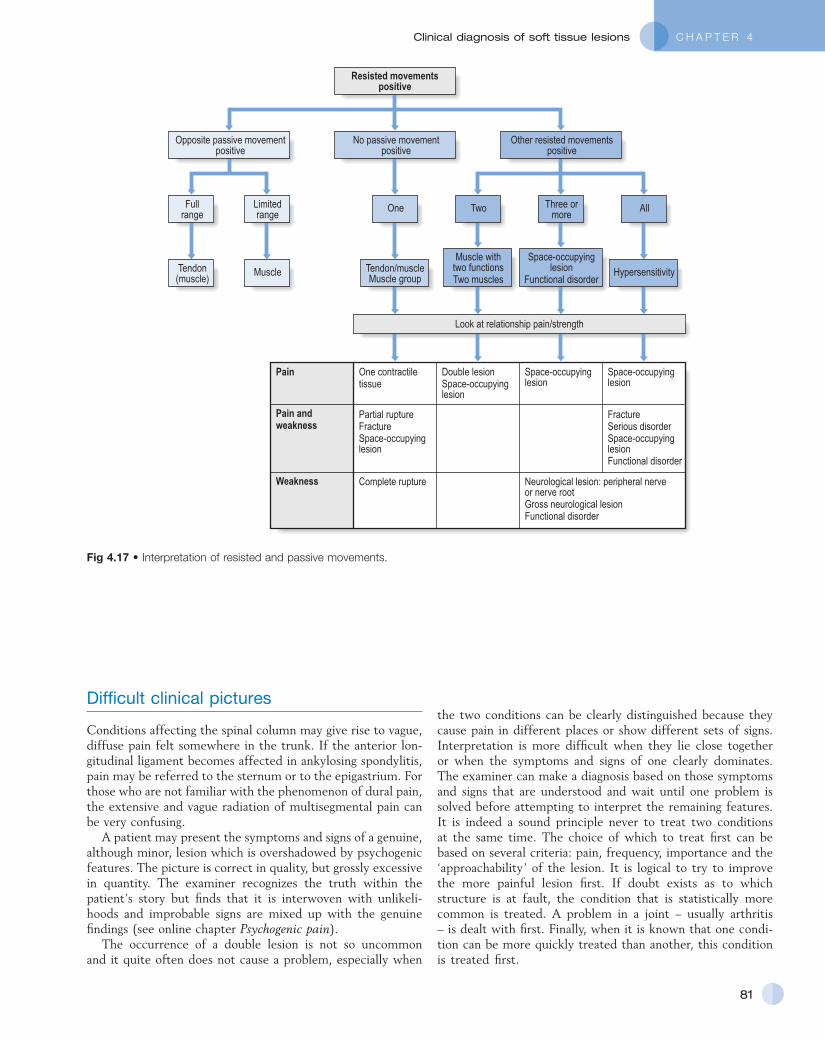
C H A P T E R 4 Clinical diagnosis of soft tissue lesions
81
the two conditions can be clearly distinguished because they cause pain in different places or show different sets of signs. Interpretation is more difficult when they lie close together or when the symptoms and signs of one clearly dominates. The examiner can make a diagnosis based on those symptoms and signs that are understood and wait until one problem is solved before attempting to interpret the remaining features. It is indeed a sound principle never to treat two conditions at the same time. The choice of which to treat first can be based on several criteria: pain, frequency, importance and the ‘approachability’ of the lesion. It is logical to try to improve the more painful lesion first. If doubt exists as to which structure is at fault, the condition that is statistically more common is treated. A problem in a joint – usually arthritis – is dealt with first. Finally, when it is known that one condi-tion can be more quickly treated than another, this condition is treated first.
Difficult clinical pictures
Conditions affecting the spinal column may give rise to vague, diffuse pain felt somewhere in the trunk. If the anterior lon-gitudinal ligament becomes affected in ankylosing spondylitis, pain may be referred to the sternum or to the epigastrium. For those who are not familiar with the phenomenon of dural pain, the extensive and vague radiation of multisegmental pain can be very confusing.
A patient may present the symptoms and signs of a genuine, although minor, lesion which is overshadowed by psychogenic features. The picture is correct in quality, but grossly excessive in quantity. The examiner recognizes the truth within the patient’s story but finds that it is interwoven with unlikeli-hoods and improbable signs are mixed up with the genuine findings (see online chapter Psychogenic pain).
The occurrence of a double lesion is not so uncommon and it quite often does not cause a problem, especially when
Fig 4.17 • Interpretation of resisted and passive movements.
Partial ruptureFractureSpace-occupyinglesion
One contractiletissue
Double lesionSpace-occupyinglesion
Space-occupyinglesion
Space-occupyinglesion
Pain
FractureSerious disorderSpace-occupyinglesionFunctional disorder
Complete rupture
Pain andweakness
Weakness Neurological lesion: peripheral nerveor nerve rootGross neurological lesionFunctional disorder
Look at relationship pain/strength
Tendon/muscleMuscle group
Muscle withtwo functionsTwo muscles
Hypersensitivity
Space-occupyinglesion
Functional disorder
Three ormore
Two AllOne
Other resisted movementspositive
No passive movementpositive
Limitedrange
Opposite passive movementpositive
Muscle
Fullrange
Tendon(muscle)
Resisted movementspositive

General Principles
82
The clinical approach in orthopaedic medicine is summa-rized in Fig. 4.18.
Psychogenic problems
The reader is referred to the online chapter Psychogenic pain.
Access the complete reference list online at www.orthopaedicmedicineonline.com
Fig 4.18 • Clinical approach in orthopaedic medicine.
Positiveand negative
responses
Soft tissue lesions
Assess function
Clinical examination
History
Symptoms
Referred pain
Inherentlikelihoods
Applied anatomy
Interpretation
Functionalexamination
Signs
Pattern
Doubt
Technicalinvestigations
Diagnosis
PrognosisNatural history Treatment Prophylaxis
Deeptransverse
friction
ManipulationMobilization
Traction
InjectionInfiltration

C H A P T E R 4Clinical diagnosis of soft tissue lesions
82.e1© Copyright 2013 Elsevier, Ltd. All rights reserved.
References
1. Takatalo J, Karppinen J, Niinimäki J, et al. Does lumbar disc degeneration on MRI associate with low back symptom severity in young Finnish adults? Spine (Phila PA 1976) 2011 Feb 24.
2. Ernst CW, Stadnik TW, Peeters E, et al. Prevalence of annular tears and disc herniations on MR images of the cervical spine in symptom-free volunteers. Eur J Radiol 2005;55(3):409–14.
3. Wood KB, Garvey TA, Gundry C, Heithoff KB. Magnetic resonance imaging of the thoracic spine. Evaluation of asymptomatic individuals. J Bone Joint Surg Am 1995;77(11):1631–38.
4. Hirano Y, Sashi R, Izumi J, et al. Comparison of the MR findings on indirect MR arthrography in patients with rotator cuff tears with and without symptoms. Radiat Med 2006;24(1):23–7.
5. Reilly P, Macleod I, Macfarlane R, et al. Dead men and radiologists don’t lie: a review of cadaveric and radiological studies of rotator cuff tear prevalence. Ann R Coll Surg Engl 2006;88(2):116–21.
6. Yamamoto A, Takagishi K, Kobayashi T, et al. Factors involved in the presence of symptoms associated with rotator cuff tears: a comparison of asymptomatic and symptomatic rotator cuff tears in the general population. J Shoulder Elbow Surg 2011 Mar 29.
7. Kim HM, Teefey SA, Zelig A, et al. Shoulder strength in asymptomatic individuals with intact compared with torn rotator cuffs. J Bone Joint Surg Am 2009;91(2):289–96.
8. Soder RB, Simões JD, Soder JB, Baldisserotto M. MRI of the knee joint in asymptomatic adolescent soccer players: a controlled study. AJR Am J Roentgenol 2011;196(1):W61–65.
9. Kaplan LD, Schurhoff MR, Selesnick H, et al. Magnetic resonance imaging of the knee in asymptomatic professional basketball players. Arthroscopy 2005;21(5):557–61.
10. Christensen HW, Nilsson N. The reliability of measuring active and passive cervical range of motion: an observer-blinded and randomized repeated-measures design. J Manipulative Physiol Ther 1998;21(5):341–7.
11. Pellecchia GL, Paolino J, Connell J. Intertester reliability of the Cyriax evaluation in assessing patients with shoulder pain. J Orthop Sports Phys Ther 1996;23(1):34–8.
12. Chesworth BM, MacDermid JC, Roth JH, Patterson SD. Movement diagram and ‘end-feel’ reliability when measuring passive lateral rotation of the shoulder in patients with shoulder pathology. Phys Ther 1998;78(6):593–601.
13. Wong A, Nansel DD. Comparisons between active vs. passive end-range assessments in subjects exhibiting cervical range of motion asymmetries. J Manipulative Physiol Ther 1992;15(3):159–63.
14. Youdas JW, Carey JR, Garrett TR. Reliability of measurements of cervical spine range of motion – comparison of three methods. Phys Ther 1991;71(2):98–104.
15. Fritz JM, Delitto A, Erhard RE, Roman M. An examination of the selective tissue tension scheme, with evidence for the concept of a capsular pattern of the knee. Phys Ther 1998;78(10):1046–56.
16. Hanchard NC, Howe TE, Gilbert MM. Diagnosis of shoulder pain by history and selective tissue tension: agreement between assessors. J Orthop Sports Phys Ther 2005;35(3):147–53.
17. Cyriax JH. Textbook of Orthopaedic Medicine, vol I: Diagnosis of Soft Tissue Lesions. 8th ed. London: Baillière Tindall; 1982. p. 45.
18. Donelson R, Silva G, Murphy K. Centralization phenomenon, its usefulness in evaluating and treating referred pain. Spine 1990;15(3):211–3.
19. Donelson R, Aprill C, Medcalf R, Grant W. A prospective study of centralisation of lumbar and referred pain. Spine 1997;22(10):1115–22.
20. Wyke W. Neurological mechanisms in the experience of pain. Acupunc Electrother Res J 1979;4:27.
21. Wyke BD. The neurology of low back pain. In: Jayson MIV, editor. The Lumbar Spine and Back Pain. 2nd ed. Kent: Pitman Medical; 1980.
22. Veys EM, Mielants H, Verbruggen G. Reumatologie. Ghent: Omega Editions; 1985. p. 21–3.
23. Hayes KW, Petersen CM. Reliability of assessing end-feel and pain and resistance sequence in subjects with painful shoulders and knees. J Orthop Sports Phys Ther 2001;31(8):432–45.
24. Greenman PE. Principles of Manual Medicine. Baltimore: William & Wilkins; 1989. p. 61–70.
25. Twomey LT, Taylor JR. Physical Therapy of the Low Back. New York: Churchill Livingstone; 1987. p. 192–7.
26. Jones MA, Jones HM. Principles of the physical examination. In: Boyling JD, Palastanga N, editors. Grieve’s Modern Manual Therapy, The Vertebral Column. Edinburgh: Churchill Livingstone; 1994. p. 491–501.
27. Panzer DM. The reliability of lumbar motion palpation. J Manipulative Physiol Ther 1992;15(8):518–24.
28. Matyas T, Bach T. The reliability of selected techniques in clinical arthrometics. Aust J Physiother 1985;31:175–99.
29. Hardy GL, Napier JK. Inter- and intratherapist reliability of passive accessory movement and technique. NZ J Physiother 1991:22–4.
30. Fjellner A, Bexander C, Faleij R, Strender LE. Interexaminer reliability in physical examination of the cervical spine. J Manipulative Physiol Ther 1999;22(8):511–6.
31. Vincent-Smith B, Gibbons P. Inter-examiner and intra-examiner reliability of the standing flexion test. Manual Therapy 1999;4(2):87–93.
32. Maher C, Latimer J. Pain or resistance – the manual therapists’ delimma. Aust J Physiother 1992;38:257–60.
33. Keating J, Bergman T, Jacobs G, Bradley D, Finer A, Larson K. Inter-examiner reliability of eight evaluative dimensions of lumbar segmental abnormality. J Manipulative Physiol Ther 1990;13:463–70.
34. Potter N, Rothstein J. Intertester reliability for selected clinical tests of the sacroiliac joint. Phys Ther 1985;65:1671–5.
35. MacRae DL. Asymptomatic intervertebral disc protrusion. Acta Radiologica 1956;46:9.
36. Hitselberger WE, Whitten RM. Abnormal myelograms in asymptomatic patients. J Neurosurg 1968;28:204.
37. Wiesel SW, Tsourmas N, Feffer HL, Citrin CM, Patronas N. A study of computer-assisted tomography: 1. The incidence of positive CAT scans in an asymptomatic group of patients. Spine 1984;9:549–51.
38. Powell MC, Wilson M, Szypryt P, Symonds EM. Prevalence of lumbar disc degeneration observed by magnetic resonance in symptomless women. Lancet 1986;13:1366–7.
39. Weinreb JC, Wolbarsht LB, Cohen JM, Brown CE, Maravilla KR. Prevalence of lumbosacral intervertebral disc abnormalities on MR images in pregnant and asymptomatic non-pregnant women. Radiology 1989;170:125–8.
40. Boden SD, Davis DO, Dina TS, Patronas MJ, Wiesel SW. Abnormal magnetic resonance scans of the lumbar spine in asymptomatic subjects. J Bone Joint Surg 1990;72A:403–8.
41. Buirski G, Silberstein M. The symptomatic lumbar disc in patients with low-back pain. Magnetic resonance imaging appearances in both a symptomatic and control population. Spine 1993;18:1808–11.
42. Matsumoto M, Fujimura Y, Suzuki N, Nishi Y, Nakamura M, Yabe Y, Shiga H. MRI of cervical intervertebral discs in asymptomatic subjects. J Bone Joint Surg (Br) 1998;80(1):19–24.
43. Humphreys SC, Hodges SD, Fisher DL, Eck JC, Covington LA. Reliability of magnetic resonance imaging in predicting disc material posterior to the posterior longitudinal ligament in the cervical spine. A prospective study. Spine 1998;23(22):2468–71.
44. Kaiser JA, Holland BA. Imaging of the cervical spine. Spine 1998;23(24):2701–12.
45. Shafaie FF, Wippold FJ 2nd, Gado M, Pilgram TK, Riew KD. Comparison of computed tomography myelography and magnetic resonance imaging in the evaluation of cervical spondylotic myelopathy and radiculopathy. Spine 1999;24(17):1781–5.

General Principles
82.e2© Copyright 2013 Elsevier, Ltd. All rights reserved.
46. Thompson WO, Debski RE, Boardman ND 3rd, Taskiran E, Warner JJ, Fu FH, Woo SL. A biomechanical analysis of rotator cuff deficiency in a cadaveric model. Am J Sports Med 1996;24(3):286–92.
47. Petersson CJ. Ruptures of the supraspinatus tendon. Cadaver dissection. Acta Orthop Scand 1984;55(1):52–6.
48. Jerosch J, Muller T, Castro WH. The incidence of rotator cuff rupture. An anatomic study. Acta Orthop Belg 1991;57(2):124–9.
49. Kummer FJ, Zuckerman JD. The incidence of full thickness rotator cuff tears in a large cadaveric population. Bull Hosp Dis 1995;54(1):30–1.
50. Jerosch J, Castro WH, Assheuer J. Age-related magnetic resonance imaging morphology of the menisci in asymptomatic individuals. Arch Orthop Trauma Surg 1996;105(3–4):199–202.
51. LaPrade RF, Burnett QM 2nd, Veenstra MA, Hodgman CG. The prevalence of abnormal magnetic resonance imaging
findings in asymptomatic knees. With correlation of magnetic resonance imaging to arthroscopic findings in symptomatic knees. Am J Sports Med 1994;22(6):739–45.
52. Cyriax JH. Textbook of Orthopaedic Medicine, vol I: Diagnosis of Soft Tissue Lesions. 8th ed. London: Baillière Tindall; 1982. p. 151.