Embed Size (px)
DESCRIPTION
FYI
Citation preview
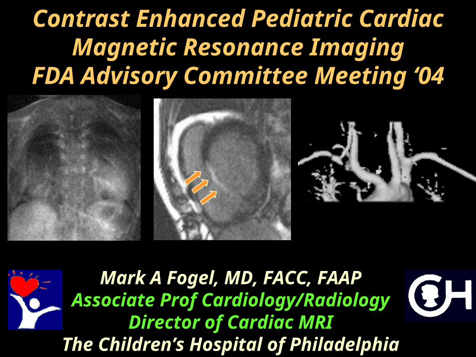
Contrast Enhanced Pediatric Cardiac Magnetic Resonance
ImagingFDA Advisory Committee Meeting
‘04
Mark A Fogel, MD, FACC, FAAPAssociate Prof
Cardiology/RadiologyDirector of Cardiac MRI
The Children’s Hospital of Philadelphia
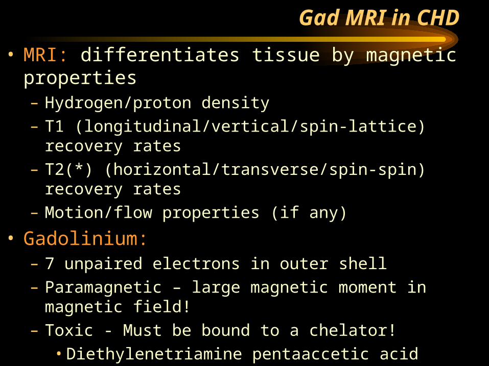
Gad MRI in CHD
• MRI: differentiates tissue by magnetic properties– Hydrogen/proton density– T1 (longitudinal/vertical/spin-lattice) recovery rates– T2(*) (horizontal/transverse/spin-spin) recovery rates– Motion/flow properties (if any)
• Gadolinium:– 7 unpaired electrons in outer shell– Paramagnetic – large magnetic moment in magnetic
field!– Toxic - Must be bound to a chelator!
• Diethylenetriamine pentaaccetic acid (DPTA)• Can be bound to large molecules (eg albumin) –
doesn’t diffuse thru capillary membrane (“blood pool agent”)
– Not yet FDA approved
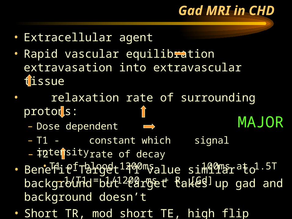
Gad MRI in CHD
– T2 - rate of decay
• Benefit-Target T1 value similar to background but target takes up gad and background doesn’t
• Short TR, mod short TE, high flip angle studies
• Extracellular agent• Rapid vascular equilibration extravasation
into extravascular tissue• relaxation rate of surrounding protons:
– Dose dependent– T1 - constant which signal intensity
• T1 of blood 1200ms 100ms at 1.5T
– 1/T1 = 1/1200 ms + R1 [Gd]
MAJOR
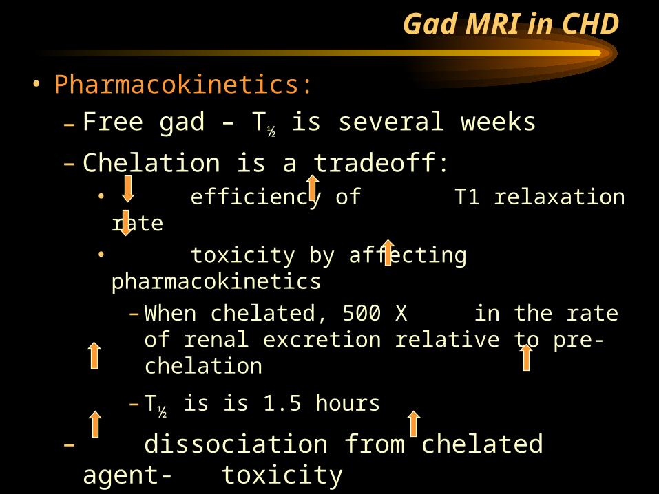
Gad MRI in CHD
• Pharmacokinetics:
– Free gad – T½ is several weeks
– Chelation is a tradeoff:• efficiency of T1 relaxation rate• toxicity by affecting pharmacokinetics
– When chelated, 500 X in the rate of renal excretion relative to pre-chelation
– T½ is is 1.5 hours
– dissociation from chelated agent- toxicity• Theory: Competing moeity – copper and zinc
– time of gad in the body- toxicity
Gad Enhanced MRI in CHD - Safety
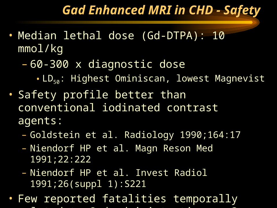
• Median lethal dose (Gd-DTPA): 10 mmol/kg– 60-300 x diagnostic dose
• LD50: Highest Ominiscan, lowest Magnevist
• Safety profile better than conventional iodinated contrast agents:– Goldstein et al. Radiology 1990;164:17– Niendorf HP et al. Magn Reson Med 1991;22:222– Niendorf HP et al. Invest Radiol 1991;26(suppl
1):S221
• Few reported fatalities temporally related to Gad administration - ? Association
• No known contraindications
Gad Enhanced MRI in CHD - Safety
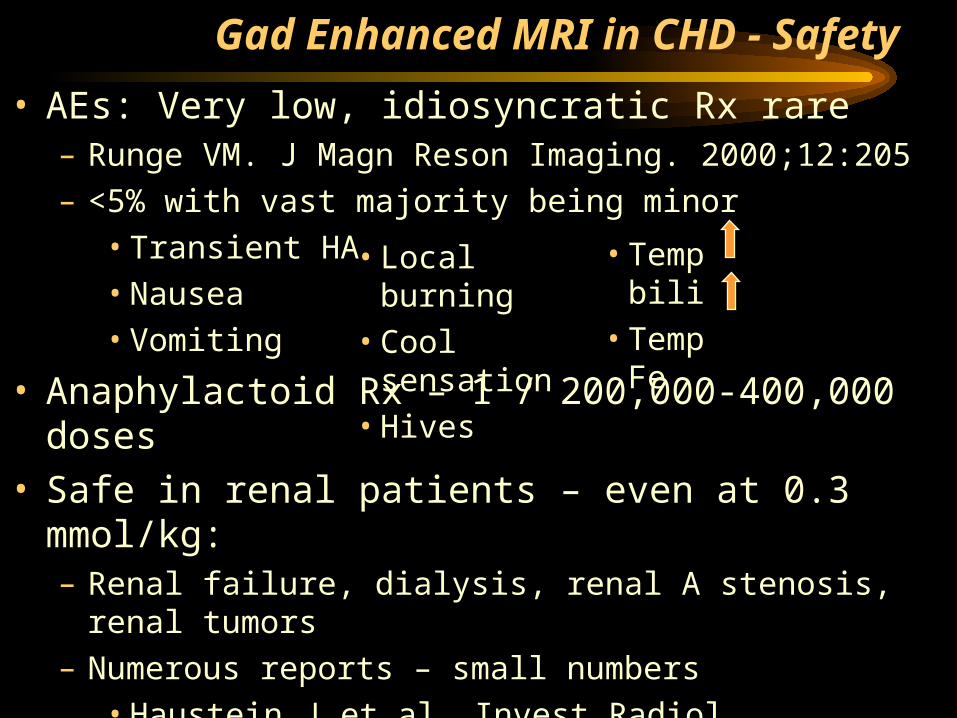
• AEs: Very low, idiosyncratic Rx rare– Runge VM. J Magn Reson Imaging. 2000;12:205 – <5% with vast majority being minor
• Transient HA• Nausea• Vomiting
• Anaphylactoid Rx – 1 / 200,000-400,000 doses
• Safe in renal patients – even at 0.3 mmol/kg:– Renal failure, dialysis, renal A stenosis, renal
tumors– Numerous reports – small numbers
• Haustein J et al. Invest Radiol 1992;27:153• Prince MR et al. J Magn Reson Imaging
1996;6:162• Rofsky NM et al. Radiology 1991;180:85
• Local burning• Cool sensation• Hives
• Temp bili• Temp Fe
Gad Enhanced MRI in CHD – Peds
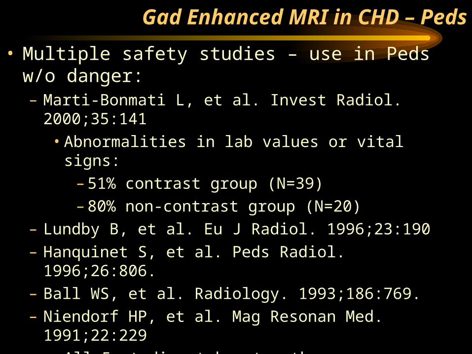
• Multiple safety studies – use in Peds w/o danger:– Marti-Bonmati L, et al. Invest Radiol. 2000;35:141
• Abnormalities in lab values or vital signs:– 51% contrast group (N=39)– 80% non-contrast group (N=20)
– Lundby B, et al. Eu J Radiol. 1996;23:190– Hanquinet S, et al. Peds Radiol. 1996;26:806.– Ball WS, et al. Radiology. 1993;186:769.– Niendorf HP, et al. Mag Resonan Med. 1991;22:229
• All 5 studies taken together:– Dose 0.1-0.2 mmol/kg– 1368 children from 15 days – 21 years of age– AEs – 2-5%, none which were serious

Gad MRI in CHD - Marketed Products
From Cardiovascular Magnetic Resonance Imaging – 2004, Martin Dunitz, Chapter 2, page 20
Gadolinium based
Gad MRI in CHD - Marketed Products
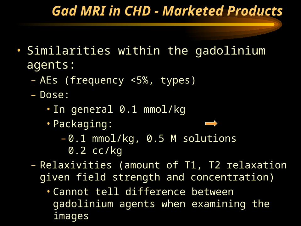
• Similarities within the gadolinium agents: – AEs (frequency <5%, types)– Dose:
• In general 0.1 mmol/kg• Packaging:
– 0.1 mmol/kg, 0.5 M solutions 0.2 cc/kg– Relaxivities (amount of T1, T2 relaxation given
field strength and concentration)• Cannot tell difference between gadolinium
agents when examining the images– Nephrotoxicity (none)
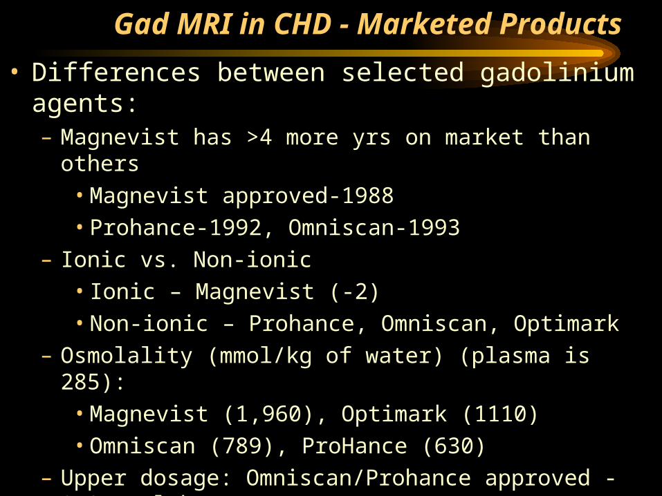
Gad MRI in CHD - Marketed Products
• Differences between selected gadolinium agents:– Magnevist has >4 more yrs on market than others
• Magnevist approved-1988• Prohance-1992, Omniscan-1993
– Ionic vs. Non-ionic• Ionic – Magnevist (-2)• Non-ionic – Prohance, Omniscan, Optimark
– Osmolality (mmol/kg of water) (plasma is 285):• Magnevist (1,960), Optimark (1110)• Omniscan (789), ProHance (630)
– Upper dosage: Omniscan/Prohance approved - 0.3 mmol/kg
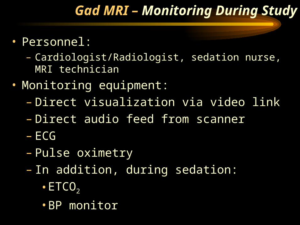
• Personnel:– Cardiologist/Radiologist, sedation nurse, MRI
technician
• Monitoring equipment:– Direct visualization via video link– Direct audio feed from scanner– ECG– Pulse oximetry– In addition, during sedation:
•ETCO2
•BP monitor
Gad MRI – Monitoring During Study
Gad Enhanced MRI in CHD
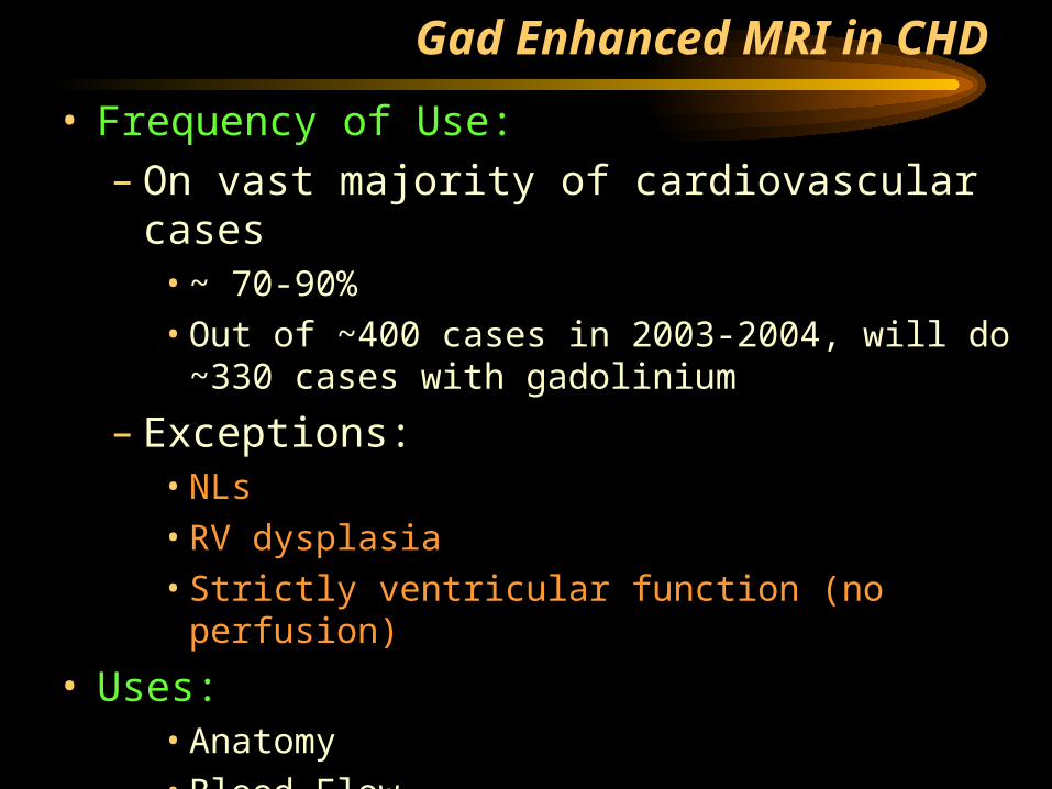
• Frequency of Use:– On vast majority of cardiovascular cases
• ~ 70-90%• Out of ~400 cases in 2003-2004, will do ~330
cases with gadolinium
– Exceptions:• NLs• RV dysplasia• Strictly ventricular function (no perfusion)
• Uses:• Anatomy• Blood Flow• Tissue Characterization
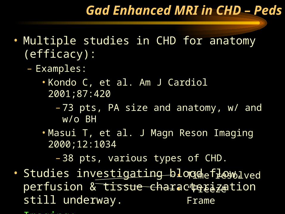
• Multiple studies in CHD for anatomy (efficacy):– Examples:
• Kondo C, et al. Am J Cardiol 2001;87:420– 73 pts, PA size and anatomy, w/ and w/o BH
• Masui T, et al. J Magn Reson Imaging 2000;12:1034
– 38 pts, various types of CHD.
• Studies investigating blood flow, perfusion & tissue characterization still underway.
• Imaging:– First pass – Delayed enhancement
Gad Enhanced MRI in CHD – Peds
• Time resolved• “Freeze Frame”
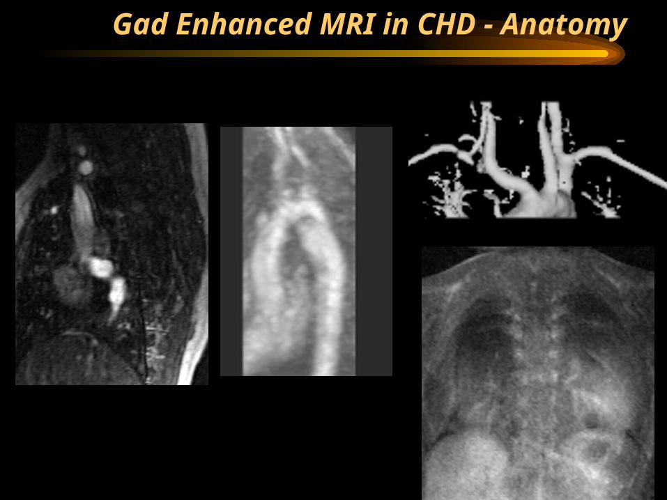
Gad Enhanced MRI in CHD - Anatomy
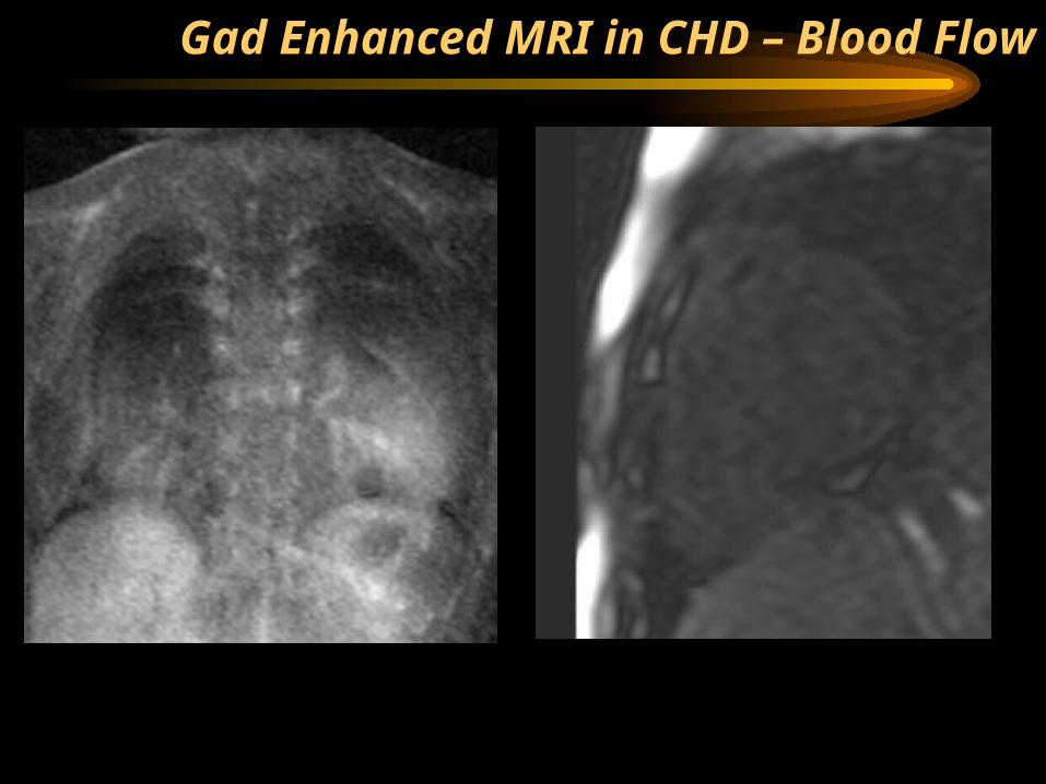
Gad Enhanced MRI in CHD – Blood Flow
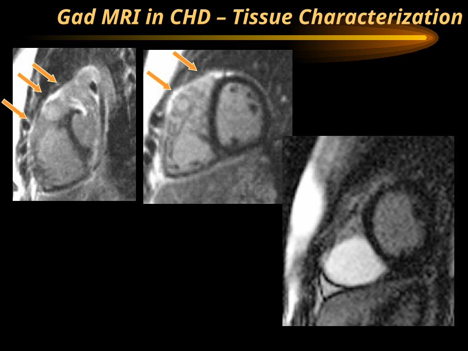
Gad MRI in CHD – Tissue Characterization
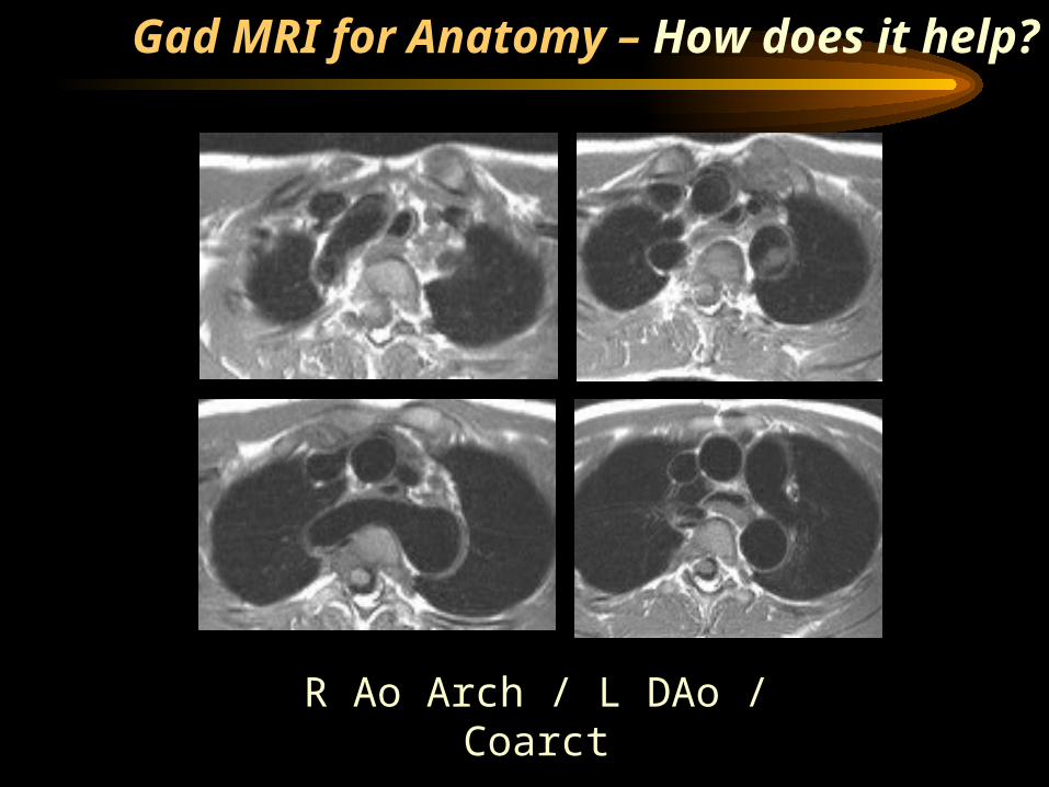
R Ao Arch / L DAo / Coarct
Gad MRI for Anatomy – How does it help?
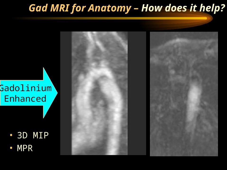
GadoliniumEnhanced
• 3D MIP• MPR
Gad MRI for Anatomy – How does it help?
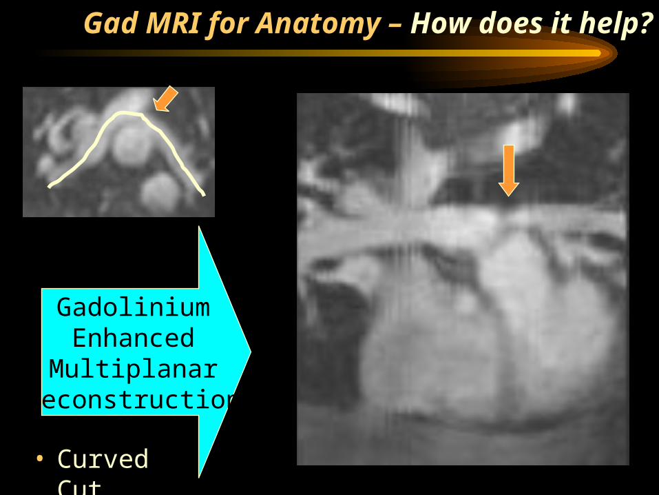
GadoliniumEnhanced
MultiplanarReconstruction
• Curved Cut
Gad MRI for Anatomy – How does it help?
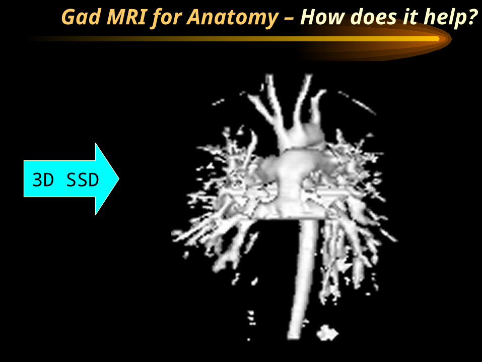
3D SSD
Gad MRI for Anatomy – How does it help?
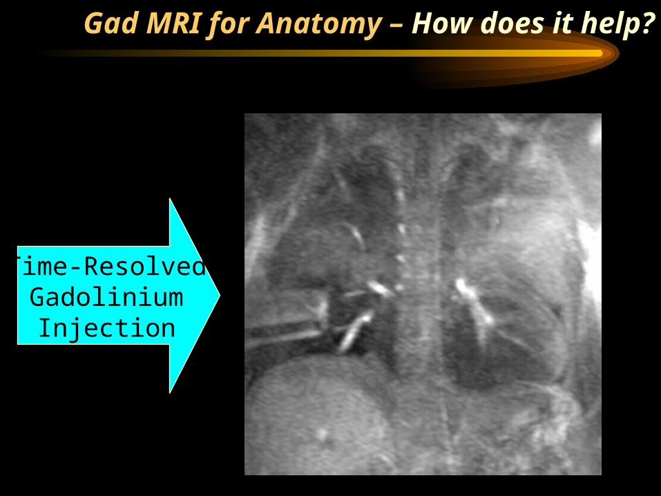
Time-ResolvedGadolinium
Injection
Gad MRI for Anatomy – How does it help?
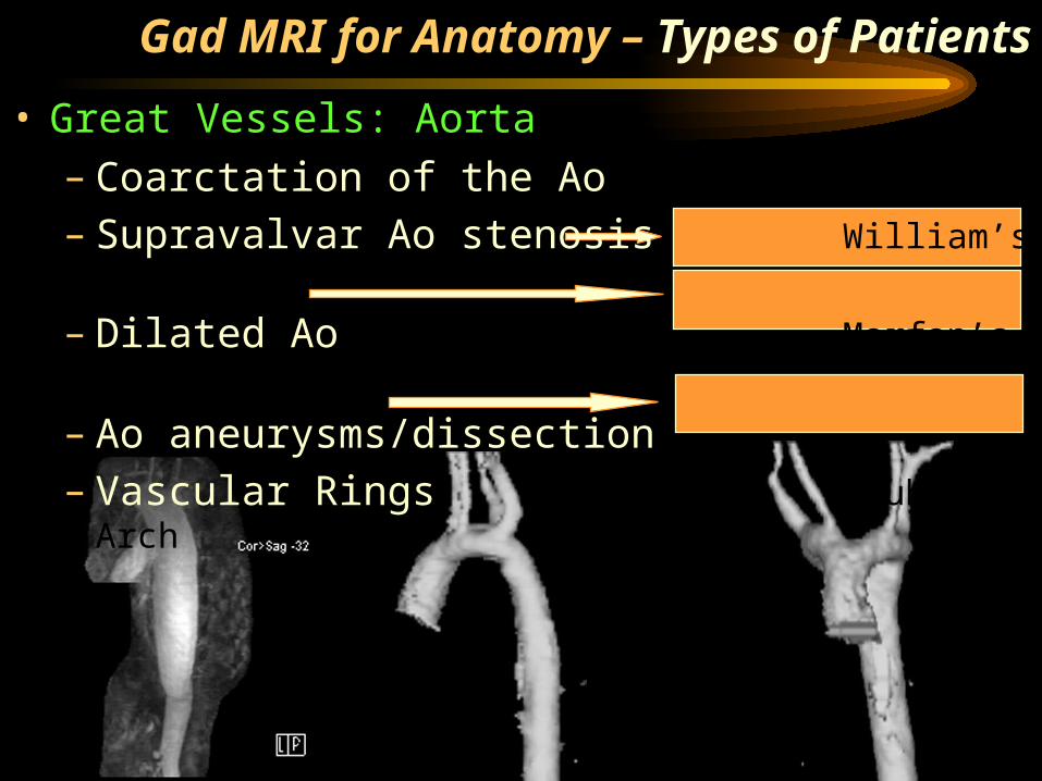
Gad MRI for Anatomy – Types of Patients
• Great Vessels: Aorta– Coarctation of the Ao– Supravalvar Ao stenosis William’s Syndrome
– Dilated Ao Marfan’s Syndrome
– Ao aneurysms/dissection– Vascular Rings Double Ao Arch
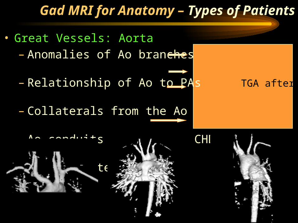
• Great Vessels: Aorta– Anomalies of Ao branches Isolated LSCA
– Relationship of Ao to PAs TGA after ASO
– Collaterals from the Ao TOF/PA
– Ao conduits for complex CHD Jump graft-Coa
– Reconstructed Aortas Ao-PA anastomosis
Gad MRI for Anatomy – Types of Patients
Gad MRI for Anatomy – Types of Patients
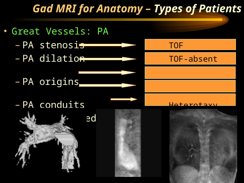
• Great Vessels: PA– PA stenosis TOF
– PA dilation TOF-absent pulm valve
– PA origins Truncus/Hemitruncus
– PA conduits Heterotaxy
– Reconstructed PAs Fontan
Gad MRI for Anatomy – Types of Patients
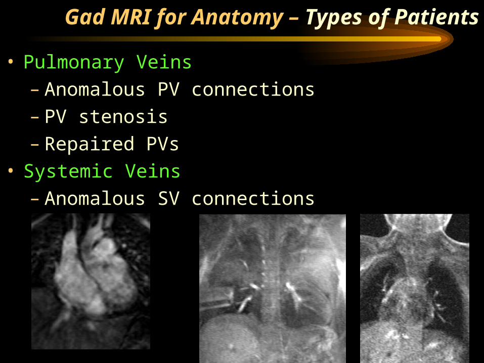
• Pulmonary Veins– Anomalous PV connections– PV stenosis– Repaired PVs
• Systemic Veins– Anomalous SV connections
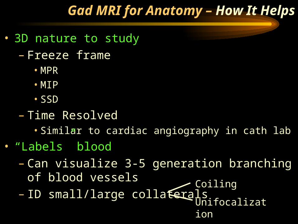
Gad MRI for Anatomy – How It Helps
• 3D nature to study– Freeze frame
• MPR• MIP• SSD
– Time Resolved• Similar to cardiac angiography in cath lab
• “Labels” blood– Can visualize 3-5 generation branching of
blood vessels– ID small/large collaterals
Coiling
Unifocalization
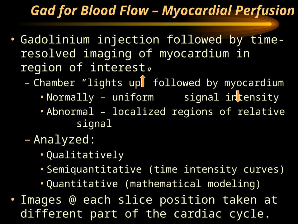
Gad for Blood Flow – Myocardial Perfusion
• Gadolinium injection followed by time-resolved imaging of myocardium in region of interest.– Chamber “lights up” followed by myocardium
• Normally – uniform signal intensity• Abnormal – localized regions of relative
signal
– Analyzed:• Qualitatively• Semiquantitative (time intensity curves)• Quantitative (mathematical modeling)
• Images @ each slice position taken at different part of the cardiac cycle.
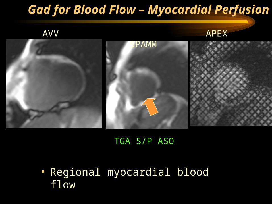
• Regional myocardial blood flow
TGA S/P ASO
AVV APEX SPAMM
Gad for Blood Flow – Myocardial Perfusion
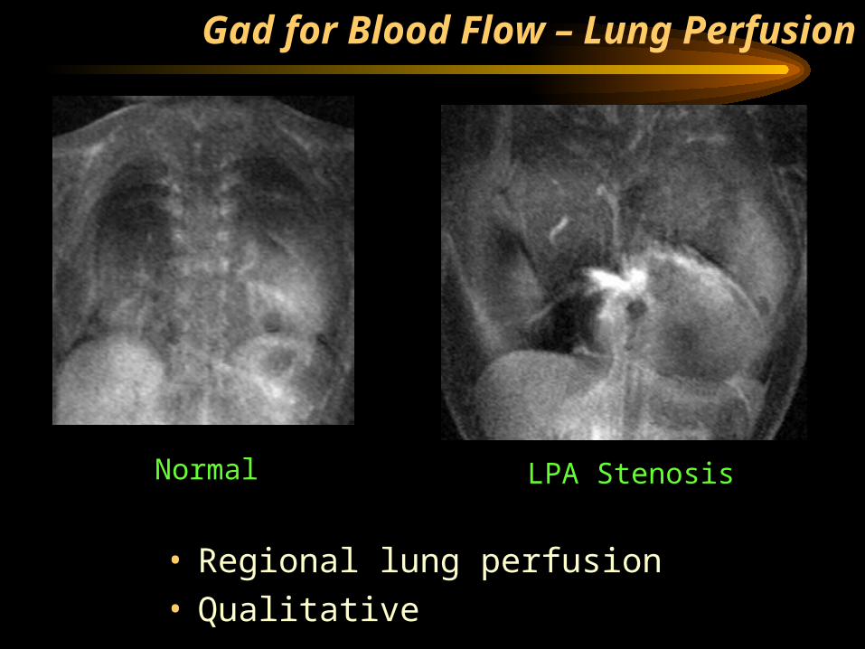
Gad for Blood Flow – Lung Perfusion
• Regional lung perfusion• Qualitative
Normal LPA Stenosis
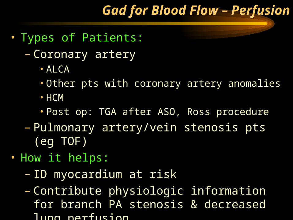
Gad for Blood Flow – Perfusion
• Types of Patients:– Coronary artery
• ALCA• Other pts with coronary artery anomalies• HCM• Post op: TGA after ASO, Ross procedure
– Pulmonary artery/vein stenosis pts (eg TOF)
• How it helps:– ID myocardium at risk– Contribute physiologic information for
branch PA stenosis & decreased lung perfusion
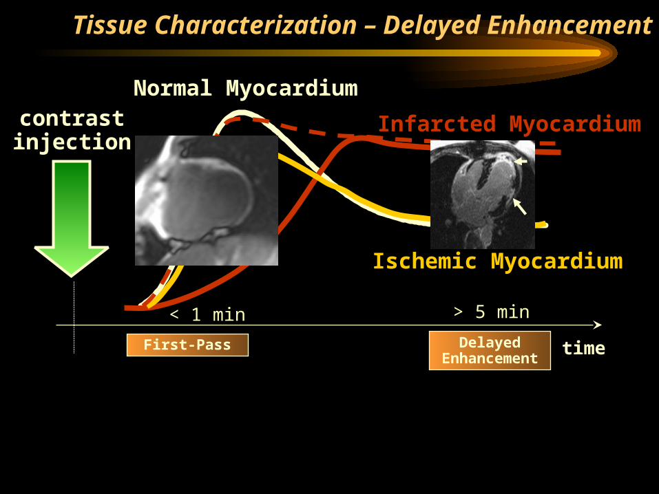
Tissue Characterization – Delayed Enhancement
time
Normal Myocardiumcontrastinjection
Infarcted Myocardium
Ischemic Myocardium
First-Pass Delayed Enhancement
< 1 min > 5 min
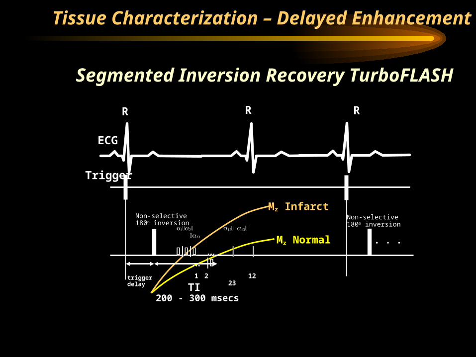
Segmented Inversion Recovery TurboFLASH
R
ECG
Trigger
R R
Non-selective180o inversion
triggerdelay
11 22 12 12 2323
TI TI 200 - 300 msecs200 - 300 msecs
Non-selective180o inversion
. . .
Mz Infarct
Mz Normal
Tissue Characterization – Delayed Enhancement
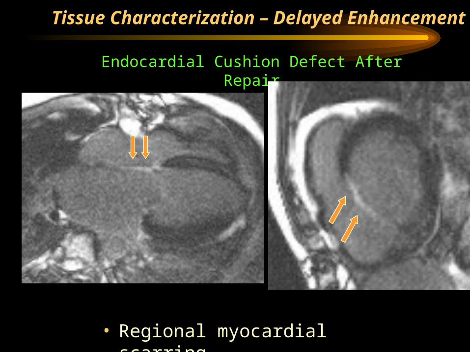
• Regional myocardial scarring
Endocardial Cushion Defect After Repair
Tissue Characterization – Delayed Enhancement
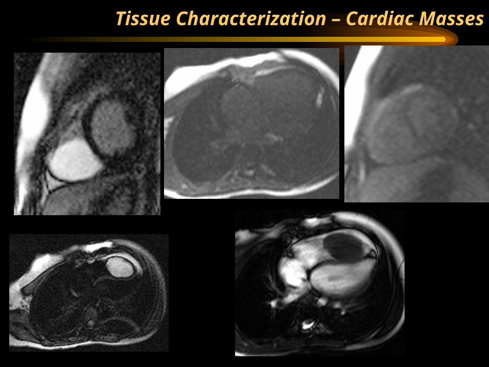
Tissue Characterization – Cardiac Masses
Tissue Characterization – Cardiac Masses
• Types of cardiac masses:– Hyperenhancement:
• Myxoma• Hemangioma• Angiosarcoma• Fibroma (slight/heterogeneous)• Pericardial cysts• Lymphoma (heterogenous)
– No enhancement• Thrombus
– Non-specific:• Lipoma• Lipomatous
hypertrophy• Rhabdomyom
a
– Not published• Liposarcoma• Leiomyosarco
ma
Tissue Characterization
• Types of Patients:– Coronary artery
• ALCA• Other pts with coronary artery anomalies• HCM• Post op patients, especially after CPB and DHCA
– Myocardial tumors/masses• How it helps:
– ID scarred myocardium– Contribute to prognosis in patients with
tumors
Gad MRI – Dosing & Administration
• Anatomy of Great Vessels:– Freeze frame: single - double dose of gad
• Neimatallah MA et al. JMRI. 1999:10:758-770.
– Time resolved: ¼ - ½ dose gad as a minimum
• Blood Flow:– Myocardial/Lung perfusion: ½ dose of gad
(minimum)• Tissue Characterization:
– Single dose of gadolinium• Administration:
– Power injector– Hand
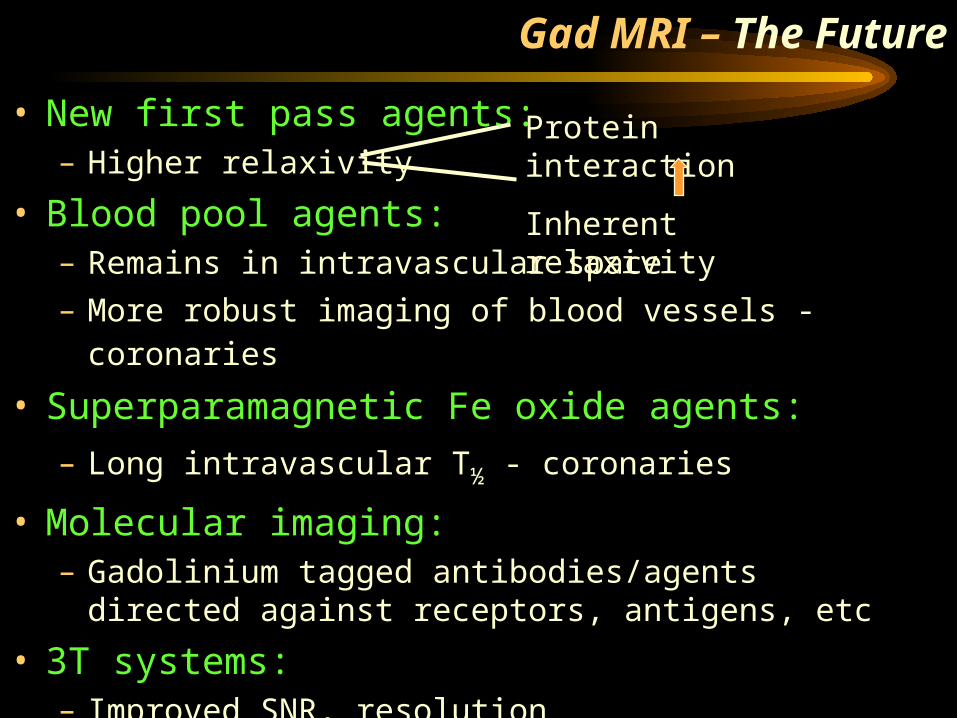
Gad MRI – The Future
• New first pass agents:– Higher relaxivity
• Blood pool agents:– Remains in intravascular space
– More robust imaging of blood vessels - coronaries • Superparamagnetic Fe oxide agents:
– Long intravascular T½ - coronaries
• Molecular imaging:– Gadolinium tagged antibodies/agents directed
against receptors, antigens, etc
• 3T systems:– Improved SNR, resolution
Protein interaction
Inherent relaxivity
“It’s hard to make predictions,
especially about the future.”
….Yogi Berra
Gad MRI – The Future
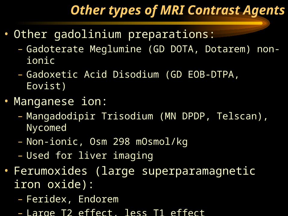
• Other gadolinium preparations:– Gadoterate Meglumine (GD DOTA, Dotarem) non-
ionic– Gadoxetic Acid Disodium (GD EOB-DTPA, Eovist)
• Manganese ion:– Mangadodipir Trisodium (MN DPDP, Telscan),
Nycomed– Non-ionic, Osm 298 mOsmol/kg– Used for liver imaging
• Ferumoxides (large superparamagnetic iron oxide):– Feridex, Endorem– Large T2 effect, less T1 effect– Liver imaging
Other types of MRI Contrast Agents


