Embed Size (px)
Citation preview
46 years old woman with chest pain
Case conference 4/1/08
Past Medical History
¢ Ischemic heart diseasel Past anterior MI by echo and NM testingl DES in 10/05l EF had improved from 45% to 50-55% in
2006l Past episodes of heart failure
¢ Diabetes mellitus, type II, diagnosed 1993
¢ Obstructive sleep apnea¢ Chronic anemia
Social history & Family history¢ Works in produce department¢ Quit smoking cigarettes 8/07 after 30
pack years¢ No alcohol, no illicit drug use¢ Family history of diabetes and ASCVD
(dad died at 60 of MI)
History of present illness
¢ One day prior to admission she noticed a few twinges of chest pain
¢ Day of admission she noted chest pain that was acute in onset, severe, substernal and radiated to both sides of chest, and positional
¢ She had noted a dry cough and fevers two weeks ago that had resolved 1 week ago
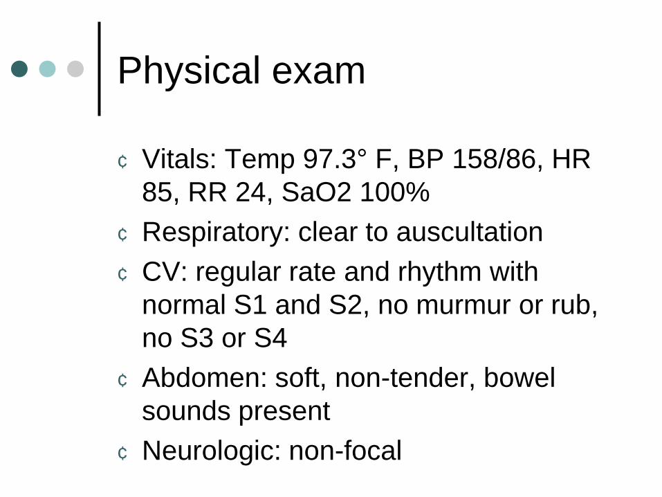
Physical exam
¢ Vitals: Temp 97.3° F, BP 158/86, HR 85, RR 24, SaO2 100%
¢ Respiratory: clear to auscultation¢ CV: regular rate and rhythm with
normal S1 and S2, no murmur or rub, no S3 or S4
¢ Abdomen: soft, non-tender, bowel sounds present
¢ Neurologic: non-focal
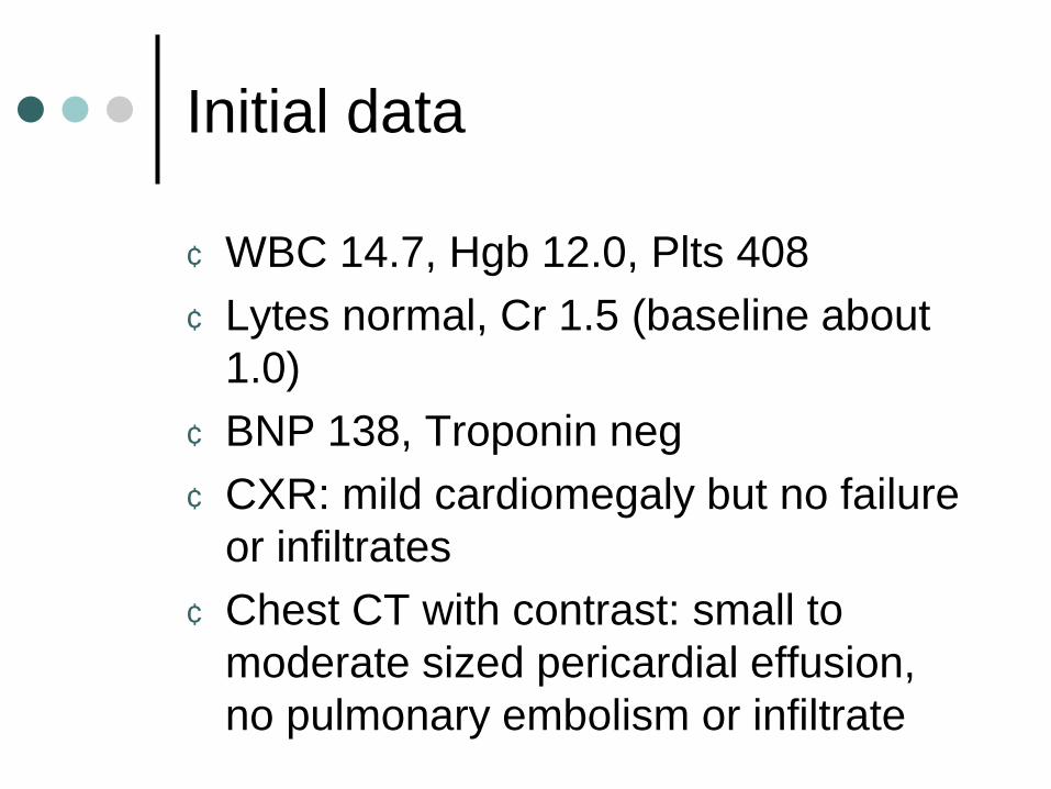
Initial data
¢ WBC 14.7, Hgb 12.0, Plts 408¢ Lytes normal, Cr 1.5 (baseline about
1.0)¢ BNP 138, Troponin neg¢ CXR: mild cardiomegaly but no failure
or infiltrates¢ Chest CT with contrast: small to
moderate sized pericardial effusion, no pulmonary embolism or infiltrate
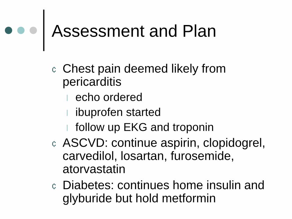
Assessment and Plan
¢ Chest pain deemed likely from pericarditisl echo orderedl ibuprofen startedl follow up EKG and troponin
¢ ASCVD: continue aspirin, clopidogrel, carvedilol, losartan, furosemide, atorvastatin
¢ Diabetes: continues home insulin and glyburide but hold metformin
Initial hospital course
¢ First contact in ED at 2212 on 11/27¢ Admitted at 0300 on 11/28¢ Still having chest pain that afternoon
requiring opiate pain medications but improving some
¢ Echocardiogram shows normal EF, small pericardial effusion without tamponade
¢ Troponin negative
11/29
¢ Pain still moderately severe and given a dose of ketorolac overnight
¢ Vitals normal¢ Exam unchanged¢ Creatinine 4.3, BUN 50, CO2 20, K
5.8¢ WBC 13.9, Hgb 10.8¢ ESR 99¢ UA: some protein, 1 RBC, 3 WBC
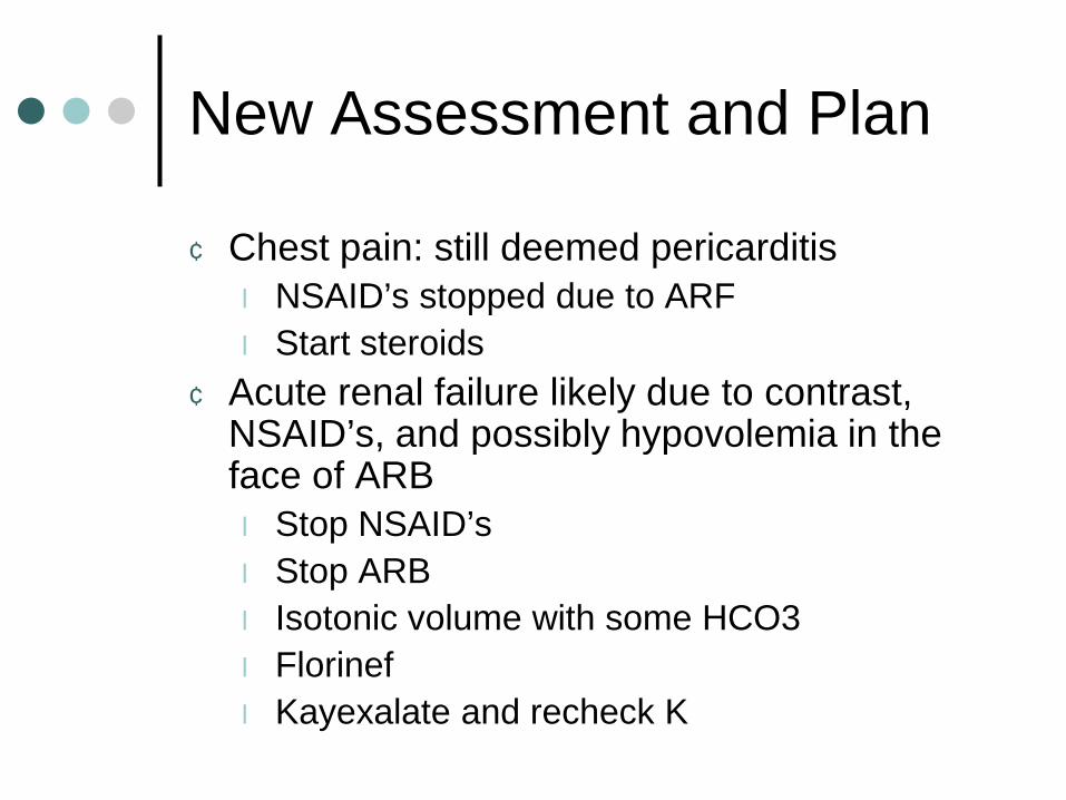
New Assessment and Plan
¢ Chest pain: still deemed pericarditisl NSAID’s stopped due to ARFl Start steroids
¢ Acute renal failure likely due to contrast, NSAID’s, and possibly hypovolemia in the face of ARBl Stop NSAID’sl Stop ARBl Isotonic volume with some HCO3l Florinefl Kayexalate and recheck K
Pericarditis course
¢ Steroids continued¢ Pain improves¢ EKG returns to baseline¢ Plan for steroid taper
Acute renal failure course
¢ Urine output increases with IVF¢ Creatinine peaks at 5.3 on 11/30 and
then improves to 1.1 on 12/4 ¢ ARB restarted and creatinine remains
stable as outpatient
Diabetes
¢ Insulin needed to be titrated up due to stopping metformin and steroids
¢ Discharged on insulin and glyburide with need for close follow up to wean insulin with steroid wean and consideration of restarting metformin
Pericarditis
Epidemiology
¢ Exact incidence and prevalence is not accurately known
¢ 0.1% of hospitalized patients¢ 5% of patients presenting to the
emergency department with non-ischemic chest pain
Etiology
¢ Idiopathic: presumed viral induced and/or autoimmune¢ Infectious: many possible infectious etiologies¢ Autoimmune: SLE, RA, etc¢ Drugs: procainamide, isoniazid, dantrolene, etc¢ Metabolic: hypothyroid, uremia, etc¢ Neoplastic: lung, breast, lymphoma, primary cardiac,
etc¢ Radiation¢ Myocardial infarction, myocarditis, or dissecting aortic
aneurysm¢ Trauma: including catheter complications
Etiology Determination
¢ Past studies have shown poor ability to come up with a diagnosis with one study reaching a firm diagnosis in 16% of the cases after an extensive workup
¢ More recent studies have shown more success with newer techniques
Diagnosis
¢ Requires 2 of the followingl Chest painl Pericardial friction rub: may be less
prevalent with an effusionl Biomarkers: troponin or CKMBl Signs of inflammation: elevated WBC,
ESR, and/or CRP
Workup
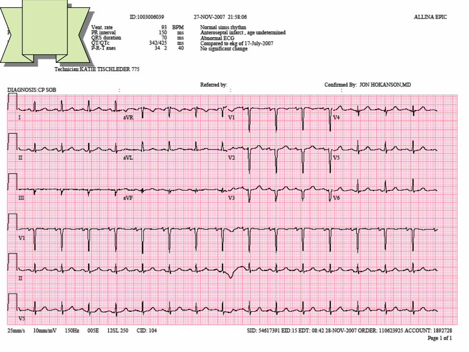
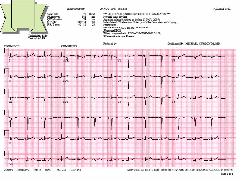
¢ EKGl Diffuse ST elevation and PR depressionl May get T wave inversion after ST and PR
segments normalize¢ Echocardiogram:l Often normall Will reveal the pericardial effusion if present
¢ CXR:l may show the pericardial effusion if presentl May be normal
Initial evaluation of percarditis¢ Acute pericarditis does not require an
extensive workup for the etiology unlessl There is a apparent associated medical or
surgical conditionl The patient is immunocompromised
¢ Additional tests that should be performedl Echocardiogram (looking for tamponade)l PPDl ANAl HIVl Blood cultures if fever present
Risk factors for severe disease¢ Subacute symptoms¢ High fever (>38.0) and leukocytosis¢ Evidence of tamponade¢ Large effusion (>20 mm)¢ Immunosuppressed state¢ On anticoagulation¢ Acute trauma¢ Failure to respond to initial therapy¢ Elevated troponin
Treatment
¢ Treat primary disease if present & known¢ Post-MI pericarditis treated with aspirin¢ NSAID’s as first line¢ Colchicinel May be added to NSAID’sl May decrease risk of recurrence
¢ Steroidsl For patients with CTD, immune-mediated, or
uremic causel If refractory to or contraindications to above
Prognosis
¢ Good long term prognosis¢ 5-28% have tamponade¢ 1% develop constrictive pericarditis¢ Recurrence ratel 15-30 % not given Colchicinel 10-15 % given Colchicinel Increased with refractory disease,
steroid therapy, inappropriate pericardiotomy