Embed Size (px)
Citation preview
news and views
1084 nature structural biology • volume 6 number 12 • december 1999
Since Anfinsen’s seminal studies, there hasbeen an enormous effort vested in under-standing how proteins are folded andunfolded in solution. In recent years, wehave also seen explosive progress in thestudy of protein folding in the cell. The con-nections between folding of proteins in vitroand in vivo, however, have been lacking. Onpage 1132 of this issue of Nature StructuralBiology, Huang et al.1 demonstrate such arare connection and in doing so providecritical insight into how proteins aretranslocated across the mitochondrialmembranes.
To elucidate the complete fold-ing pathways of a protein,researchers for many years havestudied the unfolding of proteinsafter the addition of denaturantsince unfolding is the reverse offolding. Protein unfolding is essen-tial to many cellular processes,including the degradation of pro-teins by ATP-dependent proteasesand the translocation of proteinsacross membranes. The vastmajority of proteins are synthe-sized by cytosolic ribosomes; there-fore the translocation of proteinsacross membranes is a process thatis critical to the operation of allorganelles. Mitochondria face themost daunting of translocationtasks — the post-translationalinsertion of, in some cases, afolded protein through a doubleset of membranes (for reviews seerefs 2, 3). For a folded protein to bethreaded through proteinaceouschannels that reside in mitochon-drial membranes, it first has to beunfolded on the cytosolic side ofthe membrane. Therefore, how aprotein is unfolded prior to itsimport into mitochondria is animportant biological question.
Matouschek and colleagueswere uniquely positioned toaddress this question, having pre-viously characterized in detail the
in vitro unfolding pathway of a prototypicprotein, barnase4. The unfolding pathwaywas mapped by analyzing how the addi-tion or removal of domain specific stabi-lizing factors affected the kinetics ofbarnase unfolding. By fusing barnasewith a mitochondrial presequence thattargets the protein to mitochondria, theywere able to dissect the unfolding pathwayduring mitochondrial import, utilizingmutations of barnase that affect itsunfolding, and crosslinking or ligandbinding that stabilizes specific folding
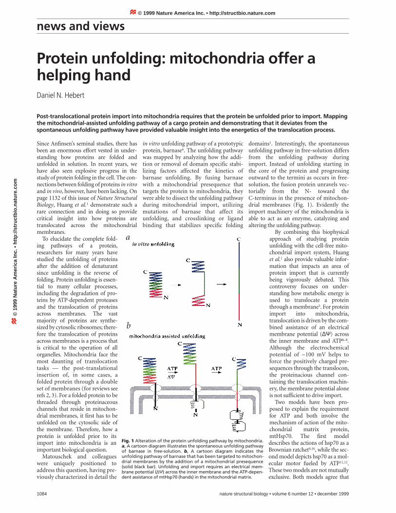
domains1. Interestingly, the spontaneousunfolding pathway in free-solution differsfrom the unfolding pathway duringimport. Instead of unfolding starting inthe core of the protein and progressingoutward to the termini as occurs in free-solution, the fusion protein unravels vec-torially from the N- toward theC-terminus in the presence of mitochon-drial membranes (Fig. 1). Evidently theimport machinery of the mitochondria isable to act as an enzyme, catalyzing andaltering the unfolding pathway.
By combining this biophysicalapproach of studying proteinunfolding with the cell-free mito-chondrial import system, Huanget al.1 also provide valuable infor-mation that impacts an area ofprotein import that is currentlybeing vigorously debated. Thiscontroversy focuses on under-standing how metabolic energy isused to translocate a proteinthrough a membrane5. For proteinimport into mitochondria,translocation is driven by the com-bined assistance of an electricalmembrane potential (∆Ψ) acrossthe inner membrane and ATP6–8.Although the electrochemicalpotential of ~100 mV helps toforce the positively charged pre-sequences through the translocon,the proteinacious channel con-taining the translocation machin-ery, the membrane potential aloneis not sufficient to drive import.
Two models have been pro-posed to explain the requirementfor ATP and both involve themechanism of action of the mito-chondrial matrix protein,mtHsp70. The first modeldescribes the actions of hsp70 as aBrownian ratchet9,10, while the sec-ond model depicts hsp70 as a mol-ecular motor fueled by ATP11,12.These two models are not mutuallyexclusive. Both models agree that
Protein unfolding: mitochondria offer ahelping handDaniel N. Hebert
Post-translocational protein import into mitochondria requires that the protein be unfolded prior to import. Mappingthe mitochondrial-assisted unfolding pathway of a cargo protein and demonstrating that it deviates from thespontaneous unfolding pathway have provided valuable insight into the energetics of the translocation process.
Fig. 1 Alteration of the protein unfolding pathway by mitochondria.a, A cartoon diagram illustrates the spontaneous unfolding pathwayof barnase in free-solution. b, A cartoon diagram indicates theunfolding pathway of barnase that has been targeted to mitochon-drial membranes by the addition of a mitochondrial presequence(solid black bar). Unfolding and import requires an electrical mem-brane potential (∆Ψ) across the inner membrane and the ATP-depen-dent assistance of mtHsp70 (hands) in the mitochondrial matrix.
a
b
© 1999 Nature America Inc. • http://structbio.nature.com©
199
9 N
atu
re A
mer
ica
Inc.
• h
ttp
://s
tru
ctb
io.n
atu
re.c
om

news and views
nature structural biology • volume 6 number 12 • december 1999 1085
mtHsp70 binds the presequence of thecargo protein and ATP regulates hsp70’sbinding cycle. The peptide binding pocketis open in the ATP-bound state and closedin the ADP-bound state. The replacementof ADP with ATP initiates the release of thebound peptide. Rebinding takes place uponrestoration of the ADP-bound state afterATP hydrolysis, which is stimulated by apartner J domain protein (for a reviewsee ref. 13). In mitochondria, the Jdomain protein (Tim44) resides in theinner membrane and positions mtHsp70at the entrance to the matrix.
The root of the discrepancy between thetwo models is in the magnitude of force thatis generated by hsp70 on the translocatingpolypeptide. The Brownian ratchet modelproposes that the binding of hsp70 to thematrix side of the membrane prevents thebackward sliding of the protein in thechannel9,10. Therefore passive diffusionwould only permit forward motion, bias-ing the movement of the protein into theorganelle. Translocation-competent sub-strates are generated by spontaneousunfolding or by cytosolic chaperones thatdelay folding. In contrast, the motor modelproposes that hsp70 provides a power-stroke that unfolds the polypeptide chainon the cis side of the outer membrane andis involved in pulling the protein across themembranes into the matrix11,12. Here, ATP-driven conformational changes in hsp70are capable of creating a force that activelypulls the protein inward.
Recent studies by Matlack et al.14 haveconvincingly demonstrated that a proteinratchet mechanism is sufficient for theimport of an unfolded protein through apurified endoplasmic reticulum (ER)translocon. As predicted by the model, thepolypeptide was able to slide backwards inthe channel in the absence of hsp70 bind-ing. However, transport also occurredwhen antibodies, which do not bind andhydrolyze ATP, instead of hsp70 were usedto bind to the translocating peptide on thetrans side of the membrane. They conclud-ed that trapping alone permitted theimport of a polypeptide chain and that aratchet mechanism was capable of translo-cating an unfolded protein across the ERtranslocon.
The post-translational import of mito-chondrial proteins, however, encounters ahigher degree of complexity since the pro-tein first has to be unfolded to an extended
structure on the cis face of the mitochon-drial membranes prior to being threadedthrough membrane pores with an internaldiameter of ~20 Å (refs 15, 16). The co-translational transport of proteins into theeukaryotic ER alleviates this requirementfor unfolding by placing the translatingribosome directly on the translocon. If themitochondrial import system relied onspontaneous unfolding to generate trans-port-competent substrates then the trans-port rate would be dependent upon thespontaneous unfolding rate, thereby pre-cluding the import of stably folded mole-cules such as barnase.
Several studies indicate that mitochon-dria play an active role in unfolding pro-teins prior to their import. Mutations inmtHsp70 inhibit protein unfolding, importand refolding in the matrix17–19. Recently,Voisine et al.20 provided additional supportfor the direct involvement of a pullingaction by mtHsp70 in mitochondrialimport. By using mutants of mtHsp70 thatpermitted the separation of the trappingand unfolding functions of mtHsp70, theydemonstrated that trapping or ratchetingalone did not permit the unfolding andsubsequent import of a folded preprotein;rather, an additional ATP-dependentpulling force was required to import a stablyfolded preprotein. The key to the powerstroke of mtHsp70 was found to depend onits interaction with Tim44 in the innermembrane to correctly position the hsp70so that the directionality of the force couldbe controlled and harnessed.
The efforts to understand the energeticsof protein import into the mitochondriahave focused on the mechanism of theimport machinery directly. Matouschekand coworkers address this concern from adifferent perspective1. While the ratchetmodel relies either on cytosolic factors tomaintain the preprotein in a loosely foldedstate or on the spontaneous unfolding ofthe polypeptide chain, the motor modelpostulates that a power-stroke can beactively involved in the unfolding of thepreprotein. Therefore monitoring theunfolding pathway of the preprotein duringimport could provide an approach to dis-criminate between the two current models.
The perturbation of the spontaneousunfolding pathway by the mitochondriaimport machinery conflicts with the cur-rent version of the ratchet model. Theseresults are, however, consistent with the
motor model with mtHsp70 providing aforce to unravel the preprotein on the cisside of the membrane. The results by Huanget al.1 clearly indicate that the importmachinery can influence the unfoldingpathway and are supportive of a motormodel. However, a caveat is that the unfold-ing pathway could also be affected simply bythe loss of freedom at the N-terminus whenit is tethered or trapped at the mitochondri-al surface. One key experiment to test thiswould be to determine whether the unfold-ing pathway is affected by immobilizationof its N-terminus.
The determination of how the mitochon-drial machinery unfolds proteins willimpact our understanding of how proteinsare unfolded by the ATPases of the degrada-tive pathway where vectorial unfolding isalso critical. Huang et al.1 have provided anew perspective to begin to address theseproblems. In the future, further biochemi-cal and biophysical experiments will have tobe performed to fully answer this importantcell biological question of how proteins areunfolded and translocated across mem-branes in the cell.
Daniel N. Hebert is in the Department ofBiochemistry and Molecular Biology, Universityof Massachusetts, Amherst, MA 01003, USA.email: [email protected]
1. Huang, S., Ratliff, K.S., Schwartz, M.P., Spenner, J.M.& Matouschek, A. Nature Struct. Biol. 6, 1132–1138(1999).
2. Schatz, G. & Dobberstein, B. Science 271, 1519–1526(1996).
3. Bauer, M.F., Hofmann, S., Neupert, W. & Brunner, W.Trends Cell Biol. in the press.
4. Matouschek, A., Kellis Jr., J.T., Serrano, L. & Fersht,A.R. Nature 342, 122–126 (1989).
5. Pilon, M. & Schekman, R. Cell 97, 679–682 (1999).6. Schleyer, M. & Neupert, W. Cell 43, 339–350 (1985).7. Hwang, S.T., Wachter, C. & Schatz, G. J. Biol. Chem.
266, 21083–21089 (1991).8. Ungermann, C., Guiard, B., Neupert, W. & Cyr, D.
EMBO J. 15, 734–744 (1996).9. Neupert, W., Hartl, F.-U., Craig, E.A. & Pfanner, N. Cell
63, 447–450 (1990).10. Simon, S.M., C.S. Peskin, & Oster, G.F. Proc. Natl.
Acad. Sci. USA 89, 3770–3774 (1992).11. Glick, B.S. Cell 80, 11–14 (1995).12. Pfanner, N. & Meijer, M. Curr. Biol. 5, 132–135 (1995).13. Bukau, B. & Horwich, A.L. Cell 92, 351–366 (1998).14. Matlack, K.E.S., Misselwitz, B., Plath, K. & Rapoport,
T.A. Cell 97, 553–564 (1999).15. Kunkele, K.P. et al. Cell 93, 1009–1019 (1998).16. Hill, K. et al. Nature 395, 516–521 (1998)17. Kang, P.J., Ostermann, J., Shilling, J., Neupert, W.,
Craig, E.A. & Pfanner, N. Nature 348, 137–143 (1990).18. Gambill, B.D. et al. J. Cell Biol. 123, 109–117 (1993).19. Voos, W., Gambill, B.D., Guiard, B., Pfanner, N. &
Craig, E.A. J. Cell Biol. 123, 119–126 (1993).20. Voisine, C., Craig, E.A., Zufall, N., von Ahsen, O.,
Pfanner, N. & Voos, W. Cell 97, 565–574 (1999).
© 1999 Nature America Inc. • http://structbio.nature.com©
199
9 N
atu
re A
mer
ica
Inc.
• h
ttp
://s
tru
ctb
io.n
atu
re.c
om


![Integrating the Healthcare Enterprise€¦ · Document Source Document ConsumerOn Entry [ITI Document Registry Document Repository Provide&Register Document Set – b [ITI-41] →](https://img.pdfslide.net/doc/110x75/5f08a1eb7e708231d422f7c5/integrating-the-healthcare-enterprise-document-source-document-consumeron-entry.jpg)


