Embed Size (px)
Citation preview
640 nature neuroscience • volume 3 no 7 • july 2000
news and views
How the cerebral cortex becomes parceledinto discrete functional subdivisions is oneof the oldest and most fundamental ques-tions in neuroscience. For some time, wehave known that the cortex is divided intodiscrete areas with distinct functions, suchas vision and motor control. Each areashows unique patterns of cellular anato-my and specific patterns of connectivitywith the thalamus: for example, the visu-al cortex is interconnected with the later-al geniculate nucleus of the thalamus,which in turn receives visual input direct-ly from the retina. How do these distinctcortical areas attain their identity duringdevelopment? Does the cortex representa conglomerate of discrete ‘cognitiveorgans’ that develop independently fromone another, arising from progenitor cellsthat ‘know’ to become visual cells ormotor cells, before the arrival of afferentinputs (the ‘protomap’ model)1? Or, is thecortex like a brand-new hard drive, capa-ble of performing a standard computa-tional analysis on whatever input itreceives, with the distinctive identities ofcortical regions being determined by theinputs (the ‘protocortex’ model)2? In sup-port of the latter model, altering thalam-ic input early in development can causedramatic changes in cortical organization.For example, when retinal input is re-routed into the auditory centers, the audi-tory cortex acquires features that normallycharacterize visual cortex2,3. However, agrowing body of evidence suggests thatthe incoming thalamic connections inter-act with a developing cortex that alreadycontains intrinsic positional information4-
6. For example, mice that lack thalamic
input altogether can still develop a sur-prisingly normal cortical arealization7,8.What provides the positional informationin the cortex, and how do factors intrin-sic to cortical progenitor cells interact withthe inputs that cortex receives?
When it comes to providing position-al information, developmental biologiststhink first about homeodomain tran-scription factors, because this large fami-ly of proteins provides positionalinformation in a host of developmentalsettings. To take one classic example, thefront end of the Drosophila embryo isspecified by a gradient of the home-odomain protein bicoid. Transcriptionfactors have the important feature thatthey activate other genes, and further-more, bicoid activates some genes at highconcentration and other genes at lowerconcentration. Therefore, a gradient ofbicoid protein can trigger a cascade ofdownstream genes (gap genes, segmenta-tion genes and others) that further subdi-vide the embryo into distinct structures.The remarkable work by Mallamaci et al.9
in this issue of Nature Neuroscience,together with a similar recentstudy by Bishop et al.10, sug-gests that cortical areas aresimilarly established in largepart by gradients of home-odomain proteins expressedin the cells that generate cere-bral cortex.
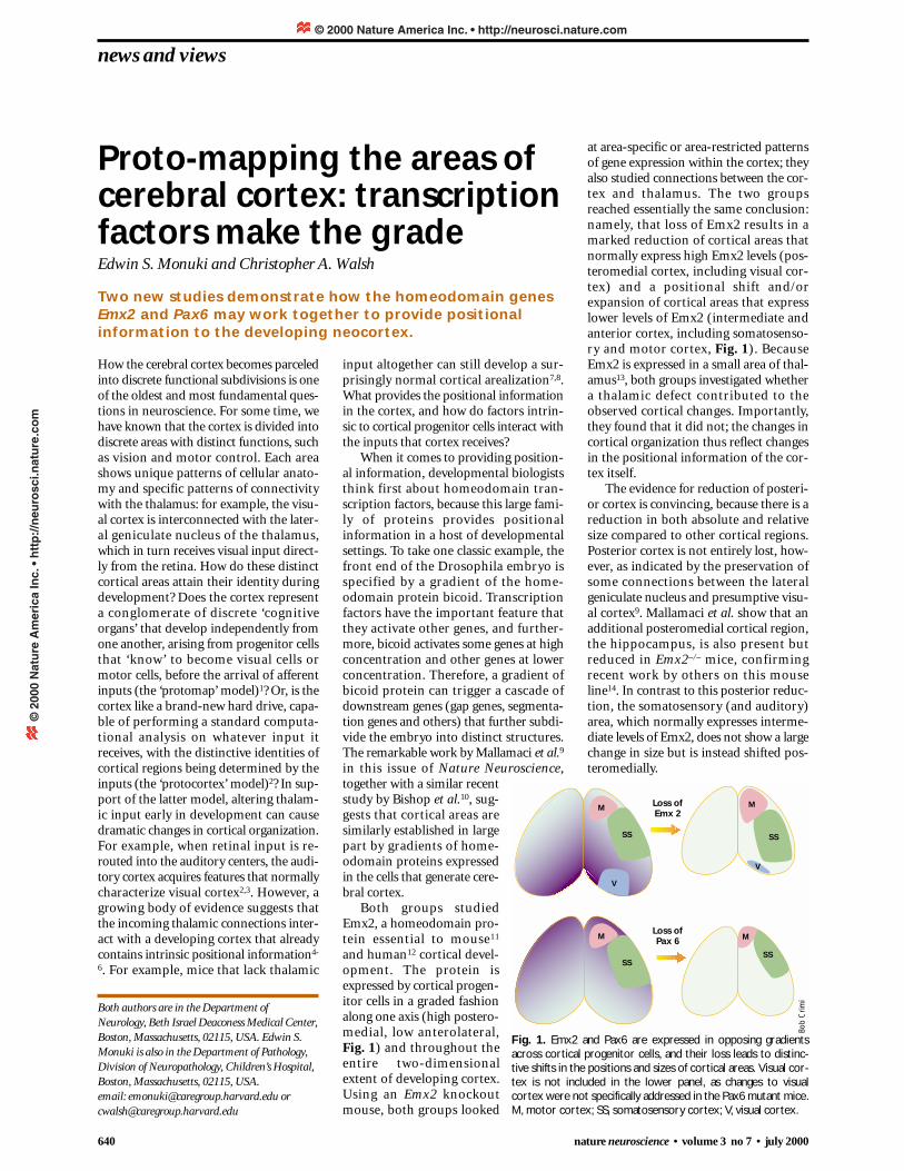
Both groups studiedEmx2, a homeodomain pro-tein essential to mouse11
and human12 cortical devel-opment. The protein isexpressed by cortical progen-itor cells in a graded fashionalong one axis (high postero-medial, low anterolateral, Fig. 1) and throughout theentire two-dimensionalextent of developing cortex.Using an Emx2 knockoutmouse, both groups looked
at area-specific or area-restricted patternsof gene expression within the cortex; theyalso studied connections between the cor-tex and thalamus. The two groupsreached essentially the same conclusion:namely, that loss of Emx2 results in amarked reduction of cortical areas thatnormally express high Emx2 levels (pos-teromedial cortex, including visual cor-tex) and a positional shift and/orexpansion of cortical areas that expresslower levels of Emx2 (intermediate andanterior cortex, including somatosenso-ry and motor cortex, Fig. 1). BecauseEmx2 is expressed in a small area of thal-amus13, both groups investigated whethera thalamic defect contributed to theobserved cortical changes. Importantly,they found that it did not; the changes incortical organization thus reflect changesin the positional information of the cor-tex itself.
The evidence for reduction of posteri-or cortex is convincing, because there is areduction in both absolute and relativesize compared to other cortical regions.Posterior cortex is not entirely lost, how-ever, as indicated by the preservation ofsome connections between the lateralgeniculate nucleus and presumptive visu-al cortex9. Mallamaci et al. show that anadditional posteromedial cortical region,the hippocampus, is also present butreduced in Emx2–/– mice, confirmingrecent work by others on this mouseline14. In contrast to this posterior reduc-tion, the somatosensory (and auditory)area, which normally expresses interme-diate levels of Emx2, does not show a largechange in size but is instead shifted pos-teromedially.
Loss ofEmx 2
Loss ofPax 6
M M
MM
SS SS
SSSS
V
V
Fig. 1. Emx2 and Pax6 are expressed in opposing gradientsacross cortical progenitor cells, and their loss leads to distinc-tive shifts in the positions and sizes of cortical areas. Visual cor-tex is not included in the lower panel, as changes to visualcortex were not specifically addressed in the Pax6 mutant mice.M, motor cortex; SS, somatosensory cortex; V, visual cortex.
Proto-mapping the areas ofcerebral cortex: transcriptionfactors make the gradeEdwin S. Monuki and Christopher A. Walsh
Two new studies demonstrate how the homeodomain genesEmx2 and Pax6 may work together to provide positionalinformation to the developing neocortex.
Both authors are in the Department ofNeurology, Beth Israel Deaconess Medical Center,Boston, Massachusetts, 02115, USA. Edwin S.Monuki is also in the Department of Pathology,Division of Neuropathology, Children’s Hospital,Boston, Massachusetts, 02115, USA.email: [email protected] [email protected]
Bob
Crim
i
© 2000 Nature America Inc. • http://neurosci.nature.com©
200
0 N
atu
re A
mer
ica
Inc.
• h
ttp
://n
euro
sci.n
atu
re.c
om
news and views
nature neuroscience • volume 3 no 7 • july 2000 641
Most intriguingly, both groups demon-strate a relative expansion in Emx2–/– miceof anterior cortical regions that normallyexpress the lowest concentration of Emx2;Bishop et al. also demonstrate an absolutetwofold expansion of anterior cortex(assessed using expression of an anteriorcortical marker, Cad8) despite an overallone-third reduction of cortical surface areain these mice. Thus, cortical areas are dif-ferentially affected in Emx2–/– mice, withposteromedial regions markedly reduced,intermediate cortex shifted, and anteriorcortex expanded. The loss of Emx2 doesnot result in complete loss of any particularcortical area, but instead results in a changeof scale of cortical areas relative to eachother. Most importantly, these changes cor-respond to the normal Emx2 expressiongradient (Fig. 1). Because changes in arealsize in the Emx2–/– mice correlate with arealposition along the Emx2 gradient, it is like-ly that the Emx2 gradient directly regulatesareal size. A further implication of thesefindings is that cortical areas are seeming-ly able to compete for space, because theareas that are least dependent on Emx2(anterior cortex) are able to expand at theexpense of those that are most dependent(posterior cortex).
This model, suggesting that parcella-tion of cortical areas is regulated by home-odomain protein gradients, is supportedby work on a second mouse mutant,which lacks the homeodomain proteinPax6. Cortical Pax6 expression is alsograded, but in a direction that opposesEmx2 (high anterolateral, low postero-medial, Fig. 1). In a fashion similar to theEmx2 mutants, markers of anterior andlateral cortical regions that normallyexpress high Pax6 levels (including motorand somatosensory cortex) are reducedbut not completely lost in Pax6-deficientmice9 (Fig. 1). Although study of the Pax6mutant line was limited (in part becausethalamic axons do not reach the cortex inthese mice), these findings support amodel in which the complementary Emx2and Pax6 gradients provide positionalinformation to the developing neocortexin much the same way that gradients ofother homeodomain proteins, such asbicoid, impart positional information inother developmental contexts.
The next set of questions involves howthese homeodomain proteins regulate theformation of specific cortical areas. Dothey influence proliferation or some otheraspect of area-specific programming?Mallamaci et al. have begun to addressthis issue by studying the tissue morphol-ogy and cell cycle kinetics of anterior,
the Emx2/Pax6 countergradient axis isclearly an important one, gradients alongother axes must also be acting to subdi-vide cortex, because functional areas arenot aligned solely along one axis. Theidentification of factors that act alongother axes, and their interactions with theEmx2/Pax6 countergradient, will also beof great future interest.
1. Rakic, P. Science 241, 170–176 (1988).
2. O’Leary, D. D. Trends Neurosci. 12, 400–406(1989).
3. Sharma, J., Angelucci, A. & Sur, M. Nature404, 841–847 (2000).
4. Barbe, M. F. & Levitt, P. J. Neurosci. 11,519–533 (1991).
5. Gitton, Y., Cohen-Tannoudji, M. & Wassef, M.J. Neurosci. 19, 4889–4898 (1999).
6. Dehay, C., Giroud, P., Berland, M., Smart, I. &Kennedy, H. Nature 366, 464–466 (1993).
7. Miyashita-Lin, E. M., Hevner, R., Wassarman,K. M., Martinez, S. & Rubenstein, J. L. Science285, 906–909 (1999).
8. Nakagawa, Y., Johnson, J. E. & O’Leary, D. D. J. Neurosci. 19, 10877–10885 (1999).
9. Mallamaci, A., Muzio, L., Chan, C.-H.,Parnvelas, J. & Boncinelli, E. Nat. Neurosci. 3,679–686 (2000).
10. Bishop, K. M., Goudreau, G. & O’Leary, D. D.Science 288, 344–349 (2000).
11. Pellegrini, M., Mansouri, A., Simeone, A.,Boncinelli, E. & Gruss, P. Development 122,3893–3898 (1996).
12. Brunelli, S. et al. Nat. Genet. 12, 94–96 (1996).
13. Simeone, A., Acampora, D., Gulisano, M.,Stornaiuolo, A. & Boncinelli, E. Nature 358,687–690 (1992).
14. Tole, S., Goudreau, G., Assimacopoulos, S. &Grove, E. A. J. Neurosci. 20, 2618–2625 (2000).
15. Vanderhaeghen, P. et al. Nat. Neurosci. 3,358–365 (2000).
intermediate and posterior cortical prog-enitors in Emx2 knockout mice. Interest-ingly, they find that the proliferative layersare significantly thicker in these mice, butthat the cell cycling times are indistin-guishable from wild-type mice. This sug-gests that Emx2 does not regulatearealization by altering proliferation in asimple way, but may instead modulateother area-specific programs; additionalexperimental work will be needed toanswer this question.
Several other outstanding questionsremain unanswered. For instance, doEmx2 and Pax6 act independently or in acombinatorial manner to impart posi-tional information? An Emx2/Pax6 dou-ble knockout might be useful to answerthis question. Also, how is the positionalinformation encoded by these home-odomain protein gradients converted intothe sharp areal boundaries of mature cor-tex? Mechanisms used to translate otherhomeodomain gradients (such as thebicoid gradient) into discrete subdivisionsmay provide insights into this problem.Another question concerns the upstreamfactors that establish the Emx2 and Pax6gradients. Secreted molecules such as theBmps, Wnts and Fgfs have been shown toact in gradients to initiate the productionof transcription factors, and they maytherefore be involved. Although thedownstream transcriptional targets ofEmx2 and Pax6 remain unknown, onepotential target may be the ephrins, whichshow graded expression withinsomatosensory cortex and affect its topo-graphic organization15. Finally, although
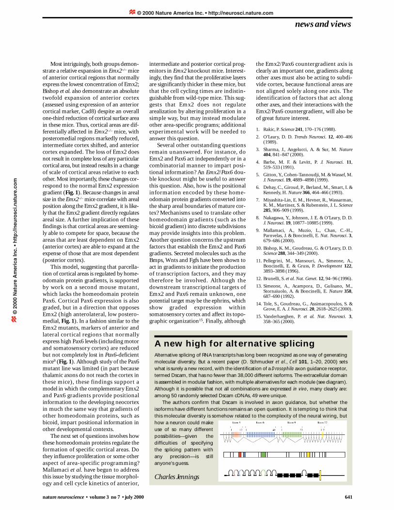
A new high for alternative splicingAlternative splicing of RNA transcripts has long been recognized as one way of generatingmolecular diversity. But a recent paper (D. Schmucker et al., Cell 101, 1–20, 2000) setswhat is surely a new record, with the identification of a Drosophila axon guidance receptor,termed Dscam, that has no fewer than 38,000 different isoforms. The extracellular domainis assembled in modular fashion, with multiple alternatives for each module (see diagram).Although it is possible that not all combinations are expressed in vivo, many clearly are:among 50 randomly selected Dscam cDNAs, 49 were unique.
The authors confirm that Dscam is involved in axon guidance, but whether theisoforms have different functions remains an open question. It is tempting to think thatthis molecular diversity is somehow related to the complexity of the neural wiring, buthow a neuron could makeuse of so many differentpossibilities—given thedifficulties of specifyingthe splicing pattern withany precision—is stillanyone’s guess.
Charles Jennings
© 2000 Nature America Inc. • http://neurosci.nature.com©
200
0 N
atu
re A
mer
ica
Inc.
• h
ttp
://n
euro
sci.n
atu
re.c
om


![Integrating the Healthcare Enterprise€¦ · Document Source Document ConsumerOn Entry [ITI Document Registry Document Repository Provide&Register Document Set – b [ITI-41] →](https://img.pdfslide.net/doc/110x75/5f08a1eb7e708231d422f7c5/integrating-the-healthcare-enterprise-document-source-document-consumeron-entry.jpg)


