Embed Size (px)
Citation preview
Mo1900
Global Profiling Study Reveals MicroRNA Dysregulation in Human GastricMetaplastic LineagesJosane F. Sousa, Ki Taek Nam, Hyuk-Joon Lee, Han-Kwang Yang, Woo Ho Kim, James R.Goldenring
Gastric cancer is one of the most common neoplasms, ranking as the second leading causeof cancer-related deaths worldwide. The predominant factor associated to gastric cancer isHelicobacter pylori infection, which leads to a chronic inflammatory response and subsequentoxyntic atrophy. Parietal cell loss result in two types of metaplasia, the intestinal metaplasia(IM) characterized by the presence of cells with goblet cell morphology and spasmolyticpolypeptide-expressing metaplasia (SPEM) that shows morphological characteristics of thedeep antral glands and express trefoil factor 2 (spasmolytic polypeptide). Although meta-plastic lesions are considered neoplastic precursors, their direct association to cancer is stillin debate. Similar to other cancers, gastric cancers are marked by global gene expressionalterations. MicroRNAs (miRNAs) are small noncoding RNAs involved in the post-transcrip-tional regulation of gene expression and an increasing number of studies has been showingtheir aberrant expression in cancer. In order to identify miRNAs involved in the early stagesof gastric cancer we performed a miRNA profiling on laser capture micro-dissected IM andSPEM cells from patient lesions. Using a qRT-PCR approach for quantitation of 754 humanmiRNAs, we identified 77 miRNAs differentially expressed in the metaplastic samples (greaterthan two-fold) in comparison to normal chief cells. The highest number of dysregulatedmiRNAs was observed in IM,which showed 45 up-regulated and 25 down-regulatedmiRNAs.In SPEM, 28 miRNAs were found up-regulated (21 of them also up-regulated in IM), whereasno down-regulation was detected. Some of the up-regulated miRNAs have already beenassociated to cancer in general (miR-26-a, miR-191, miR-155), as well as specifically togastric cancer (miR-18b, miR-196b and miR-106a). However, the regulation of some of thetop up-regulated miRNAs revealed here including miR-802, miR-922 and miR-622 are stillpoorly characterized in cancer. In a comparison with a previous mRNA profiling studyperformed on a similar group of samples, we observed that 108 out of the 568 mRNAsfound up-regulated in IM are predicted targets of the 26 microRNAs down-regulated in IM,revealing a potential mechanism for the regulation of those genes in gastric metaplasia.Particularly interesting are 4 members of the miR-30 family, with 31 predicted mRNA targetsamongst those up-regulated in IM. Although miR-30 family members have been describedas down-regulated in gastric cancer, no data on their targets in this neoplasia is currentlyavailable. An extended characterization of the microRNAs identified here will provide abetter understanding of their function in gastric metaplasia and progression to neoplasia,as well as might reveal useful early stage biomarkers or therapeutic targets.
Mo1901
Classical and Alternative Pathway Nuclear Factor-κB Signalling DifferentiallyRegulate Gastric Epithelial Responses to Helicobacter felis InfectionMichael D. Burkitt, Andrea Varro, Jorge Caamano, David M. Pritchard
INTRODUCTION: Classical pathway NF-κB signalling is implicated in the pathogenesisof several inflammation associated cancers, including colitis associated colon cancer andHelicobacter associated gastric cancer. However the role of individual NF-κB family membersand the function of alternative pathway NF-κB signalling have not previously been assessedin this context. We have therefore investigated whether abrogation of classical and alternativepathwayNF-κB signalling alteredmurine responses toHelicobacter felis infection.METHODS:6 week old female NF-κB1 null (p50-/-), NF-κB2 null (p52-/-), c-Rel null and C57BL/6 micewere infected with H. felis by oral gavage and culled 6 weeks later. Tissues were processedfor histological analysis and immunohistochemistry. RESULTS: H. felis infection of wild-type mice resulted in gastric atrophy (29% fewer parietal cells were observed in infectedthan in control mice (p<0.05, 1-way ANOVA and Holm Sidak post hoc test)), a 1.5 foldincrease in the number of Ki67 positive proliferating cells and no significant change in thenumber of active caspase 3 positive apoptotic cells. Animals with abrogated classical pathwayNF-κB signalling also developed gastric atrophy after H. felis infection. However whereasinfected c-Rel null animals showed similar parietal cell, proliferation and apoptotic indicesto infected wild-type mice, p50-/- animals developed more severe pathology with significantlyincreased inflammation scores (p<0.05, Mann-Whitney U) and amoremarked 62% reductionin parietal cell number (p<0.05, 1-way ANOVA). This was associated with significant 2.1fold and 7.6 fold increases in the number of proliferating and apoptotic cells respectively(p<0.05, 2-way ANOVA). By contrast, infected p52-/- mice showed much lower inflammationscores than wild-type mice (p<0.05, MWU) following H.felis infection and did not developgastric atrophy, with only 3% parietal cell loss (p<0.05, 1-way ANOVA). In addition, thesemice showed no significant changes in proliferation or apoptosis following infection withH. felis, and demonstrated similar proliferation and apoptotic indices to untreated wild-type mice. CONCLUSION: NF-κB1 mediated signalling protects the gastric mucosa fromHelicobacter induced atrophy, whereas alternative pathway NF-κB signalling is required forthe development of both inflammation and atrophy following infection with this organism.This supports the hypothesis that classical and alternative pathway signalling differentiallyaffect long term outcomes, including carcinogenesis, following Helicobacter felis infection inC57BL/6 mice.
Mo1902
Gastric Cancer: Increased Numbers of Myeloid-Derived Suppressor Cells AreFound in Gastric Cancer PatientsEsther W. Chang, Linda Wang, Gek Keow Lim, Belicia J. Lim, Khoon-Lin Ling
Introduction: Chronic inflammation with Helicobacter pylori infection is a major contributorto gastric carcinogenesis, with H. pylori infected individuals having a 2-3% lifetime risk ofdeveloping gastric cancer. As such, an understanding of host-pathogen factors that mitigateH. pylori induced gastric carcinogenesis is necessary. A group of newly characterized sup-pressor cells, myeloid-derived suppressor cells (MDSCs) have been found in cancer and
S-673 AGA Abstracts
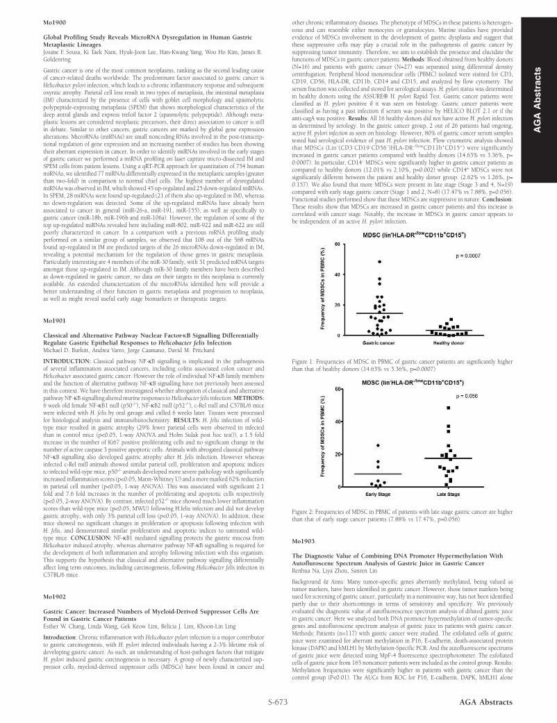
other chronic inflammatory diseases. The phenotype of MDSCs in these patients is heterogen-eous and can resemble either monocytes or granulocytes. Murine studies have providedevidence of MDSCs involvement in the development of gastric dysplasia and suggest thatthese suppressive cells may play a crucial role in the pathogenesis of gastric cancer bysuppressing tumor immunity. Therefore, we aim to establish the presence and elucidate thefunctions of MDSCs in gastric cancer patients.Methods: Blood obtained from healthy donors(N=16) and patients with gastric cancer (N=27) was separated using differential densitycentrifugation. Peripheral blood mononuclear cells (PBMC) isolated were stained for CD3,CD19, CD56, HLA-DR, CD11b, CD14 and CD15, and analyzed by flow cytometry. Theserum fraction was collected and stored for serological assays. H. pylori status was determinedin healthy donors using the ASSURE® H. pylori Rapid Test. Gastric cancer patients wereclassified as H. pylori positive if it was seen on histology. Gastric cancer patients wereclassified as having a past infection if serum was positive by HELICO BLOT 2.1 or if theanti-cagA was positive. Results: All 16 healthy donors did not have active H. pylori infectionas determined by serology. In the gastric cancer group, 2 out of 26 patients had ongoing,active H. pylori infection as seen on histology. However, 80% of gastric cancer serum samplestested had serological evidence of past H. pylori infection. Flow cytometric analysis showedthat MDSCs (Lin-(CD3-CD19-CD56-)HLA-DR-/lowCD11b+CD15+) were significantlyincreased in gastric cancer patients compared with healthy donors (14.63% vs 3.36%, p=0.0007). In particular, CD14- MDSCs were significantly higher in gastric cancer patients ascompared to healthy donors (12.01% vs 2.10%, p=0.002) while CD14+ MDSCs were notsignificantly different between the patient and healthy donor group. (2.62% vs 1.26%, p=0.157). We also found that more MDSCs were present in late stage (Stage 3 and 4, N=19)compared with early stage gastric cancer (Stage 1 and 2, N=8) (17.47% vs 7.88%, p=0.056).Functional studies performed show that these MDSCs are suppressive in nature. Conclusion:These results show that MDSCs are increased in gastric cancer patients and this increase iscorrelated with cancer stage. Notably, the increase in MDSCs in gastric cancer appears tobe independent of an active H. pylori infection.
Figure 1: Frequencies of MDSC in PBMC of gastric cancer patients are significantly higherthan that of healthy donors (14.63% vs 3.36%, p=0.0007)
Figure 2: Frequencies of MDSC in PBMC of patients with late stage gastric cancer are higherthan that of early stage cancer patients (7.88% vs 17.47%, p=0.056)
Mo1903
The Diagnostic Value of Combining DNA Promoter Hypermethylation WithAutofluroscene Spectrum Analysis of Gastric Juice in Gastric CancerRenhua Na, Liya Zhou, Sanren Lin
Background & Aims: Many tumor-specific genes aberrantly methylated, being valued astumor markers, have been identified in gastric cancer. However, those tumor markers beingused for screening of gastric cancer, particularly in a noninvasive way, has not been identifiedpartly due to their shortcomings in terms of sensitivity and specificity. We previouslyevaluated the diagnostic value of autofluorescence spectrum analysis of diluted gastric juicein gastric cancer. Here we analyzed both DNA promoter hypermethylation of tumor-specificgenes and autofluroscene spectrum analysis of gastric juice in patients with gastric cancer.Methods: Patients (n=117) with gastric cancer were studied. The exfoliated cells of gastricjuice were examined for aberrant methylation in P16, E-cadherin, death-associated proteinkinase (DAPK) and hMLH1 by Methylation-Specific PCR. And the autofluorescene spectrumsof gastric juice were detected using MpF-4 fluorescence spectrophotometer. The exfoliatedcells of gastric juice from 165 noncancer patients were included as the control group. Results:Methylation frequencies were significantly higher in patients with gastric cancer than thecontrol group (P<0.01). The AUCs from ROC for P16, E-cadherin, DAPK, hMLH1 alone
AG
AA
bst
ract
s