Embed Size (px)
Citation preview
Vol:1(2) January 2013
14 www.ijopils.com
International Journal of Pharmacy and Integrated Life Sciences “Where improvisation meets innovation”
www.ijopils.com
REVIEW ARTICLE ISSN : 2320 - 0782 V1-(I2) PG(14-29)
Transferosome: an enhancement approach for transdermal drug
delivery system
NATHJI DHAVAL*1
, PATNI CHANDRA1, SHAH HIRAL
1,
DR. CHAUDHARY SUNITA1, SANGHAVI KINJAL
1,
DR. PATEL UPENDRA1
1 Arihant School of Pharmacy & BRI, Adalaj, Gandhinagar, Gujarat, India.
ABSTRACT
A novel vesicular drug carrier system called transfersomes, which is composed of
phospholipid, surfactant, and water for enhanced transdermal delivery. Transferosome is an
ultradeformable vesicle, elastic in nature which can squeeze itself through a pore which is
many times smaller than its size owing to its elasticity. Recently, various strategies have been
used to augment the transdermal delivery of bioactives. Mainly, they include iontophoresis,
electrophoresis, sonophoresis, chemical permeation enhancers, microneedles, and vesicular
system (liposomes, niosomes, elastic liposomes such as ethosomes and transfersomes).
Among these strategies transferosomes appear promising. Transfersomes possess an
infrastructure consisting of hydrophobic and hydrophilic moieties together and as a result can
accommodate drug molecules with wide range of solubility. The transfersomal system was
much more efficient at delivering a low and high molecular weight drug to the skin in terms
of quantity and depth. Transfersomes can deform and pass through narrow constriction (from
5 to 10 times less than their own diameter) without measurable loss. The uniqueness of this
type of drug carrier system lies in the fact that it can accommodate hydrophilic, lipophilic as
well as amphiphilic drugs. Controlled release formulations can also be prepared with the help
of transferosomes. This review discusses the salient features, composition and mechanism of
action, mechanism of penetration of transfersomes, materials and method used for
preparation of transferosomes, application, scope and future of transferosomes.
KEYWORDS: Transfersomes, Transdermal, Vesicle, Ultradeformable, Osmotic gradient
Article received on : 16/12/2012 Article accepted on : 23/12/2012
Corresponding Author : Nathji Dhaval Chhotunath
Address : Arihant School of Pharmacy and Bio-Research Institute, Adalaj,
Gandhinagar, Gujarat, India.
Email ID : [email protected]
Vol:1(2) January 2013
15 www.ijopils.com
INTRODUCTION
Delivery via the transdermal route is an
interesting option in this respect because a
transdermal route is convenient and safe.
This offers several potential advantages
over conventional routes[1]
like avoidance
of first pass metabolism, predictable and
extended duration of activity, minimizing
undesirable side effects, utility of short
half-life drugs, improving physiological
and pharmacological response, avoiding
the fluctuation in drug levels, inter-and
intra-patient variations, and most
importantly, it provides patients
convenience. To date many chemical and
physical approaches have been applied to
increase the efficacy of the material
transfer across the intact skin, by use of the
penetration enhancers, enhancers,
iontophoresis, sonophoresis and the use of
colloidal carriers such as lipid vesicles
(liposomes and proliposomes) and
nonionic surfactant vesicles (niosomes and
proniosomes).
Transfersomes were developed in
order to take the advantage of
phospholipids vesicles as transdermal drug
carrier. These self-optimized aggregates,
with the ultra flexible membrane are able
to deliver the drug reproducibly either into
or through the skin, depending on the
choice of administration or application,
with high efficiency. These vesicular
transfersomes are several orders of
magnitudes more elastic than the standard
liposomes and thus well suited for the skin
penetration. Transfersomes overcome the
skin penetration difficulty by squeezing
themselves along the intracellular sealing
lipid of the stratum corneum. There is
provision or this, because of the high
vesicle deformability, which permits the
entry due to the mechanical stress of
surrounding, in a self-adapting manner.
Flexibility of transfersomes membrane is
achieved by mixing suitable surface-active
components in the proper ratios[2]
.The
resulting flexibility of transfersome
membrane minimizes the risk of complete
vesicle rupture in the skin and allows
transfersomes to follow the natural water
gradient across the epidermis, when
applied under non occlusive condition.
Transfersomes can penetrate the intact
stratum corneum spontaneously along two
routes in the intracellular lipid that differ
in their bait layers properties[3]
. The
following figure shows possible micro
routes for drug penetration across human
skin intracellular and transcellular[4]
.The
high and self-optimizing deformability of
typical composite transfersomes
membrane, which are adaptable to ambient
tress allow the ultra deformable
transfersomes to change its membrane
composition locally and reversibly, when
Dhaval et. Al. , Volume 1 – Issue 2
Vol: 1(2) January 2013 16
www.ijopils.com
it is pressed against or attracted into
narrow pore. The transfersomes
components that sustain strong membrane
deformation preferentially accumulate,
while the less adaptable molecules are
diluted at sites of great stress. This
dramatically lowers the energetic cost of
membrane deformation and permits the
resulting, highly flexible particles, first to
enter and then to pass through the pores
rapidly and efficiently. This behavior is
not limited to one type of pore and has
been observed in natural barriers such as in
intact skin[5, 6]
.
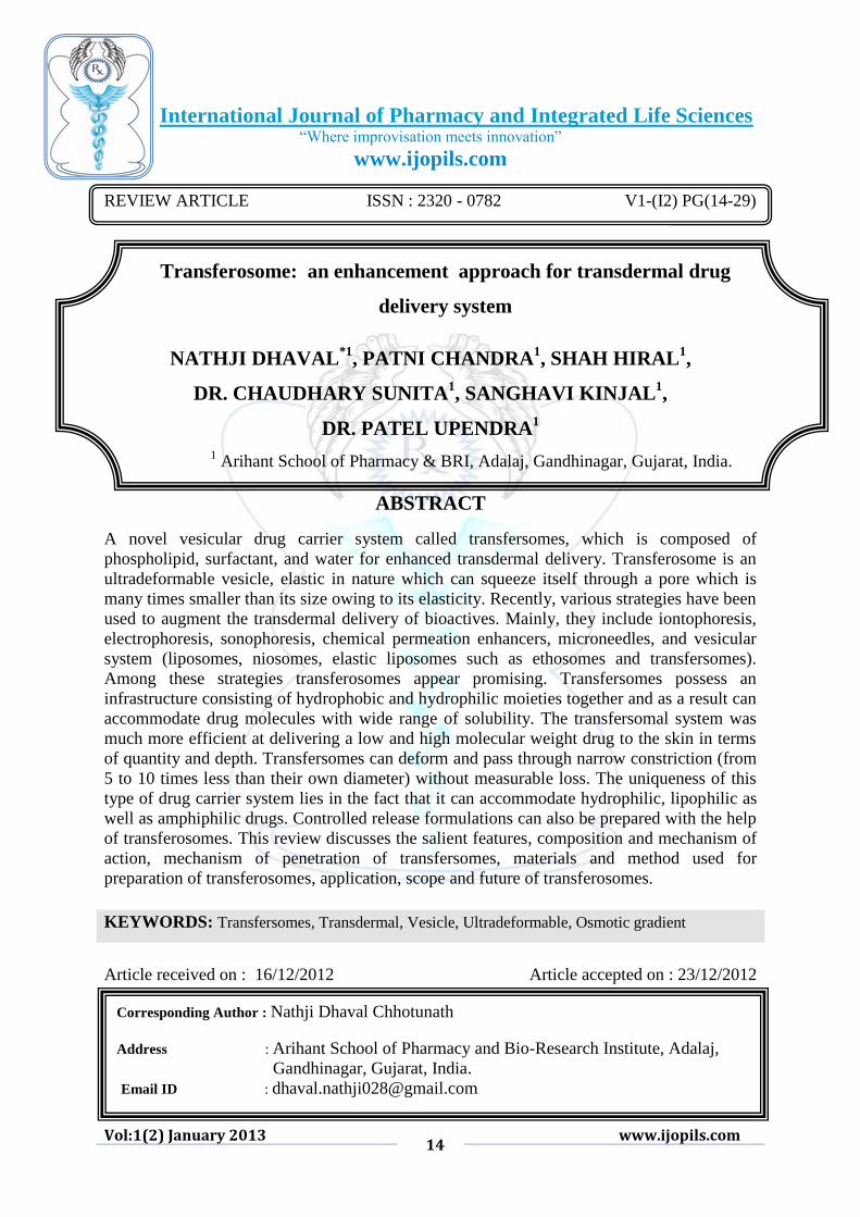
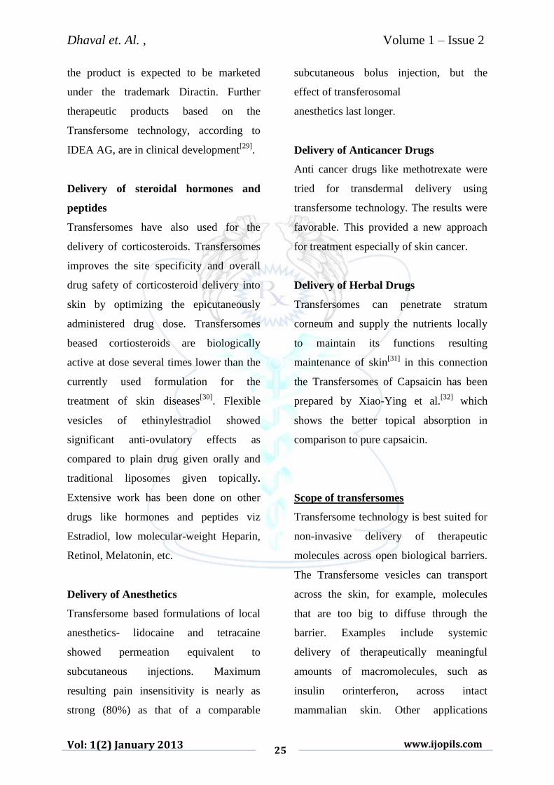
Fig 1: Undeformable Vesicle (Transferosome)
Salient features of transferosomes[7,8]
Transfersomes possess an
infrastructure consisting of
hydrophobic and hydrophilic
moieties together and as a result
can accommodate drug molecules
with a wide range of solubility as
shown in fig 1.
Transfersomes can deform and
pass through narrow constriction
(from 5 to 10 times less than their
own diameter) without measurable
loss. This high deformability gives
better penetration of intact vesicles.
They can act as a carrier for low as
well as high molecular weight
drugs e.g. analgesic, anesthetic,
corticosteroids, sex hormone,
anticancer, insulin, gap junction
protein, and albumin.
They are biocompatible and
biodegradable as they are made
from natural phospholipids similar
to liposomes.

Dhaval et. Al. , Volume 1 – Issue 2
Vol: 1(2) January 2013 17
www.ijopils.com
They have high entrapment
efficiency, in case of lipophilic
drug near to 90%
They protect the encapsulated drug
from metabolic degradation.
They act as depot, releasing their
contents slowly and gradually.
They can be used for both systemic
as well as topical delivery of drug.
Easy to scale up, as procedure is
simple, do not involve lengthy
procedure and unnecessary use of
pharmaceutically unacceptable
additives.
Limitations of transfersomes[9]
They are chemically unstable due
to their predisposition to oxidative
degradation.
Purity of natural phospholipids is
difficult to achieve so, world is
against adoption of transfersomes
as drug delivery vehicles.
These formulations are expensive.
Composition and mechanism of action
The carrier aggregate is composed of at
least one amphiphatic (such as
phosphatidylcholine), which inaqueous
solvents self-assembles into lipid bilayer
that closes into a simple lipid vesicle. By
addition of atleast one bilayer softening
component (such as a biocompatible
surfactant or an amphiphile drug) lipid
bilayer flexibility and permeability are
greatly increased. The resulting, flexibility
and permeability optimized, Transfer some
vesicle can therefore adapt its shape to
ambient easily and rapidly, by adjust in
glocal concentration of each bilayer
component to the local stress experienced
by the bilayer as shown in fig 2. In its
basic organization broadly similar to a
liposome), the Transfersome thus differs
from such more conventional vesicle
primarily by its "softer", more deformable,
and better adjustable artificial membrane.
Another beneficial consequence of strong
bilayer deformability is the increased
Transfersome affinity to bind and retain
water. An ultradeformable and highly
hydrophilic vesicle always seeks to avoid
dehydration; this may involve a transport
process related to but not identical with
forward osmosis. For example, a
Transfersome vesicle applied on an open
biological surface, such as non-occluded
skin, tends to penetrate its barrier and
migrate into the water-rich deeper strata to
secure its adequate hydration. Barrier
penetration involves reversible bilayer
deformation, but must not compromise
unacceptably either the vesicle integrity or
the barrier properties for the underlying
hydration affinity and gradient to remain
in place.

Dhaval et. Al. , Volume 1 – Issue 2
Vol: 1(2) January 2013 18
www.ijopils.com
Since it is too large to diffuse
through the skin, the Transfersome needs
to find and enforce its own route through
the organ. The Transfersome vesicles
usage in drug delivery consequently relies
on the carrier’stability to widen and
overcome the hydrophilic pores in the skin
or some other (e.g. plant cuticle) barrier.
The subsequent, gradual agent release
from the drug carrier allows the drug
molecules to diffuse and finally bind to
their target. Drug transport to an intra-
cellular action site may also involve the
carrier’s lipid bilayer fusion with the cell
membrane, unless the vesicle is taken-up
actively by the cell in the process called
endocytosis[10]
.
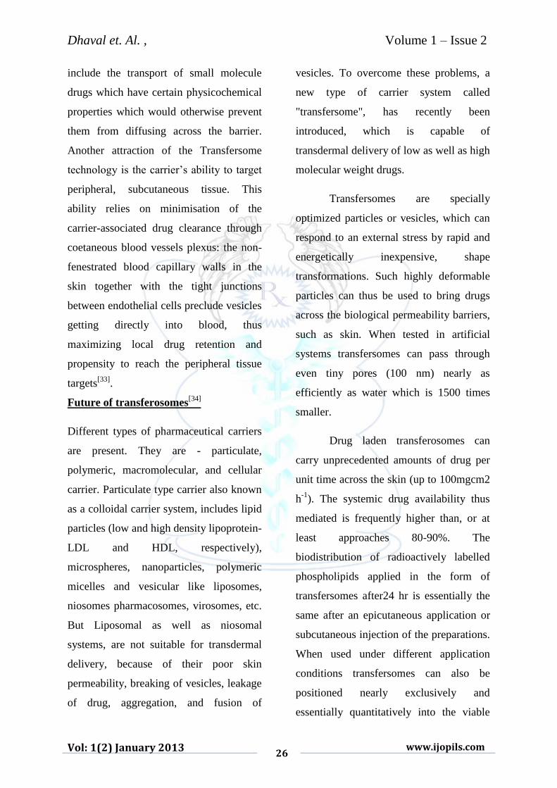
Fig 2: Diagrammatic Representation of the Stratum Corneum and the Intercellular and
Transcellular Routes of Penetration[11]
Mechanism of penetration of
transfersomes
The mechanism for penetration is the
generation of “osmotic gradient” due to
evaporation of water while applying the
lipid suspension (Transfersomes) on the
skin surface. The transport of these elastic
vesicles is thus independent of
concentration. The trans-epidermal
hydration provides the driving force for
the transport of the vesicles[12]
. As the
vesicles are elastic, they can squeeze
Dhaval et. Al. , Volume 1 – Issue 2
Vol: 1(2) January 2013 19
www.ijopils.com
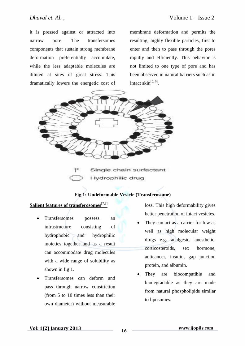
through the pores in stratum corneum
(though these pores are less than one-tenth
of the diameter of vesicles)[13]
as shown in
Fig.3.
Fig 3: Illustration of pore penetration at molecular level
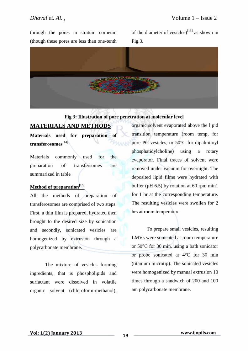
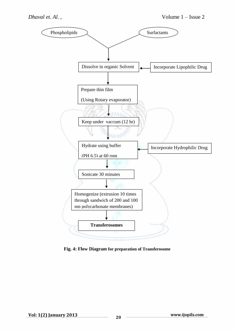
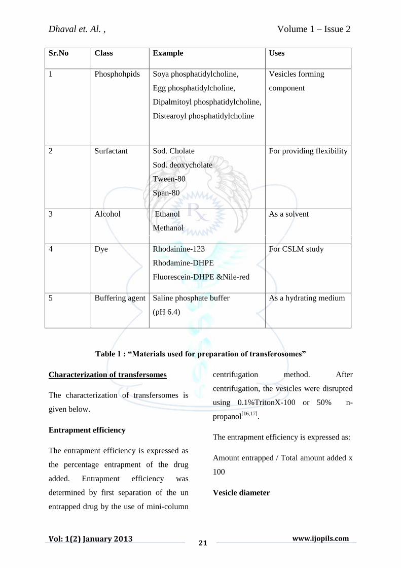
MATERIALS AND METHODS
Materials used for preparation of
transferosomes[14]
Materials commonly used for the
preparation of transfersomes are
summarized in table
Method of preparation[15]
All the methods of preparation of
transferosomes are comprised of two steps.
First, a thin film is prepared, hydrated then
brought to the desired size by sonication
and secondly, sonicated vesicles are
homogenized by extrusion through a
polycarbonate membrane.
The mixture of vesicles forming
ingredients, that is phospholipids and
surfactant were dissolved in volatile
organic solvent (chloroform-methanol),
organic solvent evaporated above the lipid
transition temperature (room temp, for
pure PC vesicles, or 50°C for dipalmitoyl
phosphatidylcholine) using a rotary
evaporator. Final traces of solvent were
removed under vacuum for overnight. The
deposited lipid films were hydrated with
buffer (pH 6.5) by rotation at 60 rpm min1
for 1 hr at the corresponding temperature.
The resulting vesicles were swollen for 2
hrs at room temperature.
To prepare small vesicles, resulting
LMVs were sonicated at room temperature
or 50°C for 30 min. using a bath sonicator
or probe sonicated at 4°C for 30 min
(titanium microtip). The sonicated vesicles
were homogenized by manual extrusion 10
times through a sandwich of 200 and 100
am polycarbonate membrane.
Dhaval et. Al. , Volume 1 – Issue 2
Vol: 1(2) January 2013 20
www.ijopils.com
Fig. 4: Flow Diagram for preparation of Transferosome
Phospholipids Surfactants
Incorporate Lipophilic Drug Dissolve in organic Solvent
Prepare thin film
(Using Rotary evaporator)
Keep under vaccum (12 hr)
Hydrate using buffer
(PH 6.5) at 60 rpm
Incorporate Hydrophilic Drug
Sonicate 30 minutes
Homogenize (extrusion 10 times
through sandwich of 200 and 100
nm polycarbonate membranes)
Transferosomes
Dhaval et. Al. , Volume 1 – Issue 2
Vol: 1(2) January 2013 21
www.ijopils.com
Sr.No Class Example Uses
1 Phosphohpids Soya phosphatidylcholine,
Egg phosphatidylcholine,
Dipalmitoyl phosphatidylcholine,
Distearoyl phosphatidylcholine
Vesicles forming
component
2 Surfactant Sod. Cholate
Sod. deoxycholate
Tween-80
Span-80
For providing flexibility
3 Alcohol
Ethanol
Methanol
As a solvent
4 Dye Rhodainine-123
Rhodamine-DHPE
Fluorescein-DHPE &Nile-red
For CSLM study
5 Buffering agent Saline phosphate buffer
(pH 6.4)
As a hydrating medium
Table 1 : “Materials used for preparation of transferosomes”
Characterization of transfersomes
The characterization of transfersomes is
given below.
Entrapment efficiency
The entrapment efficiency is expressed as
the percentage entrapment of the drug
added. Entrapment efficiency was
determined by first separation of the un
entrapped drug by the use of mini-column
centrifugation method. After
centrifugation, the vesicles were disrupted
using 0.1%TritonX-100 or 50% n-
propanol[16,17]
.
The entrapment efficiency is expressed as:
Amount entrapped / Total amount added x
100
Vesicle diameter
Dhaval et. Al. , Volume 1 – Issue 2
Vol: 1(2) January 2013 22
www.ijopils.com
Vesicle diameter can be determined using
photon correlation spectroscopy or
dynamic light scattering (DLS) method.
Samples were prepared in distilled water,
filtered through a 0.2 mm membrane filter
and diluted with filtered saline and than
size measurement done by using photon
correlation spectroscopy or dynamic light
scattering (DLS) measurements[18]
.
Confocal scanning laser microscopy
(CSLM) study[19]
Conventional light microscopy and
electron microscopy both face problem of
fixation, sectioning and staining of the skin
samples. Often the structures to be
examined are actually incompatible with
the corresponding processing techniques,
these give rise to misinterpretation, but can
be minimized by Confocal Scanning Laser
Microscopy (CSLM). In this technique
lipophilic fluorescence markers are
incorporated into the transfersomes and the
light emitted by these markers used for
following purpose.
For investigating the mechanism of
penetration of transfersomes across
the skin.
For determining histological
organization of the skin, shapes
and architecture of the skin
penetration pathways.
For comparison and differentiation
of the mechanism of penetration of
transfersomes with liposomes,
niosomes and micelles.
Different fluorescence markers used in
CSLM study are:
Fluorescein-DHPE(l,2-
dihcxadccanoyl-sn-glycero-3-
phosphoeihanolamine-N-(5-
fluresdentluocarbamoyl).
trielhylammonium salt)
Rhodamine-DHPE(l,2-
dihexadecanoyl-sn-glycero-3-
phosphoethanolaminc-N-Lissamine
rhodamine Bsulfonyl),
triethanolamine salt)
NBD-PE(1.2-dihexadecanoy-sn-
glycero-3-phosphoethanolamine-
N-(7-nitro-Benz-2-oxa-1,3 diazol-
4-yl) triethanolamine salt)
Nile red.
Degree of deformability or permeability
measurement
In the case of transferosomes the
permeability study is one of the important
and unique parameter for characterization.
The deformability study is done against
the pure water as standard. Transfersomal
preparation is passed through a large
number of pores known size (through a
sandwich or different micro porous filters,
Dhaval et. Al. , Volume 1 – Issue 2
Vol: 1(2) January 2013 23
www.ijopils.com
with pore diameter between 50 nm and
400 nm. depending on the starting
transferosomes suspension). Particle size
and size distributions are noted after each
pass by dynamic light scattering (DLS)
measurements[20]
.
In vitro drug release
In vitro drug release study is performed for
determining the permeation rate. Time
needed to attain steady state permeation
and the permeation flux at steady state and
the information from in-vitro studies are
used to optimize the formulation before
more expensive in vivo studies are
performed. For determining drug release,
transferosomes suspension is incubated at
32°C and samples are taken at different
times and the free drug is separated by
mini column centrifugation. The amount
of drug released is then calculated
indirectly from the amount of drug
entrapped at zero times as the initial
amount (100% entrapped and 0% released)
[21].
Application of transfersomes
Delivery of Insulin
Very large molecules incapable of
diffusing into skin as such can be
transported across the skin with the help of
Transfersomes. For example, insulin,
interferon can be delivered through
mammalian skin. Delivery of insulin by
Transfersomes is the successful means of
non invasive therapeutic use of such large
molecular weight drugs on the skin.
Insulin is generally administered by
subcutaneous route that is inconvenient.
Encapsulation of insulin into
Transfersomes (transfersulin) overcomes
the problems of inconvenience, larger size
(making it unsuitable for transdermal
delivery using conventional method) along
with showing 50% response as compared
to subcutaneous injection[22]
.
Carrier For Interferons & Interlukin
Transfersomes have also been used as a
carrier for interferons like leukocytic
derived interferon-α (INF-α)) is a naturally
occurring protein having antiviral,
antiproliferive and some
immunomodulatory effects. Transfersomes
as drug delivery systems have the potential
for providing controlled release of the
administred drug and increasing the
stability of labile drugs. Hafer et al[23]
studied the formulation of interleukin-2
and interferone-α containing transferosmes
for potential transdermal application. They
reported delivery of IL-2 and INF- α
trapped by Transfersomes in sufficient
concentration for immunotherapy.
Carrier For Other Proteins & Peptides

Dhaval et. Al. , Volume 1 – Issue 2
Vol: 1(2) January 2013 24
www.ijopils.com
Transfersomes have been widely used as a
carrier for the transport of other proteins
and peptides. Proteins and peptides are
large biogenic molecules which are very
difficult to transport into the body, when
given orally they are completely degraded
in the GI tract and transdermal delivery
suffers because of their large size. These
are the reasons why these peptides and
proteins still have to be introduced into the
body through injections. Various
approaches have been developed to
improve these situations. The bioavaibility
obtained from Transfersomes is somewhat
similar to that resulting from subcutaneous
injection of the same protein suspension.
Human serum albumin or gap junction
protein was found to be effective in
producing the immune response when
delivered by transdermal route
encapsulated in Transfersomes[24,25]
.
Transport of certain drug molecules that
have physicochemical which otherwise
prevent them from diffusing across stratum
corneum can be transported.
Peripheral Drug Targeting
The ability of Transfersomes to target
peripheral subcutaneous tissues is due to
minimum carrier associated drug clearance
through blood vessels in the subcutaneous
tissue. These blood vessels are non-
fenestrated and also possess tight junctions
between endothelial cells thus not allowing
vesicles to enter directly into the blood
stream. This automatically increases drug
concentration locally along with the
probability of drug to enter peripheral
tissues.
Transdermal Immunization
Since ultradeformable vesicles have the
capability of delivering the large
molecules, they can be used to deliver
vaccines topically. Transfersomes
containing proteins like integral membrane
protein, human serum albumin, gap
junction protein are used for this purpose.
Advantages of this approach are injecting
the protein can be avoided and higher IgA
levels are attained. Transcutaneous
hepatitis-B vaccine[26]
has given good
results. A 12 times higher AUC was
obtained for zidovudine as compared to
normal control administration. Selectivity
in deposition in RES (which is the usual
site for residence of HIV) was also
increased[27]
.
Delivery of NSAIDS
NSAIDS are associated with number of GI
side effects. These can be overcome by
transdermal delivery using ultradeformable
vesicles. Studies have been carried out on
Diclofenac[28]
and Ketotifen. Ketoprofen in
a Transfersome formulation gained
marketing approval by the Swiss
regulatory agency (SwissMedic) in 2007;
Dhaval et. Al. , Volume 1 – Issue 2
Vol: 1(2) January 2013 25
www.ijopils.com
the product is expected to be marketed
under the trademark Diractin. Further
therapeutic products based on the
Transfersome technology, according to
IDEA AG, are in clinical development[29]
.
Delivery of steroidal hormones and
peptides
Transfersomes have also used for the
delivery of corticosteroids. Transfersomes
improves the site specificity and overall
drug safety of corticosteroid delivery into
skin by optimizing the epicutaneously
administered drug dose. Transfersomes
beased cortiosteroids are biologically
active at dose several times lower than the
currently used formulation for the
treatment of skin diseases[30]
. Flexible
vesicles of ethinylestradiol showed
significant anti-ovulatory effects as
compared to plain drug given orally and
traditional liposomes given topically.
Extensive work has been done on other
drugs like hormones and peptides viz
Estradiol, low molecular-weight Heparin,
Retinol, Melatonin, etc.
Delivery of Anesthetics
Transfersome based formulations of local
anesthetics- lidocaine and tetracaine
showed permeation equivalent to
subcutaneous injections. Maximum
resulting pain insensitivity is nearly as
strong (80%) as that of a comparable
subcutaneous bolus injection, but the
effect of transferosomal
anesthetics last longer.
Delivery of Anticancer Drugs
Anti cancer drugs like methotrexate were
tried for transdermal delivery using
transfersome technology. The results were
favorable. This provided a new approach
for treatment especially of skin cancer.
Delivery of Herbal Drugs
Transfersomes can penetrate stratum
corneum and supply the nutrients locally
to maintain its functions resulting
maintenance of skin[31]
in this connection
the Transfersomes of Capsaicin has been
prepared by Xiao-Ying et al.[32]
which
shows the better topical absorption in
comparison to pure capsaicin.
Scope of transfersomes
Transfersome technology is best suited for
non-invasive delivery of therapeutic
molecules across open biological barriers.
The Transfersome vesicles can transport
across the skin, for example, molecules
that are too big to diffuse through the
barrier. Examples include systemic
delivery of therapeutically meaningful
amounts of macromolecules, such as
insulin orinterferon, across intact
mammalian skin. Other applications
Dhaval et. Al. , Volume 1 – Issue 2
Vol: 1(2) January 2013 26
www.ijopils.com
include the transport of small molecule
drugs which have certain physicochemical
properties which would otherwise prevent
them from diffusing across the barrier.
Another attraction of the Transfersome
technology is the carrier’s ability to target
peripheral, subcutaneous tissue. This
ability relies on minimisation of the
carrier-associated drug clearance through
coetaneous blood vessels plexus: the non-
fenestrated blood capillary walls in the
skin together with the tight junctions
between endothelial cells preclude vesicles
getting directly into blood, thus
maximizing local drug retention and
propensity to reach the peripheral tissue
targets[33]
.
Future of transferosomes[34]
Different types of pharmaceutical carriers
are present. They are - particulate,
polymeric, macromolecular, and cellular
carrier. Particulate type carrier also known
as a colloidal carrier system, includes lipid
particles (low and high density lipoprotein-
LDL and HDL, respectively),
microspheres, nanoparticles, polymeric
micelles and vesicular like liposomes,
niosomes pharmacosomes, virosomes, etc.
But Liposomal as well as niosomal
systems, are not suitable for transdermal
delivery, because of their poor skin
permeability, breaking of vesicles, leakage
of drug, aggregation, and fusion of
vesicles. To overcome these problems, a
new type of carrier system called
"transfersome", has recently been
introduced, which is capable of
transdermal delivery of low as well as high
molecular weight drugs.
Transfersomes are specially
optimized particles or vesicles, which can
respond to an external stress by rapid and
energetically inexpensive, shape
transformations. Such highly deformable
particles can thus be used to bring drugs
across the biological permeability barriers,
such as skin. When tested in artificial
systems transfersomes can pass through
even tiny pores (100 nm) nearly as
efficiently as water which is 1500 times
smaller.
Drug laden transferosomes can
carry unprecedented amounts of drug per
unit time across the skin (up to 100mgcm2
h-1
). The systemic drug availability thus
mediated is frequently higher than, or at
least approaches 80-90%. The
biodistribution of radioactively labelled
phospholipids applied in the form of
transfersomes after24 hr is essentially the
same after an epicutaneous application or
subcutaneous injection of the preparations.
When used under different application
conditions transfersomes can also be
positioned nearly exclusively and
essentially quantitatively into the viable
Dhaval et. Al. , Volume 1 – Issue 2
Vol: 1(2) January 2013 27
www.ijopils.com
skin region. Such a high efficacy of skin
passage by transfersomes is quite
reproducible even when carrier is loaded
with the peptide molecules with a
molecular weight of several KDa.
Increasing the carrier deformability
however does speed up the skin
penetration by transferosomes.
Transferosomes thus offer a
singularly good opportunity for the non-
invasive delivery of small, medium and
large sized drugs. The results of the first
human trials with the epicutaneously
applied transfersomal insulin support this
conclusion. The Transfersulin induced
systemic hypoglycemia in these
experiments is found to reach
approximately 30% of that induced by
subcutaneous insulin injections the
cumulative efficiency over a period of 12
hr being comparable (>75-100%) in both
cases. The former has a much slower
action however several unrelated
transfersulin formulations were proven to
be active hypoglycemically when applied
on the intact skin.
Multiliter quantities of sterile, well
defined transferosomes containing agent
can be and have been prepared relatively
easily. It therefore should be not before
long that the corresponding drug
formulation will find their way into clinics
to be tested for the widespread usages.
This it can be a logical conclusion that
transferosomes hold a promising future in
effective transdermal delivery.
REFERENCES
1. Shaw JE, Chandrasekaran SK,
Greaves MW, Shuster S: In
pharmacology of the skin.
Springer- Verlag, Berlin
1999;115-122.
2. Cevc G: Chem. Phys. Lipids
1991; 57:293-299.
3. Schatzlein A, Cevc G: Skin
penetration by phospholipids
vesicles, Transfersomes as
visualized by means of the
Confocal Scanning Laser
Microscopy, In
characterization, metabolisim,
and novel biological
applications. Champaign
1995;191-209.
4. Panchagnula R: Ind
J.Pharmacol 1997; 29:140-156.
5. Bain KR, Hadgkraft AJ, James
WJ, Water KA: Prediction of
percutaneous penetration. 1993;
3:226-234.
6. Cevc G, Blume G, Sehatzlein
A, Gebauer D, Paul A:
Adv.Drug.Deliv.Rev1996;
18:349-378.

Dhaval et. Al. , Volume 1 – Issue 2
Vol: 1(2) January 2013 28
www.ijopils.com
7. El-Maghraby GMM, Williams
AC, Barry BW: J. Pharm.
Pharmacol 1999; 51:1123.
8. El-Maghraby GMM, Williams
AC, Barry BW: J. Pharm.
Pharmacol 2000; 196:63.
9. Jain NK: Advances in
Controlled and Novel Drug
Delivery 2001; 426-451.
10. www.wikipedia
transferosomes.
11. Heather AE, Benson:
Transdermal Drug
Delivery:Penetration
Enhancement Techniques.
Current Drug Delivery 2005;
2:23-33.
12. Boinpally RR, Zhou SL,
Poondru S, Devraj G, Jasti BR:
European Journal of
Pharmaceutics and
Biopharmaceutics 2002; 56:
389.
13. Elsayed MM, Abdallah OY,
Naggar VF, Khalafallah NM,
Pharmazie 2007; 62:133.
14. Fry DW, White JC, Goldman
ID: J. Anal. Biochem 1978;
90:809.
15. New RRC. "Liposomes: A
practical approach". Oxford
University Press, Oxford
1990;1.
16. Gamal M, Maghraby El,
Williams AC and Barry BW: J.
Pharm. Pharmacol 1999;
51:1123.
17. Schatzlein A. and Cevc G: Brit.
J. Dermatol 1998; 138:583.
18. Cevc G and Marsh D:
Phospholipid bilayers physical
principles and models. Wiley
Intersciences, New York,
1995a:235.
19. WJ, Water KA: "Prediction of
percutaneous penetration", Vol.
3b, STS Publishing, Cardiff
1993:226.
20. Cevc G, Schatzlein A and
Blume G: J. Control. Release
1995b; 36:3.
21. Prajapati ST, Patel CG, Patel
CN: Transfersomes: A
Vesicular Carrier System for
Transdermal Drug Delivery.
Asian J Biochemical Pharma
Res 2011; 1(2):507-524.
22. Planas ME, Gonzalez P,
Rodriguez L, Sanchez S, Cevc
G: Anesthesia and Analgesia.,
1992; 75:615.
23. Hafer C, Goble R, Deering P,
Lehmer A, Breut J: Anticancer
Res 1999; 19(2c):1505.
24. Trotta M, Peira E, Carlotti ME,
Gallarate M. International

Dhaval et. Al. , Volume 1 – Issue 2
Vol: 1(2) January 2013 29
www.ijopils.com
Journal of Pharmaceutics 2004;
270:119.
25. Maghraby EI, Williams GM,
Barry BW. International
Journal of Pharmaceutics 2000;
204:159.
26. Song YK, Kim CK:
Biomaterials 2006; 27:271.
27. Oh YK, Kim MY, Shin JY,
Kim TW: Journal of Pharmacy
and Pharmacology 2006;
58:161.
28. Dubey V, Mishra D, Asthana
A, Jain NK: Biomaterials 2006;
27:3491.
29. Transfersome. Cited on 12th
April 2011.
http//www.wikipedia/Transfers
ome
30. Cevc v, Blume G, Schatzlein
A: Journal of Controlled
Release 1997; 45:211.
31. Benson HA: Expert Opin Drug
Deliv 2006; 6:727.
32. Xiao-Ying L, Luo JB, Yan ZH,
Rong HS, Huang WM:
Zhongguo Zhong Yao Za Zhi
2006; 31(12):981.
33. Jain NK: Advances in
controlled and novel drug
delivery. CBS Publ. Distrib.
New Delhi 2001:426-451.
34. Krishnan L, Sal S, Patil GB,
Sprott GD: J. Immunol 2001;
166:1885.


