Embed Size (px)
Citation preview
A broad pH range and processive chitinase from ametagenome library
S.S. Thimoteo1, A. Glogauer1,2, H. Faoro1,3, E.M. de Souza1, L.F. Huergo1,B.M. Moerschbacher4 and F.O. Pedrosa1
1Departmento de Bioquímica e Biologia Molecular, Universidade Federal do Paraná, Curitiba, PR, Brasil2Agência de Inovacão, Instituto de Tecnologia do Paraná - Tecpar, Curitiba, PR, Brasil
3Instituto Carlos Chagas, Fiocruz, Curitiba, PR, Brasil4Institute for Biology and Biotechnology of Plants, WWU Münster University, Münster, Germany
Abstract
Chitinases are hydrolases that degrade chitin, a polymer of N-acetylglucosamine linked b(1-4) present in the exoskeleton ofcrustaceans, insects, nematodes and fungal cell walls. A metagenome fosmid library from a wastewater-contaminated soil wasfunctionally screened for chitinase activity leading to the isolation and identification of a chitinase gene named metachi18A. Themetachi18A gene was subcloned and overexpressed in Escherichia coli BL21 and the MetaChi18A chitinase was purified byaffinity chromatography as a 6xHis-tagged fusion protein. The MetaChi18A enzyme is a 92-kDa protein with a conserved activesite domain of glycosyl hydrolases family 18. It hydrolyses colloidal chitin with an optimum pH of 5 and temperature of 50°C.Moreover, the enzyme retained at least 80% of its activity in the pH range from 4 to 9 and 98% at 600 mM NaCl. Thin layerchromatography analyses identified chitobiose as the main product of MetaChi18A on chitin polymers as substrate. Kineticanalysis showed inhibition of MetaChi18A activity at high concentrations of colloidal chitin and 4-methylumbelliferyl N,N0-diacetylchitobiose and sigmoid kinetics at low concentrations of colloidal chitin, indicating a possible conformational changeto lead the chitin chain from the chitin-binding to the catalytic domain. The observed stability and activity of MetaChi18A overa wide range of conditions suggest that this chitinase, now characterized, may be suitable for application in the industrialprocessing of chitin.
Key words: Chitinase; Metagenomic; Aeromonas; Kinetics
Introduction
Chitin provides structural support and protection fornumerous organisms. It is a common constituent of insectexoskeletons, crustacean shells, some algae cell wallsand many agronomically important pathogens and pestsincluding fungi and nematodes, but it is absent in higherplants and animals. Chitin is the second most abundantbiopolymer on Earth, exceeded only by cellulose (1). It iscomposed of linear chains of b-1,4-linked N-acetylglu-cosamine (GlcNAc) residues that can assemble in a crys-talline structure by many intramolecular hydrogen bondsturning insoluble, similar to cellulose.
Annually, about 6 to 8 million tonnes of crustaceanshells are wasted worldwide, from which 15–40% corre-sponds to chitin (2). The enormous amount of chitinand chitosan continuously generated in nature and fromhuman consumption requires disposal and recycling on aformidable scale. The complete enzymatic hydrolysis ofchitin to free GlcNAc involves lytic polysaccharide mono-oxygenases (3) and glycoside hydrolases [EC.3.2.1.14];
the latter are classified by amino acid sequence homologyinto families 18, 19 and 20. The chitinases of glycosylhydrolases in family 20 (GH-20) are b-(1,4)-N-acetyl-glucosaminidases that release GlcNAc monomers (4).
Families 18 (GH-18) and 19 (GH-19) chitinases differin their structure and catalytic mechanism. Family 19 chitin-ases have predominance of a-helix fold and an inverting,single-displacement catalytic mechanism, whereas family18 chitinases have a (b/a)8-barrel fold as the catalyticdomain and may have additional carbohydrate bindingmodules (CBM) or fibronectin type III-like domains (Fn3).Their catalytic mechanism is a substrate-assisted doubledisplacement with retention of substrate conformation (5).
The catalytic domain of family 18 chitinases hasconserved sequences SXGG for binding and DXDXE forhydrolysis, with the glutamate residue participating asproton donor at the -1 catalytic subsite. The catalyticdomains of family 18 can be divided into three subfamilies,A, B and C, with family A containing an additional (a+b)
Correspondence: F.O. Pedrosa: <[email protected]>
Received August 9, 2016 | Accepted October 25, 2016
Braz J Med Biol Res | doi: 10.1590/1414-431X20165658
Brazilian Journal of Medical and Biological Research (2017) 50(1): e5658, http://dx.doi.org/10.1590/1414-431X20165658ISSN 1414-431X 1/13
fold inserted between the seventh and eighth b-strand.This additional domain creates a deeper catalytic cleftfacilitating longer chitin chain hydrolyses (6).
Accessibility to the chitin chain is facilitated by the chitin-binding domains, such as the CBM and Fn3 domain. Sub-strate binding may occur at the reducing or non-reducingends of the chitin chain for exochitinases, or randomlyalong the chain for endochitinases. Both types of chitinasescan display a processive mode of action, releasing a seriesof oligomers (mostly dimers) before dissociating from thesubstrate. Usually, processive chitinases have a deep cata-lytic cleft and a path of aromatic amino acid residues fromthe chitin-binding domain to the catalytic domain. This pathhelps in the correct positioning of substrate on the catalyticsubsites, and the hydrophobic interactions give a strongbut flexible binding to guide the chitin chain into the activesite (1,7).
Traditionally, the enzymes used by the industry arederived from cultivable microorganisms. This has certainlylimited the discovery of novel enzymes with potential forindustrial applications (8), since more than 99% of micro-organisms present in the environment cannot be cultivatedusing available methods (9). To overcome this limitation,a strategy that involves the direct cloning of the totalmicrobial genomes (metagenome) from the environmentinto a cultivable host such as Escherichia coli was devel-oped (10). The metagenomic strategy has been success-fully employed to isolate and identify enzymes throughfunctional screening, PCR approaches or DNA sequencingfollowed by homology searches.
The presence and diversity of chitinases have beeninvestigated in some metagenome libraries from diverseenvironments such as soil (11), aquatic habitats (12)and extreme habitats (13) using PCR or sequencingapproaches. However, only a few works went further onthe characterization of found chitinases.
In this study, a metagenomic fosmid library was func-tionally screened for chitinase activity, and a chitinasecoding gene, namedmetachi18A, was identified and clonedinto an expression vector. The purified recombinant chi-tinase was active on a range of chitin polymers over awide range of physico-chemical conditions, suggesting thatthis novel chitinase may be suitable for biotechnologicalapplications.
Material and Methods
Bacterial strains and plasmidsEscherichia coli EPI300TM-T1R and pCC2FOS fos-
mid vector (CopyControl Fosmid Library Production Kit,Epicentre Biotechnologies, USA) were used in the meta-genomic library. E. coli DH10B and the vectors pUC18and pCR2.1 (Invitrogen Life Technologies, USA) wereused for subcloning steps. E. coli BL21(DE3) and vectorpET-28a(+) (Novagen, USA) were used as the recombi-nant protein expression system.
Chemicals and enzymesFideliTaq PCR Master Mix (USB, USA) was used for
DNA amplification. T4 DNA ligase, T4 DNA polymerase,Klenow fragment, T4 polynucleotide kinase, shrimp alka-line phosphatase (SAP), restriction enzymes and theprotein molecular mass marker were purchased fromFermentas (USA). The HiTrap Chelating HP column waspurchased from GE Healthcare (Uppsala, Sweden). Chitinfrom crab shells and Fluorimetric Chitinase Assay Kit werepurchased from Sigma-Aldrich (USA). Polyglucosamine(DA 0%) used for the preparation of partially acetylatedchitosans as well as a- and b-chitin were kindly providedby Mahtani Chitosan (India). All other chemicals used forchitinase analysis were of analytical grade.
Colloidal chitin preparationColloidal chitin was prepared according to the protocol
described by Hsu and Lockwood (14) with some modi-fications. Chitin from crab shells was ground in a ball milland selected for 40 mesh. Twenty grams of chitin powderwere gently stirred with 200 mL of 85% phosphoric acidand allowed to rest for 24 h at 4°C. Colloidal chitin waswashed with tap water four times. The pH was adjusted to7 for the last wash, and the material obtained was storedat 4°C.
Metagenomic library and screening for chitinolyticactivity
The metagenomic library constructed by Glogaueret al. (15) in fosmid pCC2FOS (around 500,000 clones)was manually screened for chitinase activity using Luria-Bertani Agar (LA) with 2% colloidal chitin. The metage-nomic library clones were collected into a single pool,which was serial diluted and plated on LA-chitin, compris-ing 50,000 colonies distributed in 50 Petri dishes (Ø=150mm). After incubation at 37°C for 7 days and at roomtemperature for another 10 days, clones with hydrolysishalos were selected. Fosmids were purified by the alka-line lysis method and the restriction cut patterns of EcoRIand BamHI were analyzed. Single-pattern fosmids wereretransformed into E. coli EPI300 and their chitinolyticactivity on LA-chitin plates was reevaluated.
Subcloning and identification of the chitinase geneThe FosChit DNA was isolated and mechanically
fragmented. The fragments were separated by agarosegel electrophoresis and the fragments of 3 and 5 Kb werepurified and cloned in pUC18 or pCR2.1 plasmid. Thissublibrary of the FosChit was screened for chitinolyticactivity on modified LA-chitin plates. Both ends of theinserts of active subclones were sequenced on an ABI3500xL Genetic Analyzer (Applied Biosystems, USA)automated sequencer using Big Dye Terminator Kit andpUC M13 primers (Applied Biosystems). The plasmids ofactive clones were also submitted to a random insertionof EZ-Tn5oKAN-24 obtained by in vitro transposon
Braz J Med Biol Res | doi: 10.1590/1414-431X20165658
Chitinase from a metagenomic library 2/13

insertion reaction with the EZTn5oKAN-24 Insertion Kit(Epicentre). Both ends of the insertion regions of 96subclones were also sequenced using the transposonforward and reverse primers provided with the inser-tion kit. Sequence assembly and editing were performedwith the Phred-Phrap-Consed software (16). The aminoacid sequences were compared with the non-redundantsequence database deposited at NCBI using BLAST.
Chitinase sequence analysesPredictions of signal peptide sequences were per-
formed using SignalP 3.0 (17). The ProtParam tool wasused to calculate the theoretical parameters of the protein(18). A multiple sequence alignment was performed usingClustalW algorithm (19). Predicted domains were ana-lyzed by BLASTp (NCBI).
Structures of proteins with sequences similar to Meta-Chi18A were acquired on Protein Data Bank (PDB) (20).The sequence and secondary structure of two chitinaseswere compared with sequence MetaChi18A. A multiplesequence alignment was performed using ClustalWalgorithm (19) and the assignments of secondary struc-tures were performed with the DSSP program (21) usingPDB entries. The visualization and edition of sequencesand secondary structures were performed on ALINEsoftware (22).
Cloning of gene metachi18AA pair of primers was designed based on the
assembled sequence of the active subclones to amplifygene metachi18A with restriction sites at both ends andto generate a chitinase with a C-terminal His-tag whenexpressed in the pET28 vector. Forward primer Meta-Chi18AFor (50 TACAACCATGGCAAGTCCAAAACCT 30)and reverse primer MetaChi18ARev (50 AGCGGAAGCTTGTACTTGCAGCTG 30) were used in a PCR reaction(FideliTaq PCR Master Mix, USB, USA) for the amplifica-tion of a 2637 bp fragment. The amplified gene was firstcloned into the pCR 2.1 vector (TA Cloning Kit, Invitro-gen, USA) according to the manufacturer’s recommen-dations, and recombinant plasmids were transformedinto E. coli DH10B competent cells by electroporation.The inserts were sequenced with M13 forward andreverse primers and with designed primers, internal tothe chitinase gene, to confirm the absence of mutationsin MetaChi18A. Recombinant plasmid was then digestedwith NcoI (cut at the MetaChi18A translation start codon)and HindIII. The insert was ligated into vector pET28a(+),which had been previously digested with the samerestriction enzymes and dephosphorylated by SAP, yield-ing plasmid pET28a-MetaChi18A – the insert of whichwas confirmed by end-sequencing using T7 promoter andT7 terminator primers. Plasmid pET28a-MetaChi18A wasthen transformed into E. coli BL21(DE3) cells to expressthe recombinant C-terminal (His)6-tagged MetaChi18Achitinase.
Overexpression and purification of recombinantMetaChi18A chitinase
E. coli BL21(DE3) cells carrying the pET28a-Meta-Chi18A plasmid were grown in 200 mL of LB medium at37°C until an OD600 of 0.5, and induced by the additionof isopropyl-b-D thiogalactopyranoside (IPTG) to a finalconcentration of 0.3 mM. The induced culture wasincubated for a further 3 h at 30°C before the harvestingof the cells by centrifugation (10,000 g for 5 min) at 4°C.The cell pellet was suspended in 20 mL of lysis buffer(20 mM Tris-HCl, pH 8.0, 150 mM NaCl) and disrupted byultrasonication in an ice bath (10 cycles of 40 s pulses,90 W, with 20 s intervals), using a Sonicators XL 2020(Heat Systems-Ultrasonics Inc., USA). The crude extractwas then centrifuged at 20,000 g for 30 min at 4°C to pelletthe cell debris. The supernatant containing the His-taggedprotein was loaded onto a HiTrap Chelating HP 5 mLcolumn (GE Healthcare, USA), previously loaded withNiCl2 100 mM and equilibrated with lysis buffer, using anÄKTA basic chromatography system (GE Healthcare).The column was washed with 5 volumes of the lysis buffer.The His-tagged protein was eluted with an increasinggradient of imidazole up to 500 mM in elution buffer. Theelution of protein was monitored at 280 nm and proteinfractions were analyzed by SDS-PAGE, pooled, dialyzed(20 mM Tris-HCl, pH 8.0, 150 mM NaCl, 50% (v/v)glycerol) and stored at –24°C until use.
Protein content determination, electrophoresis andzymogram analyses
Protein content was determined using Bradford Pro-tein Assay Reagent, following the manufacturer’s proto-col (Bio-Rad, Brazil) with bovine serum albumin as thestandard. Electrophoresis of protein samples was donewith 10% (w/v) SDS-PAGE and the gel was stainedwith Coomassie brilliant blue R-250 and destained withmethanol/acetic-acid/water (5/1/4 v/v/v). Densitometryanalysis of the stained SDS-PAGE gel was performedwith LabWorks Image Acquisition and Analysis Software4.0 (UVP BioImaging Systems, USA).
For zymogram analyses, the purified protein wasmixed with a loading buffer without reducing agent, heatedat 90°C for 5 min, and applied onto two wells of the gel.After separation, the gel was sliced in two. One part wassubmerged in Coomassie brilliant blue R-250 for proteinbands visualization and the other part was renatured. Theprotein was renatured by removing SDS with two washesof 20 mM sodium acetate buffer, pH 5.0, and incubation in50 mL of 20 mM sodium acetate buffer with 2.5% TritonX-100 (v/v) for 3 h, with gentle agitation. The gel was thenrinsed in 50 mM sodium acetate buffer, pH 5.0, andincubated in the same buffer for 20 min. A substrate layerwas prepared with 1% agar supplemented with 1%colloidal chitin in 20 mM sodium acetate buffer, pH 5.0,(23). Enzyme activity was detected by overlaying thesubstrate gel onto the polyacrylamide gel, in a Petri plate,
Braz J Med Biol Res | doi: 10.1590/1414-431X20165658
Chitinase from a metagenomic library 3/13
followed by incubation at 37°C overnight. Bands exhibitingchitinolytic activity were visualized as clearing zones onsubstrate gel.
MALDI-TOF/MS analysis of purified MetaChi18AMatrix-assisted laser desorption/ionization (MALDI)
time-of-flight (TOF) mass spectra (MS) were acquired ona MALDI-TOF/TOF Autoflex II spectrometer (Bruker Dal-tonics, Germany) in the reflector positive ion mode with anacceleration voltage of 20 kV, a delay time of 150 ns and anacquisition mass range of 800 to 3200 Da. Spots weremanually excised from SDS-PAGE and digested in-gel withsequencing grade modified trypsin (Promega, USA) asdescribed elsewhere (24). The sample was desalted usinga ZipTipC18 pipette tip (Millipore Corporation, USA) andeluted directly onto the MALDI target plate using MALDImatrix (saturated solution of a-cyano-4-hydroxycinnamicacid in 50% (v/v) acetonitrile and 0.1% TFA). Mass profileswere identified by comparing the peptide masses obtainedwith in silico digestion of the His-tagged protein sequenceusing PeptideCutter and MS- Digest tools (18).
Chitinase activity assay using4-MUF-chitooligosaccharides
Chitinase activity was tested using a fluorimetric assayfollowing the manufacturer’s protocol (Sigma CS1030).Forty nanograms of enzyme were added to 90 mL of assaybuffer containing as a substrate 0.2 mg/mL 4-MUF-GlcNAc, 4-MUF-(GlcNAc)2 or 4-MUF-(GlcNAc)3. Reac-tions were carried out in 96-well plates for 30 min at 37°Cand were terminated by adding a stop solution (400 mMsodium carbonate). Fluorescence was measured at anexcitation wavelength of 360 nm and an emission wave-length of 450 nm, in an Infinite Series M200 microplatespectrophotometer (Tecan Trading AG, Switzerland), nolater than 30 min after ending the reaction. One unit ofchitinase activity was defined as the release of 1 mmolof 4-MUF per minute at assay conditions. Effects of pHon activity were determined from pH 3–11 at standardconditions using 25 mM of each buffer, sodium citrate(pH 3), sodium acetate (pH 4–5), sodium phosphate (pH6–7), Tris-HCl (pH 8–9) and sodium carbonate (pH 10–11).The effects of temperature on activity were determinedbetween 20° and 70°C for 10 to 30 min at pH 5.
Chitinase activity assay using colloidal chitinReactions consisted of 400 mL of 10 g/L colloidal chitin
in sodium acetate buffer, pH 5, and 40 mg of enzyme.Standard conditions of incubation were pH 5 and 37°C for30 min. After incubation, the samples were centrifuged at12,000 g for 5 min, 100 mL of supernatant was added to100 mL of DNS reagent (25), boiled for 20 min, cooled atroom temperature and read at 550 nm. The standardcurve was determined with N-acetylglucosamine from 50to 800 nM. One unit of chitinase activity was defined asthe release of 1 mmol of reducing sugar per minute at
assay conditions. The pH range used in activity andstability tests was from 3 to 11 and the temperature rangewas from 20° to 70°C. Standard conditions were used foractivity tests, with varying pH or temperature values in therespective assays. After enzyme incubation for 1, 3 or 24 hat varying pH or temperature values, the remaining activityof MetaChi18A was detected using standard conditions. Allbuffers used were at a concentration of 25 mM: sodiumcarbonate (pH 11 and 10), Tris-HCl (pH 9 and 8), sodiumphosphate (pH 7 and 6), sodium acetate (pH 5 and 4), andsodium citrate (pH 3). Metal ions were added at 1, 5, or 10mM final concentrations and chloride salts of Al3+, Ca2+,Co2+, Cu2+, Fe3+, Li+, Mg2+, Mn2+, Sn2+, and Zn2+
were used.
Substrate specificityMetaChi18A activity was determined towards chito-
sans with a degree of acetylation (DA) of 10% (MWB82 kDa), 20% (MWB85 kDa), 35% (MWB88 kDa), 50%(MWB91 kDa), and 60% (MWB93 kDa); all chitosanpolymers had an average degree of polymerization (DP)of 500. And also towards colloidal chitin (DPB1000,MWB161 kDa), a- and b-chitin, activity was determinedfollowing the method of Horn and Eijsink (26). Reactionsconsisted of 40 mL of 1 g/L substrate in sodium acetatebuffer (pH 5), 1 ng/mL of enzyme and incubation at pH 5and 50°C for 1.5 h.
High-performance thin layer chromatography(HP-TLC)
HP-TLC was used to analyze the oligomers resultingfrom the enzymatic hydrolysis of commercial dimers tohexamers of GlcNAc A2-A6 (Megazyme, Ireland). Reac-tions were performed with 1.25 mg/mL of each oligomer asa substrate (in 10 mM ammonium acetate buffer pH 5) and0.75 mg/mL of MetaChi18A, for 5 to 60 min at 50°C.Mixtures containing the hydrolyzed products were loadedonto silica gel coated HP-TLC plates (Merck, Germany),carefully dried, run against the solvent (n-butanol, metha-nol, 25% ammonia, and water in a ratio of 5:4:2:1) in achromatography chamber until the solvent front reached 3/4of the TLC plate. The plate was dried and dipped into thestaining solution (30% ammonium bisulphate in water)followed by heating at 180°C (hot air gun, Black andDecker, Germany) to develop the spots on the TLC plate.Commercial glucosamine and N-acetylglucosamine oligo-mers (Carbosynth, UK and Megazyme, Ireland) were usedas standards.
Product analysis by mass spectrometryStandard reactions were incubated for 15 min, 30 min,
1, 5, and 24 h. Matrix-assisted laser desorption/ionization(MALDI) MS analysis was carried out using the protocol ofPrice and Naumann (27). Equal volumes of matrix[saturated 2,5-dihydroxybenzoic acid (2,5-DHB) in ace-tonitrile] and reaction supernatant were spotted onto the
Braz J Med Biol Res | doi: 10.1590/1414-431X20165658
Chitinase from a metagenomic library 4/13
MALDI plaque and left to dry. Mass spectra were recordedon a Bruker Daltonic OmniF instrument (USA) operating inreflection mode of positive ions, with an accelerationvoltage of 20 kV, 150 ns. The MS spectra were recordedin triplicates with matrix suppression from m/z 200 to m/z800. Excitation was set at circa 50% of maximum outputand 250 shots were accumulated.
Kinetic parameters determinationSteady-state kinetic data for MetaChi18A were ob-
tained using as substrate colloidal chitin from 0.5 to 10 mg/mL or 4-MUF-(GlcNAc)2 from 1.25 to 20 mM and 1 mM ofenzyme. Kinetic parameters were determined by measur-ing the initial rates of reaction from the increase of reduc-ing sugar concentration, as determined using the DNSreaction, using colloidal chitin as substrate, and from theincrease of MUF concentration using 4-MUF-(GlcNAc)2 assubstrate. The values of the kinetic constants were calcu-lated by non-linear least-square regression, fitting the datato equation 1, for a multiple substrate enzyme complex (28).
v0 ¼ k2 E½ � S½ �2
K þ S½ �2 þ S½ �2Ki
Eq: 1
where v0 is the reaction rate, k2 a rate constant, [E] enzymeconcentration, [S] substrate concentration, and K and Ki aredissociation constants. GraphPadPrism 5 software was usedto calculate regressions, including allosteric kinetics (V0=Vmax*[S]h/(K+ [S]h), where h is the Hill slope, Michaelis-Menten (V0 = Vmax*[S]/(Km + [S]) and MM with substrateinhibition (V0=Vmax*[S]/(Km + [S]*(1+[S]/Ki).
Statistical analysisAll experiments were performed with technical and
biological triplicates. Data are reported as means andstandard deviations.
Nucleotide sequence accession numberThe nucleotide sequence of metachi18A was depos-
ited at the GenBank database under accession No.KJ160494.
Results
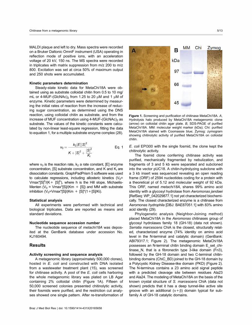
Activity screening and sequence analysisA metagenomic library (approximately 500,000 clones),
hosted in E. coli and constructed with DNA isolatedfrom a wastewater treatment plant (15), was screenedfor chitinase activity. A pool of the E. coli cells harboringthe whole metagenomic library was plated on LB Agarcontaining 2% colloidal chitin (Figure 1A). Fifteen of50,000 screened colonies presented chitinolytic activity,their fosmids were purified, and the restriction cut analy-ses showed one single pattern. After re-transformation of
E. coli EPI300 with the single fosmid, the clone kept thechitinolytic activity.
The fosmid clone conferring chitinase activity waspurified, mechanically fragmented by nebulization, andfragments of 3 and 5 kb were separated and subclonedinto the vector pUC18. A chitin-hydrolyzing subclone witha 3 kb insert was sequenced revealing an open readingframe (ORF) of 2594 nucleotides coding for a protein witha theoretical pI of 5.12 and molecular weight of 92 kDa.This ORF, named metachi18A, shares 99% amino acididentity with a glycosyl hydrolase from Aeromonas jandaei[RefSeq: WP_042029877.1] not yet characterized biochemi-cally. The closest characterized enzyme is a chitinase fromAeromonas hydrophila [DBJ: BAE87051.1] with 83% aminoacid identity (29).
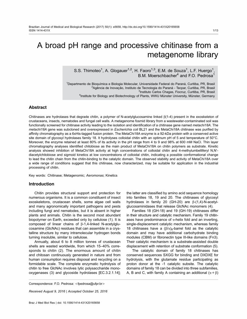
Phylogenetic analysis (Neighbor-Joining method)placed MetaChi18A in the Aeromonas chitinases group ofglycosyl hydrolases family 18 (GH-18) (data not shown).Serratia marcescens ChiA is the closest, structurally relat-ed, characterized enzyme (74% identity on amino acidlevel in the N-terminal and catalytic domain) (GenBank:ABI79317.1; Figure 2). The metagenomic MetaChi18Apossesses an N-terminal chitin binding domain E_set_chi-tinase_N, that is a fibronectin type 3-like domain (Fn3),followed by the GH-18 domain and two C-terminal chitin-binding domains (ChiC_BD) joined to the GH-18 domain bya Polycystic Kidney Disease-like domain (PKD) (Figure 2).The N-terminus contains a 23 amino acid signal peptidewith a predicted cleavage site between residues Ala23and Ala24. The modeling of MetaChi18A on the basis of theknown crystal structure of S. marcescens ChiA (data notshown) predicts that it has a deep tunnel-like active sitegroove with an additional (a+b) domain typical for sub-family A of GH-18 catalytic domains.
Figure 1. Screening and purification of chitinase MetaChi18A. A,Hydrolysis halo produced by MetaChi18A metagenomic clone(arrow) on colloidal chitin agar plate. B, SDS-PAGE of purifiedMetaChi18A. MM: molecular weight marker (kDa); Chi: purifiedMetaChi18A stained with Coomassie blue; Zymog: zymogramshowing chitinolytic activity of purified MetaChi18A on colloidalchitin.
Braz J Med Biol Res | doi: 10.1590/1414-431X20165658
Chitinase from a metagenomic library 5/13
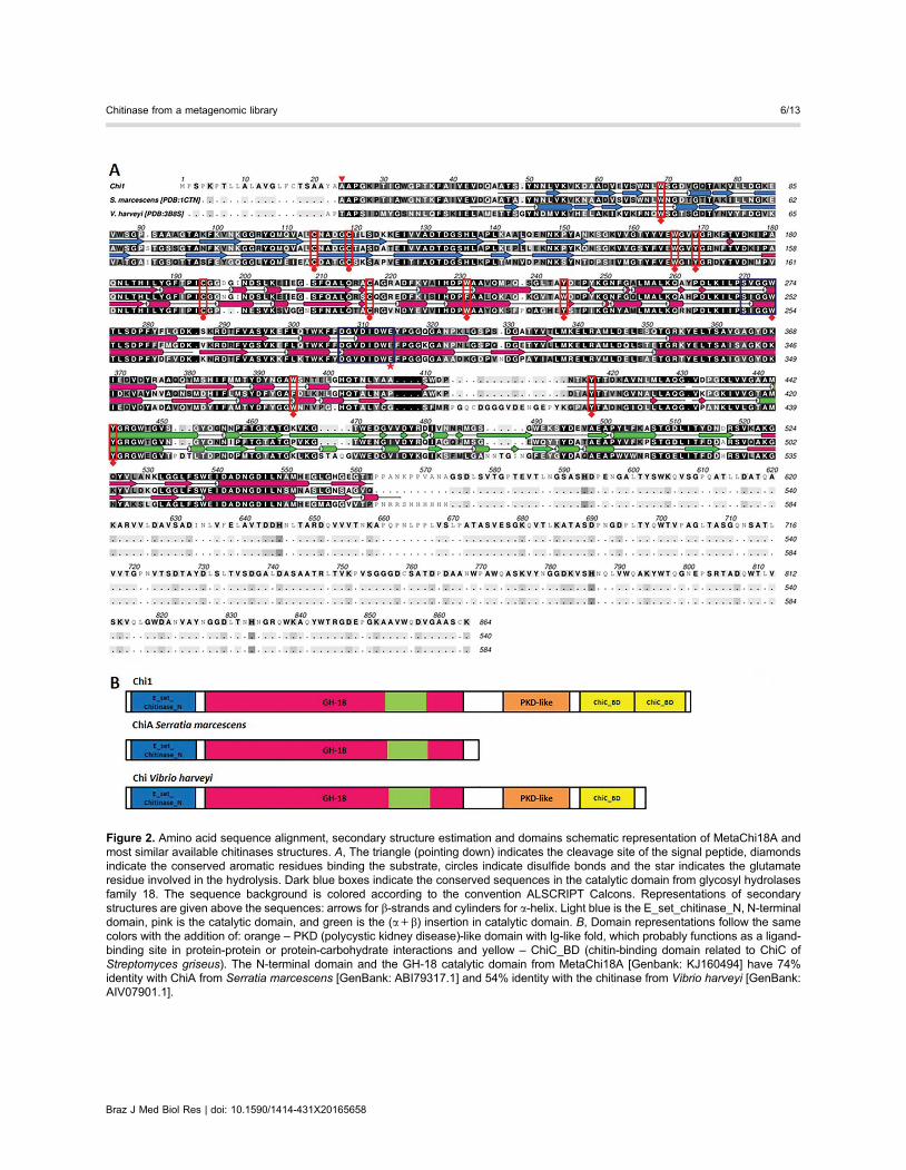
Figure 2. Amino acid sequence alignment, secondary structure estimation and domains schematic representation of MetaChi18A andmost similar available chitinases structures. A, The triangle (pointing down) indicates the cleavage site of the signal peptide, diamondsindicate the conserved aromatic residues binding the substrate, circles indicate disulfide bonds and the star indicates the glutamateresidue involved in the hydrolysis. Dark blue boxes indicate the conserved sequences in the catalytic domain from glycosyl hydrolasesfamily 18. The sequence background is colored according to the convention ALSCRIPT Calcons. Representations of secondarystructures are given above the sequences: arrows for b-strands and cylinders for a-helix. Light blue is the E_set_chitinase_N, N-terminaldomain, pink is the catalytic domain, and green is the (a+b) insertion in catalytic domain. B, Domain representations follow the samecolors with the addition of: orange – PKD (polycystic kidney disease)-like domain with Ig-like fold, which probably functions as a ligand-binding site in protein-protein or protein-carbohydrate interactions and yellow – ChiC_BD (chitin-binding domain related to ChiC ofStreptomyces griseus). The N-terminal domain and the GH-18 catalytic domain from MetaChi18A [Genbank: KJ160494] have 74%identity with ChiA from Serratia marcescens [GenBank: ABI79317.1] and 54% identity with the chitinase from Vibrio harveyi [GenBank:AIV07901.1].
Braz J Med Biol Res | doi: 10.1590/1414-431X20165658
Chitinase from a metagenomic library 6/13
Overexpression and purification of the recombinantMetaChi18A chitinase
The metachi18A gene was PCR-amplified without thesignal peptide and cloned into the pET28a expressionvector. The MetaChi18A protein carrying a C-terminal6xHis-tag was expressed in E. coli BL21 (DE3) and puri-fied by affinity chromatography. Most of the expressedprotein was insoluble (about 70% of total expressedMetaChi18A) but the protein in soluble fraction yielded180 mg of pure MetaChi18A per liter of E. coli culture. Wechose to repeat the purification to get more chitinaserather than adding components to solubilize more protein,because they could interfere on further experiments forMetaChi18A characterization. The MetaChi18A prepara-tion was homogeneous as analyzed by SDS-PAGE(Figure 1B). Zymographic analysis using colloidal chitinas substrate showed a clear band around the 97 kDaregion revealing that the purified enzyme was active(Figure 1B) and the molecular mass was as predicted fromthe translated nucleotide sequence. Peptide mass finger-printing by MALDI mass spectrometry confirmed thatthe purified enzyme was indeed MetaChi18A (data notshown).
Substrate specificitySubstrates 4-methylumbelliferyl N-acetyl-b-D-glucos-
amine (MUF-GlcNAc), 4-methylumbelliferyl N,N0-diacetyl-chitobiose (4-MUF-(GlcNAc)2) and 4-methylumbelliferylb-D-N,N0,N0 0-triacetylchitotriose (4-MUF-(GlcNAc)3) wereused for a preliminary characterization of MetaChi18Aspecificity. MetaChi18A did not show detectable activitytowards 4-MUF-GlcNAc, but the activity was 0.031 and0.015 U/mg for 4-MUF-(GlcNAc)2 and 4-MUF-(GlcNAc)3at 37°C, respectively. MetaChi18A was also active againstcolloidal chitin with an activity of 0.018 U/mg at 50°C and0.008 U/mg at 37°C.
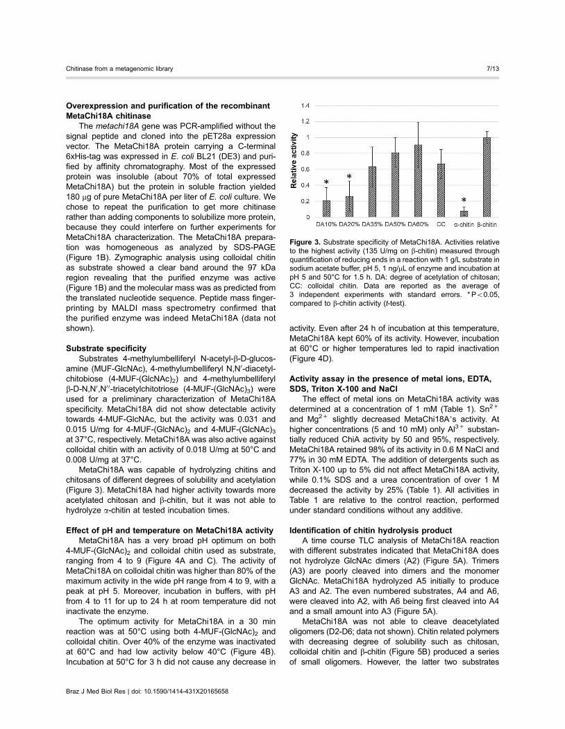
MetaChi18A was capable of hydrolyzing chitins andchitosans of different degrees of solubility and acetylation(Figure 3). MetaChi18A had higher activity towards moreacetylated chitosan and b-chitin, but it was not able tohydrolyze a-chitin at tested incubation times.
Effect of pH and temperature on MetaChi18A activityMetaChi18A has a very broad pH optimum on both
4-MUF-(GlcNAc)2 and colloidal chitin used as substrate,ranging from 4 to 9 (Figure 4A and C). The activity ofMetaChi18A on colloidal chitin was higher than 80% of themaximum activity in the wide pH range from 4 to 9, with apeak at pH 5. Moreover, incubation in buffers, with pHfrom 4 to 11 for up to 24 h at room temperature did notinactivate the enzyme.
The optimum activity for MetaChi18A in a 30 minreaction was at 50°C using both 4-MUF-(GlcNAc)2 andcolloidal chitin. Over 40% of the enzyme was inactivatedat 60°C and had low activity below 40°C (Figure 4B).Incubation at 50°C for 3 h did not cause any decrease in
activity. Even after 24 h of incubation at this temperature,MetaChi18A kept 60% of its activity. However, incubationat 60°C or higher temperatures led to rapid inactivation(Figure 4D).
Activity assay in the presence of metal ions, EDTA,SDS, Triton X-100 and NaCl
The effect of metal ions on MetaChi18A activity wasdetermined at a concentration of 1 mM (Table 1). Sn2+
and Mg2+ slightly decreased MetaChi18A’s activity. Athigher concentrations (5 and 10 mM) only Al3+ substan-tially reduced ChiA activity by 50 and 95%, respectively.MetaChi18A retained 98% of its activity in 0.6 M NaCl and77% in 30 mM EDTA. The addition of detergents such asTriton X-100 up to 5% did not affect MetaChi18A activity,while 0.1% SDS and a urea concentration of over 1 Mdecreased the activity by 25% (Table 1). All activities inTable 1 are relative to the control reaction, performedunder standard conditions without any additive.
Identification of chitin hydrolysis productA time course TLC analysis of MetaChi18A reaction
with different substrates indicated that MetaChi18A doesnot hydrolyze GlcNAc dimers (A2) (Figure 5A). Trimers(A3) are poorly cleaved into dimers and the monomerGlcNAc. MetaChi18A hydrolyzed A5 initially to produceA3 and A2. The even numbered substrates, A4 and A6,were cleaved into A2, with A6 being first cleaved into A4and a small amount into A3 (Figure 5A).
MetaChi18A was not able to cleave deacetylatedoligomers (D2-D6; data not shown). Chitin related polymerswith decreasing degree of solubility such as chitosan,colloidal chitin and b-chitin (Figure 5B) produced a seriesof small oligomers. However, the latter two substrates
Figure 3. Substrate specificity of MetaChi18A. Activities relativeto the highest activity (135 U/mg on b-chitin) measured throughquantification of reducing ends in a reaction with 1 g/L substrate insodium acetate buffer, pH 5, 1 ng/mL of enzyme and incubation atpH 5 and 50°C for 1.5 h. DA: degree of acetylation of chitosan;CC: colloidal chitin. Data are reported as the average of3 independent experiments with standard errors. * Po0.05,compared to b-chitin activity (t-test).
Braz J Med Biol Res | doi: 10.1590/1414-431X20165658
Chitinase from a metagenomic library 7/13

produced mainly dimers. MALDI-TOF-MS analyses identi-fied only diacetylchitobiose as the product of colloidal chitincleaved by MetaChi18A (data not shown).
Kinetic parametersThe kinetic constants of MetaChi18A were deter-
mined with colloidal chitin as substrate at 50°C, or 4-MUF-(GlcNAc)2 at 37°C. High concentrations of both substratesinhibited MetaChi18A activity (Figure 6). In addition,MetaChi18A exhibited a sigmoidal kinetic with colloidalchitin (Figure 6A).
Different kinetic models accounting for substrate inhibi-tion or for allosteric behavior were used to determine thekinetic parameters (Table 2) of MetaChi18A. The modelused for ChiA from Serratia marcescens (Equation 1) fitsatisfactorily to the experimental data with colloidal chitin,while for 4-MUF kinetics the Michaelis-Menten equationfor substrate inhibition fit better (Figure 6B). The KM, vmax
and KI or Hill slope values were calculated using non-linearregression analysis available on GraphPad Prism 5 orusing Equation 1, and the values for both substrates arereported in Table 2.
Discussion
Functional screening of metagenomic libraries forchitinases is still a challenge due to the heterologousexpression and to slow substrate degradation. Until now,metagenomic libraries were screened for chitinases usingsequence-based methods based on PCR or NGS, orfunctional screening methods using artificial chitin oligo-mer derivatives, that are more easily degraded. In ourwork, we were able to surmount these difficulties byscreening a metagenomic fosmid library that was pro-duced by indirect DNA extraction, therefore with highprokaryotic DNA content, and incubating plates of thesubstrate (colloidal chitin) and fosmid clones for a longertime (almost 2 months.) Even though we found 15 activeclones, the search resulted in a single fosmid. Wemanaged to save time and material by screening thepool of cells containing the whole metagenomic library.Although an estimate of 50,000 clones were checked,the redundancy of chitinase active clones shows thatfurther screening would probably not lead to a new clonediscovery.
Figure 4. Effect of pH and temperature on MetaChi18A activity and stability. Chitinolytic activity was determined using 0.2 mg/mL of4-MUF derivatives (A and B orange slashed line, Y-axis on the right) and 10 mg/mL of colloidal chitin (A and B dark blue line, Y-axis onthe left) as substrate. Enzyme concentration was 0.40 and 100 ng/mL, respectively. A, Effect of pH on the activity for 30 min at 37°C;B, effect of temperature on the activity for 30 min at pH 5; C, stability of MetaChi18A incubated at indicated pH for 1 or 24 h withoutsubstrate; D, stability of MetaChi18A incubated at the indicated temperature for 1, 3, or 24 h without substrate. The remaining activitieswere measured for 30 min at 37°C and pH 5 using 10 mg/mL of colloidal chitin. The reactions were performed in 25 mM buffers: pH 3sodium citrate; pH 4 and 5 sodium acetate; pH 6 and 7 sodium phosphate; pH 8 and 9 Tris-HCl; pH 10 and 11 sodium carbonate. Errorbars represent standard deviation. One unit of chitinase activity (U) was defined as the release of 1 mmol of reducing sugar or 4-MUF perminute.
Braz J Med Biol Res | doi: 10.1590/1414-431X20165658
Chitinase from a metagenomic library 8/13

Table 1. Effect of additives on activity of MetaChi18A.
No EDTA pre-incubation$ With EDTA pre-incubation$
Additive Relative activity (%) Additive Relative activity (%)
Al3+ 81.2±4.0 Al3+ 106.4±3.1Ca2+ 89.3±2.6 Ca2+ 108.4±10.8Co2+ 103.3±20.7 Co2+ 146.8±8.2
Cu2+ 64.3±8.9 Cu2+ 95.6±5.7Fe3+ 82.6±14.5 Fe3+ 110.5±9.1Li+ 93.8±4.5 Li+ 114.1±2.4
Mg2+ 92.1±1.5* Mg2+ 112.9±5.2Mn2+ 113.3±9.9 Mn2+ 141.0±5.2Sn2+ 72.8±1.5* Sn2+ 87.6±0.3Zn2+ 77.1±3.1 Zn2+ 93.8±9.2
EDTA (mM) NaCl (mM)
1 116.4±2.3* 50 95.5±1.9
10 101.5±0.3* 100 100.3±4.230 76.8±2.7* 300 102.9±11.960 43.0±8.1* 600 98.7±4.5
100 42.8±10.7* 900 29.3±3.4*
Triton X-100 (%) Urea (M)
1 90.5±12.3 1 69.3±1.1*5 92.3±11.1 2 52.9±5.2*
SDS 0.1% 74.0±5.1 4 21.5±2.1*
$Effect of 1 mM of the indicated chloride salt with or without 1 h of pre-incubationwith 20 mM EDTA. Chitinolytic activity was determined using 10 mg/mL ofcolloidal chitin, 100 ng/mL of MetaChi18A for 30 min at 37°C and pH 5. The activityis relative to the reaction without any additive under standard conditions.*Po0.05, compared to control (t-test).
Figure 5. Products of MetaChi18A activity on different substrates: chitooligomers (A2–A6) and polymers; b-chitin, colloidal chitin (CC)and chitosan 60% acetylated (Chtn60). MetaChi18A hydrolysis products were separated by thin-layer chromatography. A, Reaction with0.76 ng/mL MetaChi18A, 1.25 g/L oligomers in 100 mM MES buffer, pH 5, at 50°C for 5, 30, and 60 min. B, reaction with 1 ng/mLMetaChi18A, 1 g/L b-chitin, CC or Chtn60 in 10 mM ammonium acetate buffer, pH 5, at 50°C for 24 h.
Braz J Med Biol Res | doi: 10.1590/1414-431X20165658
Chitinase from a metagenomic library 9/13
Metcalfe et al. (30) showed that the diversity of thechitinolytic community of an upland pasture soil decreasedafter treatment with domestic sludge, although it increasedthe chitinolytic activity. The readily available carbon andnitrogen sources did not repress chitinases but stimulatedthe activity of specific groups of chitinolytic actinobacteria.As our metagenomic source was the soil of an industrialwaste treatment lagoon, the same effect could result in theinvariability of the chitinolytic clones we found.
In spite of the high similarity with other Aeromonaschitinases, MetaChi18A showed distinguished featureswhen compared with the closest characterized chitinasefrom Aeromonas hydrophila (29). The latter has a pHoptimum between 5 and 7.5 and shows higher activity at42°C. Its relative activity on chitosan DA 20% is only 7%lower than on colloidal chitin, while MetaChi18A has lowactivity on chitosan DA 20%. Its diverse subsite specifi-cities are also evident in the products of oligomers
hydrolysis. A. hydrophila chitinase completely hydrolyzesthe trimer into dimer and monomer, while MetaChi18Apoorly hydrolyzes it. This suggests that a few amino acidmodifications can lead to significantly divergent hydrolysisfeatures.
The structural model together with sequence analysissupport the conclusion that MetaChi18A acts processivelyon its substrate generating diacetylchitobiose as the mainproduct, as reported for ChiA and ChiB from S. marces-cens (31). Indeed, TLC and MALDI-TOF analyses iden-tified chitobiose as the single MetaChi18A product ofb-chitin and the main product of colloidal chitin hydrolysis.Moreover, MetaChi18A presents typical features of proces-sive enzymes; the insertion domain (a+b) at the catalyticdomain, a stretch of aromatic amino acid residues on itssurface, from the N-terminal substrate-binding domain tothe catalytic domain, and the conserved residues Trp166(corresponding to Trp167 in ChiA from S. marcescens),
Figure 6. Kinetic behavior of MetaChi18A. A, Colloidal chitin as substrate; B, 4-MUF-(GlcNAc)2 as substrate. Colloidal chitin (g/L) andV0 (nmol of reducing sugar/min); 4-MUF-(GlcNAc)2 (mM) and V0 (nmol of MUF/min). Reactions were performed at 25 mM sodiumacetate buffer, pH 5, at 50°C (A) or 37°C (B). The black line is the model that better fits the data: (A) allosteric kinetic+substrateinhibition, and (B) substrate inhibition; Blue dotted line: allosteric kinetic; Orange slashed line: Michaelis-Menten kinetic.
Table 2. Kinetic parameters of MetaChi18A and GH-18 chitinases on 4-MUF-(GlcNAc)2 and colloidalchitin.
Colloidal chitin KM (g/L) Ki/Hill Vmax (nmol/min) kcat (s-1)
MetaChi18A (A+SI) 98.5 1.03 g/L 627.9 14.0MetaChi18A (A) 33.2 4.12 (Hill) 62.0 1.4
MetaChi18A (MM) 4.4 – 97.2 2.2Aeromonas sp. (Jeong et al., 2012) 3.45 – 2910 –B. licheniformis (Nguyen et al., 2012) 28 – 4800 5.2
4-MUF-(GlcNAc)2 KM (mM) Ki/Hill Vmax (nmol/min) kcat (s-1)
Ch1 (SI) 65.7 1.31 mM 0.49 38.9MetaChi18A (A) 6.7 2.43 (Hill) 0.03 2.4
MetaChi18A (MM) 2.7 – 0.04 3.2S. marcescens (Honda et al., 2003) 3.5 1100 mM – 34.9
MM: Michaelis-Menten kinetic; SI: MM with substrate inhibition; A: allosteric sigmoidal (Hill slope);–: constant not provided.
Braz J Med Biol Res | doi: 10.1590/1414-431X20165658
Chitinase from a metagenomic library 10/13

Trp274 (corresponding to Trp275 in ChiA) and Trp395(corresponding to Phe396 in ChiA) (32).
Most chitinases described to date have high activity atacidic or close to neutral pH. MetaChi18A has more than80% of activity from pH 4 to 9 and remains active at 50°Cfor 24 h. The results suggest that MetaChi18A is mod-erately thermophilic, as it would be expected since it issimilar to Aeromonas chitinases. Aeromonas sp. can befound inhabiting natural soil, food and animals, but mostlyall kinds of aquatic environments. The mesophilic specieshave optimal growth at 35° to 37°C (33).
Bacterial chitinases respond differently to the pres-ence of a variety of metal ions and MetaChi18A wasnot highly affected by the metal ions tested. It has beenreported that the inhibition of chitinase by certain divalentcations occurs because they are able to form stablecomplexes with carboxylic groups of aspartic and glutamicacid residues at the active site (34), but activity assayswith EDTA and all tested ions showed that MetaChi18Ahas no necessity of a cofactor for its activity.
Concerning the resilience to higher concentrations ofNaCl, MetaChi18A remained active until 600 mM. Wecannot claim that this characteristic is related only to thepresence of high NaCl concentrations in the originalenvironment, as a wastewater treatment lagoon is a richmedium with high concentration of nutrients and micro-organisms. MetaChi18A showed high stability on differentconditions including high activity in a larger pH range. Thismay be due to the additional domains at both N- andC-terminals that keep its structure more stable avoiding itsdisturbance in these conditions. Such higher stability wasalready described for chitinases containing chitin bind-ing domain and Fn3 domain (35,36) but most chitinaseshave one or two of these domains in one side while ourMetaChi18A has four additional domains surrounding thecatalytic domain, which might protect even more its integrity.
As usual for chitinases of the GH-18 family, MetaChi18Ahad higher activity towards more acetylated substrates.Since the enzyme requires an acetylated glucosamineresidue positioned in the -1 catalytic subsite (5), the higherthe degree of acetylation (DA) the higher the abundanceof cleavage sites in the polymeric substrates. MetaChi18Awas not able to hydrolyze a-chitin presumably due to its crys-talline structure with highly packed chains making accessto the enzyme difficult. It was described that the lytic poly-saccharide monooxygenases facilitate access into a-chitinchains by producing oxidative cuts along the chain (3).
Although Cruys-Bagger et al. (37) described that proces-sive enzymes such as cellulases also show a hyperbolicrelationship between steady-state rate and substrate con-centration, the kinetic parameters are more complex andmay not be calculated directly from the Michaelis-Mentenequation. Kinetics of ChiA from S. marcescens on 4-MUF-(GlcNAc)2 had the same profile and its parameters weredetermined using Equation 1 (28). MetaChi18A and ChiAfrom S. marcescens showed similar catalytic constants (kcat):
38.9 and 34.9 s-1, respectively. On colloidal chitin, the kineticparameters of Aeromonas sp. GJ 18 and Bacillus licheni-formis chitinases were determined using Michaelis-Mentennon-linear regression (38,39). The KM for MetaChi18A wassimilar to that of Aeromonas chitinase while the kcat valuewas similar to that of Bacillus chitinase (Table 2).
Many chitinases of family GH-18 exhibit substrateinhibition by substrates of low molecular weight such as4-MUF-(GlcNAc)2. The proposed MetaChi18A active cleftcontains multi-subsites where small substrates could binddifferently leading to non-productive inhibitory binding(40). However, this inhibitory mechanism would not beexpected for inhibition caused by high concentrations ofthe polymeric colloidal chitin, where inhibition is moreprobably caused by impaired diffusion of the enzyme.MetaChi18A showed a Hill slope of 4, indicating substrate-binding cooperativity. This phenomenon may occur due tohysteresis, a delay of the enzyme in reaching its fully activeform that involves conformational changes. As MetaChi18Ais a multi-domain enzyme, the substrate should interact withbinding domains that could rearrange the conformation tofacilitate access of the catalytic domain to the substrate.
Although most kinetic studies on chitinases have usedsmall oligomers as substrates, not reflecting their nativebehavior on chitin, our study is one of few (38,39) thatreport kinetics on colloidal chitin. The data obtained reflectbetter the natural substrate, and help to elucidate howchitinases behave on polymeric substrates.
Our study was successful in the identification of achitinolytic clone by functional screening of a metagenomicfosmid library. We have succeeded in the MetaChi18Abiochemical and kinetic characterization, using colloidal chi-tin, which is a more soluble formulation of polymeric chitin.
Current limitations on the use of chitinases for bio-technological applications are their low stability and thelimited range of temperatures and pH values in which thechitinases described to date are functional. The Meta-Chi18A chitinase described in this study is a promisingalternative for industrial processing of chitin as judged byits high stability and activity under a broad range of pHvalues and temperatures.
Acknowledgments
We thank Roseli Prado, Alexsandro Albani, Valter A.de Baura, Ursula Fassin, Andrea Norra, and ClaudiaLüneberg for technical support and Dominique Gillet fromMahtani Chitosan for providing polyglucosamine. Wethank the Brazilian National Council for Scientific andTechnological Development (CNPq) and the Coordinationfor the Development of Higher Education Personnel(CAPES) for research scholarships. This work was sup-ported by the National Institute of Science and Technologyof Biological Nitrogen Fixation (INCT-FBN/CNPq/MCT573828/2008-3), and the National Program of Excellency(PRONEX/Fundacão Araucária). The authors would like
Braz J Med Biol Res | doi: 10.1590/1414-431X20165658
Chitinase from a metagenomic library 11/13

to thank the Academic Publishing Advisory Center (Centrode Assessoria de Publicacão Acadêmica, CAPA - www.
capa.ufpr.br) of the Federal University of Paraná forassistance with English language editing.
References
1. Gooday GW. Physiology of microbial degradation of chitinand chitosan. Biodegradation 1990; 1: 177–190, doi: 10.1007/BF00058835.
2. Yan N, Chen X. Sustainability: Don’t waste seafood waste.Nature 2015; 524: 155–157, doi: 10.1038/524155a.
3. Vaaje-Kolstad G, Westereng B, Horn SJ, Liu Z, Zhai H,Sorlie M, et al. An oxidative enzyme boosting the enzymaticconversion of recalcitrant polysaccharides. Science 2010;330: 219–222, doi: 10.1126/science.1192231.
4. Henrissat B, Bairoch A. New families in the classification ofglycosyl hydrolases based on amino acid sequence similarities.Biochem J 1993; 293 (Part 3): 781–788, doi: 10.1042/bj2930781.
5. van Aalten DM, Komander D, Synstad B, Gaseidnes S,Peter MG, Eijsink VG. Structural insights into the catalyticmechanism of a family 18 exo-chitinase. Proc Natl Acad SciU S A 2001; 98: 8979–8984, doi: 10.1073/pnas.151103798.
6. Li H, Greene LH. Sequence and structural analysis of thechitinase insertion domain reveals two conserved motifsinvolved in chitin-binding. PLoS One 2010; 5: e8654,doi: 10.1371/journal.pone.0008654.
7. Sørlie M, Zakariassen H, Norberg AL, Eijsink VGH. Pro-cessivity and substrate-binding in family 18 chitinases. BiocatalBiotransformation 2012; 30: 353–365, doi: 10.3109/10242422.2012.676282.
8. Leresche JE, Meyer H-P. Chemocatalysis and biocatalysis(biotransformation): some thoughts of a chemist and of abiotechnologist. Org Process Res Dev 2006; 10: 572–580,doi: 10.1021/op0600308.
9. Torsvik V, Ovreas L. Microbial diversity and function in soil:from genes to ecosystems. Curr Opin Microbiol 2002; 5:240–245, doi: 10.1016/S1369-5274(02)00324-7.
10. Handelsman J, Rondon MR, Brady SF, Clardy J, GoodmanRM. Molecular biological access to the chemistry of unknownsoil microbes: a new frontier for natural products. Chem Biol1998; 5: R245–R249, doi: 10.1016/S1074-5521(98)90108-9.
11. Stoveken J, Singh R, Kolkenbrock S, Zakrzewski M,Wibberg D, Eikmeyer FG, et al. Successful heterologousexpression of a novel chitinase identified by sequence anal-yses of the metagenome from a chitin-enriched soil sample.J Biotechnol 2015; 201: 60–68, doi: 10.1016/j.jbiotec.2014.09.010.
12. Beier S, Jones CM, Mohit V, Hallin S, Bertilsson S. Globalphylogeography of chitinase genes in aquatic metagenomes.Appl Environ Microbiol 2011; 77: 1101–1106, doi: 10.1128/AEM.01481-10.
13. Cretoiu MS, Kielak AM, Abu Al-Soud W, Sorensen SJ,van Elsas JD. Mining of unexplored habitats for novelchitinases - chiA as a helper gene proxy in metagenomics.Appl Microbiol Biotechnol 2012; 94: 1347–1358, doi: 10.1007/s00253-012-4057-5.
14. Hsu SC, Lockwood JL. Powdered chitin agar as a selectivemedium for enumeration of actinomycetes in water and soil.Appl Microbiol 1975; 29: 422–426.
15. Glogauer A, Martini VP, Faoro H, Couto GH, Muller-SantosM, Monteiro RA, et al. Identification and characterization of
a new true lipase isolated through metagenomic approach.Microb Cell Fact 2011; 10: 54, doi: 10.1186/1475-2859-10-54.
16. Ewing B, Hillier L, Wendl MC, Green P. Base-calling ofautomated sequencer traces using phred. I. Accuracy assess-ment.Genome Res 1998; 8: 175–185, doi: 10.1101/gr.8.3.175.
17. Bendtsen JD, Nielsen H, von Heijne G, Brunak S. Improvedprediction of signal peptides: SignalP 3.0. J Mol Biol 2004;340: 783–795, doi: 10.1016/j.jmb.2004.05.028.
18. Gasteiger E, Gattiker A, Hoogland C, Ivanyi I, Appel RD,Bairoch A. ExPASy: The proteomics server for in-depthprotein knowledge and analysis. Nucleic Acids Res 2003;31: 3784–3788, doi: 10.1093/nar/gkg563.
19. Thompson JD, Higgins DG, Gibson TJ. CLUSTAL W:improving the sensitivity of progressive multiple sequencealignment through sequence weighting, position-specificgap penalties and weight matrix choice. Nucleic Acids Res1994; 22: 4673–4680, doi: 10.1093/nar/22.22.4673.
20. Berman HM, Kleywegt GJ, Nakamura H, Markley JL. TheProtein Data Bank archive as an open data resource.J Comput Aided Mol Des 2014; 28: 1009–1014, doi: 10.1007/s10822-014-9770-y.
21. Kabsch W, Sander C. Dictionary of protein secondarystructure: pattern recognition of hydrogen-bonded andgeometrical features. Biopolymers 1983; 22: 2577–2637,doi: 10.1002/bip.360221211.
22. Bond CS, Schuttelkopf AW. ALINE: a WYSIWYG protein-sequence alignment editor for publication-quality alignments.Acta Crystallogr D Biol Crystallogr 2009; 65: 510–512,doi: 10.1107/S0907444909007835.
23. Tronsmo A, Harman GE. Detection and quantification ofN-acetyl-beta-D-glucosaminidase, chitobiosidase, and endo-chitinase in solutions and on gels. Anal Biochem 1993; 208:74–79, doi: 10.1006/abio.1993.1010.
24. Westermeier R, Loyland S, Asbury R. Proteomics technol-ogy. J Clin Ligando Assay 2002; 25: 242–252.
25. Miller GL. Use of dinitrosalicylic acid reagent for determi-nation of reducing sugar. Anal Chem 1959; 31: 426–428,doi: 10.1021/ac60147a030.
26. Horn SJ, Eijsink VGH. A reliable reducing end assay forchito-oligosaccharides. Carbohydr Polym 2004; 56: 35–39,doi: 10.1016/j.carbpol.2003.11.011.
27. Price NP, Naumann TA. A high-throughput matrix-assistedlaser desorption/ionization-time-of-flight mass spectrometry-based assay of chitinase activity. Anal Biochem 2011; 411:94–99, doi: 10.1016/j.ab.2010.12.027.
28. Honda Y, Kitaoka M, Tokuyasu K, Sasaki C, Fukamizo T,Hayashi K. Kinetic studies on the hydrolysis of N-acetylatedand N–deacetylated derivatives of 4-methylumbelliferylchitobioside by the family 18 chitinases ChiA and ChiBfrom Serratia marcescens. J Biochem 2003; 133: 253–258,doi: 10.1093/jb/mvg031.
29. Lan X, Zhang X, Hu J, Shimosaka M. Cloning, expression, andcharacterization of a chitinase from the chitinolytic bacteriumAeromonas hydrophila strain SUWA-9. Biosci BiotechnolBiochem 2006; 70: 2437–2442, doi: 10.1271/bbb.60169.
Braz J Med Biol Res | doi: 10.1590/1414-431X20165658
Chitinase from a metagenomic library 12/13
30. Metcalfe AC, Krsek M, Gooday GW, Prosser JI, WellingtonEM. Molecular analysis of a bacterial chitinolytic communityin an upland pasture. Appl Environ Microbiol 2002; 68:5042–5050, doi: 10.1128/AEM.68.10.5042-5050.2002.
31. Sikorski P, Sorbotten A, Horn SJ, Eijsink VG, Varum KM.Serratia marcescens chitinases with tunnel-shaped sub-strate-binding grooves show endo activity and differentdegrees of processivity during enzymatic hydrolysis ofchitosan. Biochemistry 2006; 45: 9566–9574.
32. Payne CM, Baban J, Horn SJ, Backe PH, Arvai AS, DalhusB, et al. Hallmarks of processivity in glycoside hydrolasesfrom crystallographic and computational studies of theSerratia marcescens chitinases. J Biol Chem 2012; 287:36322–36330, doi: 10.1074/jbc.M112.402149.
33. Janda JM, Abbott SL. The genus Aeromonas: taxonomy,pathogenicity, and infection. Clin Microbiol Rev 2010; 23:35–73, doi: 10.1128/CMR.00039-09.
34. Milewski S, O’Donnell RW, Gooday GW. Chemical mod-ification studies of the active centre of Candida albicanschitinase and its inhibition by allosamidin. J Gen Micro-biol 1992; 138: 2545–2550, doi: 10.1099/00221287-138-12-2545.
35. Lin FP, Juang WY, Chang KH, Chen HC. G561 site-directeddeletion mutant chitinase from Aeromonas caviae is active
without its 304 C-terminal amino acid residues. ArchMicrobiol 2001; 175: 220–225, doi: 10.1007/s002030100261.
36. Sha L, Shao E, Guan X, Huang Z. Purification and partialcharacterization of intact and truncated chitinase fromBacillus thuringiensis HZP7 expressed in Escherichia coli.Biotechnol Lett 2016; 38: 279–284, doi: 10.1007/s10529-015-1970-6.
37. Cruys-Bagger N, Elmerdahl J, Praestgaard E, Borch K,Westh P. A steady-state theory for processive cellulases.FEBS J 2013; 280: 3952–3961, doi: 10.1111/febs.12397.
38. Nguyen HA, Nguyen TH, Nguyen TT, Peterbauer CK,Mathiesen G, Haltrich D. Chitinase from Bacillus lichenifor-mis DSM13: expression in Lactobacillus plantarum WCFS1and biochemical characterisation. Protein Expr Purif 2012;81: 166–174, doi: 10.1016/j.pep.2011.10.005.
39. Jeong HC, Ju W-T, Jo K-H, Park RD. Purification andcharacterization of a 34-kDa chitobiosidase from Aeromo-nas sp. GJ-18. J Korean Soc Appl Biol Chem 2012; 55:7–12, doi: 10.1007/s13765-012-0002-7.
40. Honda Y, Kirihata M, Fukamizo T, Kaneko S, Tokuyasu K,Brzezinski R. Chitosanase-catalyzed hydrolysis of 4-methy-lumbelliferyl beta-chitotrioside. J Biochem 1999; 126:470–474, doi: 10.1093/oxfordjournals.jbchem.a022475.
Braz J Med Biol Res | doi: 10.1590/1414-431X20165658
Chitinase from a metagenomic library 13/13


![Expression of a Bacterial Chitinase (ChiB) Gene Enhances ... · Rahman (2012) [14]. Tolerance potential of the transgenic black gram carrying Bacterial chitinase gene was evaluated](https://img.pdfslide.net/doc/110x75/5e8e4c7f862d6a32fc34abea/expression-of-a-bacterial-chitinase-chib-gene-enhances-rahman-2012-14.jpg)