Embed Size (px)
Citation preview
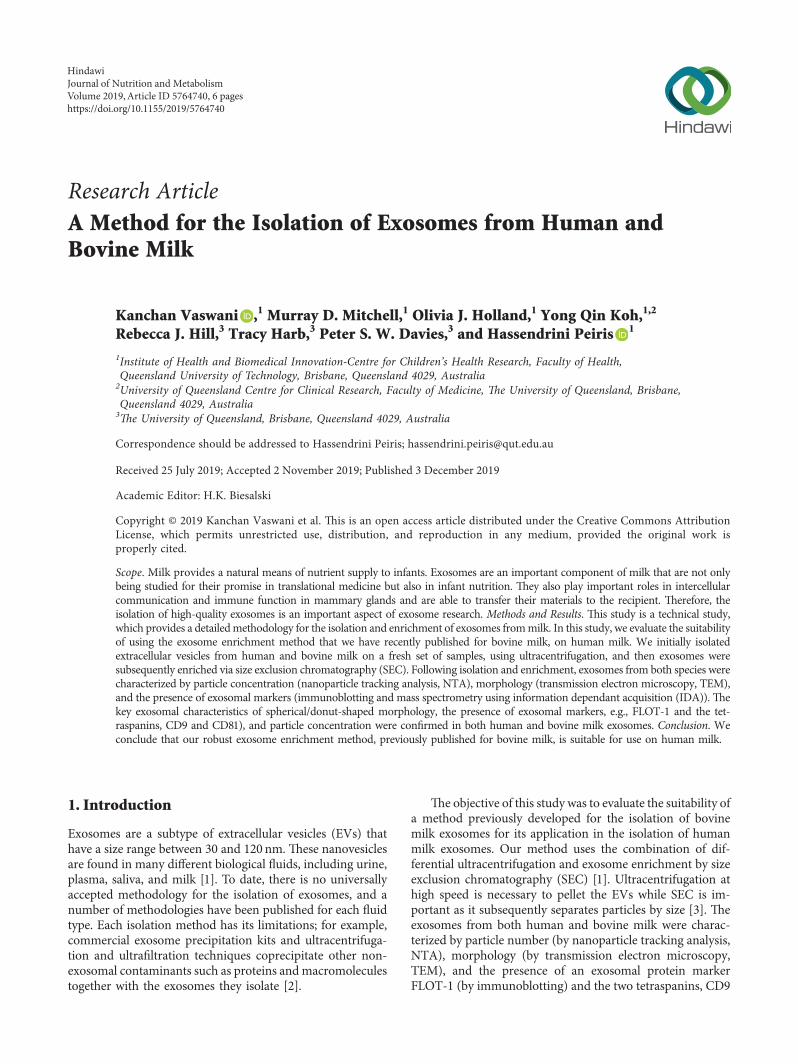
Research ArticleA Method for the Isolation of Exosomes from Human andBovine Milk
Kanchan Vaswani ,1 Murray D. Mitchell,1 Olivia J. Holland,1 Yong Qin Koh,1,2
Rebecca J. Hill,3 Tracy Harb,3 Peter S. W. Davies,3 and Hassendrini Peiris 1
1Institute of Health and Biomedical Innovation-Centre for Children’s Health Research, Faculty of Health,Queensland University of Technology, Brisbane, Queensland 4029, Australia2University of Queensland Centre for Clinical Research, Faculty of Medicine, 'e University of Queensland, Brisbane,Queensland 4029, Australia3'e University of Queensland, Brisbane, Queensland 4029, Australia
Correspondence should be addressed to Hassendrini Peiris; [email protected]
Received 25 July 2019; Accepted 2 November 2019; Published 3 December 2019
Academic Editor: H.K. Biesalski
Copyright © 2019 Kanchan Vaswani et al. ,is is an open access article distributed under the Creative Commons AttributionLicense, which permits unrestricted use, distribution, and reproduction in any medium, provided the original work isproperly cited.
Scope. Milk provides a natural means of nutrient supply to infants. Exosomes are an important component of milk that are not onlybeing studied for their promise in translational medicine but also in infant nutrition. ,ey also play important roles in intercellularcommunication and immune function in mammary glands and are able to transfer their materials to the recipient. ,erefore, theisolation of high-quality exosomes is an important aspect of exosome research. Methods and Results. ,is study is a technical study,which provides a detailedmethodology for the isolation and enrichment of exosomes frommilk. In this study, we evaluate the suitabilityof using the exosome enrichment method that we have recently published for bovine milk, on human milk. We initially isolatedextracellular vesicles from human and bovine milk on a fresh set of samples, using ultracentrifugation, and then exosomes weresubsequently enriched via size exclusion chromatography (SEC). Following isolation and enrichment, exosomes from both species werecharacterized by particle concentration (nanoparticle tracking analysis, NTA), morphology (transmission electron microscopy, TEM),and the presence of exosomal markers (immunoblotting and mass spectrometry using information dependant acquisition (IDA)). ,ekey exosomal characteristics of spherical/donut-shaped morphology, the presence of exosomal markers, e.g., FLOT-1 and the tet-raspanins, CD9 and CD81), and particle concentration were confirmed in both human and bovine milk exosomes. Conclusion. Weconclude that our robust exosome enrichment method, previously published for bovine milk, is suitable for use on human milk.
1. Introduction
Exosomes are a subtype of extracellular vesicles (EVs) thathave a size range between 30 and 120 nm. ,ese nanovesiclesare found in many different biological fluids, including urine,plasma, saliva, and milk [1]. To date, there is no universallyaccepted methodology for the isolation of exosomes, and anumber of methodologies have been published for each fluidtype. Each isolation method has its limitations; for example,commercial exosome precipitation kits and ultracentrifuga-tion and ultrafiltration techniques coprecipitate other non-exosomal contaminants such as proteins andmacromoleculestogether with the exosomes they isolate [2].
,e objective of this study was to evaluate the suitability ofa method previously developed for the isolation of bovinemilk exosomes for its application in the isolation of humanmilk exosomes. Our method uses the combination of dif-ferential ultracentrifugation and exosome enrichment by sizeexclusion chromatography (SEC) [1]. Ultracentrifugation athigh speed is necessary to pellet the EVs while SEC is im-portant as it subsequently separates particles by size [3]. ,eexosomes from both human and bovine milk were charac-terized by particle number (by nanoparticle tracking analysis,NTA), morphology (by transmission electron microscopy,TEM), and the presence of an exosomal protein markerFLOT-1 (by immunoblotting) and the two tetraspanins, CD9
HindawiJournal of Nutrition and MetabolismVolume 2019, Article ID 5764740, 6 pageshttps://doi.org/10.1155/2019/5764740

and CD81 (by information dependant acquisition, IDA massspectrometry).
2. Materials and Methods
2.1. Milk Collection. Human milk (9ml) was collected fromfour healthy donor women in compliance with the Uni-versity of Queensland Human Research Ethics Committeeand the regulations governing experimentation on humans.Human milk (9ml× 3) was used for subsequent experi-ments. Unpasteurised bovine milk (10ml× 3) was collectedfrom a healthy Holstein Friesian dairy herd located atGatton, University of Queensland. Milk was aliquoted andstored at − 80°C for later use.
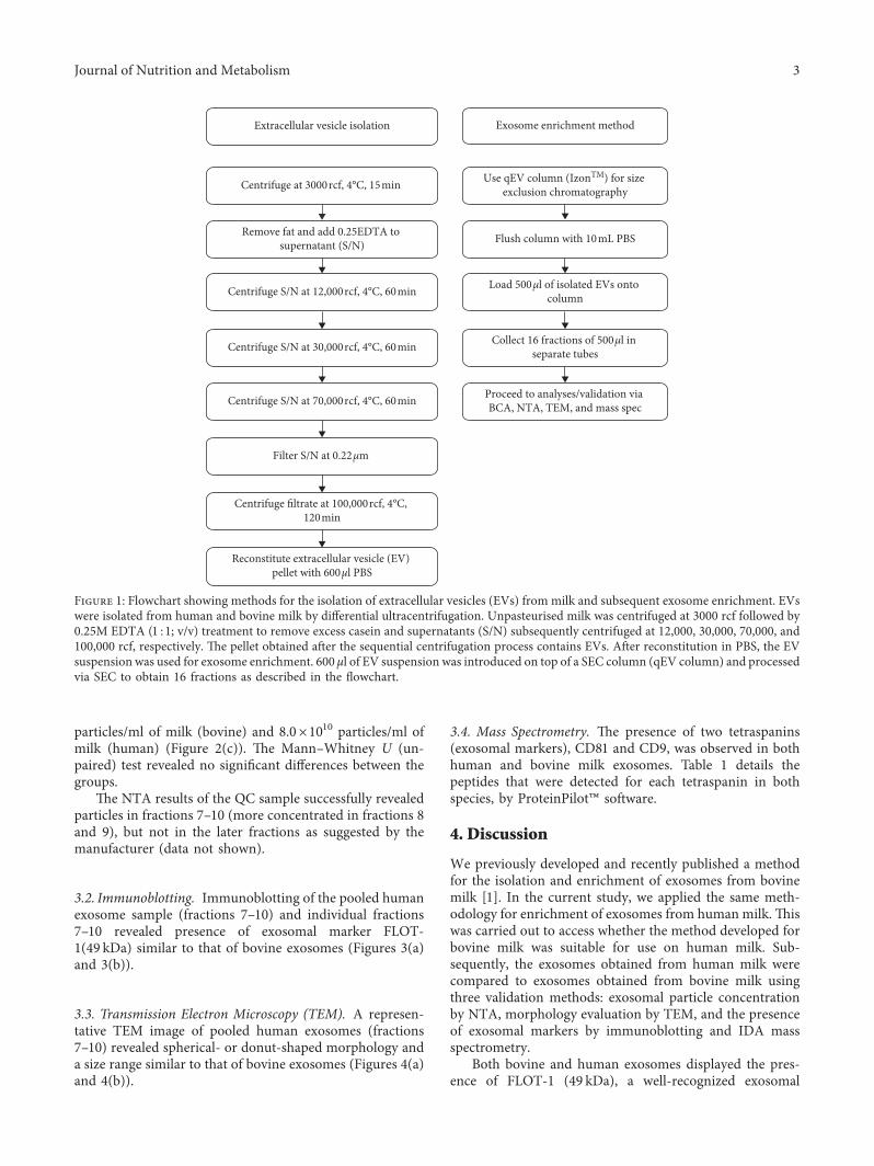
2.2. Extracellular Vesicle Isolation and Exosome Enrichment.EVs were isolated from milk by ultracentrifugation aspreviously published [1]. Briefly, bovine and human milkwere centrifuged at 3000 and 12,000 rcf to remove fatglobules, cellular debris, somatic cells, and casein. ,is wasfollowed by centrifugation steps at high speed (Figure 1).,esupernatants collected after removal of fat and casein werethen filtered at 0.2 µm (PES, Corning 431229, Sigma Aldrich,Castle Hill, NSW, Australia) and subjected to the final100,000 rcf centrifugation step. ,e pellets containing theEVs were resuspended in 600 μl PBS (Gibco, Life Tech-nologies Australia Pty Ltd). 500 μl of EVs was introduced ona washed qEV size exclusion chromatography column (qEVoriginal SP1-AUD, Izon Science Ltd, New Zealand), and500 μl fractions were collected in 16 separate tubes as permanufacturer’s instructions (Figure 1). Note that prior tostarting SEC, a single representative column was used to testthe chromatography principle using 100 nm latex beads(Malvern Panalytical Ltd, Malvern, UK) as a QC for sepa-ration of exosomes into fractions 7, 8, 9, and 10.
2.3.NanoparticleTrackingAnalysis. ,enumber of exosomesisolated from both human and bovine milk was determined bynanoparticle tracking analysis (NTA) using a MalvernNanoSight NS500, NTA 3.0 instrument as per manufacturer’sinstructions. Pooled exosomal samples (i.e., fractions 7–10)were analysed (in triplicate) to determine particle concentra-tion (particles/ml) in PBS.,en, the total yield was obtained bycalculating the concentration in particles/ml multiplied withthe total volume (2ml) of exosomal fractions (i.e., 500µlfraction 7, 500µl fraction 8, 500µl fraction 9, and 500µlfraction 10). A Mann–Whitney U (unpaired) test was carriedout between the two groups (n� 3 per group), for yield. ,eyield was then extrapolated back to the starting volumes ofmilkused (10ml, bovine milk and 9ml, human milk).
,e NTA of the QC sample revealed particles in fractions7–10.
2.4. Transmission Electron Microscopy. Human and bovineexosomal fractions (fractions 7–10) were pooled, and 5 μlfrom each sample was analysed by transmission electronmicroscopy (TEM) by negative stain. Samples were placed
on formvar-coated copper grids and viewed on a JEOL 1010microscope [1].
2.5. Immunoblotting. Exosomal markers in both pooledsamples and the individual fractions were determined byimmunoblotting for FLOT-1 (details described sub-sequently). Exosomal protein concentration was quantifiedby using a Bicinchoninic Acid reagent kit (Sigma Aldrich,Castle Hill, NSW, Australia). 10 μg of exosome protein(singular fractions and pooled fractions 7–10) was incubatedfor 10min at 70°C in reducing agent (NuPAGE SampleReducing Agent, Life Technologies Australia Pty Ltd,Mulgrave, VIC, Australia) and loading buffer (NuPAGELDS buffer, Life Technologies Australia Pty Ltd). Reducedproteins were electrophoresed and transferred onto apolyvinyl difluoride (PVDF; Bio-Rad Laboratories Pty Ltd,Australia) membrane as previously described [1]. Mem-branes were incubated for 1 hour in 2% BSA and probedovernight with primary goat polyclonal antibody anti-Flo-tillin 1 (FLOT-1) (ab13493 Abcam, Cambridge, UK) at 4°C,followed by secondary donkey anti-goat IgG-HRP (1 :1000;sc-2020, Santa Cruz Biotechnology, CA, USA). SuperSignalWest Dura-Extended Duration Substrate (,ermo FisherScientific, Australia Pty Ltd) was used for development, andthe signal visualized on X-ray film (Agfa, Mortsel, Belgium)was developed using a Konica Minolta SRX-101A processor(Konica Minolta Medical and Graphic Inc, Japan) (methodderived from [1, 4]).
2.6. Mass Spectrometry Proteomic Profiling. Exosomal frac-tions (pooled fractions 7–10) of bovine and human sampleswere sonicated for 5mins. 25 µg of exosomal protein wasused for mass spectrometry. Sample preparations wereperformed as previously published [1]. Eluted peptides weredried at RT in a vacuum evaporator for 45min. Sampleswere then reconstituted in 30 μl formic acid. ,e digestedprotein samples were analysed using the TripleTOF® 5600mass spectrometer (ABSciex, Redwood City, CA) andEksigent HPLC system to obtain initial high mass accuracysurvey MS/MS data, identifying the peptides present. An in-depth proteomic analysis was performed using the IDAexperiments on the TripleTOF® 5600 System interfaced witha nanospray source. Results were analysed usingProteinPilot™ (ABSciex, Redwood City, CA) (method de-rived from [1, 4]).
3. Results
3.1. Nanoparticle Tracking Analysis. NanoSight™ measure-ments of pooled exosome samples indicate that human milkexosomes can be successfully quantified using the sametechnique as we developed for bovine milk exosomes. ,eaverage concentration of exosomes obtained was 7.2×10¹¹particles/ml (bovine) and 3.63×10¹¹ particles/ml (human)(Figure 2(a)). ,e average yields of exosomes obtained was1.44×1012 particles (bovine) and 7.26×10¹¹ particles (hu-man) (Figure 2(b)). Average particle concentration ex-trapolating to the starting volume of milk used was 1.4×10¹¹
2 Journal of Nutrition and Metabolism
particles/ml of milk (bovine) and 8.0×10¹⁰ particles/ml ofmilk (human) (Figure 2(c)). ,e Mann–Whitney U (un-paired) test revealed no significant differences between thegroups.
,e NTA results of the QC sample successfully revealedparticles in fractions 7–10 (more concentrated in fractions 8and 9), but not in the later fractions as suggested by themanufacturer (data not shown).
3.2. Immunoblotting. Immunoblotting of the pooled humanexosome sample (fractions 7–10) and individual fractions7–10 revealed presence of exosomal marker FLOT-1(49 kDa) similar to that of bovine exosomes (Figures 3(a)and 3(b)).
3.3. Transmission Electron Microscopy (TEM). A represen-tative TEM image of pooled human exosomes (fractions7–10) revealed spherical- or donut-shaped morphology anda size range similar to that of bovine exosomes (Figures 4(a)and 4(b)).
3.4. Mass Spectrometry. ,e presence of two tetraspanins(exosomal markers), CD81 and CD9, was observed in bothhuman and bovine milk exosomes. Table 1 details thepeptides that were detected for each tetraspanin in bothspecies, by ProteinPilot™ software.
4. Discussion
We previously developed and recently published a methodfor the isolation and enrichment of exosomes from bovinemilk [1]. In the current study, we applied the same meth-odology for enrichment of exosomes from human milk. ,iswas carried out to access whether the method developed forbovine milk was suitable for use on human milk. Sub-sequently, the exosomes obtained from human milk werecompared to exosomes obtained from bovine milk usingthree validation methods: exosomal particle concentrationby NTA, morphology evaluation by TEM, and the presenceof exosomal markers by immunoblotting and IDA massspectrometry.
Both bovine and human exosomes displayed the pres-ence of FLOT-1 (49 kDa), a well-recognized exosomal
Collect 16 fractions of 500µl in separate tubes
Load 500µl of isolated EVs onto column
Flush column with 10mL PBS
Exosome enrichment methodExtracellular vesicle isolation
Use qEV column (IzonTM) for size exclusion chromatography
Reconstitute extracellular vesicle (EV) pellet with 600µl PBS
Centrifuge filtrate at 100,000rcf, 4°C, 120min
Filter S/N at 0.22µm
Centrifuge S/N at 70,000rcf, 4°C, 60min
Centrifuge S/N at 30,000rcf, 4°C, 60min
Centrifuge S/N at 12,000rcf, 4°C, 60min
Remove fat and add 0.25EDTA to supernatant (S/N)
Centrifuge at 3000rcf, 4°C, 15min
Proceed to analyses/validation via BCA, NTA, TEM, and mass spec
Figure 1: Flowchart showing methods for the isolation of extracellular vesicles (EVs) from milk and subsequent exosome enrichment. EVswere isolated from human and bovine milk by differential ultracentrifugation. Unpasteurised milk was centrifuged at 3000 rcf followed by0.25M EDTA (1 :1; v/v) treatment to remove excess casein and supernatants (S/N) subsequently centrifuged at 12,000, 30,000, 70,000, and100,000 rcf, respectively. ,e pellet obtained after the sequential centrifugation process contains EVs. After reconstitution in PBS, the EVsuspension was used for exosome enrichment. 600 μl of EV suspension was introduced on top of a SEC column (qEV column) and processedvia SEC to obtain 16 fractions as described in the flowchart.
Journal of Nutrition and Metabolism 3
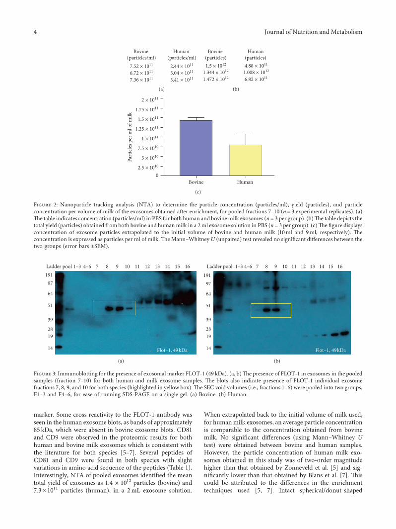
marker. Some cross reactivity to the FLOT-1 antibody wasseen in the human exosome blots, as bands of approximately85 kDa, which were absent in bovine exosome blots. CD81and CD9 were observed in the proteomic results for bothhuman and bovine milk exosomes which is consistent withthe literature for both species [5–7]. Several peptides ofCD81 and CD9 were found in both species with slightvariations in amino acid sequence of the peptides (Table 1).Interestingly, NTA of pooled exosomes identified the meantotal yield of exosomes as 1.4 × 10¹² particles (bovine) and7.3×1011 particles (human), in a 2mL exosome solution.
When extrapolated back to the initial volume of milk used,for human milk exosomes, an average particle concentrationis comparable to the concentration obtained from bovinemilk. No significant differences (using Mann–Whitney Utest) were obtained between bovine and human samples.However, the particle concentration of human milk exo-somes obtained in this study was of two-order magnitudehigher than that obtained by Zonneveld et al. [5] and sig-nificantly lower than that obtained by Blans et al. [7]. ,iscould be attributed to the differences in the enrichmenttechniques used [5, 7]. Intact spherical/donut-shaped
19197
64
51
39
2819
14
Ladder pool 4–6
Flot–1, 49kDa
7 8 9 10 11 12 13 14 15 161–3
(a)
19197
64
51
39
2819
14 Flot–1, 49kDa
Ladder pool 4–6 7 8 9 10 11 12 13 14 15 161–3
(b)
Figure 3: Immunoblotting for the presence of exosomal marker FLOT-1 (49 kDa). (a, b),e presence of FLOT-1 in exosomes in the pooledsamples (fraction 7–10) for both human and milk exosome samples. ,e blots also indicate presence of FLOT-1 individual exosomefractions 7, 8, 9, and 10 for both species (highlighted in yellow box).,e SEC void volumes (i.e., fractions 1–6) were pooled into two groups,F1–3 and F4–6, for ease of running SDS-PAGE on a single gel. (a) Bovine. (b) Human.
Bovine (particles/ml)
7.52 × 10¹¹
6.72 × 10¹¹
7.36 × 10¹¹
Human (particles/ml)
2.44 × 10¹¹
5.04 × 10¹¹
3.41 × 10¹¹
(a)
Bovine (particles)1.5 × 10¹²
1.344 × 10¹²
1.472 × 10¹²
Human (particles) 4.88 × 10¹¹
1.008 × 10¹²
6.82 × 10¹¹
(b)
Bovine Human
Part
icle
s per
ml o
f milk
02.5 × 10¹⁰
5 × 10¹⁰
7.5 × 10¹⁰
1 × 10¹¹
1.25 × 10¹¹
1.5 × 10¹¹
1.75 × 10¹¹
2 × 10¹¹
(c)
Figure 2: Nanoparticle tracking analysis (NTA) to determine the particle concentration (particles/ml), yield (particles), and particleconcentration per volume of milk of the exosomes obtained after enrichment, for pooled fractions 7–10 (n� 3 experimental replicates). (a),e table indicates concentration (particles/ml) in PBS for both human and bovinemilk exosomes (n� 3 per group). (b),e table depicts thetotal yield (particles) obtained from both bovine and humanmilk in a 2ml exosome solution in PBS (n� 3 per group). (c),e figure displaysconcentration of exosome particles extrapolated to the initial volume of bovine and human milk (10ml and 9ml, respectively). ,econcentration is expressed as particles per ml of milk. ,eMann–Whitney U (unpaired) test revealed no significant differences between thetwo groups (error bars ±SEM).
4 Journal of Nutrition and Metabolism
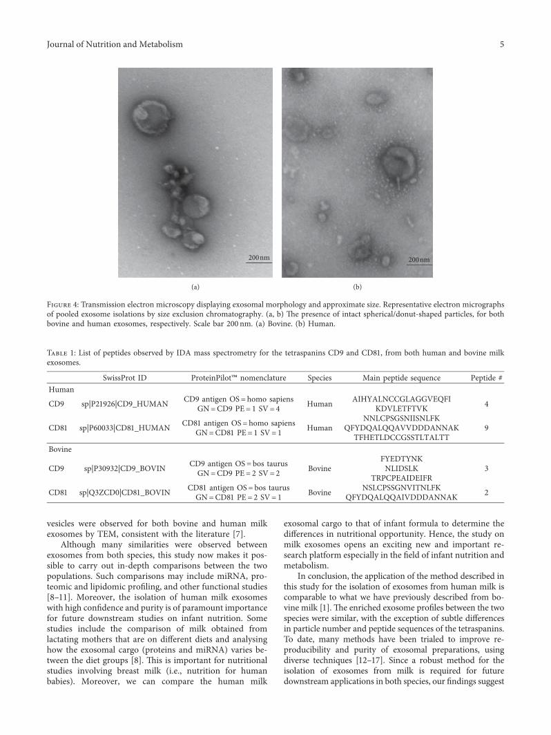
vesicles were observed for both bovine and human milkexosomes by TEM, consistent with the literature [7].
Although many similarities were observed betweenexosomes from both species, this study now makes it pos-sible to carry out in-depth comparisons between the twopopulations. Such comparisons may include miRNA, pro-teomic and lipidomic profiling, and other functional studies[8–11]. Moreover, the isolation of human milk exosomeswith high confidence and purity is of paramount importancefor future downstream studies on infant nutrition. Somestudies include the comparison of milk obtained fromlactating mothers that are on different diets and analysinghow the exosomal cargo (proteins and miRNA) varies be-tween the diet groups [8]. ,is is important for nutritionalstudies involving breast milk (i.e., nutrition for humanbabies). Moreover, we can compare the human milk
exosomal cargo to that of infant formula to determine thedifferences in nutritional opportunity. Hence, the study onmilk exosomes opens an exciting new and important re-search platform especially in the field of infant nutrition andmetabolism.
In conclusion, the application of the method described inthis study for the isolation of exosomes from human milk iscomparable to what we have previously described from bo-vine milk [1]. ,e enriched exosome profiles between the twospecies were similar, with the exception of subtle differencesin particle number and peptide sequences of the tetraspanins.To date, many methods have been trialed to improve re-producibility and purity of exosomal preparations, usingdiverse techniques [12–17]. Since a robust method for theisolation of exosomes from milk is required for futuredownstream applications in both species, our findings suggest
200nm
(a)
200nm
(b)
Figure 4: Transmission electron microscopy displaying exosomal morphology and approximate size. Representative electron micrographsof pooled exosome isolations by size exclusion chromatography. (a, b) ,e presence of intact spherical/donut-shaped particles, for bothbovine and human exosomes, respectively. Scale bar 200 nm. (a) Bovine. (b) Human.
Table 1: List of peptides observed by IDA mass spectrometry for the tetraspanins CD9 and CD81, from both human and bovine milkexosomes.
SwissProt ID ProteinPilot™ nomenclature Species Main peptide sequence Peptide #Human
CD9 sp|P21926|CD9_HUMAN CD9 antigen OS� homo sapiensGN�CD9 PE� 1 SV� 4 Human AIHYALNCCGLAGGVEQFI
KDVLETFTVK 4
CD81 sp|P60033|CD81_HUMAN CD81 antigen OS� homo sapiensGN�CD81 PE� 1 SV� 1 Human
NNLCPSGSNIISNLFKQFYDQALQQAVVDDDANNAK
TFHETLDCCGSSTLTALTT9
Bovine
CD9 sp|P30932|CD9_BOVIN CD9 antigen OS� bos taurusGN�CD9 PE� 2 SV� 2 Bovine
FYEDTYNKNLIDSLK
TRPCPEAIDEIFR3
CD81 sp|Q3ZCD0|CD81_BOVIN CD81 antigen OS� bos taurusGN�CD81 PE� 2 SV� 1 Bovine NSLCPSSGNVITNLFK
QFYDQALQQAIVDDDANNAK 2
Journal of Nutrition and Metabolism 5
a standardized methodology for milk exosome isolation cannow be established for human milk exosomes.
Data Availability
,e data used to support the findings of this study are in-cluded within the article and also available upon request.Since large data sets are not available, there is no datarepository.
Conflicts of Interest
,e authors declare no conflicts of interest.
Acknowledgments
,is project has been granted funding through the 2018Child and Adolescent Health Program Funding SupportScheme, Institute of Biomedical Innovation, QueenslandUniversity of Technology, to Yong Qin Koh. ,e authorswould also like to acknowledge the funding provided by theAustralian Research Council, ARC. ,e expertise of Prof.Michael McGowan and the technical staff at UQ GattonFarms was invaluable.
References
[1] K. Vaswani, Y. Q. Koh, F. B. Almughlliq, H. N. Peiris, andM. D.Mitchell, “Amethod for the isolation and enrichment ofpurified bovine milk exosomes,” Reproductive Biology, vol. 17,2017.
[2] T. Baranyai, K. Herczeg, Z. Onodi et al., “Isolation of exo-somes from blood plasma: qualitative and quantitativecomparison of ultracentrifugation and size exclusion chro-matography methods,” PLoS One, vol. 10, 2015.
[3] A. Gamez-Valero, M. Monguio-Tortajada, L. Carreras-Pla-nella, M. Franquesa, K. Beyer, and F. E. Borras, “Size-ex-clusion chromatography-based isolation minimally altersextracellular vesicles’ characteristics compared to pre-cipitating agents,” Scientific Reports, vol. 6, 2016.
[4] Y. Q. Koh, Exosomes and their potential roles in dairy cowfertility, Ph.D. thesis, Faculty of Medicine, ,e University ofQueensland, Brisbane, Queensland 4029, Australia, 2019.
[5] M. I. Zonneveld, A. R. Brisson, M. J. van Herwijnen et al.,“Recovery of extracellular vesicles from human breast milk isinfluenced by sample collection and vesicle isolation pro-cedures,” Journal of Extracellular Vesicles, vol. 3, 2014.
[6] C. Admyre, S. M. Johansson, K. R. Qazi et al., “Exosomes withimmune modulatory features are present in human breastmilk,” 'e Journal of Immunology, vol. 179, 2007.
[7] K. Blans, M. S. Hansen, L. V. Sorensen et al., “Pellet-freeisolation of human and bovine milk extracellular vesicles bysize-exclusion chromatography,” Journal of ExtracellularVesicles, vol. 6, 2017.
[8] H. Izumi, N. Kosaka, T. Shimizu, K. Sekine, T. Ochiya, andM. Takase, “Bovine milk contains microRNA and messengerRNA that are stable under degradative conditions,” Journal ofDairy Science, vol. 95, 2012.
[9] T. Hata, K. Murakami, H. Nakatani, Y. Yamamoto,T. Matsuda, and N. Aoki, “Isolation of bovine milk-derivedmicrovesicles carrying mRNAs andmicroRNAs,” Biochemicaland Biophysical Research Communications, vol. 396, 2010.
[10] M. J. van Herwijnen, M. I. Zonneveld, S. Goerdayal et al.,“Comprehensive proteomic analysis of human milk-derivedextracellular vesicles unveils a novel functional proteomedistinct from other milk components,” Molecular & CellularProteomics, vol. 15, 2016.
[11] T. A. Reinhardt, J. D. Lippolis, B. J. Nonnecke, and R. E. Sacco,“Bovine milk exosome proteome,” Journal of Proteomics,vol. 75, 2012.
[12] T. Yamada, Y. Inoshima, T. Matsuda, and N. Ishiguro,“Comparison of methods for isolating exosomes from bovinemilk,” Journal of Veterinary Medical Science, vol. 74, 2012.
[13] R. Munagala, F. Aqil, J. Jeyabalan, and R. C. Gupta, “Bovinemilk-derived exosomes for drug delivery,” Cancer Letters,vol. 371, 2016.
[14] P. Li, M. Kaslan, S. H. Lee, J. Yao, and Z. Gao, “Progress inexosome isolation techniques,” 'eranostics, vol. 7, 2017.
[15] F. Liu, O. Vermesh, V. Mani et al., “,e exosome total iso-lation chip,” ACS Nano, vol. 11, 2017.
[16] M. Somiya, Y. Yoshioka, and T. Ochiya, “Biocompatibility ofhighly purified bovine milk-derived extracellular vesicles,”Journal of Extracellular Vesicles, vol. 7, 2018.
[17] J. Webber and A. Clayton, “How pure are your vesicles?,”Journal of Extracellular Vesicles, vol. 2, 2013.
6 Journal of Nutrition and Metabolism
Stem Cells International
Hindawiwww.hindawi.com Volume 2018
Hindawiwww.hindawi.com Volume 2018
MEDIATORSINFLAMMATION
of
EndocrinologyInternational Journal of
Hindawiwww.hindawi.com Volume 2018
Hindawiwww.hindawi.com Volume 2018
Disease Markers
Hindawiwww.hindawi.com Volume 2018
BioMed Research International
OncologyJournal of
Hindawiwww.hindawi.com Volume 2013
Hindawiwww.hindawi.com Volume 2018
Oxidative Medicine and Cellular Longevity
Hindawiwww.hindawi.com Volume 2018
PPAR Research
Hindawi Publishing Corporation http://www.hindawi.com Volume 2013Hindawiwww.hindawi.com
The Scientific World Journal
Volume 2018
Immunology ResearchHindawiwww.hindawi.com Volume 2018
Journal of
ObesityJournal of
Hindawiwww.hindawi.com Volume 2018
Hindawiwww.hindawi.com Volume 2018
Computational and Mathematical Methods in Medicine
Hindawiwww.hindawi.com Volume 2018
Behavioural Neurology
OphthalmologyJournal of
Hindawiwww.hindawi.com Volume 2018
Diabetes ResearchJournal of
Hindawiwww.hindawi.com Volume 2018
Hindawiwww.hindawi.com Volume 2018
Research and TreatmentAIDS
Hindawiwww.hindawi.com Volume 2018
Gastroenterology Research and Practice
Hindawiwww.hindawi.com Volume 2018
Parkinson’s Disease
Evidence-Based Complementary andAlternative Medicine
Volume 2018Hindawiwww.hindawi.com
Submit your manuscripts atwww.hindawi.com