Embed Size (px)
Citation preview
ABSTRACT
A case of a pigmented calcifying cystic odontogenic tu-mor (CCOT) localized in the mandible occuring in a 16-year-old white female - is reported, and the characteris-tic histomorphologic features of CCOT and the pathoge-nesis of melanin and melanocytes present in the pigmen-ted type is discussed under the light of histochemicaland immunohistochemical data.
Key words: Odontogenic tumor, calcifying odontogeniccyst, pigment, melanin, melanocytes
ÖZET
On alt› yafl›nda bir kad›n hastada mandibula yerleflimlipigmentli tipte kalsifiye kistik odontojenik tümör olgu-su sunulmaktad›r. Kalsifiye kistik odontojenik tümöriçin tan›mlanm›fl karakteristik histomorfolojik özellik-ler ve ayr›ca pigmentli tipte mevcut melanin ve melano-sitlerin patogenezi histokimyasal ve immünhistokimya-sal veriler eflli¤inda tart›fl›lmaktad›r.
Anahtar sözcükler: Odontojenik tümör, kalsifiye odon-tojenik kist, pigment, melanin, melanosit
INTRODUCTION
The calcifying odontogenic cyst, renamedas calcifying cystic odontogenic tumor (CCOT)(1) was first described by Gorlin et al. (2) as adistinct entity, which has a histologic resemb-lance to cutaneous calcifying epithelioma ofMalherbe. In an analysis of 392 odontogenic tu-mors, Daley et al. (3) found that CCOT forms4.6% of all odontogenic tumors. The characte-ristic histology of CCOT includes an epitheliallining showing a well-defined basal layer of co-lumnar cells, a zone of loose edematous cellsbearing some resemblance to stellate reticulum,abnormal keratinization producing ghost cellsand a few small calcifications (1,4). A rare vari-ant of CCOT, in which melanin is present in theepithelium, can also be seen in literature (2,5-8).
A pigmented variant of CCOT is describedhere with its histomorphologic, histochemicaland immunohistochemical features.
CASE REPORT
Clinical SummaryA 16 year-old white female was referred to
Istanbul University, Istanbul Faculty of Den-tistry because of a painless swelling in her lefthalf of the mandible. Panoramic radiograms de-monstrated a well-defined unilocular radiolu-cent liquid containing cyst localised betweenleft canine and the second premolar in the man-dible (Figure 1). The lesion was enucleated andthe postoperative course was uneventful.
Pathological FindingsA well-demarcated, 2x1x1 cm cystic mass
was seen in gross examination. The cut surfacewas dark-brown in color. Light microscopic
A pigmented calcifying cystic odontogenictumor
Pigmentli kalsifiye kistik odontojenik tümör
Yasemin ÖZLÜK1, Yersu KAPRAN1, Ahmet Bülent KAT‹BO⁄LU2, Cem TANYEL2
Istanbul University, Istanbul Faculty of Medicine, Department of Pathology1, Istanbul University, Istanbul Faculty ofDentistry, Department of Oral Surgery2, ISTANBUL
Corresponding Author: Dr. Yasemin Özlük, ‹stanbul T›pFakültesi Temel Bilimler Binas› Patoloji Anabilim Dal›, 34390,Çapa-‹stanbul
111
Turkish Journal of Pathology 2007;23(2):111-115
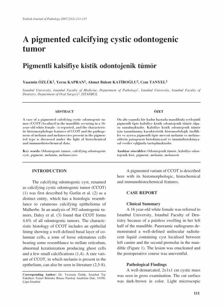
examination demonstrated a cyst displayingcharacteristic features for the CCOT, such as awell-defined basal cell layer, an epithelial com-ponent in varying thickness resembling stellatereticulum of enamel organ, eosinophilic ghostcells protruding into cystic cavity and small cal-cifications (Figure 2).
Abundant deposition of a dark-brown pig-
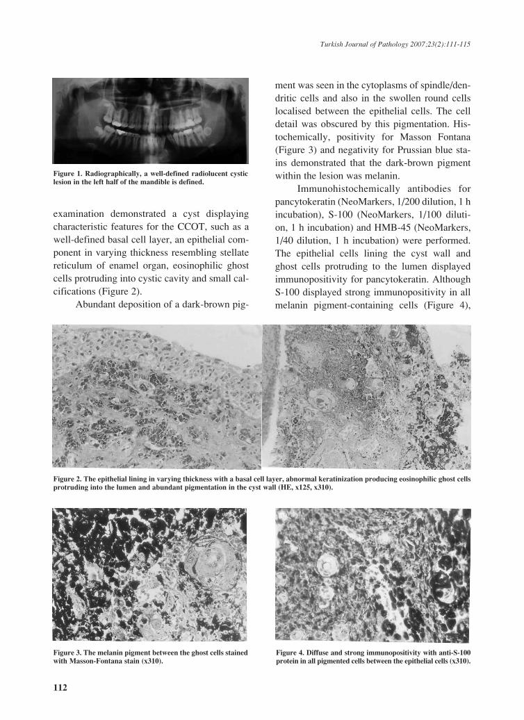
ment was seen in the cytoplasms of spindle/den-dritic cells and also in the swollen round cellslocalised between the epithelial cells. The celldetail was obscured by this pigmentation. His-tochemically, positivity for Masson Fontana(Figure 3) and negativity for Prussian blue sta-ins demonstrated that the dark-brown pigmentwithin the lesion was melanin.
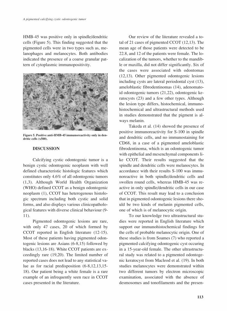
Immunohistochemically antibodies forpancytokeratin (NeoMarkers, 1/200 dilution, 1 hincubation), S-100 (NeoMarkers, 1/100 diluti-on, 1 h incubation) and HMB-45 (NeoMarkers,1/40 dilution, 1 h incubation) were performed.The epithelial cells lining the cyst wall andghost cells protruding to the lumen displayedimmunopositivity for pancytokeratin. AlthoughS-100 displayed strong immunopositivity in allmelanin pigment-containing cells (Figure 4),
Figure 2. The epithelial lining in varying thickness with a basal cell layer, abnormal keratinization producing eosinophilic ghost cellsprotruding into the lumen and abundant pigmentation in the cyst wall (HE, x125, x310).
Figure 3. The melanin pigment between the ghost cells stainedwith Masson-Fontana stain (x310).
Figure 4. Diffuse and strong immunopositivity with anti-S-100protein in all pigmented cells between the epithelial cells (x310).
Figure 1. Radiographically, a well-defined radiolucent cysticlesion in the left half of the mandible is defined.
112
Turkish Journal of Pathology 2007;23(2):111-115
HMB-45 was positive only in spindle/dendriticcells (Figure 5). This finding suggested that thepigmented cells were in two types such as, me-lanophages and melanocytes. Both antibodiesindicated the presence of a coarse granular pat-tern of cytoplasmic immunopositivity.
DISCUSSION
Calcifying cystic odontogenic tumor is abenign cystic odontogenic neoplasm with welldefined characteristic histologic features whichconstitutes only 4.6% of all odontogenic tumors(1,3). Although World Health Organization(WHO) defined CCOT as a benign odontogenicneoplasm (1), CCOT has heterogenous histolo-gic spectrum including both cystic and solidforms, and also displays various clinicopatholo-gical features with diverse clinical behaviour (9-11).
Pigmented odontogenic lesions are rare,with only 47 cases, 20 of which formed byCCOT reported in English literature (12-15).Most of these patients having pigmented odon-togenic lesions are Asians (6-8,15) followed byblacks (13,16-18). White CCOT patients are ex-ceedingly rare (19,20). The limited number ofreported cases does not lead to any statistical va-lue as for racial predisposition (6-8,12,13,15-18). Our patient being a white female is a rareexample of an infrequently seen race in CCOTcases presented in the literature.
Our review of the literature revealed a to-tal of 21 cases of pigmented CCOT (12,13). Themean age of those patients were detected to be22.8, and 12 of the patients were female. The lo-calization of the tumors, whether to the mandib-le or maxilla, did not differ significantly. Six ofthe cases were associated with odontomas(12,13). Other pigmented odontogenic lesionsincluding cysts are lateral periodontal cyst (13),ameloblastic fibrodentinomas (14), adenomato-id odontogenic tumors (21,22), odontogenic ke-ratocysts (23) and a few other types. Althoughthe lesion type differs, histochemical, immuno-histochemical and ultrastructural methods usedin studies demonstrated that the pigment is al-ways melanin.
Takeda et al. (14) showed the presence ofpositive immunoreactivity for S-100 in spindleand dendritic cells, and no immunostaining forCD68, in a case of a pigmented ameloblasticfibrodentinoma, which is an odontogenic tumorwith epithelial and mesenchymal components li-ke CCOT. Their results suggested that thespindle and dendritic cells were melanocytes. Inaccordance with their results S-100 was immu-noreactive in both spindle/dendritic cells andswollen round cells, whereas HMB-45 was re-active in only spindle/dendritic cells in our caseof CCOT. This result may lead to a conclusionthat in pigmented odontogenic lesions there sho-uld be two kinds of melanin pigmented cells,one of which is of melanocytic origin.
To our knowledge two ultrastructural stu-dies were reported in English literature whichsupport our immunohistochemical findings forthe cells of probable melanocytic origin. One ofthese studies is from Soames (7) who reported apigmented calcifying odontogenic cyst occuringin a 15-year-old female. The other ultrastructu-ral study was related to a pigmented odontoge-nic keratocyst from Macleod et al. (19). In bothstudies melanocytes were demonstrated withintwo different tumors by electron microscopicexamination, associated with the absence ofdesmosomes and tonofilaments and the presen-
Figure 5. Positive anti-HMB-45 immunoreactivity only in den-dritic cells (x500).
113
A pigmented calcifying cystic odontogenic tumor
ce of melanised melanosomes and/or premela-nosomes in various stages of development incytoplasms and cytoplasmic processes betweensurrounding cells (7,19).
The origin of melanocytes present in odon-togenic lesions is not known and their patholo-gic significance is still of interest. An interestingobservation is that melanin containing intra-os-seous lesions, except malignant melanoma me-tastases, are all localized in the jaw bone. Theprobable pathogenesis of this localization is spe-culative. It is generally known that melanocytesare normally present in clinically non-pigmen-ted oral mucosa, therefore the presence of mela-nocytes can be anticipated in odontogenic lesi-ons since the dental lamina originates from theprimitive oral lining. Lawson et al. (24) havediscovered melanocytes in the dental lamina andouter enamel epithelium during 12-18 weeks ofgestation which were apparently more commonin blacks than in whites, suggesting a racial pre-disposition for pigmented odontogenic lesions.
Another theory of melanocytic origin inodontogenic lesions is the migration of mela-nocytes through the mesenchyme rather than theectoderm (25). Takeda et al. (25) displayed me-lanocytes in mesenchymal tissue around thedental anlage in dog fetuses. They could notshow any melanocytes both in the oral epitheli-um and in the epithelial element of the dentalanlage.
It is a striking feature that all pigmentedodontogenic lesions, but not cysts, have an epit-helial component accompanied by a mesench-ymal component with dentin formation and cal-cification. Some factors such as the presence ofmesenchymal component or the hard tissue for-mation, that can activate the melanocytes alre-ady present in the epithelium might have playeda role. More future investigations must be per-formed to illuminate the origin of melanocytesin odontogenic lesions.
REFERENCES
1. Prætorius F, Ledesma-Montes. Calcifying cystic odon-togenic tumour. In: Barnes L. Eveson JW, Reichart P,Sidransky D, eds. Pathology and Genetics of Head andNeck Tumors. Lyon: IARC Pres, 2005; p.313.
2. Gorlin RJ, Pindborg JJ, Redman RS, Williamson JJ,Hansen LS. The calcifying odontogenic cyst: a new en-tity and possible analogue of the cutaneous calcifyingepithelioma of Malherbe. Cancer 1964;17:723-729.
3. Daley TD, Wysocki GP, Pringle GA. Relative inciden-ce of odontogenic tumors and oral and jaw cysts in aCanadian population. Oral Surg Oral Med Oral Pathol1994;77:276-280.
4. Cawson RA, Binnie WH, Speight PM, Barrett AW,Wright JM. Calcifying odontogenic cyst (odontogenicghost cell cyst and tumor, Gorlin cyst). In: CawsonRA, Binnie WH, Speight PM, Barrett AW, Wright JM(eds). Lucas’s Pathology of Tumors of the Oral Tissu-es. 5th ed. London, Churchill Livingstone 1998, p.59-63.
5. Buchner A. The central (intraosseous) calcifying odon-togenic cyst: an analysis of 215 cases. J Oral Maxillo-fac Surg 1991;49:330-339.
6. Takeda Y, Kuroda M, Suzuki A, Fujioka Y. Pigmentedvariant of calcifying odontogenic cyst: report of an ad-ditional case and review of the literature. Acta Pathol1985;35:1023-1027.
7. Soames JV. A pigmented calcifying odontogenic cyst.Oral Surg Oral Med Oral Pathol 1982;53:395-400.
8. Takeda Y, Suzuki A, Yamamoto H. Histopathologicstudy of epithelial components in the connective tissu-e wall of unilocular type of calcifying odontogeniccyst. J Oral Pathol Med 1990;19:108-113.
9. Li TJ, Yu SF. Clinicopathologic spectrum of the so-called calcifying odontogenic cysts: a study of 21 intra-osseous cases with reconsideration of the terminologyand classification. Am J Surg Pathol 2003;27:372-384.
10. Hong SP, Ellis GL, Hartman KS. Calcifying odontoge-nic cyst: a review of ninety-two cases with reevaluati-on of their nature as cysts or neoplasms, the nature ofghost cells, and subclassification. Oral Surg Oral MedOral Pathol 1991;72:56-64.
11. Wright BA, Bhardwaj AK, Murphy D. Recurrent cal-cifying odontogenic cyst. Oral Surg 1984;58:579-583.
12. Han PP, Nagatsuka H, Siar CH, Tsujigiwa H, GunduzM, Tamamura R, et al. A pigmented calcifying cysticodontogenic tumor associated with compound odonto-ma: a case report and review of literature. Head FaceMed 2007;3:35.
13. Buchner A, David R, Carpenter W, Leider A. Pigmen-ted lateral periodontal cyst and other pigmented odon-togenic lesions. Oral Dis 1996;2:299-302.
14. Takeda Y, Sato H, Satoh M, Nakamura S, YamamotoH. Pigmented ameloblastic fibrodentinoma: a novelmelanin-pigmented intraosseous odontogenic lesion.Virchow Arch 2000;437:454-458.
15. Takeda Y, Yamomoto H. Case report of a pigmenteddentigerous cyst and a review of the literature on pig-mented odontogenic cysts. J Oral Sci 2000;42:43-46.
16. Lurie HI. Congenital melanocarcinoma, melanoticadamantinoma, retinal anlage tumor, progonoma, and
114
Turkish Journal of Pathology 2007;23(2):111-115
pigmented epulis of infancy. Cancer 1961;14:1090-1108.
17. Petri WH, Stump TE. Calcifying odontogenic cyst: re-port of three cases. J Oral Surg 1976;34:1105-1108.
18. Richardson JF, Balogh K, Merk F, Booth D. Pigmen-ted odontogenic tumor of jawbone: a previously undes-cribed neoplastic potential. Cancer 1974;34:1244-1251.
19. Macleod RI, Fanibunda KB, Soames JV. A pigmentedodontogenic keratocyst. Br J Oral Maxillofac Surg1985;23:216-219.
20. Abrams AM, Howell FY. The calcifying odontogeniccyst. Oral Surg 1968;25:594-606.
21. Takeda Y. Pigmented adenomatoid odontogenic tu-mor, report of an undescribed case and review of the li-terature of pigmented intra-osseous odontogenic lesi-
ons. Virchow Arch 1989;415:571-575.22. Warter A, George-Diolomb G, Chazal M, Ango A.
Melanin in a dentigerous cyst and associated adenoma-toid odontogenic tumor. Cancer 1990;66:786-788.
23. Brannon RB. The odontogenic keratocyst: a clinicopat-hologic study of 312 cases. Oral Surg 1997;43:233-255.
24. Lawson W, Abaci IF, Zak FG. Studies on melanocytes.V. The presence of melanocytes in the human dentalprimordium: an explanation for pigmented lesions ofthe jaws. Oral Surg Oral Med Oral Pathol1976;42:375-380.
25. Takeda Y, Uyeno K. The existence and distribution ofmelanocytes in the periodontal ligament of the mongreldog. Arch Histol Cytol 1993;56:417-421.
115
A pigmented calcifying cystic odontogenic tumor


![Ghost cell odontogenic carcinoma: A rare case report and ... · PDF fileGhost cell odontogenic carcinoma [GCOC] is a rare malignant odontogenic epithelial tumor with features of calcifying](https://img.pdfslide.net/doc/110x75/5a9cd2d97f8b9a335c8b5251/ghost-cell-odontogenic-carcinoma-a-rare-case-report-and-cell-odontogenic-carcinoma.jpg)


![Short Case Report · Calcifying odontogenic cysts (COCs), or Gorlin’s cysts, are rare lesions that account for less than 1% of all odontogenic cysts [1]. This article details two](https://img.pdfslide.net/doc/110x75/5fea60342725d22f6c4de3eb/short-case-report-calcifying-odontogenic-cysts-cocs-or-gorlinas-cysts-are.jpg)