Embed Size (px)
Citation preview

A Quick Tour of Congenital Heart Disease
Chris Longhurst, MDTuesday, April 18, 2023
Introduction Present in 0.8% of North American
and European children Most common category of
congenital structural malformation Commonly divided into
noncyanotic (L R) and cyanotic (R L) categories based on direction of shunting
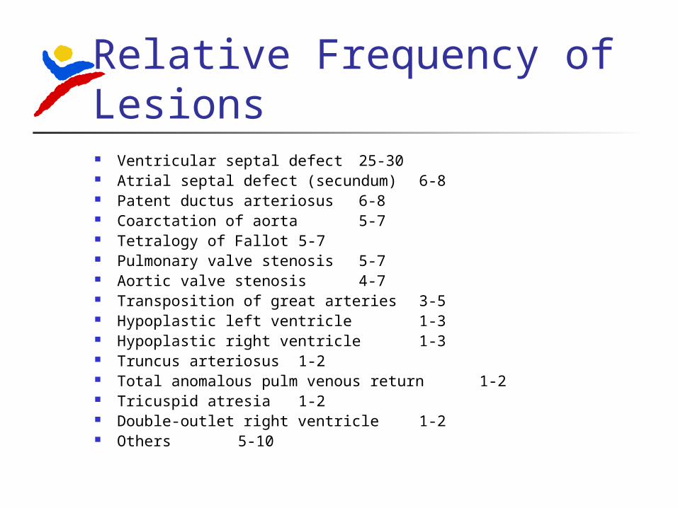
Relative Frequency of Lesions Ventricular septal defect 25-30 Atrial septal defect (secundum) 6-8 Patent ductus arteriosus 6-8 Coarctation of aorta 5-7 Tetralogy of Fallot 5-7 Pulmonary valve stenosis 5-7 Aortic valve stenosis 4-7 Transposition of great arteries 3-5 Hypoplastic left ventricle 1-3 Hypoplastic right ventricle 1-3 Truncus arteriosus 1-2 Total anomalous pulm venous return 1-2 Tricuspid atresia 1-2 Double-outlet right ventricle 1-2 Others 5-10
Noncyanotic CHD (L R) Atrial septal defects (ASD) Ventricular septal defects (VSD) Patent ductus arteriosus (PDA) Obstruction to blood flow
Pulmonic stenosis (PS) Aortic stenosis (AS) Aortic coarctation
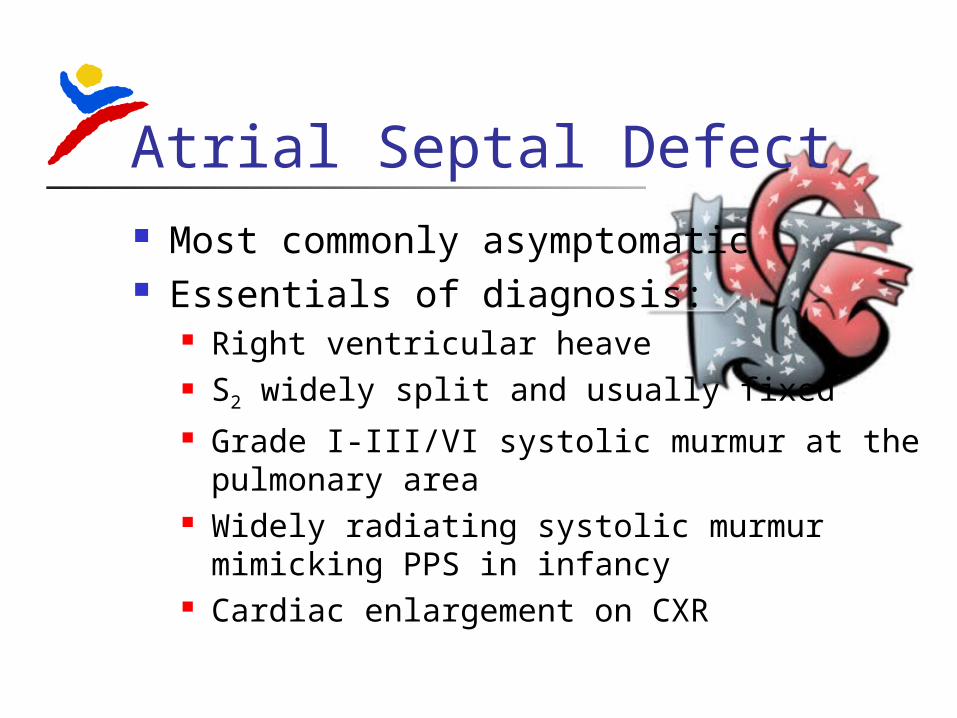
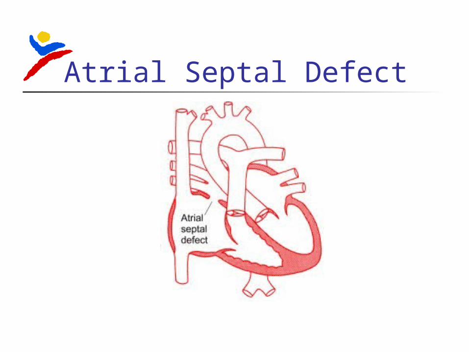
Atrial Septal Defect Most commonly asymptomatic Essentials of diagnosis:
Right ventricular heave S2 widely split and usually fixed Grade I-III/VI systolic murmur at the
pulmonary area Widely radiating systolic murmur mimicking
PPS in infancy Cardiac enlargement on CXR
Atrial Septal Defect
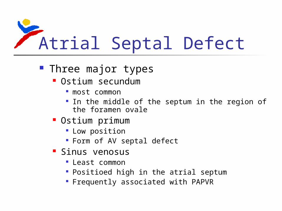
Atrial Septal Defect Three major types
Ostium secundum most common In the middle of the septum in the region of the
foramen ovale Ostium primum
Low position Form of AV septal defect
Sinus venosus Least common Positioed high in the atrial septum Frequently associated with PAPVR
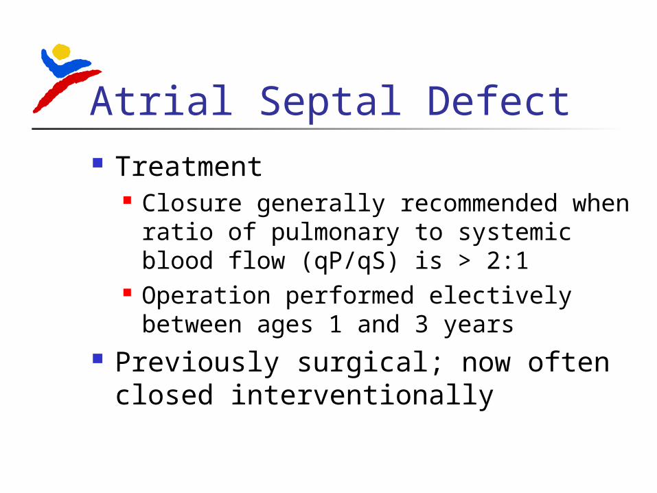
Atrial Septal Defect Treatment
Closure generally recommended when ratio of pulmonary to systemic blood flow (qP/qS) is > 2:1
Operation performed electively between ages 1 and 3 years
Previously surgical; now often closed interventionally
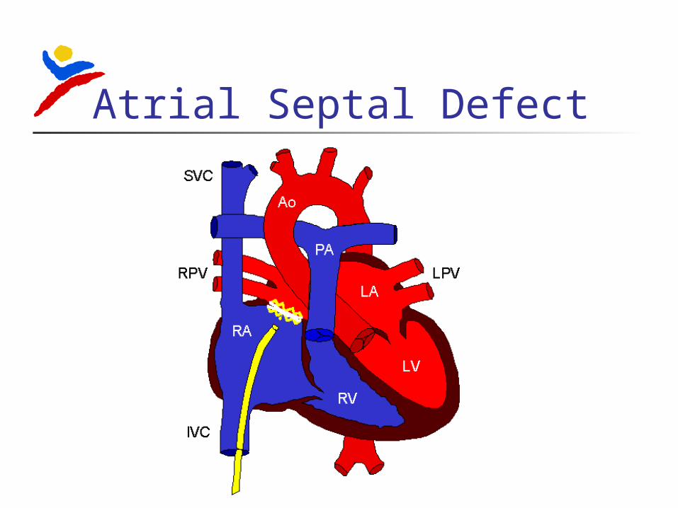
Atrial Septal Defect

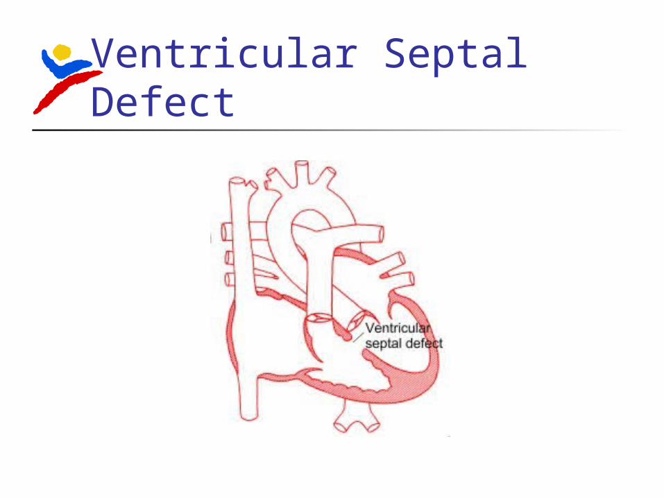
Ventricular Septal Defect Single most common congenital
heart malformation, accounting for almost 30% of all CHD
Defects can occur in both the membranous portion of the septum (most common) and the muscular portion
Ventricular Septal Defect

Ventricular Septal Defect Three major types Small, hemodynamically
insignificant Between 80% and 85% of all
VSDs < 3 mm in diameter All close spontanously
50% by 2 years 90% by 6 years 10% during school years
Muscular close sooner than membranous
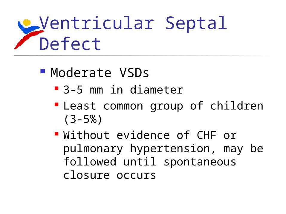
Ventricular Septal Defect Moderate VSDs
3-5 mm in diameter Least common group of children (3-
5%) Without evidence of CHF or
pulmonary hypertension, may be followed until spontaneous closure occurs

Ventricular Septal Defect Large VSDs with normal PVR
6-10 mm in diameter Usually requires surgery, otherwise… Will develop CHF and FTT by age 3-6
months
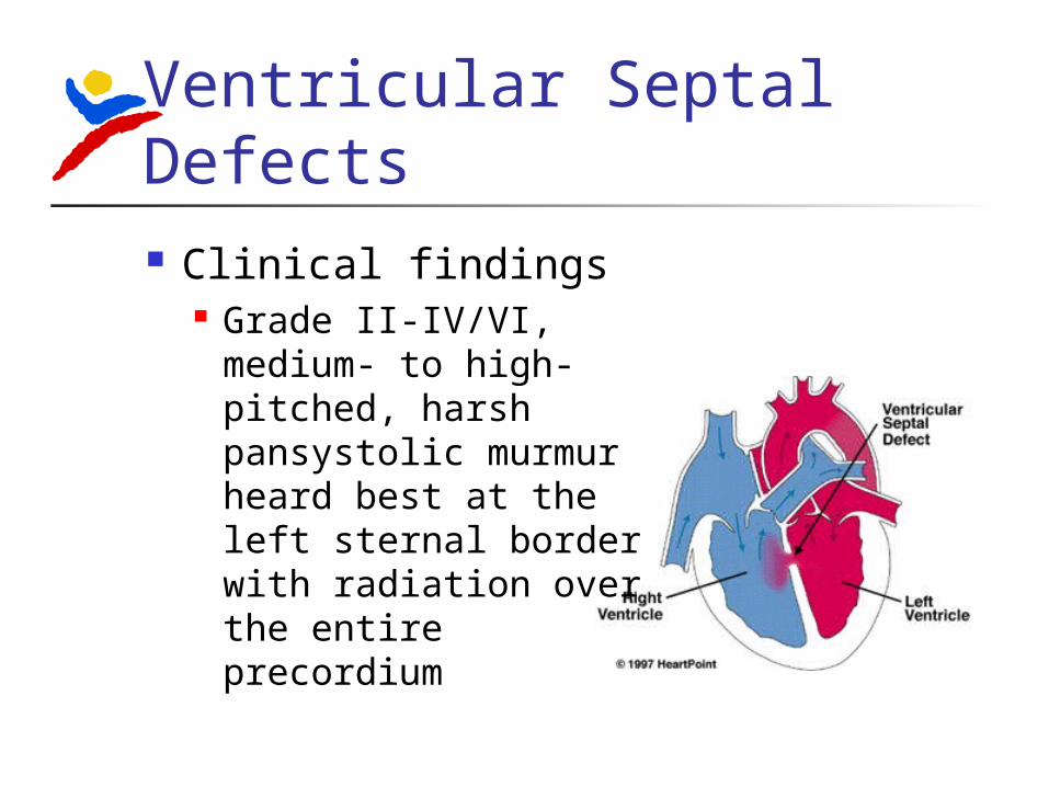
Ventricular Septal Defects Clinical findings
Grade II-IV/VI, medium- to high-pitched, harsh pansystolic murmur heard best at the left sternal border with radiation over the entire precordium
Ventricular Septal Defect Treatment
Indicated for closure of a VSD associated with CHF and FTT or pulmonary hypertension
Patients with cardiomegaly, poor growth, poor exercise tolerance, or other clinical abnormalities and a qP/qS > 2:1 typically undergo surgical repair at 3-6 mo
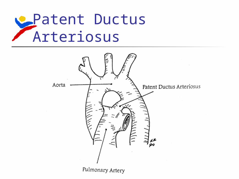
Patent Ductus Arteriosus Persistence of normal fetal vessel joining
the pulmonary artery to the aorta Closes spontaneously in normal term
infants at 3-5 days of age Epi facts
Accounts for about 10% of all cases of CHD Higher incidence of PDA in infants born at
high altitudes (> 10,000 feet) More common in females
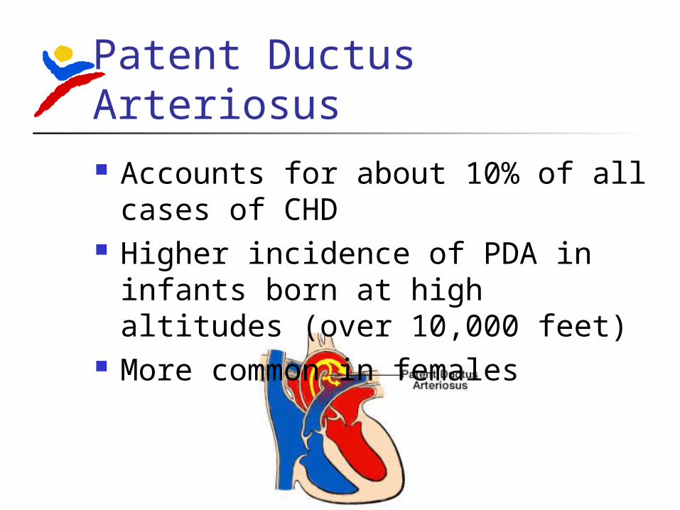
Patent Ductus Arteriosus Accounts for about 10% of all
cases of CHD Higher incidence of PDA in infants
born at high altitudes (over 10,000 feet)
More common in females
Patent Ductus Arteriosus
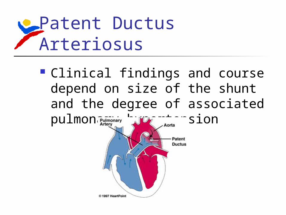
Patent Ductus Arteriosus Clinical findings and course
depend on size of the shunt and the degree of associated pulmonary hypertension

Patent Ductus Arteriosus Pulses are bounding and pulse
pressure is widened Characteristically has a rough
“machinery” murmur which peaks at S2 and becomes a decrescendo murmur and fades before the S1
Patent Ductus Arteriosus Treatment consists of surgical
correction when the PDA is large except in patients with pulmonary vascular obstructive disease
Transcatheter closure of small defects has become standard therapy
In preterm infants indomethacin is used (80-90% success in infants > 1200 grams)
Cyanotic CHD (R L) Tetralogy of Fallot (TOF) Tricuspid atresia (TA) Total anomalous pulmonary venous
return (TAPVR) Truncus arteriosus Transposition of the great vessels Hypoplastic left heart syndrome (HLH) Pulmonary atresia (PA) / critical PS Double outlet right ventricle (DORV)
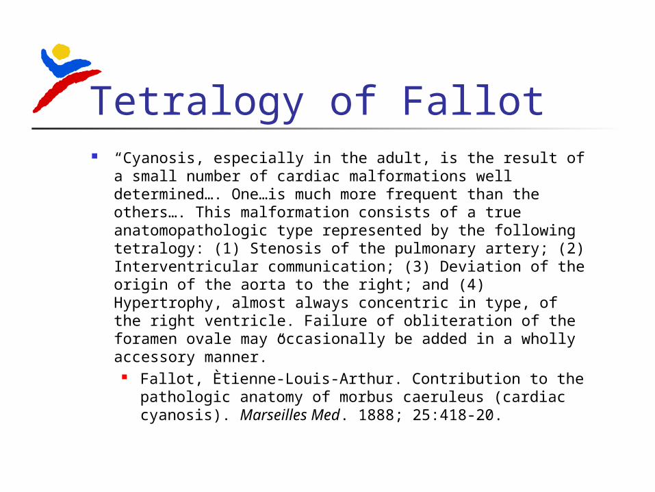
Tetralogy of Fallot “Cyanosis, especially in the adult, is the result of a small number
of cardiac malformations well determined…. One…is much more frequent than the others…. This malformation consists of a true anatomopathologic type represented by the following tetralogy: (1) Stenosis of the pulmonary artery; (2) Interventricular communication; (3) Deviation of the origin of the aorta to the right; and (4) Hypertrophy, almost always concentric in type, of the right ventricle. Failure of obliteration of the foramen ovale may occasionally be added in a wholly accessory manner.”
Fallot, Ètienne-Louis-Arthur. Contribution to the pathologic anatomy of morbus caeruleus (cardiac cyanosis). Marseilles Med. 1888; 25:418-20.

Tetralogy of Fallot
Tetralogy of Fallot Most common cyanotic lesion (7 to
10% of all CHD) Typical features
Cyanosis after the neonatal period Hypoxemic spells during infancy Right-sided aortic arch in 25% of all
patients Systlic ejection murmur at the upper
LSB
Tetralogy of Fallot Clinical findings vary depending on
degree of RVOFT obstruction Most patients are cyanotic by 4
months and it is usually progressive
Hypoxemic spells (“tet spells”) are one of the hallmarks of severe tetralogy

Tetralogy of Fallot
Tetralogy of Fallot Tet spells most commonly start
around 4 to 6 months of age and are charcterized by
1. Sudden onset or deepening of cyanosis2. Sudden onset of dyspnea3. Alterations of consciousness4. Decrease in intensity of systolic murmur
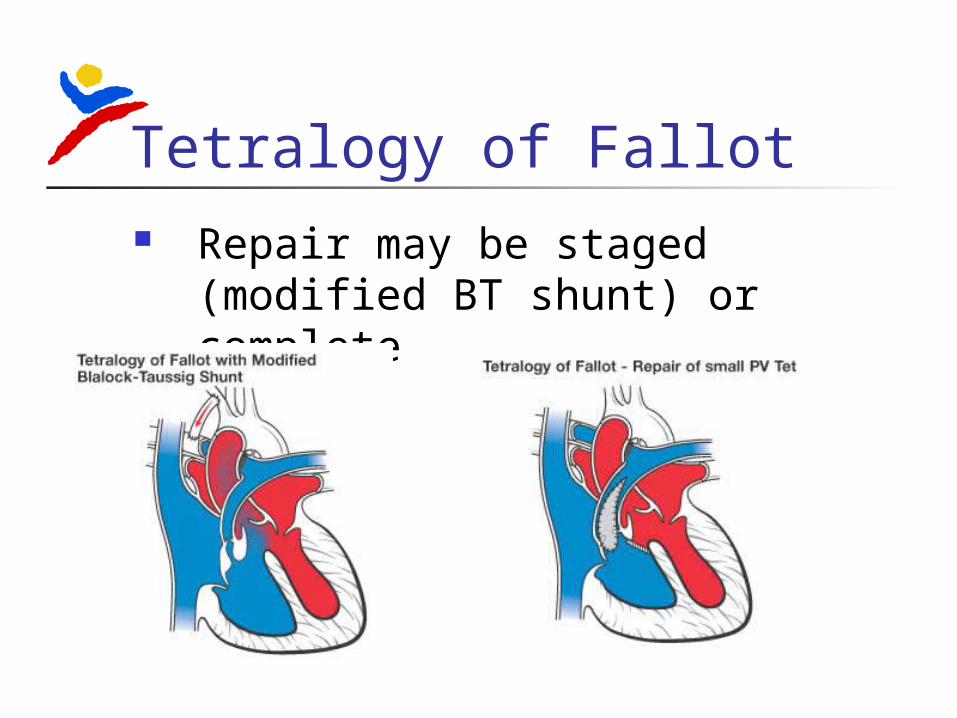
Tetralogy of Fallot Repair may be staged (modified
BT shunt) or complete