Embed Size (px)
Citation preview

METHODOLOGY Open Access
A robust and cost-effective approach tosequence and analyze complete genomesof small RNA virusesKiril M. Dimitrov1†, Poonam Sharma1†, Jeremy D. Volkening2, Iryna V. Goraichuk1,3, Abdul Wajid4,5,Shafqat Fatima Rehmani4, Asma Basharat4, Ismaila Shittu6, Tony M. Joannis6, Patti J. Miller1
and Claudio L. Afonso1*
Abstract
Background: Next-generation sequencing (NGS) allows ultra-deep sequencing of nucleic acids. The use ofsequence-independent amplification of viral nucleic acids without utilization of target-specific primers providesadvantages over traditional sequencing methods and allows detection of unsuspected variants and co-infectingagents. However, NGS is not widely used for small RNA viruses because of incorrectly perceived cost estimatesand inefficient utilization of freely available bioinformatics tools.
Methods: In this study, we have utilized NGS-based random sequencing of total RNA combined with barcodemultiplexing of libraries to quickly, effectively and simultaneously characterize the genomic sequences of multipleavian paramyxoviruses. Thirty libraries were prepared from diagnostic samples amplified in allantoic fluids and theirtotal RNAs were sequenced in a single flow cell on an Illumina MiSeq instrument. After digital normalization, datawere assembled using the MIRA assembler within a customized workflow on the Galaxy platform.
Results: Twenty-eight avian paramyxovirus 1 (APMV-1), one APMV-13, four avian influenza and two infectiousbronchitis virus complete or nearly complete genome sequences were obtained from the single run.The 29 avian paramyxovirus genomes displayed 99.6% mean coverage based on bases with Phred quality scores of30 or more. The lower and upper quartiles of sample median depth per position for those 29 samples were 2984and 6894, respectively, indicating coverage across samples sufficient for deep variant analysis. Sample processingand library preparation took approximately 25–30 h, the sequencing run took 39 h, and processing throughthe Galaxy workflow took approximately 2–3 h. The cost of all steps, excluding labor, was estimated to be106 USD per sample.
Conclusions: This work describes an efficient multiplexing NGS approach, a detailed analysis workflow, andcustomized tools for the characterization of the genomes of RNA viruses. The combination of multiplexingNGS technology with the Galaxy workflow platform resulted in a fast, user-friendly, and cost-efficient protocolfor the simultaneous characterization of multiple full-length viral genomes. Twenty-nine full-length or near-full-length APMV genomes with a high median depth were successfully sequenced out of 30 samples. Theapplied de novo assembly approach also allowed identification of mixed viral populations in some of thesamples.
Keywords: Newcastle disease virus, Next-generation sequencing, Multiplexing, Galaxy, De novo assembly,Multiplexing, Complete genomes, Mixed infection, Avian paramyxovirus
* Correspondence: [email protected]†Equal contributors1Exotic and Emerging Avian Viral Diseases Research Unit, Southeast PoultryResearch Laboratory, US National Poultry Research Center, AgriculturalResearch Service, USDA, 934 College Station Road, Athens, GA 30605, USAFull list of author information is available at the end of the article
© The Author(s). 2017 Open Access This article is distributed under the terms of the Creative Commons Attribution 4.0International License (http://creativecommons.org/licenses/by/4.0/), which permits unrestricted use, distribution, andreproduction in any medium, provided you give appropriate credit to the original author(s) and the source, provide a link tothe Creative Commons license, and indicate if changes were made. The Creative Commons Public Domain Dedication waiver(http://creativecommons.org/publicdomain/zero/1.0/) applies to the data made available in this article, unless otherwise stated.
Dimitrov et al. Virology Journal (2017) 14:72 DOI 10.1186/s12985-017-0741-5

BackgroundConventional laboratory methods like enzyme-linkedimmunosorbent assay, nucleic acid hybridization tech-nique, and polymerase chain reaction (PCR) are allcommon and inexpensive diagnostic and research toolsutilized in virology [1]. However, as these assays arehighly dependent on reagents (primers, probes, anti-bodies) developed from previously known and character-ized viruses, they might be ineffective for the identificationof new viral variants, new pathogens or a mixed popula-tion of pathogens that have high genetic divergence fromthose described previously [1, 2]. Next-generation sequen-cing (NGS) technologies enable large numbers of samplesto undergo parallel sequencing, and can be used for thedetection and characterization of multiple agents fromone sample. The use of sequence-independent amplifica-tion of viral nucleic acids eliminates the need for priorknowledge of genomic sequences and provides advantagesover traditional methods such as PCR amplification ormicroarray hybridization dependent on target-specificprimers [2, 3]. NGS technologies allow screening ofclinical and environmental samples for the presence ofviral pathogens, including previously unknown viruses [4].This has led to the discovery of numerous viral pathogens[4–6], including 2009 pandemic influenza A, a novel pegi-virus, Canine bocavirus 3, and a novel hepacivirus [7–10].The majority of previously employed techniques used
virus enrichment prior to cDNA synthesis and librarypreparation, or used specific primers for amplification.Virion enrichment steps such as centrifugation, poly-ethylene glycol precipitation, ultrafiltration, chloroformtreatment or nuclease treatment have been used forRNA viruses [3, 11, 12]. Other techniques, such asgDNA depletion and host RNA depletion to enrich forviral RNA [13] and DNase pretreatment of the allantoicfluid to enrich for viral particles, have also beendescribed [14]. Different methods have been employed forproducing cDNA following enrichment, including se-quence-independent single primer amplification(SISPA) and universal primers which have been usedfor sequencing RNA viruses [4, 15] including Newcas-tle disease virus (NDV) [11] and avian paramyxovirus(APMV) 4 and 6 [16].The genus Avulavirus of the family Paramyxoviridae,
order Mononegavirales, consists of 14 known avian para-myxovirus serotypes (APMV 1 – 14) [17–19]. Of these,APMV-1, synonymous with Newcastle disease virus, isthe most widely characterized and studied due to theeconomic importance of Newcastle disease (ND) causedby virulent strains of the virus. Newcastle disease is oneof the most significant poultry diseases and infects bothwild and domestic avian hosts. NDV has a single-stranded, non-segmented, negative-sense RNA genomeconsisting of six genes in order of 3’ to 5’: nucleocapsid
(NP), phosphoprotein (P), matrix (M), fusion (F),hemagglutinin-neuraminidase (HN), and polymerase (L),coding for these six structural proteins and at least oneadditional V protein [20–22]. Newcastle disease viruseshave three genome sizes – 15186, 15192 and 15198 nucle-otides, and are genetically grouped into two divergentclasses that are further classified in genotypes [23–25].Newcastle disease viruses are constantly evolving
and different genetic groups undergo simultaneousevolutionary changes in different geographical loca-tions [22, 26] making the available genetic makeupinformation outdated. These evolutionary changespresent challenges for prompt diagnosis. Some currentlyvalidated methods are target-oriented and might fail to de-tect new viral genetic variants [27–29]. Lack of completegenetic information for many NDV isolates further ham-pers the better understanding of Newcastle disease evolu-tion and epidemiology. Furthermore, mixed viral infectionare not uncommon in animals, and in the case of poultry,they are quite frequent [30]. Efficient and accurate identi-fication of these pathogens is essential for the develop-ment of adequate disease control strategies. Thesechallenges require an approach that provides de novo,rapid and high-quality genetic characterization of full-length viral genomes.Until recently, genome sequencing of small RNA
viruses, including NDV, has been performed using over-lapping genome amplification with primer pairs. Thisapproach is laborious, depends on preexisting informa-tion, and produces very low depth. NGS advances pro-vide tools for deep sequencing of multiple viral strains ina short time. However, the simultaneous cost- and time-effective sequencing and characterization of a largenumber of NDV genomes has not yet been reported.The aim of the current study was to utilize sequence-independent NGS technologies applied to viral nucleicacids for the simultaneous and rapid characterization ofmultiple NDV genomes. We demonstrate a straightfor-ward, efficient protocol for multiplexed sequencing usinga single flow cell on the Illumina MiSeq platformcoupled with a detailed customized Galaxy workflow forde novo assembly that allows for quick and accurategeneration of near-full-length, or full-length, genome se-quences of dozens of isolates, simultaneously. Further-more, we report the efficient detection and completesequencing of contaminant RNA viruses.
MethodsVirus propagationTwenty nine NDV and one APMV-13 isolates were sub-mitted to the Southeast Poultry Research Laboratory ofthe USDA in Athens, Georgia, USA. The viruses wereisolated in Pakistan (n = 15), Nigeria (n = 9) and Ukraine(n = 6) between 2003 and 2015. Viruses were propagated
Dimitrov et al. Virology Journal (2017) 14:72 Page 2 of 14

in 9-to-11-day-old specific-pathogen-free (SPF) embryo-nating chicken eggs [31]. The background informationof the 30 isolates used in the study is summarized inAdditional file 1: Table S1.
RNA isolationRNA from each sample was extracted from allantoicfluids. Two milliliters (equal volumes of 0.25 ml) of eachsample were aliquoted into 8 microtubes, each contain-ing 0.75 ml of TRIZOL LS (Invitrogen, USA). After5 min of incubation, 0.2 ml of chloroform was added toeach tube and shaken vigorously. After 10 min ofadditional incubation at room temperature, tubes werecentrifuged at 12000 × g for 15 min at 4 °C. The aqueousphase from all eight tubes was removed and pooled foreach sample. Two milliliters of aqueous layer of eachsample were treated with 4 μl Turbo DNase 2U/μl(Ambion, USA) for 15 min at 37 °C and then placed onice. The extraction proceeded using the QIAamp® ViralRNA Mini Kit (Qiagen, USA) according to the manufac-turer’s instructions. Briefly, DNase-treated aqueousphase was passed through a spin column for RNA ab-sorption on the QIAamp silica membrane followed bywashing with 2 ml of provided buffers AW1 and AW2using the QIAvac 24 Plus vacuum manifold (Qiagen,USA). The RNA was eluted in 50 μl buffer AVE. ElutedRNA was quantified using a Qubit® RNA HS Assay Kitin a Qubit® fluorometer (ThermoFisher Scientific, USA)and stored at -20 °C until further use.
NDV RNA captureA set of three biotinylated oligonucleotides designedfrom consensus of alignment of 330 available NDVgenomes were used for NDV RNA capture (Oligo 1 –5’- AGA GAA TCT GTG AGG TAC GA/3Bio -3’ atnucleotide position 8; Oligo 2 – 5’ -TTC TCA AGTCAT CGT GAC AG/3Bio -3’ at position 5905; Oligo 3 –5’ - CCC TGC ATC TCT CTA CAG/3Bio -3’ at position12226) (GenBank accession number AF431744). RNAcapture reactions were performed using 50 μl RNA incu-bated with 167 μl 6X saline-sodium phosphate-EDTAbuffer (900 mM NaCl, 60 mM NaH2PO4, 60 mMNa2EDTA), 2 μl RNaseOUT 40 U/μl (Invitrogen, USA)and 1.5 μl 100 μM mix of the three primers. Reactionswere performed at 70 °C for 5 min, followed by 15 minat 55 °C. Two hundred fifty microliters of 1X bindingand wash buffer solution (2 M NaCl) containing 12.5 μl(0.05%) Sera-Mag beads (magnetic streptavidin-coatedbeads, GE Healthcare Life Sciences, USA) was preparedfor each sample and mixed with the capture reactionproducts for binding. Washing was done on a magneticstand with 500 μl of 0.5X binding and wash buffer onetime and twice with bead wash buffer (5 M NaCl, 1 MTris-HCl [pH 7.5], 0.5 M EDTA, and 0.01% Tween® 20)
to remove unbound RNA. Viral RNA was recovered byadding 19 μl of 10 mM Tris-HCl (pH 7.5) to the beadmixture and incubating at 65 °C for 5 min followed bycooling on ice until the next step. In a separate experi-ment, a comparison with three known NDV was per-formed and the libraries were prepared side-by-side withand without the capture step, while all remaining stepsof the library preparation were identical.
Reverse transcriptionReverse transcription reactions were performed usingthe M-MLV Reverse Transcriptase (Moloney MurineLeukemia Virus Reverse Transcriptase, Invitrogen, USA)and 10 μl RNA (concentration varied from below250 pg/μl to 55 ng/μl), 1.0 μl Random Primers mix(3 μg/μl) (Invitrogen, USA) and following manufacturer’sinstruction. The cDNA products were purified using60 μl Agencourt® RNAClean® XP beads (BeckmanCoulter, USA) as per manufacturer’s instructions. Puri-fied cDNA were recovered in 15 μl of 10 mM Tris-Cl(pH 7.5) and quantified using a Qubit® ssDNA Assay Kit(ThermoFisher Scientific, USA) on the Qubit®fluorometer.
Library preparation, quality and quantity assessmentDNA libraries (n = 30, one library for each sample), wereprepared for deep sequencing using 1 ng purified cDNA(0.2 ng/μl in molecular grade water) and the Nextera XTDNA Library Preparation Kit (Illumina, USA) followingthe manufacturer's protocol. The resulting dsDNA prod-ucts were purified with 30 μl Agencourt® AMPure® XPbeads (Beckman Coulter, USA) by incubating at roomtemperature for 5 min followed by two washes with200 μl 80% ethanol on a magnetic stand. The tubes withthe beads were air dried for 10 to 15 min at roomtemperature and the library products were recovered in52.5 μl of Resuspension Buffer. The quality and fragmentlength distribution for each library was assessed usingthe Agilent High Sensitivity DNA Kit (Agilent Technolo-gies, USA) on the Agilent 2100 Bioanalyzer (AgilentTechnologies, Germany). The Qubit® fluorometer andthe Qubit®dsDNA HS Assay Kit were used for measuringthe concentration of the libraries.
Equimolar dilution and pooling of the NGS librariesAll libraries for NGS were diluted to a 4 nM concentra-tion, based on their determined concentrations and frag-ment sizes. Equal volumes of 5 μl of each library werepooled and denatured with NaOH (0.2 N final concen-tration) for 5 min. The pooled mixture was vortexed andspun briefly and incubated at room temperature for5 min. The pool was further diluted to 20 pM concen-tration with chilled HT1 hybridization buffer (Illumina,USA). Using the same buffer, the final concentration of
Dimitrov et al. Virology Journal (2017) 14:72 Page 3 of 14
the library pool was diluted to 10 pM. Control library(3% PhiX library, Illumina, USA) was added and the poolwas snap-chilled on ice. The library pool (600 μl) wasloaded in the flow cell of the 500 cycle MiSeq ReagentKit v2 (Illumina, USA) and pair-end sequencing (2 ×250 bp) was performed on the Illumina MiSeq instru-ment (Illumina, USA). After automated cluster gener-ation in MiSeq, the sequencing reads were processedand all statistical data generated by the instrument werecollected and summarized.
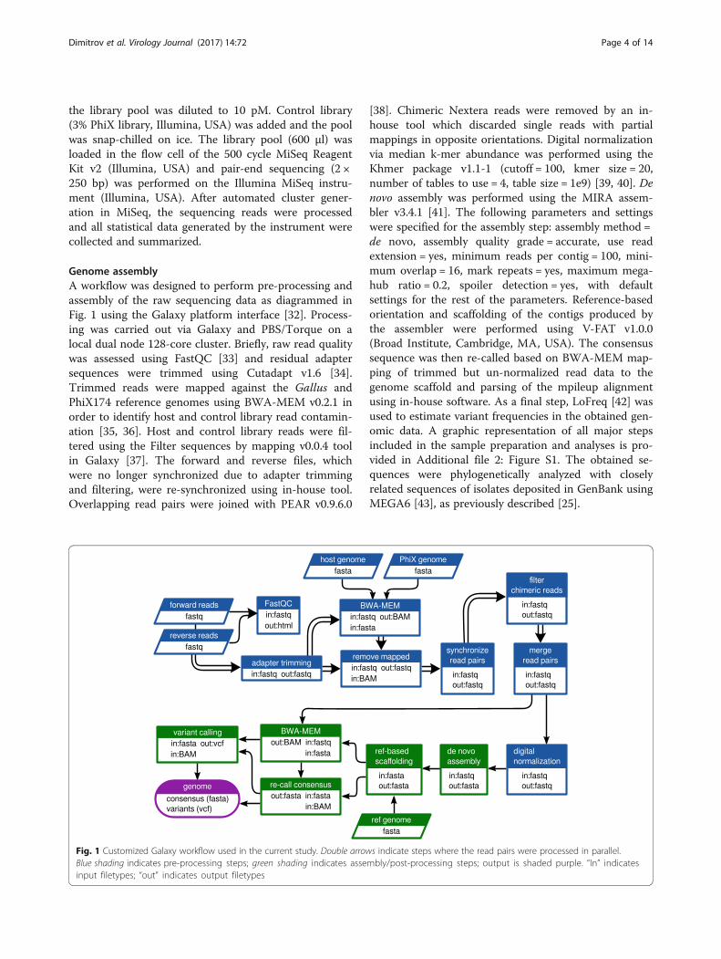
Genome assemblyA workflow was designed to perform pre-processing andassembly of the raw sequencing data as diagrammed inFig. 1 using the Galaxy platform interface [32]. Process-ing was carried out via Galaxy and PBS/Torque on alocal dual node 128-core cluster. Briefly, raw read qualitywas assessed using FastQC [33] and residual adaptersequences were trimmed using Cutadapt v1.6 [34].Trimmed reads were mapped against the Gallus andPhiX174 reference genomes using BWA-MEM v0.2.1 inorder to identify host and control library read contamin-ation [35, 36]. Host and control library reads were fil-tered using the Filter sequences by mapping v0.0.4 toolin Galaxy [37]. The forward and reverse files, whichwere no longer synchronized due to adapter trimmingand filtering, were re-synchronized using in-house tool.Overlapping read pairs were joined with PEAR v0.9.6.0
[38]. Chimeric Nextera reads were removed by an in-house tool which discarded single reads with partialmappings in opposite orientations. Digital normalizationvia median k-mer abundance was performed using theKhmer package v1.1-1 (cutoff = 100, kmer size = 20,number of tables to use = 4, table size = 1e9) [39, 40]. Denovo assembly was performed using the MIRA assem-bler v3.4.1 [41]. The following parameters and settingswere specified for the assembly step: assembly method =de novo, assembly quality grade = accurate, use readextension = yes, minimum reads per contig = 100, mini-mum overlap = 16, mark repeats = yes, maximum mega-hub ratio = 0.2, spoiler detection = yes, with defaultsettings for the rest of the parameters. Reference-basedorientation and scaffolding of the contigs produced bythe assembler were performed using V-FAT v1.0.0(Broad Institute, Cambridge, MA, USA). The consensussequence was then re-called based on BWA-MEM map-ping of trimmed but un-normalized read data to thegenome scaffold and parsing of the mpileup alignmentusing in-house software. As a final step, LoFreq [42] wasused to estimate variant frequencies in the obtained gen-omic data. A graphic representation of all major stepsincluded in the sample preparation and analyses is pro-vided in Additional file 2: Figure S1. The obtained se-quences were phylogenetically analyzed with closelyrelated sequences of isolates deposited in GenBank usingMEGA6 [43], as previously described [25].
Fig. 1 Customized Galaxy workflow used in the current study. Double arrows indicate steps where the read pairs were processed in parallel.Blue shading indicates pre-processing steps; green shading indicates assembly/post-processing steps; output is shaded purple. “In” indicatesinput filetypes; “out” indicates output filetypes
Dimitrov et al. Virology Journal (2017) 14:72 Page 4 of 14

ResultsNucleic acids quantification and libraries fragment sizeThe nucleic acid concentrations obtained at differentsteps throughout the preparation of the libraries for se-quencing are summarized in Additional file 3: Table S2.The lowest detected RNA concentration was 2 ng/μl andthe maximum was 55 ng/μl. After RNA purification, theRNA concentrations of five samples were below the de-tection limit of Qubit (250 pg/μl); however, these sam-ples resulted in sufficient cDNA quantity to be furtherprocessed in library preparation. The generated librarieshad a relatively narrow combined distribution of meanfragment lengths (mean 351 bp, standard deviation30 bp, with 26 of 30 libraries within the range of 334 to371 bp) (see Additional file 3: Table S2). It was observedthat the true fragment length distributions observedpost-sequencing were shorter than expected based onBioanalyzer reports, even after counting for adapterlength (Table S2). As a result, a large proportion (morethan 90% in nearly all libraries) of read pairs overlappedat the ends. The source of the discrepancy with the Bioa-nalyzer estimates is still unclear.
Summarized statistics of the sequencing runA summary of the sequencing run statistics as estimatedby the MiSeq instrument is provided in Table 1. A clus-ter density of 917 +/- 19 K/mm2 and 92.34% of the clus-ters passing the chastity filter yielded a total of 8.4Gigabases of data. Of 17.7 million total reads, 96.31%passed the instrument quality control filter. Almost 80%of the bases were assigned Phred quality scores equal orgreater to Q30 (Q30 score is equivalent to an expectederror rate of 0.001). The fraction of reads in the pool
assigned to each sample varied from 0.0007 to 7.16%(mean 3.2 ± 1.4%).
Optimization of the assembly/analysis workflowIn order to take advantage of the overlapping reads, amerging step was introduced to produce longer pseudo-reads and to reduce complexity of the assembly task. Anessential optimization was made by reducing the esti-mated coverage depth to a level that would still produceoptimal assemblies. Two techniques for data reductionwere investigated. Random sub-sampling resulted in lossof specific regions in the genome with reproducibly lowcoverage (data not shown). Digital normalization, whichaims to down-sample high-coverage regions while pre-serving reads from low-coverage areas, provided meansfor decreasing the number of used reads to an optimallevel without loss of data, and thus, was incorporatedinto the customized Galaxy workflow prior to assembly.In order to determine an optimal target depth for assem-bly, preliminary test assemblies using the Velvet assem-bler v1.2.10 [44] were performed on a geometricprogression of sampling depths from 10x to 10000x (theapproximate depth of the raw data) with an additionaloptimization of the velvetg “cov_cutoff” parameter foreach depth (parameter used to low coverage nodes). Theresults indicated that optimal (in this case, full-length)assembly occurred over a range of approximately oneorder of magnitude (100x to 1000x). Below and abovethis range, fragmentation began to occur (Fig. 2).
Data analysisThe final outputs of the analysis workflow for each sam-ple included a consensus genome scaffold (.fasta), a fileof all assembled contigs (.fasta), a variant frequency callfile (variant call format or .vcf ) and a set of summaryTable 1 Statistics of next-generation sequencing of 30 avian
paramyxovirus isolates in a single run
Data Results
Cluster density (K/mm2)a 917 +/- 19
Clusters passing filterb 92.34%
Total number of reads 17762176
Pass-filter readsc 16403251
Percentage of reads passing filter 96.31%
≥ Q30d 77.9%
Lowest representation for any indexe 0.0007%
Highest representation for any indexe 7.16%a shows number of clusters per square millimeter (optimal cluster density is1000–1200, can vary with chemistry)b indicates the purity of the signals detected from the clusters (i.e. signalspassing chastity filter that is the ratio of the brightest base intensity dividedby the sum of the brightest and second brightest base intensities and thefiltration process removes the least reliable clusters from the imageanalysis results)c reads passing filter (about 15 million reads are expected from an optimallyclustered flow cell)d percentage of bases with Phred quality score equal or greater to 30e percentage of pass-filter reads assigned to any index
Fig. 2 Analysis of Newcastle disease virus genome assembly atvarious read depths. Shown are the longest contig produced ateach read depth as a fraction of the full genome length. Subsamplesup to 200x were generated using digital normalization. Above 200x,additional reads were added using random subsampling (due toissues with high median cutoffs in the kh-mer package). At eachsubsampling depth, the final velvetg assembly was optimized formaximum contig length based on the “cov_cutoff” parameter
Dimitrov et al. Virology Journal (2017) 14:72 Page 5 of 14

statistics on the run and the assembly. An in-depth sum-mary of the outputs from all samples is presented inTables 2 and 3, including detailed information on readquality and depth distributions and genome coverageper sample. A total of 29 full-length or near-full-lengthAPMV genomes (99.56% mean genome coverage) wereobtained from 30 libraries with only one sample (#1005)having coverage below 99% and nine samples having100% coverage (Tables 2 and 3). The lower and upperquartiles of median depth per position of the sequencingresults were 2984 and 6894 respectively, allowing for ac-curate detection of low-frequency single nucleotide vari-ants. In fact, all but one NDV samples had a medianread depth of at least 2583 (the exception, sample 1005,was found to consist of approximately 98% avian influ-enza virus reads after host filtering). In addition to NDVgenome assembly, the de novo strategy allowed for thedetection of full-length and near-full-length genomes ofavian influenza virus (AIV) in libraries of isolates 998,1005, 1009 and 1011 [45], as well as infectious bronchitisvirus (IBV) in samples 1003 and 1009. The coverage ofthe two obtained IBV genomes was 85.78 and 99.37%,while the sequenced AIV genes had coverages rangingbetween 92.23 and 100%, and two complete AIV ge-nomes were sequenced (see Table 3). The estimated me-dian depths for the IBV (5 and 22) and AIV (from 35 to1274) isolates were lower (Table 3), most likely reflectingthe lower titer of these viruses in the samples. Sample959 was identified as a member of the novel APMVserotype 13 and the median depth for this sample was3484. The host reads were between 0.1 and 5.4% (aver-age 1.3%) of all reads per sample. No data was obtainedfrom the library of sample 688 (only 0.0007% of the rawreads were assigned to this sample). The results fromthe comparison of the three NDV libraries preparedside-by-side with and without the capture step showedidentical accuracy, not significantly affected overallcoverage and near full-length and full-length genomes wereobtained using both approaches. However, the number ofNDV-specific reads decreased by approximately 30% whenthe capture step was not performed (see Table 4).While the high-throughput workflow sometimes
resulted in short segments of missing data at the gen-ome termini and/or at one short internal gap, completesequences for all coding regions of the 29 APMV posi-tive samples were obtained directly from the workflow.Nearly all of the short missing regions occurred at eitherthe termini (a common issue in viral NGS sequencing)[46] or at one specific intergenic location in the genomebetween genes N and P which displayed extremely lowcoverage in all analyzed samples (possibly as a resultof high GC content – 76%). For the purpose of sub-mitting full-length NDV sequences to GenBank, wesequenced the termini using a previously described
protocol [47] and primers designed for the currentstudy (see Additional file 4: Table S3). The internalgaps, where necessary, were sequenced using PCRand Sanger sequencing (for primers sequences seeAdditional file 4: Table S3). This additional work wasnot included in the time/cost estimates, as it was per-formed to submit complete NDV sequences to Gen-Bank and would not be necessary for a full analysisof the coding regions.
Time and cost estimatesThe time and cost estimates for all steps are summarizedin Additional file 5: Table S4. Assuming the addition ofthe first reagent as the start and the final dilutions of thesamples as the end of the procedure, the approximatetime taken for preparing 30 samples was 25 to 30person-hours. The sequencing run (500 cycle kit) lasted39 h. Submission of the raw data to the customizedGalaxy workflow and data analysis on the cluster tookan additional 2 to 3 h. The average cost of all steps,including all reagents but excluding labor, depreciationand maintenance of equipment, was estimated to beapproximately 106 USD per sample.
DiscussionNext-generation sequencing has been previously de-scribed for whole-genome sequencing of NDV by ourteam and others [48–55]; however, this study is the firstreport that demonstrates robust simultaneous genomiccharacterization of multiple NDV viruses in a singleNGS run. The study further demonstrates the addedbenefit of conducting random non-targeted sequencingwith an optimized de novo assembly workflow for identi-fication of mixed viral infections. In contrast to previouswork, here an optimized and customized workflow thatemploys publically available tools and produced consist-ently high quality assemblies of complete genomes isdescribed in details. This study also provides detailedstatistical and sequencing information that allows qualityand quantity assessment of the obtained results.Our findings demonstrate that the described chemistry
and bioinformatics approach is sufficiently robust to ob-tain and distinguish the complete genomes of completelydifferent types of RNA viruses during a mixed infection.In addition to the conclusive results with NDV andAPMV-13 (family Paramyxoviridae), the complete ornear complete genomes of four avian influenza and twoinfectious bronchitis viruses, which were co-infectingfive samples originally identified as Newcastle diseaseviruses alone, were also obtained. Infectious bronchitisviruses belong to the family Coronaviridae and aresingle-stranded positive-sense RNA viruses with genomesize of approximately 27,5 to 28 kb, excluding the poly(A) tail, which includes ten open reading frames [56].
Dimitrov et al. Virology Journal (2017) 14:72 Page 6 of 14

Table
2Summaryof
sequ
encing
andassemblydata
of25
avianparamyxoviru
sisolates
Isolate
numbe
r%
PFreadsa
Num
berof
raw
read
pairs
Num
berof
filteredread
pairs
b
Forw
ard
read
quality
cReverseread
quality
cIden
tified
virus
Finalcoveragede
pthc
Num
berof
readsused
for
consen
susd
Con
sensus
nucleo
tide
leng
th
Missing
positio
nsat
5'en
deLeng
thof
internalgaps
Missing
positio
nsat
3'en
de
Percen
tcoverage
e
1002
2.49
409193
405137
2|37|38|38|38
2|36|37|38|38
NDV
0|3680|6088|7868|18004
390740
15124
6899.55
1004
2.67
437755
432361
2|37|38|38|38
2|34|37|38|38
NDV
0|4185|6151|7909|14329
422150
15125
6799.56
1007
4.20
688524
681691
2|37|38|38|38
2|36|37|38|38
NDV
0|817|3648|7368|19348
665220
15125
6799.56
994
1.39
227500
226196
2|37|38|38|38
2|36|37|38|38
NDV
0|1758|2756|4186|14276
219609
15121
7199.53
995
1.38
226050
224416
2|36|37|38|38
2|34|36|37|38
NDV
0|2162|2995|4197|9101
216240
15110
8299.46
996
1.53
251238
250338
2|37|38|38|38
2|34|37|37|38
NDV
0|2383|3175|4411|9167
242158
15104
2068
99.42
1001
3.79
621655
618002
2|37|38|38|38
2|36|37|38|38
NDV
0|4653|6784|9510|21623
594361
15122
7099.54
997
1.72
281376
266251
2|37|38|38|38
2|35|37|38|38
NDV
0|1937|3026|4030|9645
233775
15104
2266
99.42
999
1.70
279207
272105
2|37|38|38|38
2|36|37|38|38
NDV
0|1728|2583|4496|11915
241105
15108
8499.45
1000
1.74
285079
280596
2|37|38|38|38
2|35|37|38|38
NDV
0|2338|3107|4441|8796
241724
15117
7599.51
959
3.60
590494
588212
2|37|38|38|38
2|37|38|38|38
APM
V-13
1|2250|3484|5801|23998
531520
16126
2099.88
960
4.34
711909
709377
2|37|37|38|38
2|37|37|38|38
NDV
0|3738|5631|7347|19491
628837
15135
2532
99.62
961
4.61
756197
753487
2|36|37|38|38
2|35|37|38|38
NDV
0|4585|6509|8206|14939
674667
15167
2599.84
962
3.35
548833
547622
2|37|38|38|38
2|35|37|38|38
NDV
2|4617|6340|8772|14719
529553
15192
100.00
967
3.71
607876
597902
2|37|38|38|38
2|36|37|38|38
NDV
2|2673|3928|6881|26072
562657
15192
100.00
968
4.46
731692
727838
2|37|38|38|38
2|35|37|38|38
NDV
0|2507|4136|7393|22877
636218
15167
1999.87
695
7.16
1156516
1129415
2|36|37|38|38
2|35|37|38|38
NDV
2|1678|3827|6301|32862
955829
15192
100.00
714
3.98
653603
643246
2|37|37|38|38
2|36|37|38|38
NDV
1|2082|3513|6724|35052
570825
15192
100.00
715
3.63
594802
580223
2|37|38|38|38
2|36|37|38|38
NDV
10|2608|5633|8881|28809
526885
15192
100.00
720
4.05
663757
657982
2|37|37|38|38
2|35|37|38|38
NDV
13|3821|6267|9252|27439
525313
15192
100.00
861
3.51
576077
574864
2|37|38|38|38
2|35|37|38|38
NDV
6|3930|6419|8881|27153
559394
15192
100.00
867
4.11
673510
668781
2|37|38|38|38
2|36|37|38|38
NDV
0|4284|6079|9028|23441
647586
15176
1699.89
892
3.92
642753
642250
2|37|38|38|38
2|35|37|38|38
NDV
1|5147|7049|9922|18221
618591
15180
1299.92
913
4.06
665640
661350
2|36|37|38|38
2|35|37|38|38
NDV
0|2278|3784|7011|31842
566280
15192
100.00
688
0.01
nodata
NA
NA
NA
NA
NA
NA
NA
NA
NA
NA
NAno
tap
plicab
leathefractio
nof
read
sassign
edto
each
sampleou
tof
alln
umbe
rof
read
sthat
passed
filter(i.e.
pass-filter
read
s)bthenu
mbe
rof
paire
dread
sremaining
afterho
stan
dinternal
controlfiltering
cnu
mbe
rsrepresen
tdistrib
ution(m
inim
um|low
erqu
artile|m
edian|u
pper
quartile|m
axim
um)
dnu
mbe
rsof
paire
dread
sused
tore-callthe
final
consen
susforeach
sequ
ence
ethemissing
nucleo
tides
attheen
dsan
dthefractio
nof
theexpe
cted
fullge
nomeleng
thcoveredby
theconsen
susscaffold
(i.e.
notcontaining
unkn
ownnu
cleo
tides)
Dimitrov et al. Virology Journal (2017) 14:72 Page 7 of 14

Table
3Summaryof
sequ
encing
andassemblydata
offivesamples
that
wereiden
tifiedto
have
mixed
popu
latio
nsof
New
castlediseasevirus(NDV)
andothe
ravianviruses
Isolate
numbe
r%
PFreadsa
Num
berof
raw
read
pairs
Num
berof
filteredread
pairs
b
Forw
ard
read
quality
cReverse
read
quality
cIden
tified
virus
Finalcoveragede
pthc
Num
berof
readsused
for
consen
susd
Con
sensus
nucleo
tide
leng
th
Missing
positio
nsat
5'en
deLeng
thof
internalgaps
Missing
positio
nsat
3'en
de
Percen
tcoverage
f
1003
3.33
546797
540519
2|37|38|38|38
2|36|37|38|38
NDV
0|4229|6090|8762|18055
525597
15127
6599.57
IBV
g0|2|5|8|44
904
23711
269
3644
1885.78
1005
3.44
564101
123024
2|36|37|38|38
2|33|36|37|38
NDV
h0|7|13|19|36
1161
14494
343
272
8395.41
AIV
i-PB2
2|685|1003|1446|2774
16804
2283
100.00
AIV-PB1
2|1845|2896|3432|4634
38261
2334
100.00
AIV-PA
68|1180|1496|1970|4721
23466
2151
100.00
AIV-HA
12|595|1057|1598|2604
14687
1683
100.00
AIV-NP
25|886|1306|1867|2563
11252
1497
100.00
AIV-NA
11|416|838|1030|1692
7707
1410
100.00
AIV-M1,M2
11|210|811|1357|2195
5699
982
100.00
AIV-NEP,N
S115|396|787|1222|1707
3796
838
100.00
1009
3.21
526425
519854
2|37|38|38|38
2|36|37|38|38
NDV
0|4216|6887|9403|18079
485669
15127
6599.57
IBV
0|15|22|35|92
3743
27469
136
1918
99.37
AIV-PB2
0|137|175|272|545
2791
2283
100.00
AIV-PB1
3|370|651|835|1177
7692
2324
2098.99
AIV-PA
0|88|117|183|446
1675
2151
100.00
AIV-HA
0|113|256|342|578
2399
1683
100.00
AIV-NP
0|145|277|333|523
2117
1485
100.00
AIV-NA
4|118|225|317|637
2226
1410
100.00
AIV-M1,M2
2|46|132|183|246
793
958
2496.71
AIV-NEP,N
S10|47|83|103|139
368
838
100.00
1011
3.44
565083
538171
2|37|38|38|38
2|36|37|38|38
NDV
1|3272|5386|7121|16101
499378
15192
100.00
AIV-PB2
0|181|290|462|1254
6242
2283
100.00
AIV-PB1
2|323|482|609|1077
7728
2334
100.00
AIV-PA
8|127|172|220|437
2743
2151
100.00
AIV-HA
3|217|449|685|906
5306
1683
100.00
AIV-NP
3|180|380|605|758
4314
1497
100.00
AIV-NA
3|74|98|153|242
1383
1410
100.00
AIV-M1,M2
4|59|255|372|579
2092
982
100.00
AIV-NEP,N
S13|29|70|137|216
516
808
100.00
Dimitrov et al. Virology Journal (2017) 14:72 Page 8 of 14

Table
3Summaryof
sequ
encing
andassemblydata
offivesamples
that
wereiden
tifiedto
have
mixed
popu
latio
nsof
New
castlediseasevirus(NDV)
andothe
ravianviruses
(Con
tinued)
998
1.80
295662
294217
2|37|38|38|38
2|35|37|38|38
NDV
0|2248|3275|4719|11528
280492
15103
2069
99.41
AIV-PB2
0|22|33|48|78
477
2246
3498.51
AIV-PB1
0|34|56|85|125
698
2217
6097.36
AIV-PA
2|28|55|106|178
767
2141
1099.53
AIV-HA
0|9|18|29|53
204
1643
4097.65
AIV-NP
0|24|43|82|126
430
1456
356
97.26
AIV-NA
1|21|28|41|83
272
1304
3175
92.23
AIV-M1,M2
0|8|28|65|87
196
923
5692.62
AIV-NEP,N
S10|9|17|28|40
94810
100.00
athefractio
nof
read
sassign
edto
each
sampleou
tof
alln
umbe
rof
read
sthat
passed
filter(i.e.
pass-filter
read
s)bthenu
mbe
rof
paire
dread
sremaining
afterho
stan
dinternal
controlfiltering
cnu
mbe
rsrepresen
tdistrib
ution(m
inim
um|low
erqu
artile|m
edian|u
pper
quartile|m
axim
um)
dnu
mbe
rsof
paire
dread
sused
tore-callthe
final
consen
susforeach
sequ
ence
eforavianinflu
enza
viruses,themissing
nucleo
tides
referto
thebe
ginn
ingan
dtheen
dof
thecoding
sequ
encesof
thege
nes
fthefractio
nof
theexpe
cted
fullge
nomeleng
thcoveredby
theconsen
susscaffold
(i.e.
notcontaining
unkn
ownnu
cleo
tides),foravianinflu
enza
gene
s,thecoverage
represen
tscompa
rison
tothecoding
sequ
ences
ofthege
neson
lygInfectious
bron
chitisvirus
hcoverage
depthan
dnu
mbe
rof
read
sused
tore-callthe
final
consen
susforthisNDVisolatewereim
pacted
bythepresen
ceof
influ
enza
virusAin
thesample(in
fluen
zaread
swereestim
ated
tobe
approxim
ately
98%
ofallreads,d
atano
tshow
n)iAvian
influ
enza
virus;PB
2=segm
ent1po
lymerasePB
2;PB
1=segm
ent2po
lymerasePB
1;PA
=segm
ent3po
lymerasePA
;HA=segm
ent4he
mag
glutinin;N
P=segm
ent5nu
cleo
capsid
protein;
NA=segm
ent6
neuram
inidase;
M1,
M2=segm
ent7matrix
protein1an
dmatrix
protein2;
NEP
=segm
ent8nu
clearexpo
rtproteinan
dno
nstructuralp
rotein
1
Dimitrov et al. Virology Journal (2017) 14:72 Page 9 of 14

The avian influenza viruses belong to the family Ortho-myxoviridae and have genomes containing eight seg-ments of single-stranded, negative-sense RNA that codefor 10 or 11 proteins, depending on the strain [57].Despite the diverse nature of the RNA present in sam-ples with mixed populations, the procedure describedhere successfully produced complete genomes of theseviruses.Our results also demonstrate the capability of the
methodology to produce quality libraries and assem-blies without any physical or mechanical enrichment.The cDNA and dsDNA concentrations were notfound to be proportional to the initial total RNA con-centrations. The introduced nuclease step aided diges-tion of host nucleic acids resulting in low averagenumber (see Tables 2 and 3) of host-associated readsper sample. The abundance of host nucleic acids maypose a problem in obtaining sufficient numbers ofviral reads for optimal viral genome assembly [4]. Toavoid or decrease problems caused by contaminationwith host sequences others have developed method-ologies for enrichment of target viral RNAs. We havenot utilized any pretreatment or purification; however,a target-specific capture step with biotinylated oligosdesigned from three different conservative regions ofthe NDV genome was tested. The comparison of re-sults from samples with and without the RNA capturestep presented here demonstrates that the primarytradeoff comes in the form of approximately 30% re-duced depth of coverage, although the coverage wasstill sufficient for proper consensus re-calling (seeTable 4). The ability of the capture step to reducehost sequences and other non-target RNA and to im-prove downstream assembly and analysis should befurther assessed on different sample types (e.g. clinicalsamples, formalin-fixed paraffin-embedded samples)that may contain less amounts of viral RNA. For egg-grown viruses with high viral titers the observed de-crease of reads without the RNA capture step wasnot essential for obtaining complete coverage withsufficient depth. In clinical diagnostic samples, how-ever, the number of NDV sequencing reads is oftensignificantly lower, and introducing the RNA capturestep could improve the final results.
There is a clear difference between the presentedapplication of NGS and the use of this approach in diag-nostics. Here we describe the use of high-titer egg-grown viruses for production of high quality and deepdata useful for detailed genomic characterization andrare variant analyses. However, the use of this NGS tech-nology for diagnostics is more complex. It requires toclearly establish sensitivity, specificity and limit of detec-tion based on the nature of samples and these are be-yond the scope of the current work. The describedmethodology has been successfully transferred for usewith clinical samples and optimization studies are inprogress in our lab.Prior to the production run, considerable time was
spent optimizing the assembly and analysis workflow forthe task at hand. While some of the steps in the work-flow are fairly standard procedures in NGS analyses (QCsummarization, adapter trimming, contaminant read fil-tering), others were tailored to the specific characteris-tics of the data being generated. The most criticaloptimization, however, was reducing estimated coveragedepth to a level that would produce optimal assemblies.It has previously been shown that, past a certain level,increasing read depth can decrease de novo assemblyquality [58]. This effect can have significant conse-quences when working with massively deep sequencingdata such as viral population studies that can easily ex-ceed 10000x sample coverage. Digital normalization hasbeen included in similar workflows by others [59, 60]but is often overlooked in naïve approaches to high-coverage de novo assembly. As the assembler used inour workflow (MIRA) is relatively resource-intensiveoverlap-layout-consensus (OLC) assembler, we chose atarget (100x) at the lower end of the empirically deter-mined optimal range to incorporate as a cut-off into thecustomized production workflow. Graph-based assem-blers such as Velvet utilize de Bruijn graph algorithmsand assemble data by representing the genome by a setof short k-mer sequences [44]. Notably, graph-basedassemblers are less resource-demanding and can be suc-cessfully utilized with limited computational resources.However, for graph-based assemblers, the k-mer size isan essential parameter [44, 59, 60] and the optimal valuehas to be determined depending on the characteristics of
Table 4 Comparison of differences in number of reads and genome coverage of three samples prepared with and without captureof NDV RNA
Virusdesignation
Number of reads % fewer readswithout capture
Identity ofconsensussequences
Missing sequences at genome termini and internal gaps (in number of nucleotides)
Withcapture
Withoutcapture
With capture Without capture
5′ gaps 3′ 5′ gaps 3′
691 403515 283501 29.7 100% 20 0 0 26 0 0
698 363962 262452 27.9 100% 0 0 0 25 0 0
901 415661 285405 31.3 100% 0 94 0 22 84 0
Dimitrov et al. Virology Journal (2017) 14:72 Page 10 of 14

the sequence reads, while this is avoided using an OLCassembler. In our hands, MIRA consistently producesquality assemblies with minimal tuning needed. Add-itionally, due to the potential skewing effects of digitalnormalization and V-FAT scaffolding on the proportionof nucleotide variant frequencies, the post-assembly stepto re-map the un-normalized data to the genome scaf-fold allowed for proper consensus re-calling and precisevariant analysis.One important aspect to the use of NGS approaches
in mainstream viral sequencing studies is the capacity tomultiplex samples in order to reduce costs. The timeand cost summary for all steps (summarized inAdditional file 5: Table S4) demonstrate that the simul-taneous processing of 30 samples requires approximateone hour of operator time per sample with a cost of ap-proximately 106 USD per sample. Those values, althoughstill high for diagnostics purposes, are 15 to 20 times lower(based on internal estimates) compared to the cost ofprimer-based sequencing with Sanger technologies.Furthermore, the demonstrated lower and upper quartilesof median depth per position (2984 and 6894, respectively),allow for accurate consensus re-calling and rare variantanalysis. The final output not only includes the consensusgenome sequence but also produced a variant call formatfile (https://samtools.github.io/hts-specs/VCFv4.2.pdf) anddemonstrate that the protocol could be used for researchon viral quasispecies and evolutionary studies (Dimitrov etal., in preparation). As previously reported by Gould et al.,the 1998 Newcastle disease outbreak in Australia was pre-ceded by several months of circulation of mutant quasispe-cies of the virulent cleavage site [61]. These viral variantswere undetected in the Sanger consensus sequences butcould have been detected and properly quantified if themethodology described here was available at that time. Theobtained genomic coverages (see Tables 2 and 3) illustratethe ability of the described protocol in generating full-length or near-full-length RNA virus genomes. Al-though very short internal gaps were present due tocomplete absence of coverage in the raw data andshort sequences at the genome termini were missing,all coding sequences (commonly used in genetic stud-ies) were obtained.The total turnaround time for the entire testing (sam-
ple preparation, sequencing and analyses) was approxi-mately 72 h, of which most of the time consisted oflibrary preparation and the sequence run. The durationof the sequencing run could be reduced by approxi-mately 15 h by using a 300-cycle configuration withoutany anticipated drop-off in assembly quality based onour observed fragment length distributions, althoughthis assumption is untested. The time taken for dataanalysis may be expected to vary somewhat based on theavailable computational resources in a lab, although in
our protocol this represents a small fraction of the totalturnaround time to begin with.The obtained results were phylogenetically consistent
with preliminary studies of the tested viruses (data notshown) and expectations based on previous research.Almost all of the samples from Pakistan were of sub-genotype VIIi which is currently circulating in Pakistanand may be causing a new panzootic [25, 62]. Ukrainiansamples were of different sub-genotypes (II, VIg andVIId) that have been reported to be isolated frompigeons in Ukraine [25] and also in Europe [63]. TheNigerian samples were of genotypes XIVb and XVIIawhich have been reported to circulate in Nigeria since2006 [64] and previously un-sequenced full-lengthgenomes of these sub-genotypes have been reported byus [48, 49]. In addition, and demonstrating the broadapplicability and the advantages of the de novo approachdescribed here, the first complete APMV-13 genomewas obtained [17] and avian influenza and infectiousbronchitis viruses populations were identified. Phylogen-etic analyses of the obtained NDV and IBV sequencesare presented in Additional file 6: Figures S2 and S3).
ConclusionIn summary, a robust chemistry and bioinformaticsprotocol utilizing publicly available tools to sequenceand analyze complete genomes from small RNA virusesis described. Thirty-five full-length or near-full-lengthavian RNA viral genomes with a high median coveragedepth were successfully sequenced out of 30 samples.The applied de novo approach allowed identification ofmixed viral populations in some of the samples. Thecombination of multiplexing NGS technology with thecustomized Galaxy workflow platform enabled a quickturnaround time and cost-efficient methodology for sim-ultaneous characterization of multiple viral genomes.
Additional files
Additional file 1: Table S1. Background information of the avianparamyxovirus isolates used in this study. (DOCX 15 kb)
Additional file 2: Figure S1. Major processing steps used in thecurrent study (PDF 222 kb)
Additional file 3: Table S2. Nucleic acid concentrations and libraryfragment size distributions of thirty virus isolates used in the study.(DOCX 18 kb)
Additional file 4: Table S3. Sequences of primers used for sequencinginternal gaps and missing termini. (DOCX 13 kb)
Additional file 5: Table S4. Time and cost analysis of next-generationsequencing of thirty avian paramyxovirus isolates. (DOCX 15 kb)
Additional file 6: Figure S2. Phylogenetic analysis based on thecomplete genome coding sequence of Newcastle disease virus isolatesstudied here and selected closely related sequences from GenBank.Figure S3. Phylogenetic analysis based on the hypervariable regionof the spike protein gene of Infectious bronchitis virus studied hereand selected closely related sequences from GenBank. (PDF 142 kb)
Dimitrov et al. Virology Journal (2017) 14:72 Page 11 of 14

AcknowledgementsThe authors gratefully acknowledge Dawn Williams-Coplin and Tim Olivierfor their technical assistance. We appreciate critical and constructive reviewsprovided by three anonymous reviewers. The mention of trade names orcommercial products in this publication is solely for the purpose of providingspecific information and does not imply recommendation or endorsementby the U.S. Department of Agriculture. The USDA is an equal opportunityprovider and employer.
FundingThis work was supported by USDA/ARS CRIS 6040-32000-072, U.S. Defense ThreatReduction Agency BAA projects FRCALL12-6-2-0005 and ARS#715 FRCALL 12-6-1-0046, and U.S. Department of State Biosecurity Engagement Program NDV 31063.
Availability of data and materialsThe complete genome sequences (n = 28) of the Newcastle diseasevirus isolates obtained in this study were submitted to GenBank and areavailable under the accession numbers KY171989 to KY171995, KY076030to KY076039, KY076043, KU295453 to KU295455, KX496962 to KX496964,KY042127, KX236101, KU133362, and KT948996. The sequence of theAPMV-13 is available under accession number KX119151. The nucleotidesequences of the four H9N2 AIVs have been deposited in GenBankunder accession numbers KU042891 to KU042922. The sequences of thetwo IBV are available under accession numbers KY588134 and KY588135.All software tools utilized in the workflow that are not described else-where, along with their Galaxy wrappers, can be found online at https://github.com/jvolkening/b2b-utils.
Authors’ contributionsCLA, PS, KMD, and PJM conceived this project. AW, SFR, IS, and TMJcoordinated field sampling efforts and isolated viruses. PS, IVG, and AWprepared and sequenced the viral isolates. JDV developed the Galaxyworkflow and associated software tools. PS, KMD, JDV, and CLA conductedanalyses of the data. PS, KMD, JDV, PJM, and CLA wrote the manuscript.All authors read and approved the final manuscript.
Competing interestsThe authors declare that they have no competing interests.
Consent for publicationNot applicable.
Ethics approval and consent to participateNot applicable.
Publisher’s NoteSpringer Nature remains neutral with regard to jurisdictional claims inpublished maps and institutional affiliations.
Author details1Exotic and Emerging Avian Viral Diseases Research Unit, Southeast PoultryResearch Laboratory, US National Poultry Research Center, AgriculturalResearch Service, USDA, 934 College Station Road, Athens, GA 30605, USA.2BASE2BIO, 1945 Arlington Drive, Oshkosh, WI 54904, USA. 3NationalScientific Center Institute of Experimental and Clinical Veterinary Medicine,83 Pushkinskaya Street, Kharkiv 61023, Ukraine. 4Quality OperationsLaboratory (QOL), University of Veterinary and Animal Sciences, Syed AbdulQadir Jilani Road, Lahore 54000, Pakistan. 5Institute of Biochemistry andBiotechnology, University of Veterinary and Animal Sciences, Syed AbdulQadir Jilani Road, Lahore 54000, Pakistan. 6Regional Laboratory for AnimalInfluenza and other Transboundary Animal Diseases, National VeterinaryResearch Institute, PMB01, Vom 930010, Plateau State, Nigeria.
Received: 16 February 2017 Accepted: 29 March 2017
References1. Wu Q, Ding SW, Zhang Y, Zhu S. Identification of viruses and viroids by
next-generation sequencing and homology-dependent and homology-independent algorithms. Annu Rev Phytopathol. 2015;53:425–44.http://doi.org/10.1146/annurev-phyto-080614-120030.
2. Victoria JG, Kapoor A, Li L, Blinkova O, Slikas B, Wang C, Naeem A, Zaidi S,Delwart E. Metagenomic analyses of viruses in stool samples from childrenwith acute flaccid paralysis. J Virol. 2009;83:4642–51. http://doi.org/10.1128/jvi.02301-08.
3. Allander T, Emerson SU, Engle RE, Purcell RH, Bukh J. A virus discoverymethod incorporating DNase treatment and its application to theidentification of two bovine parvovirus species. Proc Natl Acad Sci U S A.2001;98:11609–14. http://dx.doi.org/10.1073/pnas.211424698.
4. Neill JD, Bayles DO, Ridpath JF. Simultaneous rapid sequencing of multipleRNA virus genomes. J Virol Methods. 2014;201:68–72. http://doi.org/10.1016/j.jviromet.2014.02.016.
5. Reuter G, Pankovics P, Boros A. Identification of a novel astrovirus in adomestic pig in Hungary. Arch Virol. 2011;156:125–8. http://doi.org/10.1007/s00705-010-0827-5.
6. Cholleti H, Hayer J, Abilio AP, Mulandane FC, Verner-Carlsson J, Falk KI,Fafetine JM, Berg M, Blomstrom AL. Discovery of novel viruses inmosquitoes from the Zambezi valley of Mozambique. PLoS One. 2016;11:e0162751. http://doi.org/10.1371/journal.pone.0162751.
7. Chiu CY. Viral pathogen discovery. Curr Opin Microbiol. 2013;16:468–78.http://dx.doi.org/10.1016/j.mib.2013.05.001.
8. Chandriani S, Skewes-Cox P, Zhong W, Ganem DE, Divers TJ, Van BlaricumAJ, Tennant BC, Kistler AL. Identification of a previously undescribeddivergent virus from the Flaviviridae family in an outbreak of equine serumhepatitis. Proc Natl Acad Sci U S A. 2013;110:E1407–15. http://dx.doi.org/10.1073/pnas.1219217110.
9. Li L, Pesavento PA, Leutenegger CM, Estrada M, Coffey LL, Naccache SN,Samayoa E, Chiu C, Qiu J, Wang C, et al. A novel bocavirus in canine liver.Virol J. 2013;10:54. http://dx.doi.org/10.1186/1743-422x-10-54.
10. Lauck M, Sibley SD, Lara J, Purdy MA, Khudyakov Y, Hyeroba D, Tumukunde A,Weny G, Switzer WM, Chapman CA, et al. A novel hepacivirus with anunusually long and intrinsically disordered NS5A protein in a wild Old Worldprimate. J Virol. 2013;87:8971–81. http://dx.doi.org/10.1128/jvi.00888-13.
11. Djikeng A, Halpin R, Kuzmickas R, Depasse J, Feldblyum J, Sengamalay N,Afonso C, Zhang X, Anderson NG, Ghedin E, Spiro DJ. Viral genomesequencing by random priming methods. BMC Genomics. 2008;9:5.http://dx.doi.org/10.1186/1471-2164-9-5.
12. Thurber RV, Haynes M, Breitbart M, Wegley L, Rohwer F. Laboratoryprocedures to generate viral metagenomes. Nat Protoc. 2009;4:470–83.http://dx.doi.org/10.1038/nprot.2009.10.
13. Marston DA, McElhinney LM, Ellis RJ, Horton DL, Wise EL, Leech SL, David D,de Lamballerie X, Fooks AR. Next generation sequencing of viral RNAgenomes. BMC Genomics. 2013;14:444. http://dx.doi.org/10.1186/1471-2164-14-444.
14. Rosseel T, Ozhelvaci O, Freimanis G, Van Borm S. Evaluation of convenientpretreatment protocols for RNA virus metagenomics in serum and tissuesamples. J Virol Methods. 2015;222:72–80. http://dx.doi.org/10.1016/j.jviromet.2015.05.010.
15. Zhao J, Liu J, Vemula SV, Lin C, Tan J, Ragupathy V, Wang X, Mbondji-WonjeC, Ye Z, Landry ML, Hewlett I. Sensitive detection and simultaneousdiscrimination of influenza a and B viruses in nasopharyngeal swabs in asingle assay using next-generation sequencing-based diagnostics. PLoSOne. 2016;11:e0163175. http://doi.org/10.1371/journal.pone.0163175.
16. Rosseel T, Lambrecht B, Vandenbussche F, van den Berg T, Van Borm S.Identification and complete genome sequencing of paramyxoviruses inmallard ducks (Anas platyrhynchos) using random access amplification andnext generation sequencing technologies. Virol J. 2011;8:463. http://dx.doi.org/10.1186/1743-422x-8-463.
17. Goraichuk I, Sharma P, Stegniy B, Muzyka D, Pantin-Jackwood MJ, GerilovychA, Solodiankin O, Bolotin V, Miller PJ, Dimitrov KM, Afonso CL. Completegenome sequence of an avian paramyxovirus representative of putativeNew serotype 13. Genome Announc. 2016;4:e00729–16. http://dx.doi.org/10.1128/genomeA.00729-16.
18. Afonso CL, Amarasinghe GK, Banyai K, Bao Y, Basler CF, Bavari S, Bejerman N,Blasdell KR, Briand FX, Briese T, et al. Taxonomy of the order Mononegavirales:update 2016. Arch Virol. 2016;161:2351–60. http://dx.doi.org/10.1007/s00705-016-2880-1.
19. Thampaisarn R, Bui VN, Trinh DQ, Nagai M, Mizutani T, Omatsu T, Katayama Y,Gronsang D, Le DH, Ogawa H, Imai K. Characterization of avian paramyxovirusserotype 14, a novel serotype, isolated from a duck fecal sample inJapan. Virus Res. 2016;228:46–57. http://dx.doi.org/10.1016/j.virusres.2016.11.018.
Dimitrov et al. Virology Journal (2017) 14:72 Page 12 of 14

20. Chambers P, Millar NS, Bingham RW, Emmerson PT. Molecular cloning ofcomplementary DNA to Newcastle disease virus, and nucleotide sequenceanalysis of the junction between the genes encoding the haemmaglutinin-neuraminidase and the large protein. J Gen Virol. 1986;67:475–86. http://dx.doi.org/10.1099/0022-1317-67-3-475.
21. Miller PJ, Koch G. Newcastle disease. In: Swayne DE, Glisson JR, McDougaldLR, Nolan LK, Suarez DL, Nair V, editors. Diseases of poultry. 13th ed.Hoboken: Wiley-Blackwell; 2013. p. 89–138.
22. Miller PJ, Decanini EL, Afonso CL. Newcastle disease: evolution of genotypesand the related diagnostic challenges. Infect Genet Evol. 2010;10:26–35.http://dx.doi.org/10.1016/j.meegid.2009.09.012.
23. Czeglédi A, Ujvári D, Somogyi E, Wehmann E, Werner O, Lomniczi B. Thirdgenome size category of avian paramyxovirus serotype 1 (Newcastledisease virus) and evolutionary implications. Virus Res. 2006;120:36–48.http://dx.doi.org/10.1016/j.virusres.2005.11.009.
24. Diel DG, da Silva LH, Liu H, Wang Z, Miller PJ, Afonso CL. Genetic diversityof avian paramyxovirus type 1: proposal for a unified nomenclature andclassification system of Newcastle disease virus genotypes. Infect GenetEvol. 2012;12:1770–9. http://dx.doi.org/10.1016/j.meegid.2012.07.012.
25. Dimitrov KM, Ramey AM, Qiu X, Bahl J, Afonso CL. Temporal, geographic,and host distribution of avian paramyxovirus 1 (Newcastle disease virus).Infect Genet Evol. 2016;39:22–34. http://dx.doi.org/10.1016/j.meegid.2016.01.008.
26. Miller PJ, Kim LM, Ip HS, Afonso CL. Evolutionary dynamics of Newcastledisease virus. Virology. 2009;391:64–72. http://dx.doi.org/10.1016/j.virol.2009.05.033.
27. Rue CA, Susta L, Brown CC, Pasick JM, Swafford SR, Wolf PC, Killian ML,Pedersen JC, Miller PJ, Afonso CL. Evolutionary Changes Affecting RapidDiagnostic of 2008 Newcastle Disease Viruses Isolated from Double-CrestedCormorants. J Clin Microbiol. 2010. http://dx.doi.org/10.1128/JCM.02213-09
28. Kim LM, Afonso CL, Suarez DL. Effect of probe-site mismatches on detectionof virulent Newcastle disease viruses using a fusion-gene real-time reversetranscription polymerase chain reaction test. J Vet Diagn Invest. 2006;18:519–28. PMID:17121078.
29. Khan TA, Rue CA, Rehmani SF, Ahmed A, Wasilenko JL, Miller PJ, Afonso CL.Phylogenetic and biological characterization of Newcastle disease virusisolates from Pakistan. J Clin Microbiol. 2010;48:1892–4. http://dx.doi.org/10.1128/JCM.00148-10.
30. Allison AB, Gottdenker NL, Stallknecht DE. Wintering of neurotropicvelogenic Newcastle disease virus and West Nile virus in double-crestedcormorants (Phalacrocorax auritus) from the Florida Keys. Avian Dis. 2005;49:292–7. http://dx.doi.org/10.1637/7278-091304R.
31. Alexander DJ, Swayne DE. Newcastle disease virus and other avianparamyxoviruses. In: Swayne DE, Glisson JR, Jackwood MW, Pearson JE, ReedWM, editors. A Laboratory Manual for the Isolation and Identification ofAvian Pathogens. 4th ed. Kennett Square: The American Association ofAvian Pathologists; 1998. p. 156–63.
32. Afgan E, Baker D, Van den Beek M, Blankenberg D, Bouvier D, Čech M,Chilton J, Clements D, Coraor N, Eberhard C. The Galaxy platform foraccessible, reproducible and collaborative biomedical analyses: 2016 update.Nucleic Acids Res. 2016;gkw343. http://doi.org/10.1093/nar/gkw343
33. Andrews S. FastQC A Quality Control tool for High Throughput SequenceData. http://www.bioinformatics.babraham.ac.uk/projects/fastqc/. Accessed 1Feb 2017.
34. Martin M. Cutadapt removes adapter sequences from high-throughputsequencing reads. EMBnet Journal. 2011;17:10–2. http://dx.doi.org/10.14806/ej.17.1.200.
35. Li H, Durbin R. Fast and accurate long-read alignment with Burrows-Wheeler transform. Bioinformatics. 2010;26:589–95. http://dx.doi.org/10.1093/bioinformatics/btp698.
36. Li H. Aligning sequence reads, clone sequences and assembly contigs withBWA-MEM. arXiv preprint arXiv:13033997. 2013.
37. Cock PJ. Galaxy tool for filtering reads by mapping. http://toolshed.g2.bx.psu.edu/view/peterjc/seq_filter_by_mapping. Accessed 1 Feb 2017.
38. Zhang J, Kobert K, Flouri T, Stamatakis A. PEAR: a fast and accurate IlluminaPaired-End reAd mergeR. Bioinformatics. 2014;30:614–20. http://dx.doi.org/10.1093/bioinformatics/btt593.
39. Crusoe MR, Alameldin HF, Awad S, Boucher E, Caldwell A, Cartwright R,Charbonneau A, Constantinides B, Edvenson G, Fay S, et al. The khmersoftware package: enabling efficient nucleotide sequence analysis.F1000Res. 2015;4:900. http://dx.doi.org/10.12688/f1000research.6924.1.
40. Brown CT, Howe A, Zhang Q, Pyrkosz AB, Brom TH. A reference-freealgorithm for computational normalization of shotgun sequencing data.arXiv preprint arXiv:12034802. 2012.
41. Chevreux B, Wetter T, Suhai S. Genome sequence assembly using tracesignals and additional sequence information. In: computer science andbiology; Hanover. Germany. 1999;99:45–56.
42. Wilm A, Aw PP, Bertrand D, Yeo GH, Ong SH, Wong CH, Khor CC, Petric R,Hibberd ML, Nagarajan N. LoFreq: a sequence-quality aware, ultra-sensitivevariant caller for uncovering cell-population heterogeneity from high-throughput sequencing datasets. Nucleic Acids Res. 2012;40:11189–201.http://dx.doi.org/10.1093/nar/gks918.
43. Tamura K, Stecher G, Peterson D, Filipski A, Kumar S. MEGA6: molecularevolutionary genetics analysis version 6.0. Mol Biol Evol. 2013;30:2725–9.http://dx.doi.org/10.1093/molbev/mst197.
44. Zerbino DR, Birney E. Velvet: algorithms for de novo short read assemblyusing de Bruijn graphs. Genome Res. 2008;18:821–9. http://doi.org/10.1101/gr.074492.107.
45. Lee DH, Swayne DE, Sharma P, Rehmani SF, Wajid A, Suarez DL, Afonso C.H9N2 low pathogenic avian influenza in Pakistan (2012-2015). Vet RecOpen. 2016;3:e000171. http://doi.org/10.1136/vetreco-2016-000171.
46. Alfson KJ, Beadles MW, Griffiths A. A new approach to determiningwhole viral genomic sequences including termini using a single deepsequencing run. J Virol Methods. 2014;208:1–5. http://doi.org/10.1016/j.jviromet.2014.07.023.
47. Brown PA, Briand F-X, Guionie O, Lemaitre E, Courtillon C, Henry A, Jestin V,Eterradossi N. An alternative method to determine the 5’ extremities ofnon-segmented, negative sense RNA viral genomes using positivereplication intermediate 3’ tailing: Application to two members of theParamyxoviridae family. J Virol Methods. 2013;193:121–7. http://dx.doi.org/10.1016/j.jviromet.2013.05.007.
48. Shittu I, Sharma P, Joannis TM, Volkening JD, Odaibo GN, Olaleye DO,Williams-Coplin D, Solomon P, Abolnik C, Miller PJ, et al. Complete genomesequence of a genotype XVII Newcastle disease virus, isolated from anapparently healthy domestic duck in Nigeria. Genome Announc. 2016;4:e01716–15. http://dx.doi.org/10.1128/genomeA.01716-15.
49. Shittu I, Sharma P, Volkening JD, Solomon P, Sulaiman LK, Joannis TM,Williams-Coplin D, Miller PJ, Dimitrov KM, Afonso CL. Identification andcomplete genome sequence analysis of a genotype XIV Newcastle diseasevirus from Nigeria. Genome Announc. 2016;4:e01581–15. http://dx.doi.org/10.1128/genomeA.01581-15.
50. Yurchenko KS, Sivay MV, Glushchenko AV, Alkhovsky SV, Shchetinin AM,Shchelkanov MY, Shestopalov AM. Complete Genome Sequence of aNewcastle Disease Virus Isolated from a Rock Dove (Columba livia) in theRussian Federation. Genome Announc. 2015; 3e01514-14. http://dx.doi.org/10.1128/genomeA.01514-14.
51. Yurchenko KS, Sobolev IA, Glushchenko AV, Shestopalov AM. Completegenome sequence of genotype Ib Newcastle disease virus isolated from amallard (anas platyrhynchos) in Russia. Genome Announc. 2015;3. http://dx.doi.org/10.1128/genomeA.01414-15.
52. Van Borm S, Rosseel T, Steensels M, van den Berg T, Lambrecht B. What’s ina strain? Viral metagenomics identifies genetic variation and contaminatingcircoviruses in laboratory isolates of pigeon paramyxovirus type 1. Virus Res.2013;171:186–93. http://doi.org/10.1016/j.virusres.2012.11.017.
53. Shabbir MZ, Akhtar S, Tang Y, Yaqub T, Ahmad A, Mustafa G, Alam MA,Santhakumar D, Nair V, Munir M. Infectivity of wild bird-origin avianparamyxovirus serotype 1 and vaccine effectiveness in chickens. J Gen Virol.2016;97:3161–73. http://doi.org/10.1099/jgv.0.000618.
54. Satharasinghe DA, Murulitharan K, Tan SW, Yeap SK, Munir M, Ideris A, OmarAR. Detection of inter-lineage natural recombination in avian paramyxovirusserotype 1 using simplified deep sequencing platform. Front Microbiol.2016;7:1907. http://dx.doi.org/10.3389/fmicb.2016.01907.
55. Van Borm S, Rizotto LS, Ullmann LS, Scagion GP, Malossi CD, Simao RM, Araujo JrJP, Cordeiro IM, Keid LB, Oliveira TM, et al. Complete genome sequence of avaccinal Newcastle disease virus strain isolated from an Owl (Rhinoptynx clamator).Genome Announc. 2016;4. http://dx.doi.org/10.1128/genomeA.01243-16.
56. Jackwood MW, de Wit S. Infectious bronchitis. In: Swayne DE, Glisson JR,McDougald LR, Nolan LK, Suarez DL, Nair V, editors. Diseases of poultry.13th ed. Hoboken: Wiley-Blackwell; 2013. p. 139–59.
57. Swayne DE, Suarez DL, Sims LD. Influenza. In: Swayne DE, Glisson JR,McDougald LR, Nolan LK, Suarez DL, Nair V, editors. Diseases of poultry.13th ed. Hoboken: Wiley-Blackwell; 2013. p. 181–218.
Dimitrov et al. Virology Journal (2017) 14:72 Page 13 of 14

58. Junemann S, Prior K, Albersmeier A, Albaum S, Kalinowski J, Goesmann A,Stoye J, Harmsen D. GABenchToB: a genome assembly benchmarktuned on bacteria and benchtop sequencers. PLoS One. 2014;9:e107014.http://doi.org/10.1371/journal.pone.0107014.
59. Wan Y, Renner DW, Albert I, Szpara ML. VirAmp: a galaxy-based viralgenome assembly pipeline. Gigascience. 2015;4:19. http://doi.org/10.1186/s13742-015-0060-y.
60. Zheng Y, Gao S, Padmanabhan C, Li R, Galvez M, Gutierrez D, Fuentes S,Ling KS, Kreuze J, Fei Z. VirusDetect: An automated pipeline for efficientvirus discovery using deep sequencing of small RNAs. Virology. 2017;500:130–8. http://dx.doi.org/10.1016/j.virol.2016.10.017.
61. Gould AR, Kattenbelt JA, Selleck P, Hansson E, La-Porta A, Westbury HA.Virulent Newcastle disease in Australia: molecular epidemiological analysisof viruses isolated prior to and during the outbreaks of 1998-2000. Virus Res.2001;77:51–60. http://dx.doi.org/10.1016/S0168-1702(01)00265-9.
62. Miller PJ, Haddas R, Simanov L, Lublin A, Rehmani SF, Wajid A, Bibi T,Khan TA, Yaqub T, Setiyaningsih S, Afonso CL. Identification of new sub-genotypes of virulent Newcastle disease virus with potential panzooticfeatures. Infect Genet Evol. 2015;29:216–29. http://dx.doi.org/10.1016/j.meegid.2014.10.032.
63. Alexander DJ. Newcastle disease in the European Union 2000 to 2009.Avian Pathol. 2011;40:547–58. http://dx.doi.org/10.1080/03079457.2011.618823.
64. Snoeck CJ, Owoade AA, Couacy-Hymann E, Alkali BR, Okwen MP, AdeyanjuAT, Komoyo GF, Nakouné E, Le Faou A, Muller CP. High genetic diversity ofNewcastle disease virus in poultry in West and Central Africa: cocirculationof genotype XIV and newly defined genotypes XVII and XVIII. J ClinMicrobiol. 2013;51:2250–60. http://dx.doi.org/10.1128/JCM.00684-13.
• We accept pre-submission inquiries
• Our selector tool helps you to find the most relevant journal
• We provide round the clock customer support
• Convenient online submission
• Thorough peer review
• Inclusion in PubMed and all major indexing services
• Maximum visibility for your research
Submit your manuscript atwww.biomedcentral.com/submit
Submit your next manuscript to BioMed Central and we will help you at every step:
Dimitrov et al. Virology Journal (2017) 14:72 Page 14 of 14