Embed Size (px)
Citation preview
Instructions for use
Title A sulfated glycosaminoglycan array for molecular interactions between glycosaminoglycans and growth factors or anti-glycosaminoglycan antibodies
Author(s) Takada, Wataru; Fukushima, Masao; Pothacharoen, Peraphan; Kongtawelert, Prachya; Sugahara, Kazuyuki
Citation Analytical Biochemistry, 435(2): 123-130
Issue Date 2013-04-15
Doc URL http://hdl.handle.net/2115/52650
Type article (author version)
Additional Information There are other files related to this item in HUSCAP. Check the above URL.
File Information text Takada20120908 submitted Word.pdf
Hokkaido University Collection of Scholarly and Academic Papers : HUSCAP
1
Analytical Biochemistry (20120908)
A sulfated glycosaminoglycan array for molecular interactions between
glycosaminoglycans and growth factors or anti-glycosaminoglycan antibodies
Short title
Sulfated GAG array for interaction analyses
Wataru Takada1, Masao Fukushima
1, Peraphan Pothacharoen
2, Prachya Kongtawelert
2,
and Kazuyuki Sugahara3,*
1Sumitomo Bakelite Co., Ltd., Tokyo, 140-0002, Japan
2Thailand Excellence Ctr. for Tissue Engineering and Stem Cells, Dept. of Biochem.,
Faculty of Med., Chiang Mai Univ., Chiang Mai, 50200, Thailand
3Lab. of Proteoglycan Signaling and Therapeutics, Frontier Res. Ctr. for Post-Genomic
Sci. and Technol., Hokkaido Univ. Grad. School of Life Sci., Sapporo, 001-0021, Japan
*Corresponding author
2
Abstract
Glycosaminoglycans (GAGs) take part in numerous biological processes by binding to
protein molecules and functionally regulating protein-ligand interactions; therefore,
molecular interactions of GAGs have been studied by several methods including surface
plasmon resonance, enzyme-linked immunosorbent assays, and GAG microarrays. To
achieve rapid, sensitive and high-throughput screening of GAG interactions, we have
developed a novel microarray, in which GAGs including chondroitin sulfate, heparan
sulfate, and heparin were immobilized. The microarray is made from cyclic polyolefin
substrate coated with metacrylate polymers, which have phospholipid groups as side
chains. The polymer has aminooxy groups also, which specifically react with aldehyde
groups at the reducing termini of GAG chains, whereas the phospholipid groups prevent
non-specific adsorption of proteins. Thus, minute amounts of GAGs can be chemically
immobilized on the surface with low non-specific binding of proteins. Using this array,
interactions between GAGs and antibodies against chondroitin or heparan sulfate and
heparin-binding growth factors were examined. The results were in agreement with
previously reported specificities, suggesting that the GAG array is useful for
high-throughput interaction analyses between GAGs and functional proteins in
miniscule amounts, and can be applied to both basic studies of GAGs and the
development of diagnostic methods for metabolic diseases involving GAGs.
Keywords: Antibodies; Glycosaminoglycans; Growth factors; Microarray; Molecular
interactions
3
Introduction
Glycosaminoglycans (GAGs1) are very long linear polysaccharides that are
present in almost all tissues in the animal kingdom, but are not found in plants. GAGs
are extremely diverse in molecular weight and the degree and position of sulfation,
among other properties. On the basis of their diverse structure, GAGs are involved in
various biological processes such as cell proliferation, cell differentiation, cancer
metastasis, viral infection, nerve regeneration and differentiation of stem cells through
intermolecular interactions with different functional proteins. Therefore, it is important
to investigate the molecular mechanisms of interactions between GAGs and specific
proteins under various physiological and pathological conditions.
To analyze the interactions of GAGs, by ELISA for example, the target GAGs
are immobilized on microtiter plates and allowed to react with a specimen. Generally,
however, these analyses are time-consuming and need large sample volumes; therefore,
rapid high-throughput analytical techniques with high sensitivity for small amounts of
samples are desirable.
As a high-throughput analysis technology, microarray has become popular in
recent years. In microarray analysis, a number of ligands are immobilized on a substrate,
enabling the analysis of multiple interactions at one time. Microarray is superior in that
a comprehensive analysis of an interaction can be carried out at one time with minute
amounts of samples. In addition, microarrays can be used for various materials,
although development has mainly progressed in DNA and protein microarray so far.
In preparing a microarray, highly reactive functional groups such as amino
groups and thiol groups are generally used to immobilize the ligand. However, sugar
chains have many hydroxyl groups that are less reactive than their functional groups;
4
thus, it is difficult to ensure that only the specific hydroxyl group is selectively
immobilized. Many of the sugar chain microarrays reported so far have been generated
using an immobilization method similar to established microarrays, such as DNA and
protein microarrays. That is, linkers with a reactive functional group are introduced at
the reducing end of a sugar chain, which is then chemically immobilized on the
activated glass substrate.
For example, Consortium for Functional Glycomics adopted a method for
glycans that were aminoethylated at the reducing end for immobilization on a glass
surface activated with N-hydroxysuccinimide. This array is called the “Printed glycan
array” [1]. Similarly, Shin et al. reported a method of introducing a maleimide group to
glycans via an alkyl chain spacer and bonding with a substrate presenting a thiol group
[2]. Furthermore, Wong et al. constructed a microarray by reaction of an azide group
introduced via a spacer at the reducing end of glycans with an alkyne on the surface of
substrate [3]. In each of these methods, however, it is necessary to introduce linkers to
all glycans; thus, they require considerable man-hours and cost. Furthermore, as the
numbers of glycans increase, it becomes more difficult to obtain the glycan library itself
that is required to prepare a microarray.
In this study, we have combined surface treatment techniques [4, 5] and a
“Glycoblotting method” [6] to develop a novel plastic GAG microarray, in which free
GAG chains are immobilized easily and efficiently by trapping their reducing end. In
addition, we demonstrate that this microarray can be applied to the analysis of
interactions between GAGs and growth factors or antibodies.
In the Glycoblotting method [6], glycans released from glycoproteins and
glycolipids have a hemiacetal group equivalent to an aldehyde group at their reducing
5
end in contrast to biomolecules such as nucleotides, peptides, amino acids, and lipids,
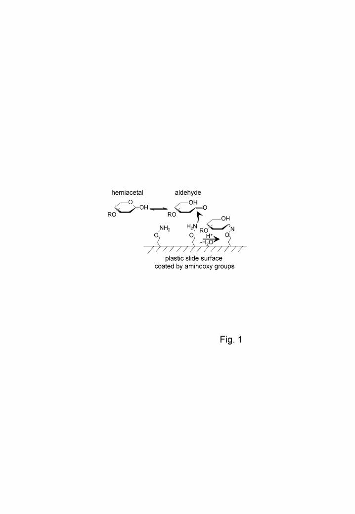
which have no aldehyde group. Therefore, as shown in Fig.1, if the hemiacetal group
can be selectively captured, then glycans can be recovered specifically, differentiating
them chemically from other biomolecules. For example, Furukawa et al. synthesized
functional polymers with hydrazide groups (hydrazide beads) by suspension
polymerization and made a specific collection of glycans [6].
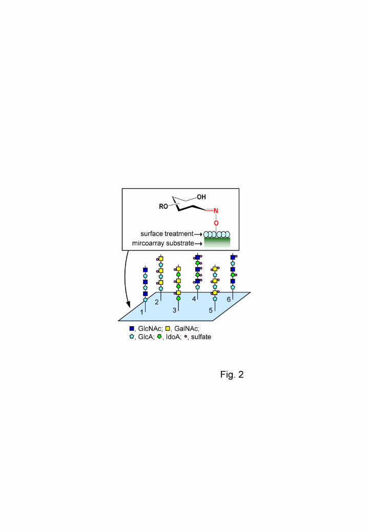
Using the above-described method, a special treatment was applied to the
surface of a plastic substrate (polyolephin). Aminooxy groups were introduced because
they bind more strongly to aldehyde groups present at the reducing termini of the
glycans than hydrazide groups [6]. Thus, after contacting the substrate surface, glycans
with a reducing end could be easily immobilized by applying heat (Fig. 2).
Furthermore, another feature incorporated in the surface treatment of the
substrate is the suppression of non-specific adsorption of biomolecules such as proteins.
When analyzing the interaction, non-specific adsorption of the analyte to the substrate
generates background noise and results in a decrease in detection sensitivity. Therefore,
a blocking treatment is generally used to suppress non-specific adsorption by a solution
such as BSA or skim milk; in such an approach, however, verification of the blocking
solution may be needed to check that it responds sufficiently.
As shown in Supplementary Fig. S1, the coating with methacrylic polymer
prevented efficiently non-specific adsorption of hydrophobic, basic or glycoproteins.
The polymer that we have developed contains phospholipid groups, which are superior
at preventing non-specific adsorption of proteins as compared with general blocking
solution. In this study a glass-like substrate made of cyclic polyolefin was molded into a
slide by Sumitomo Bakelite as the base of the GAG microarray, and was coated by
6
methacrylic polymer [8, 9] for immobilization of glycans. As a result, surface treatment
by this polymer has enabled the detection of interactions via glycans at a high S/N ratio
without a blocking step. Generally, the interaction between a glycan and a protein is
much weaker than that between an antigen and antibody reaction; thus, suppressing the
background noise is especially significant in the analysis of interactions of glycans. The
utility of this GAG microarray as a tool for interaction analysis is described below.
Materials and Methods
Materials
Chondroitin sulfate A (CS-A), CS-B (dermatan sulfate), CS-C, CS-D, CS-E,
heparan sulfate (derived from bovine kidney; HS), heparin (derived from pig intestines;
Hep), and chondroitin (desulfated CS-A; Chn) were purchased from Seikagaku Corp.
(Tokyo, Japan). The hyaluronan oligosaccharide preparation (HA-oligo; molecular mass
of less than 10,000 Da) was obtained from Kewpie Corp. (Tokyo, Japan). Cy3-maltose,
which was used as a grid marker in preparing the GAG microarray, was synthesized by
NARD Institute, Ltd. (Hyogo, Japan).
Recombinant human midkine (hMK), pleiotrophin (hPTN), FGF-1 (acidic FGF,
aa 16-155; hFGF-1), VEGF165 (hVEGF), and HGF (hHGF) were purchased from R&D
Systems (Minneapolis, MN, USA). Recombinant human FGF-2 (basic FGF; hFGF-2)
was purchased from PEPROTECH (Rocky Hill, NJ, USA).
Anti-CS monoclonal antibody (anti-CS mAb; clone CS-56), anti-CS mAb
(clone MO-225), anti-HS mAb (clone F58-10E4) were purchased from Seikagaku Corp.
Anti-hMK mAb, anti-hPTN mAb, and anti-hFGF-1 mAb were purchased from Santa
Cruz Biotechnology (Santa Cruz, CA, USA). Anti-FGF-2 mAb was purchased from
7
PEPROTECH. Anti-hVEGF mAb and anti-hHGF mAb were purchased from R&D
Systems.
Cy3-AffiniPure F(ab')2 fragment of goat anti-mouse IgM was purchased from
Jackson ImmunoResearch (West Grove, PA, USA). Cy3-labeled anti-mouse IgG was
purchased from GE Healthcare UK Ltd. (Little Chalfont, UK). In addition, mouse
monoclonal Ab WF6 (mAb WF6) was prepared by a previously described method [10].
Preparation of the GAG microarray
A glass-like substrate made of cyclic polyolefin was molded into a slide by
Sumitomo Bakelite as the base of the GAG microarray. The size of the slide was 75-mm
long, 25-mm wide and 1-mm thick, and was coated by methacrylic polymer [8, 9] for
immobilization of glycan. The slide was then immersed in 2 M HCl for 4 h at 37 °C, so
that aminooxy groups were present on the surface of the substrate. The slide was
washed twice with water, and dried by centrifugation.
Nine kinds of GAG were immobilized on the substrate: CS-A, CS-B, CS-C,
CS-D, CS-E, HS, Hep, Chn (desulfated CS-A), and HA-oligo. They were prepared at a
concentration of 0.9 mg/mL in spotting buffer (100 mM acetate buffer, pH 5.0,
containing 0.01% Triton X-100 and 0.01% polyvinyl alcohol [Mw = 1,500] at a final
concentration). Next, the GAG solutions were spotted (n = 3) on the substrate using a
BioChip Arrayer (Filgen, Aichi, Japan) [11]. About 1 ~ 10 nL of GAG solution was
applied per spot, corresponding to a GAG weight of 0.9 ~ 9 ng. This meant that, for a
GAG with an average molecular mass of 20,000 Da, the amount on the array would be
0.05 ~ 0.5 pmol. After spotting, the substrate was placed in an oven for 1 h at 80 °C to
immobilize the GAGs. After the reaction, it was washed once with water, and then
8
immersed in an aqueous solution of 10 mg/mL succinic anhydride for 1 h at room
temperature to cap unreacted aminooxy groups. Finally, it was washed twice with water,
and dried by centrifugation.
Assay with commercial reagents
Binding assays for anti-GAG antibodies
Binding assays for anti-GAG antibodies were performed as follows: (1) A
hybridization cover (Sumitomo Bakelite, Tokyo, Japan) was mounted onto a slide, and
defined concentrations of anti-GAG antibody in a reaction buffer (50 mM Tris-HCl, pH
7.4, containing 100 mM NaCl, 1 mM CaCl2, 1 mM MnCl2, 1 mM MgCl2, 0.05% Tween
20) (70 L) were added. (2) The slide was incubated at room temperature for 2 h. (3)
The hybridization cover was removed and the slide was washed with washing buffer (50
mM Tris-HCl, pH 7.4, containing 100 mM NaCl, 1 mM CaCl2, 1 mM MnCl2, 1 mM
MgCl2) for 2 min (once) and twice with water for 1 min. (4) The slide was dried by
centrifugation (2,000 rpm, 2 min, room temperature). (5) A new hybridization cover was
mounted onto the slide and 5 g/mL Cy3-labeled goat anti-mouse IgM (70 L) was
added. (6) The slide was incubated at room temperature for 1 h. (7) The hybridization
cover was removed, and the slide was washed with washing buffer for 2 min (once) and
twice with water for 1 min. (8) The slide was dried by centrifugation (2,000 rpm, 2 min,
room temperature), and the fluorescent intensity of the spots was measured.
Binding assays for growth factors
Binding assays for growth factors were performed as described for anti-GAG
antibodies except that 5 g/mL mouse anti-human growth factor antibody (70 L) was
9
used instead of anti-GAG antibody, and 5 g/mL Cy3-labeled goat anti-mouse IgG (70
L) was used instead of Cy3-labeled goat anti-mouse IgM.
Results
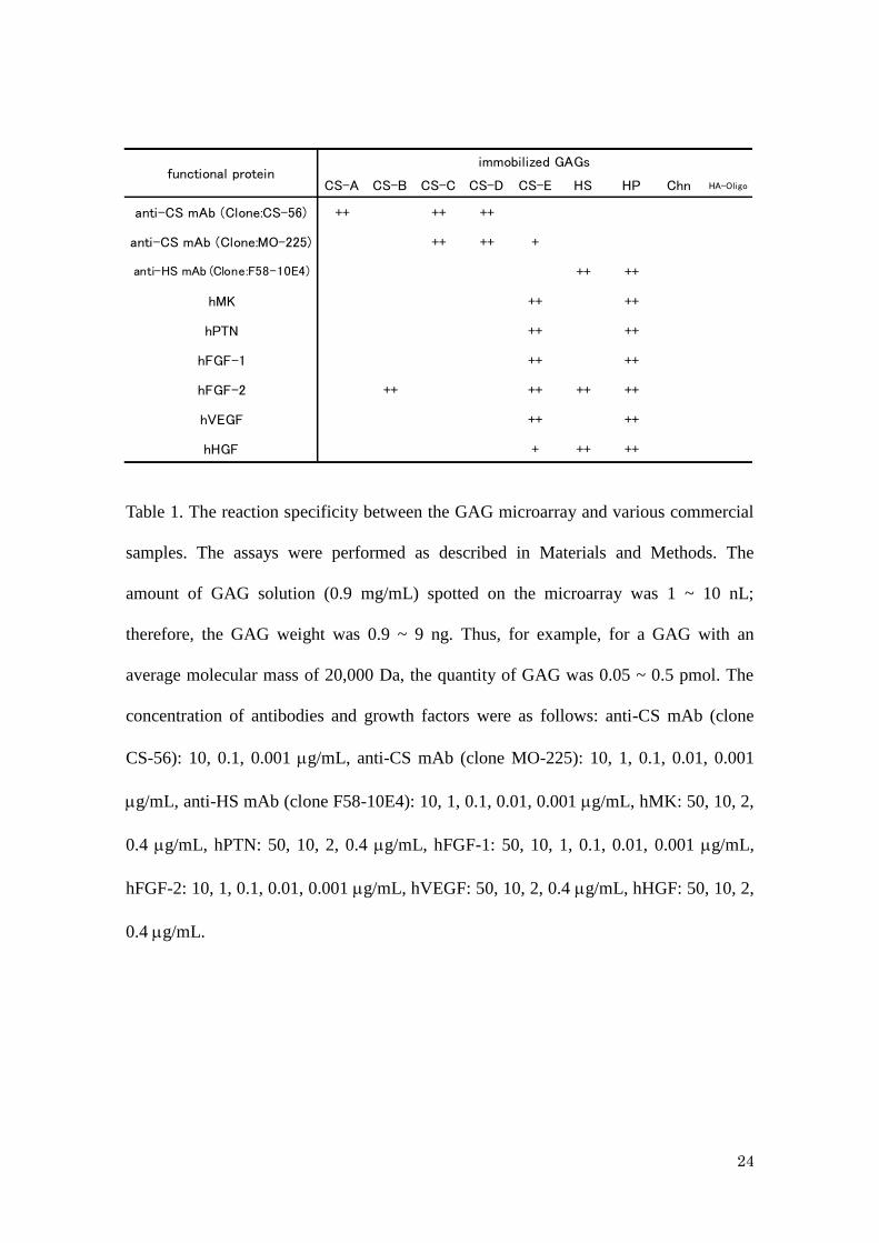
Interaction with commercially available antibodies and growth factors using GAG
microarray
In order to investigate the performance of the newly developed GAG
microarray, we examined its substrate specificity for commercially available antibodies
(3 kinds) and growth factors (6 kinds). As described in the Materials and Methods, the
amount of GAG on the microarray was 0.05 ~ 0.5 pmol. The concentrations of
antibodies and growth factors were varied as described in the legend of Table 1.
Interactions between the antibodies or growth factors and GAGs are indicated in Table 1
(observed interactions are described as “+”), and the main results for each sample are
summarized below.
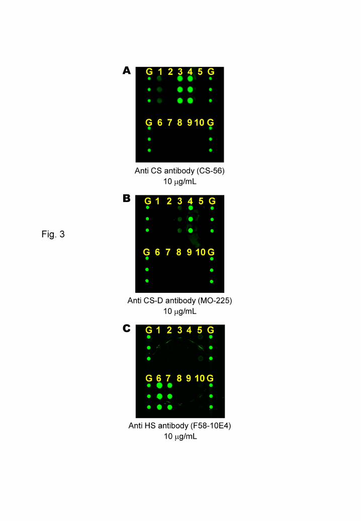
Antibodies
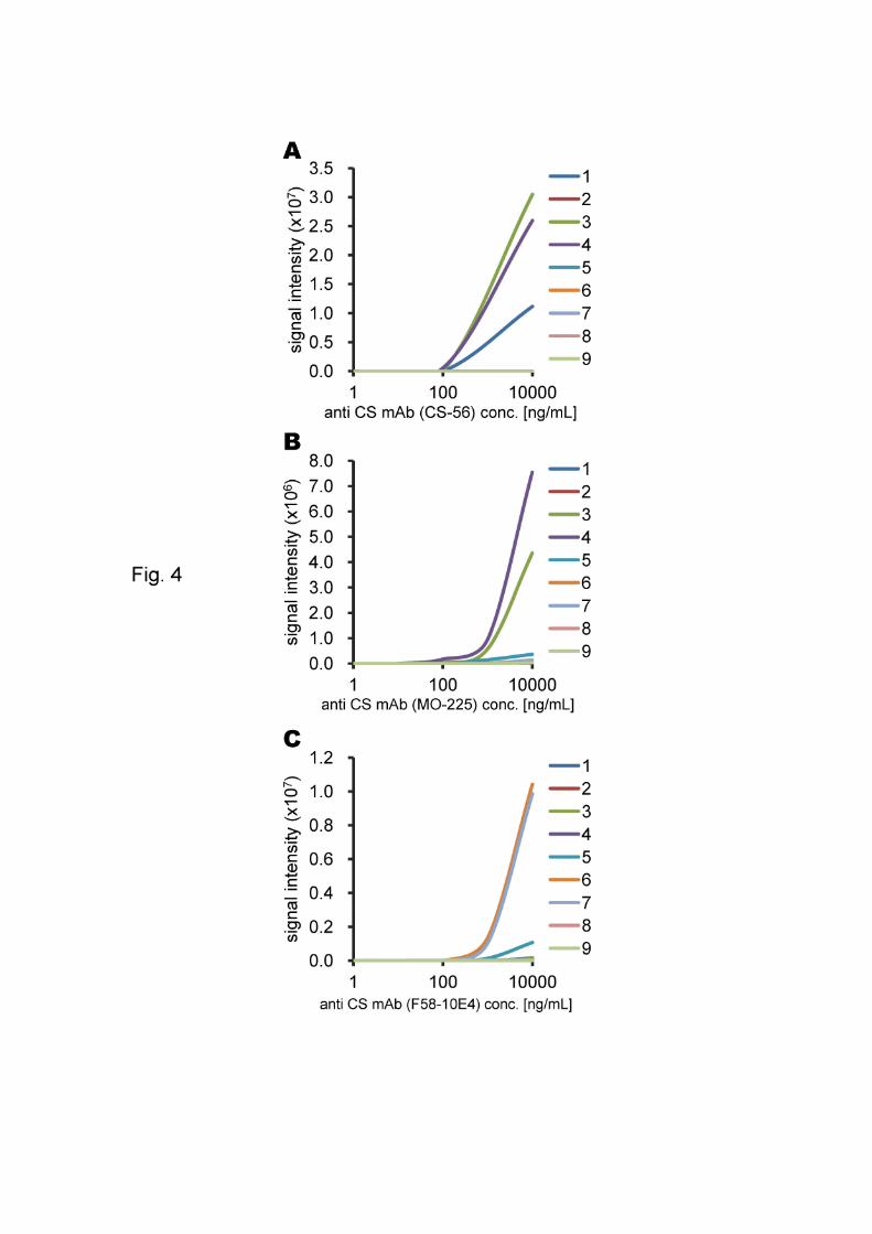
Anti-CS mAb (clone CS-56) reacted with CS-A, CS-C and CS-D (Fig. 3-A).
The detection limit varied between 1 and 100 ng/mL of antibody for CS-C and CS-D,
and between 0.1 and 10 g/mL for CS-A (Fig. 4-A). Anti-CS mAb (clone MO-225)
reacted with CS-C, CS-D and CS-E (Fig. 3-B). The detection limit varied between 0.1
and 1 g/mL of antibody for CS-C and CS-D, and between 1 and 10 g/mL for CS-E
(Fig. 4-B). Anti-HS mAb (clone F58-10E4) reacted with HS and Hep (Fig. 3-C) with
detection limits of 0.1 ~ 1 g/mL of antibody for both GAGs (Fig. 4-C).
10
Growth factors
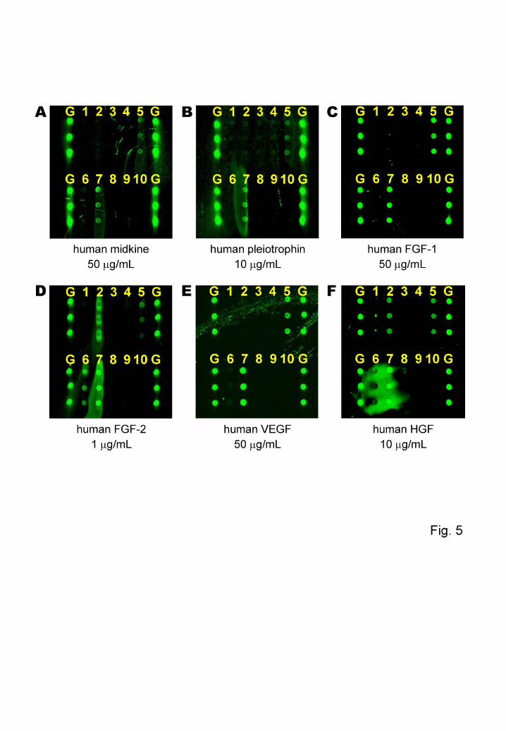
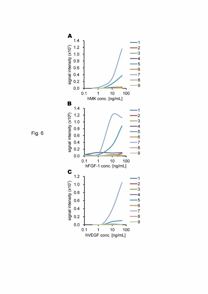
The growth factor hMK reacted with CS-E and Hep (Fig. 5-A) with a detection
limit of 0.4 ~ 2 g/mL of growth factor for Hep, and 2 ~ 10 g/mL for CS-E (Fig. 6-A).
For hPTN, interactions were observed with CS-E and Hep (Fig. 5-B). In this assay,
signals were observed for Hep even at a concentration of 0.4 g/mL, which was the
minimum growth factor concentration tested. By contrast, the detection limit for CS-E
was 10 ~ 50 g/mL of growth factor.
hFGF-1 reacted with CS-E and Hep (Fig. 5-C). The detection limit varied
between 1 and 10 g/mL of growth factor for Hep and between 10 ~ 50 g/mL for CS-E
(Fig. 6-B). The growth factor hFGF-2 reacted with CS-B, CS-E, HS and Hep (Fig. 5-D).
In this assay, the detection limit varied between 0.01 and 0.1 g/mL of growth factor for
Hep, and between 0.1 and 1 g/mL for CS-B, CS-E and HS. For CS-A, signals were
observed at a growth factor concentration of 10 g/mL, which was the maximum
concentration tested, but it was judged that this was a nonspecific reaction.
hVEGF reacted with CS-E and Hep (Fig. 5-E) with detection limits of 0.4 ~ 2
g/mL of growth factor for Hep and 2 ~ 10 g/mL for CS-E (Fig. 6-C). For hHGF,
interactions were observed with CS-B, CS-E, HS and Hep (Fig. 5-F). In this assay,
signals were observed for Hep even at a growth factor concentration of 0.4 g/mL,
which was the minimum concentration tested. In addition, signal levels were higher at
10 g/mL than at 50 g/mL. For the other GAGs that interacted, the detection limit was
around 10 g/mL of growth factor.
Interaction with mAb WF6 using GAG microarray
11
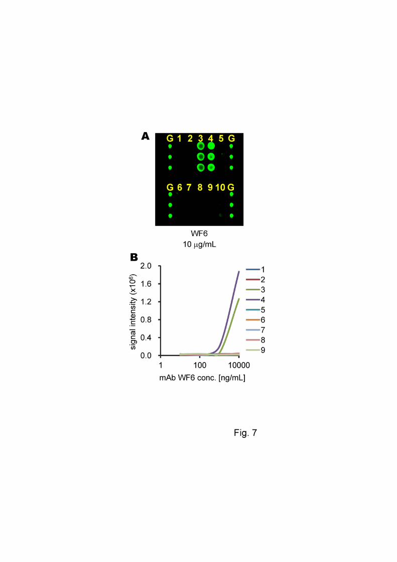
For mAb WF6, which was recently generated for screening of diseases
including osteoarthritis, rheumatoid arthritis and ovarian cancer [10, 12], the assay was
carried out on the GAG microarray in the same manner as described above. Specific
reactions with CS-C and CS-D were observed for mAb WF6 as shown in Fig. 7(A). In
addition, these interactions demonstrated a concentration dependence as shown in Fig.
7(B).
Discussion
Growth factors function in the regulation of various physiological and
cytological processes, and act as signals between cells by specifically binding to a
receptor protein on the surface of a target cell. In general, most growth factors have
specific interactions with GAGs, and we therefore considered that the utility of the
GAG microarray as an analytical tool would be shown if we could confirm these
specific interactions on the microarray.
With regard to the antibodies tested, anti-CS mAb (clone CS-56) is specific for
CS and is widely used in immunohistochemical studies of various tissues [13, 14]. It
mainly recognizes CS-A and CS-C [15], and has subsequently been found to recognize
sequences containing the A-D sequence [16]. Given these facts, the present results (in
which it reacted with CS-A, CS-C and CS-D) are reasonable.
Similarly, anti-CS mAb (clone MO-225) is also specific for CS and is widely
used in immunohistochemical studies of various tissues. It recognizes the determinant
on the CS (dual structure of D-GlcA-2-O-sulfate(1-3)GalNAc-6-O-sulfate: D-unit)
[17], and has also been found to recognize sequences containing the A-D sequence [16].
Therefore, the present results (in which it reacted with CS-C, CS-D and CS-E) are also
12
reasonable. Anti-HS mAb (clone F58-10E4) recognizes GlcNS in HS [18, 19]. It does
not bind to HA, CS, CS-B, or keratan sulfate. Consistent with this, clone F58-10E4
reacted with HS and Hep in the present study.
Regarding the growth factors tested, hMK is an ~13-kDa protein with an
abundance of cysteine and basic amino acids, and consists of two kringle domains at the
C-terminus and N-terminus. It is mainly the C-terminal kringle domain that plays a role
in the activity of hMK, and this domain contains two Hep-binding sites [20-22]. hMK
binds strongly to regions of over-sulfated HS, as well as the tri-sulfated structure and
E-type structure of CS [23-26]. Given these observations, the present results (in which
hMK reacted with CS-E and Hep) are reasonable. hPTN is a protein forms a unique
gene family with hMK, and about 50% of amino acid sequence is shared between hPTN
and hMK [20]. In keeping with this, hPTN reacted with CS-E and Hep on the present
GAG microarray.
The FGF family comprises polypeptides of 17 ~ 26 kDa, such as FGF-1 and
FGF-2; at least 22 kinds of FGFs have been confirmed in human. It is considered that
most FGF proteins that are secreted outside the cell bind HS chains that exist in the
extracellular matrix [27, 28]. FGF-1 is an ~17-kDa Hep-binding growth factor, and the
presence of many 6-O-sulfate groups is important to interact specifically with Hep/HS.
By contrast, FGF-2 does not need these groups [29]. In particular, a size of more than 10
residues and a structure rich in iduronic acid(2-O-sulfate)-GlcNS(6-O-sulfate) are
required for the specific interaction between FGF-1 and HS [30]. In addition, FGF-1
does not bind to CS-E [31]. In the present results, however, the observed binding
between hFGF-1 and CS-E differs from previous reports and will be the subject of
future investigation. Other results (in which hFGF-1 reacted with Hep) are reasonable.
13
FGF-2 is an ~18-kDa Hep-binding growth factor of 154 amino acids [32]. FGF-2 binds
HS as strong as Hep if the HS has more than 10 sugar residues and has a structure rich
in iduronic acid(2-O-sulfate)-GlcNS [33, 34]. In addition, FGF-2 binds to CS-B
containing iduronic acid, and highly sulfated CS-E [31, 35]. Therefore, the present
results (in which FGF-2 reacted with CE-B, CS-E, HS and Hep) are sound.
VEGF is an ~22-kDa Hep-binding growth factor, which has a dimer structure
consisting of subunits and a Hep-binding site in the C-terminus. VEGF has subfamilies
termed A to E, and it binds one of 3 kinds of VEGF receptors (VEGF-R1, -R2, -R3) to
transfer its signal [36]. VEGF exists as an isoform of 5 different sizes (VEGF121,
VEGF145, VEGF165, VEGF189, VEGF206, where the subscript indicates the number of
constituent amino acids) [37]. VEGF-A165, which exists most in vivo, binds to
VEGF-R1 and -R2, but not to VEGF-R3. It is known that VEGF specifically binds to
CS-E [38]. In keeping with these observations, hVEGF reacted with CS-E and Hep in
the present study. HGF is a Hep-binding growth factor. It is heterodimer, in which an
~69-kDa -chain (heavy chain) and ~34-kDa -chain (light chain) are bound by an S-S
bond [39]. It binds to CS-B [40, 41]. In the present results, hHGF was found to react
with CS-B, CS-E, HS and Hep.
Because the above data confirmed that the GAG microarray that we developed
was able to detect interactions properly, we examined the specific interaction of an
antibody that is commercially unavailable. WF6 is a useful antibody that can be used to
diagnose ovarian cancer, osteoarthritis, and rheumatoid arthritis. It has reaction
specificity with CS-C and CS-D, and it recognizes their special 8-sugar sequence [42].
The present results, in which WF6 interacted with CS-C and CS-D, were consistent with
previously published characteristics [42]. Thus, these results collectively showed that
14
the GAG microarray that we have developed can be used as a tool for the analysis of the
interactions between GAGs and protein.
As described above, we immobilized GAGs chemically on the substrate by first
applying a surface treatment (presenting aminooxy groups) and then applying the
Glycoblotting method. This means that we achieved not only the specific
immobilization of glycans such as GAGs but also position-selective immobilization by
binding GAGs at their reducing end. In addition, because there was no need to make
modifications to the GAGs, this method has many benefits. For example, it would be
possible to immobilize non-modified GAGs extracted from a biological sample after
treatment with weak alkali such as 0.5 M LiOH to release GAGs from the core protein
or peptide [43, 44]. In addition, by making this microarray, the interaction analysis
could be carried out with a very small amount of GAGs and proteins. Because GAGs
extracted from natural products are often available in only tiny amounts or are
expensive, it is a considerable advantage that a small quantity of sample is used.
The present microarray is based on a plastic substrate. Plastic has the advantage
that it can be processed into various shapes such as a slide glasses, microplates, and
microfluidic formats with a fine flow channel. As a result, a plastic substrate can be
provided in a format suitable for the measurement needs and can be easily
mass-produced at low cost. In the future, when use of microarrays for diagnostic
purposes becomes widespread, we think that the plastic-based microarray will become
mainstream because it is inexpensive and disposable.
On the basis of the present screening results, a more detailed analysis using the
same type of GAGs with different degrees of sulfation and molecular weight would
bring deeper knowledge of molecular biology. In the future, we will also develop such
15
GAG microarrays as a variation of the technique.
As mentioned above, using the newly developed GAG microarray enabled us
to detect multiple interactions with a small amount of sample and without modification
of the GAGs, and to expedite the interaction analysis. As a result, this microarray will
contribute to future studies of the biological functions of GAGs.
Conclusions
To achieve rapid, sensitive and high-throughput screening for molecular
interactions of GAGs, we have developed a novel plastic microarray that can be
immobilized with various GAGs at their reducing end without modification. Using this
microarray, we confirmed specific interactions between GAGs and commercially
available growth factors and antibodies. Furthermore, we demonstrated the specificity
toward CS-C and CS-D of an antibody (WF6) that can be applied to the diagnosis of
ovarian cancer, osteoarthritis, and rheumatoid arthritis [10, 12].
This microarray will be useful for developing diagnostic methods for diseases,
in which growth factors, cytokines, and/or chemokines are secreted, because only
minute amounts of serum samples are required. It can be expected that this microarray
will accelerate molecular biological research and drug discovery involving GAGs for a
variety of pathological conditions.
Conflict of Interest
The authors declare no conflict of interest.
Acknowledgements
16
This work was supported, in part, by Grants-in-aid for Scientific Research on
Innovative Areas (24110501) (to K.S.) from the Ministry of Education, Culture, Sports,
Science, Technology of Japan (MEXT), and by the Japan-Thailand Research
Cooperative Program (to K. S., P. P., and P. K.) from the Japan Society for the
Promotion of Science and the National Research Council of Thailand (JSPS-NRCT).
17
References
[1] O. Blixt, S. Head, T. Mondala, C. Scanlan, M.E. Huflejt, R. Alvarez, M.C. Bryan, F.
Fazio, D. Calarese, J. Stevens, N. Razi, D.J. Stevens, J.J. Skehel, I. Die, D.R. Burton, I.
A. Wilson, R. Cummings, N. Bovin, C-H. Wong, J.C. Paulson, Printed covalent glycan
array for ligand profiling of diverse glycan binding proteins, Proc. Natl. Acad. Sci. USA
101 (2004) 17033-17038.
[2] S. Park, I. Shin, Fabrication of carbohydrate chips for studying protein-carbohydrate
interactions, Angew. Chem. Int. Ed. 41 (2002) 3180-3182.
[3] F. Fazio, M.C. Bryan, O. Blixt, J.C. Paulson, C-H. Wong, Synthesis of sugar arrays
in microtiter plate, J. Am. Chem. Soc. 124 (2002) 14397-14402.
[4] H. Tanaka, WO Patent WO2000039582.
[5] H. Tanaka, Patent JP 2004-275862.
[6] J-I. Furukawa, Y. Shinohara, H. Kuramoto, Y. Miura, H. Shimaoka, M. Kurogochi,
M. Nakano, S-I. Nishimura, Comprehensive approach to structural and functional
glycomics based on chemoselective glycoblotting and sequential tag conversion,
Anal.Chem. 80 (2008) 1094–1101.
[7] Y. Miura, Y. Shinohara, J-I. Furukawa, N. Nagahori. S-I. Nishimura, Rapid and
simple solid-phase esterification of sialic acid residues for quantitative glycomics by
mass spectrometry, Chem. Eur. J. 13 (2007) 4797-4804.
[8] T. Matsumoto, Y. Fukunishi, H. Kuramoto, K. Fujiwara, S. Funaoka, Patent JP
2007-326920.
[9] S-I. Nishimura, H. Shimaoka, WO Patent WO2005097844.
[10] P. Pothacharoen, S. Siriaunkgul, S. Ong-Chai, J. Supabandhu, P. Kumja, C.
Wanaphirak, K. Sugahara, T. Hardingham, P. Kongtawelert, Raised serum chondroitin
18
sulfate epitope level in ovarian epithelial cancer, J. Biochem. 140 (2006) 517-524.
[11] D. Rose, Microfluidic technologies and instrumentation for printing DNA
microarrays. In: M. Schena (ed), “Microarray Biochip Technology”, Eaton Publishing
ISBN1-8812-9937-6 (2000) Chapter 2.
[12] P. Pothacharoen, S. Teekachunhatean, W. Louthrenoo, W. Yingsung, S. Ong-Chai,
T. Hardingham, P. Kongtawelert, Raised chondroitin sulfate epitopes and hyaluronan in
serum from rheumatoid arthritis and osteoarthritis patients. Osteoarthritis Cartilage, 14
(2006) 299-301.
[13] J.M. Sorreli, D.A. Carrino, A.I. Caplan, Regulated expression of chondroitin
sulfates at sites of epithelial-mesenchymal interaction: spatio-temporal patterning
identified with anti-chondroitin sulfate monoclonal antibodies, Int. J. Dev. Neurosci. 14
(1996) 233-248.
[14] Y. Shimazaki, I. Nagata, M. Ishii, M. Tanaka, T. Marunouchi, T. Hata, N. Maeda,
Developmental change and function of chondroitin sulfate deposited around cerebellar
Purkinje cells, J. Neurosci. Res. 82 (2005) 172-183.
[15] Z. Avnur, B. Geiger, Immunocytochemical localization of native
chondroitin-sulfate in tissues and cultured cells using specific monoclonal antibody,
Cell 38 (1984) 811-822.
[16] Y. Ito, M. Hikino, Y. Yajima, T. Mikami, S. Sirko, A. von Holst, A. Faissner, S.
Fukui, K. Sugahara, Structural characterization of the epitopes of the monoclonal
antibodies 473HD, CS-56, and MO-225 specific for chondroitin sulfate D-type using
the oligosaccharide library, Glycobiology 15 (2005) 593-603.
[17] M. Yamagata, K. Kimata, Y. Oike, K. Tani, N. Maeda, K. Yoshida, Y. Shimomura,
M. Yoneda, S. Suzuki, A monoclonal antibody that specifically recognizes a glucuronic
19
acid 2-sulfate-containing determinant in intact chondroitin sulfate chain, J. Biol. Chem.
262 (1987) 4146-4152.
[18] David, X.M. Bai, B. Van der Schueren, J.J. Cassiman, H. Van den Berghe,
Developmental changes in heparan sulfate expression: in situ detection with mAbs, J.
Cell Biol. 119 (1992) 961-975.
[19] X.M. Bai, B. Van der Schueren, J.J. Cassiman, H. Van den Berghe, G. David,
Differential expression of multiple cell-surface heparan sulfate proteoglycans during
embryonic tooth development, J. Histochem. Cytochem. 42 (1994) 1043-1054.
[20] T. Muramatsu, Midkine (MK), the product of a retinoic acid responsive gene, and
pleiotrophin constitute a new protein family regulating growth and differentiation, Int. J.
Dev. Biol. 37 (1993) 183-188.
[21] T. Muramatsu, Midkine and pleiotrophin: Two related proteins involved in
development, survival, inflammation and tumorigenesis, J. Biochem. 132 (2002)
359-371.
[22] Wiley Encyclopedia Mol. Med. (2002) 2086-2088, ISBN 978-0-4712-0307-0.
[23] N. Kaneda, A.H. Talukdera, M. Ishihara, S. Hara, K. Yoshida, T. Muramatsu,
Structural characteristics of heparin-like domain required for interaction of midkine
with embryonic neurons, Biochem. Biophys. Res. Commun. 220 (1996) 108-112.
[24] C. Ueoka, N. Kaneda, I. Okazaki, S. Nadanaka, T. Muramatsu, K. Sugahara,
Neuronal cell adhesion, mediated by the heparin-binding neuroregulatory factor
midkine, is specifically inhibited by chondroitin sulfate E, J. Biol. Chem. 275 (2000)
37407-37413.
[25] P. Zou, K. Zou, H. Muramatsu, K. Ichihara-Tanaka, O. Habuchi, S. Ohtake, S.
Ikematsu, S. Sakuma, T. Muramatsu, Glycosaminoglycan structures required for strong
20
binding to midkine, a heparin-binding growth factor, Glycobiology 13 (2003) 35-42.
[26] K. Zou, H. Muramatsu, S. Ikematsu, S. Sakuma, R.H.M. Salama, T. Shinomura, K.
Kimata, T. Muramatsu, A heparin-binding growth factor, midkine, binds to a chondroitin
sulfate proteoglycan, PG-M/versican, Eur. J. Biochem. 267 (2000) 4046-4053.
[27] S.R. Yu, M. Burkhardt, M. Nowak, J. Ries, Z. Petrasek, S. Scholpp, P. Schwille, M.
Brand, Fgf8 morphogen gradient forms by a source-sink mechanism with freely
diffusing molecules, Nature 461 (2009) 533-536.
[28] J.R. Bishop, M. Schuksz, J.D. Esko, Heparan sulphate proteoglycans fine-tune
mammalian physiology, Nature 446 (2007) 1030-1037.
[29] M. Ishihara, P.N. Shaklee, Z. Yang, W. Liang, Z. Wei, R.J. Stack, K. Holme,
Structural features in heparin which modulate specific biological activities mediated by
basic fibroblast growth factor, Glycobiology 4 (1994) 451-458.
[30] M. Ishihara, Structural requirements in heparin for binding and activation of FGF-1
and FGF-4 are different from that for FGF-2, Glycobiology 4 (1994) 817-824.
[31] S.S. Deepa, Y. Umehara, S. Higashiyama, N. Itoh, K. Sugahara, Specific molecular
interactions of oversulfated chondroitin sulfate E with various heparin-binding growth
factors, J. Biol. Chem. 277 (2002) 43707-43716.
[32] A. Yayon, M. Klagsbrun, J.D. Esko, P. Leder, D.M. Ornitz, Cell surface,
heparin-like molecules are required for binding of basic fibroblast growth factor to its
high affinity receptor, Cell 64 (1991) 841-848.
[33] J.E. Turnbull, D.G. Fernig, Y. Ke, M.C. Wilkinson, J.T. Gallagher, Identification of
the basic fibroblast growth factor binding sequence in fibroblast heparan sulfate, J. Biol.
Chem. 267 (1992) 10337-10341.
[34] H. Habuchi, S. Suzuki, T. Saito, T. Tamura, T. Harada, K. Yoshida, K. Kimata,
21
Structure of a heparan sulphate oligosaccharide that binds to basic fibroblast growth
factor, Biochem. J. 285 (1992) 805-813.
[35] K.R. Taylor, J.A. Rudisill, R.L. Gallo, Structural and sequence motifs in dermatan
sulfate for promoting fibroblast growth factor-2 (FGF-2) and FGF-7 activity, J. Biol.
Chem. 280 (2005) 5300–5306.
[36] K. Holmes, O.L. Roberts, A.M. Thomas, M.J. Cross, Vascular endothelial growth
factor receptor-2: structure, function, intracellular signalling and therapeutic inhibition,
Cell. Signal. 19 (2007) 2003-2012.
[37] E. Tischer, R. Mitchell, T. Hartman, M. Silva, D. Gospodarowicz, J.C. Fiddes, J.A.
Abraham, The human gene for vascular endothelial growth factor. Multiple protein
forms are encoded through alternative exon splicing, J. Biol. Chem. 266 (1991)
11947-11954.
[38] G.B. ten Dam, E.M.A. van de Westerlo, A. Purushothaman, R.V. Stan, J. Bulten,
F.C.G.J. Sweep, L.F. Massuger, K. Sugahara, T.H. van Kuppevelt, Antibody GD3G7
selected against embryonic glycosaminoglycans defines chondroitin sulfate-E domains
highly up-regulated in ovarian cancer and involved in VEGF binding, Am. J. Pathol.
171 (2007) 1324-1333.
[39] T. Nakamura, T. Nishizawa, M. Hagiya, T. Seki, M. Shimonishi, A. Sugimura, K.
Tashiro, S. Shimizu, Molecular cloning and expression of human hepatocyte growth
factor, Nature 342 (1989) 440-443.
[40] M. Lyon, J.A. Deakin, J.T. Gallagher, The Mode of action of heparan and dermatan
sulfates in the regulation of hepatocyte growth factor/scatter factor, J. Biol. Chem. 277
(2002) 1040-1046.
[41] F. Li, A.K. Shetty, K. Sugahara, Neuritogenic activity of chondroitin/dermatan
22
sulfate hybrid chains of embryonic pig brain and their mimicry from shark liver, J. Biol.
Chem. 282 (2007) 2956-2966.
[42] P. Pothacharoen, K. Kalayanamitra, S.S. Deepa, S. Fukui, T. Hattori, N. Fukushima,
T. Hardingham, P. Kongtawelert, K. Sugahara, Two related but distinct chondroitin
sulfate mimetope octasaccharide sequences recognized by monoclonal antibody WF6, J.
Biol. Chem. 282 (2007) 35232-35246.
[43] D. Heinegard, Hyaluronidase digestion and alkaline treatment of bovine tracheal
cartilage proteoglycans. Isolation and characterisation of different keratan sulfate
proteins, Biochim. Biophys. Acta 285 (1972) 193-207.
[44] H. Sakaguchi, M. Watanabe, C. Ueoka, E. Sugiyama, T. Taketomi, S. Yamada, K.
Sugahara, Isolation of reducing oligosaccharide chains from the chondroitin/dermatan
sulfate-protein linkage region and preparation of analytical probes by fluorescent
labeling with 2-aminobenzamide, J. Biochem. 129 (2001) 107-118.
23
Abbreviations:
GAG, glycosaminoglycan; ELISA, enzyme-linked immunosorbent assay; BSA, bovine
serum albumin; S/N, signal/noise; CS, chondroitin sulfate; HS, heparan sulfate; Hep,
heparin; Chn, chondroitin; HA, hyaluronan; MK, midkine; PTN, pleiotrophin; FGF,
fibroblast growth factor; VEGF, vascular endothelial growth factor; HGF, hepatocyte
growth factor; mAb, monoclonal antibody; Cy3, cyanine 3; Ig, immunoglobulin; Mw,
molecular weight; GlcA, D-glucuronic acid; GalNAc, N-acetyl-D-galactosamine; GlcNS,
N-sulfated-D-glucosamine
24
Table 1. The reaction specificity between the GAG microarray and various commercial
samples. The assays were performed as described in Materials and Methods. The
amount of GAG solution (0.9 mg/mL) spotted on the microarray was 1 ~ 10 nL;
therefore, the GAG weight was 0.9 ~ 9 ng. Thus, for example, for a GAG with an
average molecular mass of 20,000 Da, the quantity of GAG was 0.05 ~ 0.5 pmol. The
concentration of antibodies and growth factors were as follows: anti-CS mAb (clone
CS-56): 10, 0.1, 0.001 g/mL, anti-CS mAb (clone MO-225): 10, 1, 0.1, 0.01, 0.001
g/mL, anti-HS mAb (clone F58-10E4): 10, 1, 0.1, 0.01, 0.001 g/mL, hMK: 50, 10, 2,
0.4 g/mL, hPTN: 50, 10, 2, 0.4 g/mL, hFGF-1: 50, 10, 1, 0.1, 0.01, 0.001 g/mL,
hFGF-2: 10, 1, 0.1, 0.01, 0.001 g/mL, hVEGF: 50, 10, 2, 0.4 g/mL, hHGF: 50, 10, 2,
0.4 g/mL.
CS-A CS-B CS-C CS-D CS-E HS HP Chn HA-Oligo
anti-CS mAb (Clone:CS-56) ++ ++ ++
anti-CS mAb (Clone:MO-225) ++ ++ +
anti-HS mAb (Clone:F58-10E4) ++ ++
hMK ++ ++
hPTN ++ ++
hFGF-1 ++ ++
hFGF-2 ++ ++ ++ ++
hVEGF ++ ++
hHGF + ++ ++
immobilized GAGsfunctional protein
25
Figure Legends
Fig. 1. Using their aldehyde group at the reducing terminus, GAGs were covalently and
chemoselectively linked to the NH2 of an aminooxy group on a solid surface via oxime
bond formation under weak acidic conditions [6, 7].
Fig. 2. Patterns of a GAG microarray based on the glycoblotting method. The aminooxy
group present on the surface of the substrate reacts chemically and selectively with the
hemiacetal group at the reducing end of the sugar chain. 1, HA-oligo; 2, CS-A; 3, CS-B;
4, Hep; 5, CS-E; 6, HS, GlcNAc, N-acetyl-D-glucosamine; GalNAc,
N-acetyl-D-galactosamine; GlcA, D-glucuronic acid; IdoA, L-iduronic acid.
Fig. 3. Images of the fluorescence scanning of slides. The slides were imaged at 10-mm
resolution on a Typhoon TRIO+ variable mode imager (GE Healthcare UK Ltd., Little
Chalfont, UK) with a green (532 nm) laser and a 580 BP 30 filter at a PMT voltage of
600V and normal sensitivity. Lane 1, CS-A; lane 2, CS-B; lane 3, CS-C; lane 4, CS-D;
lane 5, CS-E; lane 6, HS; lane 7, Hep; lane 8, Chn; lane 9, HA-oligo; lane 10, buffer
only; lane G, grid marker (Cy3-maltose).
Fig. 4. The correlation between antibody concentration and signal intensity. The slides
were imaged at 10-mm resolution on a Typhoon TRIO+ variable mode imager (GE
Healthcare) with a green (532 nm) laser and a 580 BP 30 filter at a PMT voltage of
600V and normal sensitivity. Lane 1, CS-A; lane 2, CS-B; lane 3, CS-C; lane 4, CS-D;
lane 5, CS-E; lane 6, HS; lane 7, Hep; lane 8, Chn; lane 9, HA-oligo; lane 10, buffer
only; lane G, grid marker (Cy3-maltose).
26
Fig. 5. Images of the fluorescence scanning of slides. The slides were imaged at 10-mm
resolution on a Typhoon TRIO+ variable mode imager (GE Healthcare) with a green
(532 nm) laser and a 580 BP 30 filter at a PMT voltage of 600V and normal sensitivity.
Lane 1, CS-A; lane 2, CS-B; lane 3, CS-C; lane 4, CS-D; lane 5, CS-E; lane 6, HS; lane
7, Hep; lane 8, Chn; lane 9, HA-oligo; lane 10, buffer only; lane G, grid marker
(Cy3-maltose).
Fig. 6. The correlation between each growth factor concentration and signal intensity.
Representative examples are shown. The slides were imaged at 10-mm resolution on a
Typhoon TRIO+ variable mode imager (GE Healthcare) with a green (532 nm) laser
and a 580 BP 30 filter at a PMT voltage of 600V and normal sensitivity. Lane 1, CS-A;
lane 2, CS-B; lane 3, CS-C; lane 4, CS-D; lane 5, CS-E; lane 6, HS; lane 7, Hep; lane 8,
Chn; lane 9, HA-oligo; lane 10, buffer only; lane G, grid marker (Cy3-maltose).
Fig. 7. The specificity and concentration dependency of mAb WF6. (A) Images of the
fluorescence scanning of slides. (B) The correlation between antibody concentration and
signal intensity. The slides were imaged at 10-mm resolution on a Typhoon TRIO+
variable mode imager (GE Healthcare) with a green (532 nm) laser and a 580 BP 30
filter at a PMT voltage of 600V and normal sensitivity. Lane 1, CS-A; lane 2, CS-B;
lane 3, CS-C; lane 4, CS-D; lane 5, CS-E; lane 6, HS; lane 7, Hep; lane 8, Chn; lane 9,
HA-oligo; lane 10, buffer only; lane G, grid marker (Cy3-maltose).

27


![Sulfated zirconia[1]](https://img.pdfslide.net/doc/110x75/5568f2ecd8b42aff2e8b4932/sulfated-zirconia1.jpg)