Embed Size (px)
Citation preview
Proc. Natl. Acad. Sci. USAVol. 86, pp. 5257-5261, July 1989Biochemistry
cDNA isolated from a human T-cell library encodes a member ofthe protein-tyrosine-phosphatase family
(trosine phosphorylation/dephosphorylation/phosphatase/T-cell cDNA)
DEBORAH E. COOL*, NICHOLAS K. TONKSt, HARRY CHARBONNEAUt, KENNETH A. WALSHt,EDMOND H. FISCHERt, AND EDWIN G. KREBS***Howard Hughes Medical Institute, SL-15; and tDepartment of Biochemistry, SJ-70, University of Washington, Seattle, WA 98195
Contributed by Edwin G. Krebs, April 17, 1989
ABSTRACT A human peripheral T-ceil cDNA library wasscreened with two labeled synthetic oligonucleotides encodingregions of a human placenta protein-tyrosine-phosphatase(protein-tyrosine-phosphate phosphohydrolase, EC 3.1.3.48).One positive clone was isolated and the nucleotide sequence wasdetermined. It contained 1305 base pairs ofopen reading framefollowed by a TAA stop codon and 978 base pairs of 3'untranslated end, although a poly(A)+ tail was not found. Aninitiator methionine residue was predicted at position 61, whichwould result in a protein of 415 amino acid residues (Mr,48,400). This was supported by the synthesis of a Mr 48,000protein in an in vitro reticulocyte lysate translation system usingRNA transcribed from the cloned cDNA and T7 RNA poly-merase. The deduced amino acid sequence was compared toother known proteins revealing 65% identity to the low MrPTPase lB isolated from placenta. In view of the high degreeof similarity, the T-cell cDNA likely encodes a newly discoveredprotein-tyrosine-phosphatase, thus expanding this family ofgenes.
Protein-tyrosine-phosphatases (PTPases; protein-tyrosine-phosphate phosphohydrolase, EC 3.1.3.48) are a family ofproteins described in the accompanying paper (1) that spe-cifically remove phosphatate from tyrosyl residues in pro-teins. Two forms of the enzyme have been purified to homo-geneity: a low Mr enzyme (Mr, 37,000) from human placenta(PTPase 1B) and a high Mr transmembrane receptor-linkedglycoprotein (Mr, 180,000-220,000) designated the leukocytecommon antigen, CD45, found in hematopoietic cells. Thecomplete amino acid sequence of the low Mr PTPase 1B ispresented in the preceding article (1); sequences ofhuman (2),rat (3), and mouse (4) CD45 were deduced from cDNAsisolated from lymphocyte libraries. CD45 contains in its cy-toplasmic segment two -"300-residue domains, I and II, whichbear 37% and 32% sequence identity to PTPase 1B, respec-tively (5).The precise role of protein tyrosine phosphorylation in
mediating signal transduction, cell cycle, and transformationremains to be established. An essential component of theseinvestigations will be to discern the effects of PTPases. Forinstance, injection of PTPase 1B into oocytes delayed mat-uration induced by insulin and blocked insulin-stimulated S6peptide phosphorylation (ref. 6 and unpublished data). Like-wise, CD45 was shown to effect T- and B-cell activation whenconjugated to cell-surface antigens including CD2, CD3,CD4, and CD19 (7).To further our understanding of the structure, regulation,
and tissue distribution of PTPases, a cDNA was isolated froma human T-cell cDNA library. Synthetic oligonucleotidesencoding conserved amino acid segments among the PTPase
domains were used as probes under low-stringency hybrid-ization conditions. This manuscript reports the identificationof a single cDNA clone that encodes a low Mr protein§displaying 65% identity to the placenta PTPase 1B.1
MATERIALSAll restriction and modifying enzymes and in vitro transcrip-tion and translation systems were purchased from Strata-gene. The human peripheral T-cell cDNA library (8) wasprovided by R. Perlmutter (University of Washington). Oli-gonucleotides were synthesized with an Applied Biosystems380A DNA Synthesizer (Howard Hughes Medical InstituteDNA Synthesis Facility, University of Washington). Radio-nucleotides were from NEN. Sequenase was provided byUnited States Biochemical.
Screening of a cDNA Library with Synthetic Oligonucleo-tides. A set of complementary overlapping oligonucleotideswere synthesized for each of the protein segments Lys-120-Asn-139 and Gly-209-Phe-225 from the human placentaPTPase 1B (1). Prediction ofthe DNA sequence was based onthe optimal codon choice for human amino acid sequencedata (9). Oligonucleotides in set 1 (5'-AAGTGTGCACAG-TACTGGCCGCAGAAGGAA-3' and 5'-GTTGGTATCCT-CAAAGATCATCTCCTTCTCTTCCTTCTGC-3') or in set 2(5'-GGTCCTGTGGTGGTGCACTGCAGTGCTGGT-3' and5'-AAGGTTCCAGTGCGCCAATACCAGCACTG-3') wereannealed and labeled using [a-32P]dATP and [a-32P]dCTP andthe large fragment ofDNA polymerase I (Klenow fragment),producing radioactive double-stranded DNA with a specificactivity of 4 x 10⁷ cpm/pmol bp.
AgtlO recombinant cDNA phage (500,000) prepared fromhuman peripheral T-cell poly(A)+ mRNA (8) was plated at adensity of 50,000 phage per plate. Duplicate nitrocellulosefilter lifts were taken from each plate and hybridized with theoligonucleotide probes (2.5 x 10⁶ cpm per filter) in 20oformamide/5 x SSC (1x SSC is 150mM NaCI/15mM sodiumcitrate)/2.5x Denhardt's solution (10)/1 mM sodium pyro-phosphate/50 mM sodium phosphate buffer, pH 6.8, at 370Cfor 18 hr. The filters were washed in 2x SSC/0.2% SDS at420C and subjected to autoradiography for 3 days with anintensifier screen at -700C.DNA Sequence Analysis. An EcoRI fragment isolated from
a recombinant phage was ligated into the Bluescript plasmidvector (Stratagene) using T4 DNA ligase under reactionconditions suggested by the supplier. The DNA in the ligationmixture was transfected into an Escherichia coli host (XL-
Abbreviation: PTPase, protein-tyrosine-phosphatase.tTo whom reprint requests should be addressed.§The protein encoded by this cDNA is designated here as the T-cellPTPase; however, the protein itself has not been isolated anddirectly demonstrated to possess intrinsic PTPase activity.IThe sequence reported in this paper has been deposited in theGenBank data base (accession no. M25393).
5257
The publication costs of this article were defrayed in part by page chargepayment. This article must therefore be hereby marked "advertisement"in accordance with 18 U.S.C. §1734 solely to indicate this fact.
5258 Biochemistry: Cool et al. Proc. Natl. Acad. Sci. USA 86 (1989)
Blue, Stratagene) and colonies containing the correct DNA translation system in the presence of [35S]methionine andinserts were selected and amplified. Closed circular plasmid protein synthesis was allowed to proceed for 30 min. TheDNA was purified by CsCl density centrifugation. The DNA control reaction mixture contained mRNA produced from(1 ,g) was denatured and hybridized to synthetic deoxyri- linearized vector. The products were analyzed on a 10%obonucleotide primers (15-mers). The DNA sequence was SDS/polyacrylamide gel (12) and subjected to autoradiogra-determined by the chain-termination method of Sanger et al. phy for 18 hr.(11) using the Sequenase reaction mixtures recommended by Northern Blot Analysis. Total RNA was extracted (13) fromthe supplier. monkey brain, spleen, and thymus; human RNA was fromIn Vitro Translation of T-Cell PTPase mRNA. The Blue- placenta and T cells. Poly(A)+ mRNA was purified by oli-
script plasmid containing the T-cell cDNA was made linear go(dT) column chromatography as described (10). Poly(A)+by HindIII restriction endonuclease digestion. mRNA was mRNA (10 ,ug) from brain, spleen, thymus, and placenta andsynthesized in vitro from 1 ug of plasmid DNA using the T7 20 ,g of the total T-cell mRNA were subjected to Northernpolymerase promoter and Stratagene's assay condition. The blot analysis using 32P-labeled cDNA insert from the T-cellDNA was degraded with DNase I and the RNA was purified. clone as a probe. The hybridization conditions were the sameThe mRNA (1 ug) was added to 20 ,ul of a rabbit reticulocyte as those described for the screening of the library except that
1 GGG GGG CCT GAG CCT CTC CGC CGG CGC AGG CTC TGC TCG CGC CAG
46 CTC GCT CCC GCA GCC ATG CCC ACC ACC ATC GAG CGG GAG TTC GAAMet Pro Thr Thr Ile Glu Arg Glu Phe Glu
91 GAG TTG GAT ACT CAG CGT CGC TGG CAG CCG CTG TAC TTG GAA ATTGlu Leu Asp Thr Gln Arg Arg Trp Gln Pro Leu Tyr Leu Glu Ile
901 GGA GAT TCT AGT ATA CAG AAA CGA TGG AAA GAA CTT TCT AAG GAAGly Asp Ser Ser Ile Gln Lys Arg Trp Lys Glu Leu Ser Lys Glu
946 GAC TTA TCT CCT GCC TTT GAT CAT TCA CCA AAC AAA ATA ATG ACTAsp Leu Ser Pro Ala Phe Asp His Ser Pro Asn Lys Ile Met Thr
991 GAA AAA TAC AAT GGG AAC AGA ATA GGT CTA GAA GAA GAA AAA CTGGlu Lys Tyr Asn Gly Asn Arg Ile Gly Leu Glu Glu Glu Lys Leu
136 CGA AAT GAG TCC CAT GAC TAT CCT CAT AGA GTG GCC AAG TTT CCA 1036 ACA GGT GAC CGA TGT ACA GGA CTT TCC TCT AAA ATG CAA GAT ACAArg Asn Glu Ser His Asp Tyr Pro His Arg Val Ala Lys Phe Pro Thr Gly Asp Arg Cys Thr Gly Leu Ser Ser Lys Met Gln Asp Thr
181 GAA AAC AGA AAT CGA AAC AGA TAC AGA GAT GTA AGC CCA TAT GAT 1081 ATG GAG GAG AAC AGT GAG AGT GCT CTA CGG AAA CGT ATT CGA GAGGlu Asn Arg Asn Arg Asn Arg Tyr Arg Asp Val Ser Pro Tyr Asp Met Glu Glu Asn Ser Glu Ser Ala Leu Arg Lys Arg Ile Arg Glu
226 CAC AGT CGT GTT AAA CTG CAA AAT GCT GAG AAT GAT TAT ATT AAT 1126 GAC AGA AAG GCC ACC ACA GCT CAG AAG GTG CAG CAG ATG AAA CAGHis Ser Arg Val Lys Leu Gln Asn Ala Glu Asn Asp Tyr Ile Asn Asp Arg Lys Ala Thr Thr Ala Gln Lys Val Gln Gln Met Lys Gln
271 GCC AGT TTA GTT GAC ATA GAA GAG GCA CAA AGG AGT TAC ATC TTA 1171 AGG CTA AAT GAG AAT GAA CGA AAA AGA AAA AGG TGG TTA TAT TGGAla Ser Leu Val Asp Ile Glu Glu Ala Gln Arg Ser Tyr Ile Leu Arg Leu Asn Glu Asn Glu Arg Lys Arg Lys Arg Trp Leu Tyr Trp
316 ACA CAG GGT CCA CTT CCT AAC ACA TGC TGC CAT TTC TGG CTT ATG 1216 CAA CCT ATT CTC ACT AAG ATG GGG TTT ATG TCA GTC ATT TTG GTTThr Gln Gly Pro Leu Pro Asn Thr Cys Cys His Phe Trp Leu Met Gln Pro Ile Leu Thr Lys Met Gly Phe Met Ser Val Ile Leu Val
361 GTT TGG CAG CAG AAG ACC AAA GCA GTT GTC ATG CTG AAC CGC ATT 1261 GGC GCT TTT GTT GGC TGG ACA CTG TTT TTT CAG CAA AAT GCC CTAVal Trp Gln Gln Lys Thr Lys Ala Val Val Met Leu Asn Arg Ile Gly Ala Phe Val Gly Trp Thr Leu Phe Phe Gln Gln Asn Ala Leu
406 GTG GAG AAA GAA TCG GTT AAA TGT GCA CAG TAC TGG CCA ACA GAT 1306 TAA ACA ATT AAT TTT GCC CAG CAA GCT TCT GCA CTA GTA ACT GACVal Glu Lys Glu Ser Val Lys Cys Ala Gln Tyr Trp Pro Thr Asp END
451 GAC CAA GAG ATG CTG TTT AAA GAA ACA GGA TTC AGT GTG AAG CTCAsp Gln Glu Met Leu Phe Lys Glu Thr Gly Phe Ser Val Lys Leu
496 TTG TCA GAA GAT GTG AAG TCG TAT TAT ACA GTA CAT CTA CTA CAALeu Ser Glu Asp Val Lys Ser Tyr Tyr Thr Val His Leu Leu Gln
541 TTA GAA AAT ATC AAT AGT GGT GAA ACC AGA ACA ATA TCT CAC TTTLeu Glu Asn Ile Asn Ser Gly Glu Thr Arg Thr Ile Ser His Phe
586 CAT TAT ACT ACC TGG CCA GAT TTT GGA GTC CCT GAA TCA CCA GCTHis Tyr Thr Thr Trp Pro Asp Phe Gly Val Pro Glu Ser Pro Ala
631 TCA TTT CTC AAT TTC TTG TTT AAA GTG AGA GAA TCT GGC TCC TTGSer Phe Leu Asn Phe Leu Phe Lys Val Arg Glu Ser Gly Ser Leu
676 AAC CCT GAC CAT GGG CCT GCG GTG ATC CAC TGT AGT GCA GGC ATTAsn Pro Asp His Gly Pro Ala Val Ile His Cys Ser Ala Gly Ile
721 GGG CGC TCT GGC ACC TTC TCT CTG GTA GAC ACT TGT CTT GTT TTGGly Arg Ser Gly Thr Phe Ser Leu Val Asp Thr Cys Leu Val Leu
766 ATG GAA AAA GGA GAT GAT ATT AAC ATA AAA CAA GTG TTA CTG AACMet Glu Lys Gly Asp Asp Ile Asn Ile Lys Gln Val Leu Leu Asn
811 ATG AGA AAA TAC CGA ATG GGT CTT ATT CAG ACC CCA GAT CAA CTGMet Arg Lys Tyr Arg Met Gly Leu Ile Gln Thr Pro Asp Gln Leu
856 AGA TTC TCA TAC ATG GCT ATA ATA GAA GGA GCA AAA TGT ATA AAGArg Phe Ser Tyr Met Ala Ile Ile Glu Gly Ala Lys Cys Ile Lys
1351 AGT GCT ACA TTA ATC ATA GGG GTT TGT CTG CAG CAA ACG CCT CAT1396 ATC CCA AAA ACG GTG CAG TAG AAT AGA CAT CAA CCA GAT AAG TGA1441 TAT TTA CAG TCA CAA GCC CAA CAT CTC AGG ACT CTT GAC TGC AGG1486 TTC CTC TGA ACC CCA AAC TGT AAA TGG CTG TCT AAA ATA AAG ACA1531 TTC ATG TTT GTT AAA AAC TGG TAA ATT TTG CAA CTG TAT TCA TAC1576 ATG TCA AAC ACA GTA TTT CAC CTG ACC AAC ATT GAG ATA TCC TTT1621 ATC ACA GGA TTT GTT TTT GGA GGC TAT CTG GAT TTT AAC CTG CAC1666 TTG ATA TAA GCA ATA AAT ATT GTG GTT TTA TCT ACG TTA TTG GAA1711 AGA AAA TGA CAT TTA AAT AAT GTG TGT AAT GTA TAA TGT ACT ATT1756 GAC ATG GGC ATC AAC ACT TTT ATT CTT AAG CAT TTC AGG GTA AAT1801 ATA TTT TAT AAG TAT CTA TTT AAT CTT TTG TAG TTA ACT GTA CTT1846 TTT AAG AGC TCA ATT TGA AAA ATC TGT TAC TAA AAA AAA AAA TTG1891 TAT GTC GAT TGA ATT GTA CTG GAT ACA TTT TCC ATT TTT CTA AAG1936 AGA AGT TTG ATA TGA GCA GTT AGA AGT TGG AAT AAG CAA TTT CTA1981 CTA TAT ATT GCA TTT CTT TTA TGT TTT ACA GTT TTC CCC ATT TTA2026 AAA AGA AAA GCA AAC AAA GAA ACA AAA GTT TTT CCT AAA AAT ATC2071 TTT GAA GGA AAA TTC TCC TTA CTG GGA TAG TCA GGT AAA CAG TTG2116 GTC AAG ACT TTG TAA AGA AAT TGG TTT CTG TAA ATC CCA TTA TTG2161 ATA TGT TTA TTT TTC ATG AAA ATT TCA ATG TAG TTG GGG TAG ATT2206 ATG ATT TAG GAA GCA AAA GTA AGA AGC AGC ATT TTA TGA TTC ATA2251 ATT TCA GTT TAC TAG ACT GAA GTT TTG AAG TAA ACC C
E X H S E
7 --7-7 - -7/- --7 / --
-7
17- ;Z,- 17 17 I~z-- ll 0 K
0.2 KBP
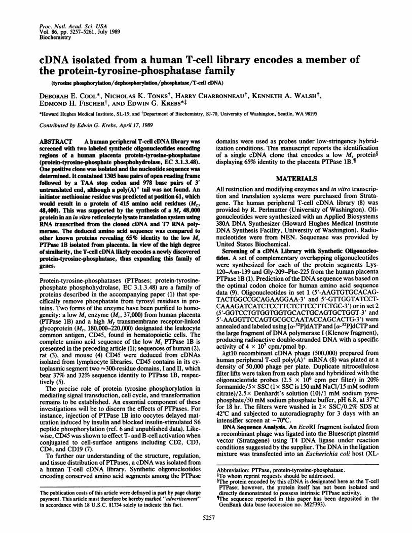
FIG. 1. Sequencing strategy, nucleotide, and deduced amino acid sequence of human T-cell cDNA T-cell PTPase. The predicted amino acidsequence of the open reading frame is shown below the nucleotide sequence. The oligonucleotide sequences used for screening the library areindicated by dots [e.g., between nucleotides 425 and 479 (probe 1), and 689 and 737 (probe 2)]. The TAA stop codon is located at nucleotide1306 followed by a 3' untranslated end containing two possible polyadenylylation sites AATAAA at 1521 and 1677. The schematic diagram belowthe nucleotide sequence depicts the sequencing strategy used. Open bar, open reading frame; solid bar, 3' untranslated end. Arrows indicatethe length of sequence obtained from different sequencing oligonucleotide primers. E, EcoRI; H, HindIII; S, Sst I; X, Xba I. The scale at thebottom represents 200 nucleotides (in kbp).
Proc. Natl. Acad. Sci. USA 86 (1989) 5259
the blot was washed in 0.lx SSC/0.2% SDS at 500C. The gelwas exposed to film for 3 days with an intensifier screen at-700C.Southern Blot Analysis. Human genomic DNA was cleaved
with the restriction endonucleases BamHI, EcoRI, and Hin-dIII. The blot was hybridized to the labeled insert of thecDNA and washed as described for the Northern blot anal-ysis and subjected to autoradiography for 3 days with anintensifier screen at -70'C. It was then reprobed with thelabeled cDNA using the same hybridization conditions asabove, but washed under less stringent conditions, such as2x SSC/0.2% SDS and 450C.
RESULTSIsolation and DNA Sequence Analysis of a Human T-Cell
cDNA Encoding an Isoform of Placenta PTPase 1B. Twosynthetic 32P-labeled oligonucleotides representing differentsegments of the low Mr placenta PTPase 1B were used asprobes to screen duplicate plaque lifts containing 500,000recombinant phage from a AgtlOcDNA library prepared fromhuman peripheral T-cell mRNA (8). Although many recom-binant phage hybridized to each probe, only one overlappingpositive clone bound to both oligonucleotides. Restrictionenzyme analysis of the purified recombinant cDNA revealeda single EcoRI cDNA insert 2.3 kilobase pairs (kbp) long (Fig.1). The entire nucleotide sequence ofthe EcoRI fragment wasobtained in duplicate on both strands. The sequencing strat-egy is shown in Fig. 1; short regions of DNA sequencerepresented by the overlapping arrows were obtained withdifferent primers in the extension reactions. Sequence anal-ysis (Fig. 1) shows that the T-cell PTPase cDNA contains anopen reading frame of 1305 nucleotides. A consensus se-
quence [CC(G)CCAUG(G)] for eukaryotic initiation sitesdescribed by Kozak (14) is found at nucleotides 56-64encoding a putative initiator methionine. The open readingframe terminates with a TAA stop codon followed by 978 bpof 3' untranslated end. However, neither a polyadenylylationsite nor a 3' poly(A)+ tail was observed. There are twopossible AATAAA polyadenylylation signals (15) at sites 213and 369 bp past the stop codon (nucleotides 1521-1526 and1677-1682, respectively).
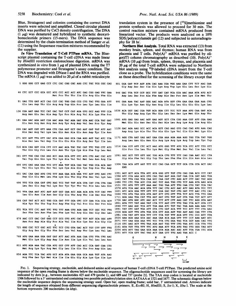
Identification of an Initiator Methionine Residue. mRNAwas synthesized in vitro from the T7 polymerase promoter inthe Bluescript vector in which the T-cell cDNA was sub-cloned. Using the rabbit reticulocyte lysate translation sys-tem, a protein product with an estimated Mr of 48,000 wasproduced (Fig. 2). Since its apparent Mr agreed closely withthat predicted from the cDNA sequence, it is probable thatthe putative initiator methionine at nucleotide 61 is beingrecognized as a translation initiator codon in the in vitrosystem. The translation reaction was carried out in thepresence of [35S]methionine and the band of labeled proteinwas excised and counted. The amount ofprotein synthesized,estimated at 2.5 pg, was not sufficient to detect PTPaseactivity under our assay conditions (16).The T-Cell cDNA Sequence Is Present in Other Tissues.
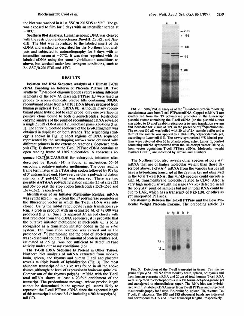
Northern blot analysis of mRNA extracted from monkeybrain, spleen, and thymus and human T cell and placentareveals multiple bands of hybridization (Fig. 3). The mostabundant transcript of -2.3 kb was found in all the abovetissues, although the level ofexpression in brain was quite low.Comparison of the thymus poly(A)+ mRNA with the T-celltotal mRNA shows at least a 20-fold enrichment of thetranscript. The predominant message, whose precise lengthcannot be determined in the agarose gel, seems likely torepresent the T-cell PTPase cDNA since the expected lengthofthis transcript is at least 2.5 kb including a 200-base poly(A)+tail (17).
A B
_..-200- 96
_.- 68
-.E- 45
EL- 30
FIG. 2. SDS/PAGE analysis of the 35S-labeled protein followingtranslation in vitro from T-cell PTPase mRNA. Capped mRNA (1 ,ug)synthesized from the T7 polymerase promoter in the Bluescriptplasmid vector containing the T-cell cDNA (or the plasmid alone)was added to 25 ,ul of a rabbit reticulocyte in vitro translation systemand incubated for 30 min at 30°C in the presence of [35S]methionine.The extract (10 ,ul) was boiled with 20 Al of 2x sample buffer and athird of the sample was applied to a 10%o SDS/polyacrylamide gel,according to Laemmli (12). The newly synthesized 35S-labeled pro-teins were detected after 16 hr of autoradiography. Lanes: 1, controlcontaining mRNA synthesized from the Bluescript vector DNA; 2,from vector containing T-cell PTPase cDNA. Molecular weightmarkers (x10-3) are indicated by arrows and numbers.
The Northern blot also reveals other species of poly(A)+mRNA that are of higher molecular weight than those de-scribed above. Poly(A)+ mRNA from the various tissues allhave a hybridizing transcript at the 28S marker not observedin the total T-cell RNA; this 4.7-kb species could encode ahigh Mr transmembrane molecule related to CD45. Also, avery high molecular weight message (>7 kb) detected in allthe poly(A)+ purified samples but not in total RNA could bedue to LAR, which has a transcript of 8 kb (18), or other asyet unreported PTPases.
Relationship Between the T-Cell PTPase and the Low Mo-lecular Weight Placenta Enzyme. The preceding article (1)
Br Sp Th Tc Pl
4.7-....
FIG. 3. Detection of the T-cell transcript in tissue. Ten micro-grams of poly(A)' mRNA from monkey brain, spleen, or thymus andfrom human placenta mRNA and 20 ug of total human T-cell RNAwere subjected to electrophoresis in a 1% formaldehyde-agarose geland transferred to nitrocellulose paper. The RNA blot was hybrid-ized with 32P-labeled cDNA insert from T-cell PTPase and subjectedto autoradiography for 3 days. Br, brain; Sp, spleen; Th, thymus; Tc,T cell; P1, placenta. The 28S and 18S ribosomal bands are indicatedand correspond to 4.7- and 1.9-kb transcript lengths, respectively.
Biochemistry: Cool et al.
Proc. Natl. Acad. Sci. USA 86 (1989)
TCPTP: RPTEIUZFUWIQRRWQPLYLEIRNESHDYPHRVAKFPENRNRNRYRDVSPYDHSRVK:.:::.: : .: .:: :I:.: :::: ::.::::.:::
PTP1B: MEKEFEQIDKSGSWAAIYQDIRHEASDFPCRVAKLPKNKNRNRYRDVSPFDHSRIK
TCPTP: LQNAENDYINASLVDIEEAQRSYILTQGPLPNTCCEFWLMVWQQKTKAVVMLNRIVEKES.LHQEDMDYINAL . . .....IKNEEAQRSYW . ....Q..H M R.GP..CH.----WKG-
PTPlB: LHQEDNDYINASLIKMEEAQRSYILTQGPLPNTCGHFWD-NWEQKSRGVVLEMRVMEKGS
TCPTP:
PTP1B:
VKCAQYWP-TDDQEIMLFKETGFSVKLLSEDVKSYYTVHLLQLENINSGETRTISHFHYT1I
LKCAQYWPVKEEKEMIFEDTNLKLTLISEDIKSYYTVR0TILHFHYTT
TCPTP: WPDFGVPESPASFLNFLFKVRESGSLNPDHGPAVIHCSAGIGRSGSLVDTCLVEKG
PTP1B: WPDFGVPESPASFLNFLFKVRESGSLSPEHGPVVVHCSAGIGRSGTFCLADTCLLIJUR
TCPTP:
PTP1B:
TCPTP:
DD---INIKQVLLNMRKYRMLIQTPDQLRFSYMAIIEGKCIK(GDSSIQKRKLSE. :: : ::: ::::::: :.::::: : ::::.: ::::: ::
KDPSDI Y L E&~MI TDLR SYLAVI GKINGSVDWE
LSPAFDHSPNKI PKYNNRIGLEEKLTGDRCTGLSS TWSESR
PTP1B: LTPPPRITPI
TCPTP:
OPPPRPPKRILEPHN
PILTUFNLVILVGAFV _-LFFQWA
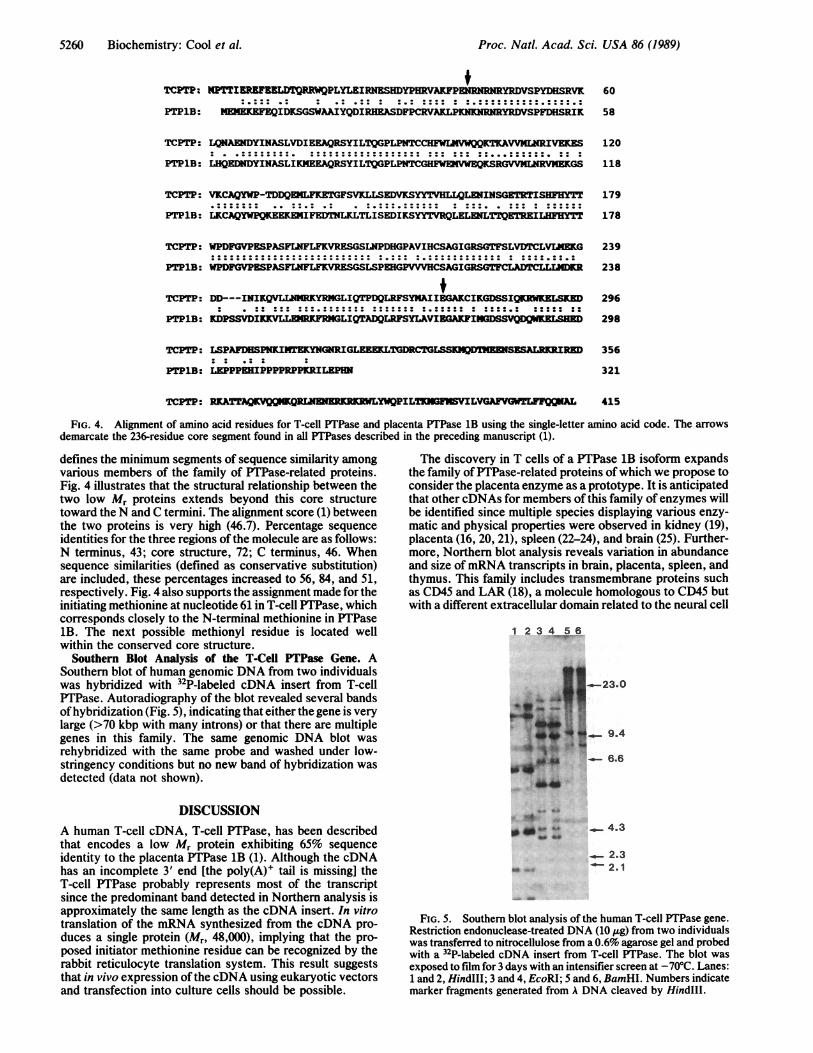
FIG. 4. Alignment of amino acid residues for T-cell PTPase and placenta PTPase 1B using the single-letter amino acid code. The arrowsdemarcate the 236-residue core segment found in all PTPases described in the preceding manuscript (1).
defines the minimum segments of sequence similarity amongvarious members of the family of PTPase-related proteins.Fig. 4 illustrates that the structural relationship between thetwo low Mr proteins extends beyond this core structuretoward the N and C termini. The alignment score (1) betweenthe two proteins is very high (46.7). Percentage sequenceidentities for the three regions of the molecule are as follows:N terminus, 43; core structure, 72; C terminus, 46. Whensequence similarities (defined as conservative substitution)are included, these percentages increased to 56, 84, and 51,respectively. Fig. 4 also supports the assignment made for theinitiating methionine at nucleotide 61 in T-cell PTPase, whichcorresponds closely to the N-terminal methionine in PTPase1B. The next possible methionyl residue is located wellwithin the conserved core structure.
Southern Blot Analysis of the T-Cell PTPase Gene. ASouthern blot of human genomic DNA from two individualswas hybridized with 32P-labeled cDNA insert from T-cellPTPase. Autoradiography of the blot revealed several bandsofhybridization (Fig. 5), indicating that either the gene is verylarge (>70 kbp with many introns) or that there are multiplegenes in this family. The same genomic DNA blot wasrehybridized with the same probe and washed under low-stringency conditions but no new band of hybridization wasdetected (data not shown).
DISCUSSIONA human T-cell cDNA, T-cell PTPase, has been describedthat encodes a low Mr protein exhibiting 65% sequenceidentity to the placenta PTPase 1B (1). Although the cDNAhas an incomplete 3' end [the poly(A)+ tail is missing] theT-cell PTPase probably represents most of the transcriptsince the predominant band detected in Northern analysis isapproximately the same length as the cDNA insert. In vitrotranslation of the mRNA synthesized from the cDNA pro-duces a single protein (Mr, 48,000), implying that the pro-posed initiator methionine residue can be recognized by therabbit reticulocyte translation system. This result suggeststhat in vivo expression of the cDNA using eukaryotic vectorsand transfection into culture cells should be possible.
The discovery in T cells of a PTPase 1B isoform expandsthe family of PTPase-related proteins ofwhich we propose toconsider the placenta enzyme as a prototype. It is anticipatedthat other cDNAs for members of this family of enzymes willbe identified since multiple species displaying various enzy-matic and physical properties were observed in kidney (19),placenta (16, 20, 21), spleen (22-24), and brain (25). Further-more, Northern blot analysis reveals variation in abundanceand size ofmRNA transcripts in brain, placenta, spleen, andthymus. This family includes transmembrane proteins suchas CD45 and LAR (18), a molecule homologous to CD45 butwith a different extracellular domain related to the neural cell
1 2 3 4 5 6
.0
9.4
4.3
2.340--4 2.1
FIG. 5. Southern blot analysis of the human T-cell PTPase gene.Restriction endonuclease-treated DNA (10 ,ug) from two individualswas transferred to nitrocellulose from a 0.6% agarose gel and probedwith a 32P-labeled cDNA insert from T-cell PTPase. The blot was
exposed to film for 3 days with an intensifier screen at -70°C. Lanes:1 and 2, HindIII; 3 and 4, EcoRI; 5 and 6, BamHI. Numbers indicatemarker fragments generated from A DNA cleaved by HindIIl.
60
58
120
118
179
178
239
238
296
298
356
321
415
5260 Biochemistry: Cool et al.
W.* A*.sl. aPC m
0 04W do
so so
Proc. Natl. Acad. Sci. USA 86 (1989) 5261
adhesion molecule N-CAM (26). The existence of multipleforms may suggest differences in substrate specificity andfunction.The low Mr placenta enzyme displays unusually high
specific activity and affinity toward artificial substrates (16).By contrast, CD45 has only 1% of the activity of PTPase 1Bunder our standard assay conditions (24). This could be dueto loss of activity during purification, differing substratespecificity, lack of a ligand binding to the external domain or,as in the case of the protein tyrosine kinases, to the fact thatthe receptor-linked forms seem to be intrinsically less activethan their cytoplasmic counterparts (24). With 65% sequenceidentity between the two low Mr proteins and up to 85%sequence similarity within the 236-residue core described inthe preceding article (1) as shown in Fig. 4, it could beanticipated that the T-cell PTPase protein would exhibitsimilar enzymatic activity to the placenta enzyme. However,these two proteins display significant differences in theircarboxyl termini. The placenta enzyme is smaller (Mr, 37,000vs. 48,000) and ends with a segment in which 10 of 21 residuesare prolyl. The T-cell protein has an extension (Mr, 11,000)with a highly charged segment (Lys-284 to Lys-390) followedby a region of 25 uncharged residues. It will be interesting todetermine whether this C-terminal extension possesses someregulatory function such as exerting a negative influence onenzyme activity. Cleavage of such a structure in a posttrans-lational event or during purification might derepress theenzyme and contribute to the high specific activity ofPTPase1B. Alternatively, it would serve to localize the protein inspecific intracellular elements or compartments.The role of low Mr PTPases in cell signaling, growth, and
transformation is not known. They may be responsible forensuring the transient nature of tyrosine phosphorylationevents in response to certain external stimuli. Consideringthe discrepancy between the activity of the protein tyrosinekinases and phosphatases (20), it would be expected that thelatter are under tight control or confined to specific compart-ments within the cell.Many multigene families, such as those for the serine
proteases (27, 28) or protein kinase (29), arose from dupli-cations of an ancestral gene. These families can often becharacterized by the degree of conservation of both thenumber and position of introns in the coding region (28,30-32). The intron/exon gene organization has been de-scribed completely for CD45 (33) but only partially for LAR(18). Frequency and position of introns present in segmentsof the gene encoding the homologous cytoplasmic regions ofCD45 and LAR are not totally conserved. For example,within CD45, there are seven introns in domain I and six indomain II, with evidence of intron sliding (34) in one of theconserved insertions. Furthermore, LAR has two fewerintrons than CD45 in the PTPase-related cytoplasmic do-mains between exons III and VIII (18). Determination of thegene structure of T-cell PTPase may help to establish theevolutionary relationship between the low Mr and the integralmembrane proteins.
Cell signaling through certain hormone and growth factorreceptors, or transformation by a number of oncogenicretroviruses involves the phosphorylation of proteins ontyrosyl residues. Overexpression ofa PTPase should cause aperturbation of the system in favor of the dephosphorylatedstate and thus may help clarify the role of protein tyrosinephosphorylation in the control of cellular processes.
We are especially grateful to Richard Olsgaard for his assistancein sequencing the cDNA described in this paper and for other
essential technical help. We thank Dr. Roger Perlmutter for provid-ing us with the T-cell cDNA library. We are grateful to Dr. S.McKnight for his critical review of this manuscript and CarmenWestwater for typing it. This work was funded by Grants DK07902and GM 15731 from the National Institutes of Health, the MuscularDystrophy Association, and Howard Hughes Medical Institute.D.E.C. is supported as a Postdoctoral Fellow by the Canadian HeartFoundation.
1. Charbonneau, L., Tonks, N. K., Kumar, S., Diltz, C. D.,Harrylock, M., Cool, D. E., Krebs, E. G., Fischer, E. H. &Walsh, K. A. (1989) Proc. Natl. Acad. Sci. USA 86,5252-5256.
2. Streuli, M., Hall, L. R., Saga, Y., Schlossman, S. F. & Saito,H. (1987) J. Exp. Med. 166, 1548-1566.
3. Thomas, M. L., Barclay, A. N., Gagnon, J. & Williams, A. F.(1985) Cell 41, 83-93.
4. Thomas, M. L., Reynolds, P. J., Chain, A., Ben-Neriah, Y. &Trowbridge, I. S. (1987) Proc. Natl. Acad. Sci. USA 84,5360-5363.
5. Charbonneau, H., Tonks, N. K., Walsh, K. A. & Fischer,E. H. (1988) Proc. NatI. Acad. Sci. USA 85, 7182-7186.
6. Tonks, N. K., Charbonneau, H., Diltz, C. D., Kuman, S.,Cicirelli, M. F., Krebs, E. G., Walsh, K. A. & Fischer, E. H.(1989) Adv. Protein Phosphatases 5, 149-180.
7. Ledbetter, J. A., Tonks, N. K., Fischer, E. H. & Clark, E. A.(1988) Proc. Nati. Acad. Sci. USA 85, 8628-8632.
8. Littman, D. R., Thomas, Y., Maddon, P. J., Chess, L. & Axel,R. (1985) Cell 40, 237-246.
9. Lathe, R. (1985) J. Mol. Biol. 183, 1-12.10. Maniatis, T., Fritsch, E. H. & Sambrook, J. (1982) Molecular
Cloning:A Laboratory Manual (Cold Spring Harbor Lab., ColdSpring Harbor, NY).
11. Sanger, F., Nicklen, S. & Coulson, A. R. (1977) Proc. Natl.Acad. Sci. USA 74, 5463-5467.
12. Laemmli, U. K. (1970) Nature (London) 227, 680-685.13. Cathala, G., Savouret, J. F., Mendez, B. L., Karin, M., Mar-
tial, J. A. & Baxter, J. D. (1983) DNA 2, 329-335.14. Kozak, M. (1984) Nucleic Acids Res. 12, 857-873.15. Proudfoot, J. M. & Brownlee, G. G. (1976) Nature (London)
265, 211-214.16. Tonks, N. K., Diltz, C. D. & Fischer, E. H. (1988) J. Biol.
Chem. 263, 6722-6730.17. Perry, R. P. (1976) Annu. Rev. Biochem. 45, 605-629.18. Streuli, M., Krueger, N. X., Hall, L. R., Schlossman, S. F. &
Saito, H. (1988) J. Exp. Med. 168, 1523-1530.19. Shriner, C. L. & Brautigan, D. L. (1984) J. Biol. Chem. 259,
11383-11390.20. Tonks, N. K., Diltz, C. D. & Fischer, E. H. (1988) J. Biol.
Chem. 263, 6731-6737.21. Roome, J., O'Hare, T., Pilch, P. F. & Brautigan, D. L. (1988)
Biochem. J. 256,493-500.22. Brunati, A. M. & Pinna, L. A. (1985) Biochem. Biophys. Res.
Comm. 133, 929-936.23. Tung, H. Y. L. & Reed, L. J. (1987) Anal. Biochem. 261,
412-419.24. Tonks, N. K., Charbonneau, H., Diltz, C. D., Fischer, E. H.
& Walsh, K. A. (1988) Biochemistry 27, 8695-8701.25. Ingebritsen, T. S., Lewis, S. K., Ingebritsen, V. M., Jena,
V. P., Hiriyanna, K. T., Jones, S. W. & Erikson, R. L. (1989)Adv. Protein Phosphatases 5, 121-147.
26. Edelman, G. M. (1988) Biochemistry 27, 3533-3543.27. Neurath, H. (1984) Science 224, 350-357.28. MacGillivray, R. T. A., Cool, D. E., Fung, M. R., Guinto,
E. R., Keschinsky, M. L. & Van Oost, B. A. (1988) Genet.Eng. 10, 265-330.
29. Hanks, S. K., Quinn, A. M. & Hunter, T. (1988) Science 241,42-52.
30. Doolittle, R. F. (1985) Trends Biochem. Sci. 10, 233-237.31. Gilbert, W. (1985) Science 228, 823-824.32. Furie, B. & Furie, B. C. (1988) Cell 53, 505-518.33. Hall, L. R., Streuli, M., Schlossman, S. F. & Saito, H. (1988)
J. Immunol. 141, 2781-2787.34. Craik, C. S., Rutter, W. J. & Fletterick, R. (1983) Science 220,
1125-1129.
Biochemistry: Cool et A


![Docking interactions in protein kinase and phosphatase ...interacting protein–protein motifs for MAP kinases and tyrosine phosphatases [12,13]. Docking interactions in protein phosphatases](https://img.pdfslide.net/doc/110x75/60ee63efe2bdd8639d7712a5/docking-interactions-in-protein-kinase-and-phosphatase-interacting-proteinaprotein.jpg)