Embed Size (px)
Citation preview
Acquired Deficiency of A20 Results in Rapid Apoptosis,Systemic Inflammation, and Abnormal HematopoieticStem Cell FunctionAkiko Nagamachi1., Yuichiro Nakata2., Takeshi Ueda2, Norimasa Yamasaki2, Yasuhiro Ebihara3,
Kohichiro Tsuji3, Zen-ichiro Honda4, Keiyo Takubo5, Toshio Suda5, Hideaki Oda6, Toshiya Inaba1,
Hiroaki Honda2*
1Department of Molecular Oncology, Research Institute of Radiation Biology and Medicine, Hiroshima University, Minami-ku, Hiroshima, Japan, 2Department of Disease
Model, Research Institute of Radiation Biology and Medicine, Hiroshima University, Minami-ku, Hiroshima, Japan, 3Division of Cellular Therapy, Advanced Clinical Research
Center, The Institute of Medical Science, The University of Tokyo, Minato-ku, Tokyo, Japan, 4Health Care Center and Graduate School of Humanities and Sciences, Institute
of Environmental Science for Human Life, Ochanomizu University, Bunkyo-ku, Tokyo, Japan, 5Department of Cell Differentiation, The Sakaguchi Laboratory of
Developmental Biology, Keio University School of Medicine, Shinjuku-ku, Tokyo, Japan, 6Department of Pathology, Tokyo Women’s Medical University, Shinjuku-ku,
Tokyo, Japan
Abstract
A20 is a negative regulator of NF-kB, and mutational loss of A20 expression is involved in the pathogenesis of autoimmunediseases and B-cell lymphomas. To clarify the role of A20 in adult hematopoiesis, we generated conditional A20 knockoutmice (A20flox/flox) and crossed them with Mx–1Cre (MxCre+) and ERT2Cre (ERT2Cre+) transgenic mice in which Cre is induciblyactivated by endogenous interferon and exogenous tamoxifen, respectively. A20flox/flox MxCre+ (A20Mx) mice spontaneouslyexhibited myeloid proliferation, B cell apoptosis, and anemia with overproduction of pro-inflammatory cytokines. Bonemarrow transplantation demonstrated that these changes were caused by hematopoietic cells. NF-kB was constitutivelyactivated in A20Mx hematopoietic stem cells (HSCs), which caused enhanced cell cycle entry and impaired repopulatingability. Tamoxifen stimulation of A20flox/flox ERT2Cre+ (A20ERT2) mice induced fulminant apoptosis and subsequentmyeloproliferation, lymphocytopenia, and progressive anemia with excessive production of pro-inflammatory cytokines, asobserved in A20Mx mice. These results demonstrate that A20 plays essential roles in the homeostasis of adult hematopoiesisby preventing apoptosis and inflammation. Our findings provide insights into the mechanism underlying A20 dysfunctionand human diseases in which A20 expression is impaired.
Citation: Nagamachi A, Nakata Y, Ueda T, Yamasaki N, Ebihara Y, et al. (2014) Acquired Deficiency of A20 Results in Rapid Apoptosis, Systemic Inflammation, andAbnormal Hematopoietic Stem Cell Function. PLoS ONE 9(1): e87425. doi:10.1371/journal.pone.0087425
Editor: Kevin D. Bunting, Emory University, United States of America
Received August 25, 2013; Accepted December 23, 2013; Published January 31, 2014
Copyright: � 2014 Nagamachi et al. This is an open-access article distributed under the terms of the Creative Commons Attribution License, which permitsunrestricted use, distribution, and reproduction in any medium, provided the original author and source are credited.
Funding: The authors have no support or funding to report.
Competing Interests: The authors have declared that no competing interests exist.
* E-mail: [email protected]
. These authors equally contributed to this work.
Introduction
NF-kB plays fundamental roles in various physiological and
pathological processes, such as immunity, apoptosis, inflammation,
and cancer [1,2,3]. In an unstimulated state, NF-kB is sequestered
in the cytoplasm by binding to IkB proteins. Upon activation by
external stimuli, IkB proteins are phosphorylated by the IkBkinase (IKK) complex and then degraded by ubiquitination. NF-
kB is released and translocates to the nucleus where it drives the
expression of target genes [1,2,3].
A20, also known as tumor necrosis factor alpha-induced protein
3 (TNFAIP3), now emerges as a major negative regulator of NF-
kB signaling [4,5]. A20 comprises an ovarian tumor (OTU)
domain at its N-terminus and seven Zn-finger motifs. The OTU
domain is predicted to have deubiquitinating protease activity, and
the Zn finger motifs possess E3 ubiquitin ligase and ubiquitin-
binding activities [4,5]. Thus, A20, acting as a ubiquitin-modifying
protein, may participate in a negative feedback loop controlling
NF-kB signaling [4,5]. The most compelling evidence that A20
plays an essential role in inhibiting inflammation are results of a
gene knockout experiment in which A20 deficient mice prema-
turely died because of severe systemic inflammation and cachexia
[6].
A20 is involved in various human diseases, including hemato-
poietic malignancies. Frequent loss of A20 expression in B-cell
lymphomas caused by biallelic deletions and/or point mutations
[7,8] indicates that A20 functions as a tumor suppressor in the
hematopoietic system. Moreover, single nucleotide polymorphisms
in A20 are associated with autoimmune and inflammatory
diseases, such as systemic lupus erythematosus (SLE) [9,10,11],
rheumatoid arthritis (RA) [12,13], and Crohn’s disease [14].
An approach to determine whether there is a causative
association between A20 mutations and pathogenesis employs
mice to target A20 in a tissue-specific manner. A number of A20
conditional knockout (cKO) mice have been generated for this
purpose. For example, B cell-specific deletion of A20 using a
CD19–Cre transgene results in hyper-responsiveness of B cells and
PLOS ONE | www.plosone.org 1 January 2014 | Volume 9 | Issue 1 | e87425
causes autoimmune disease similar to SLE [15,16,17]. Deletion of
A20 from dendritic or myeloid cells using CD11c–Cre or LysM–Cre
transgenes, respectively, also induced autoimmune disease. The
former exhibited an SLE-like phenotype [18], and the latter
developed an RA-like disease [19]. Moreover, Villin–Cre transgenic
mice harboring a deletion of A20 from their epithelial intestinal
cells showed susceptibility to dextran sodium sulfate-induced colitis
[20].
Although these studies provide important insights into the role
of A20 as a suppressor of tumorigenesis and autoimmunity, its
role(s) in the normal functioning of the hematopoietic system of
adults remains to be determined. To address this issue, we created
mice in which A20 expression can be inducibly and preferentially
ablated in hematopoietic cells.
Materials and Methods
MiceThe detailed procedures for constructing the targeting vector
and generating the A20flox/flox mice are described in Text S1 (A20
cKO mice have been deposited in RIKEN BioResource Center
(http://www.brc.riken.jp/inf/en/index.shtml, RBRC05494).
A20flox/flox mice were crossed with Mx–1Cre (MxCre+) transgenic
mice [21] and ERT2Cre (ERT2Cre+) transgenic mice (C57BL/6-
Gt(ROSA)26Sortm1(cre/Est1)Arte, purchased from Taconic) to gener-
ate A20flox/flox MxCre+ and A20flox/flox ERT2Cre+ mice, respectively.
Mice backcrossed with the C57BL/6-Ly5.2 background at least
seven times were used here. This study was carried out in strict
accordance with the recommendations in the Guide for the Care
and Use of Laboratory Animals of the Hiroshima University
Animal Research Committee. The protocol was approved by the
Committee on the Ethics of Animal Experiments of the Hiroshima
University (Permit Number: A13-13). All mice were maintained
according to the guidelines of the Institute of Laboratory Animal
Science of Hiroshima University, all surgeries were performed
under sodium pentobarbital anesthesia and all efforts were made
to minimize suffering.
Western Blotting, Flow Cytometry, and HistopathologyWestern blotting, flow cytometry, and histopathology were
performed as previously described [22,23,24]. Antibodies and a
staining kit used in these analyses are listed in Table S1.
Measurement of Serum Cytokine ConcentrationConcentrations of pro-inflammatory cytokines (TNF-a, IFN-c,
GM-CSF, IL-1b, and IL-6) were measured using a BD Cytometric
Bead Array Flex Set Kit (BD Biosciences, San Diego, CA, USA)
according to the manufacturer’s instructions.
Colony Formation AssayThe colony formation assay was performed as previously
described [25].
Bone Marrow Transplantation (BMT), CompetitiveRepopulation, and Cell Cycle AnalysesTransplantation of bone marrow (BM) cells, competitive
repopulation, and short-term BrdU incorporation assays were
performed as previously described [24].
Results
A20 flox/flox MxCre+ Mice Exhibited Severe Inflammation, BLymphocyte Apoptosis, and Premature DeathTo conditionally ablate A20 function, we generated mice in
which exon 3 of A20 was flanked by two loxP sites (A20flox/flox mice,
Fig. S1A and S1B). To examine the role of A20 in hematopoietic
homeostasis, we crossed A20flox/flox mice with Mx–1Cre transgenic
(MxCre+) mice in which Cre is placed under the control of IFN-
responsiveMx–1 promoter [21]. Lack of A20 expression in A20flox/
flox MxCre+ mice was confirmed by western blotting of spleen
extracts prepared from A20flox/flox MxCre2 and A20flox/flox MxCre+
mice (hereafter referred to as control and A20Mx mice, respectively)
using an anti-A20 antibody (left panel of Fig. S1C).
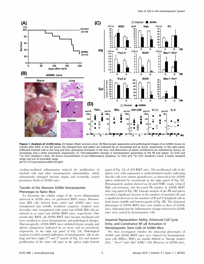
Although A20Mx mice were apparently normal at birth, they
exhibited spontaneous emaciation and cachexia without stimula-
tion by polyinosinic:polycytidylic acid (pIpC), which is a strong
and transient inducer of IFN, and most mice died within six
months after birth (Fig. 1A). Hematological analysis of moribund
mice revealed anemia, proliferation of myeloid cells, and reduction
of B lymphoid cells in the peripheral blood (PB) (Table S2). The
macroscopic appearance of the mice was uniformly characterized
by massive hepatomegaly and enlarged spleens (indicated by an
arrowhead and an arrow, respectively, in the left panel of Fig. 1B),
which were frequently associated with lymph node (LN) swelling
(Table S2). Pathological analysis revealed infiltration of the lung
and liver by hematopoietic cells (indicated by arrows in the right
top and middle panels of Fig. 1B), formation of granulomas in the
liver (indicated by an arrowhead in the right middle panel of
Fig. 1B), and destruction of spleen architecture caused by the
proliferation of white blood cells (indicated by a white arrowhead
in the right bottom panel of Fig. 1B). Higher magnification of the
spleen showed that the most of the proliferated cells were with
segmented or multi-lobulated nuclei, strongly suggesting that these
cells were of myeloid in origin (indicated by arrowheads in the left
panel of Fig. S2).
We then analyzed the time-dependent changes in hematopoietic
parameters in control and A20Mx mice. The white blood cell count
(WBC), hemoglobin concentration (Hgb), and platelet (Plt)
number in the PB, and the absolute numbers of B lymphoid
(B220+), T lymphoid (Thy1.2+), and myeloid (Mac1+Gr1+) cells in
the PB and spleen were analyzed at three and six months after
birth. Although no significant difference was observed in the WBC
count and Plt number between the two groups, A20Mx mice
exhibited progressive anemia (top panels of Fig. 1C). In addition,
lineage analysis of WBCs in the PB and spleen revealed that the
numbers of myeloid cells were significantly increased, whereas
those of B lymphoid cells were significantly decreased in A20Mx
mice compared with control mice (middle and bottom panels of
Fig. 1C). To clarify the mechanism of B-cell reduction in A20Mx
mice, spleens of A20Mx mice and control littermates were
subjected to and anti-B-cell and TUNEL double staining. As
shown in Fig. S3, a significant portion of B lymphocytes in the
A20Mx spleen was positive for TUNEL, indicating that the B cell
reduction in A20Mx mice was due to apoptosis.
Because A20 inhibits NF-kB signaling and suppresses inflam-
matory pathway activity [4,5], we reasoned that the aforemen-
tioned findings were caused by sustained inflammatory responses.
Therefore, serum concentrations of pro-inflammatory cytokines,
including TNF-a, IFN-c, GM-CSF, IL-1b, and IL-6, were
measured and compared between the two groups. The concen-
trations of these cytokines were higher in A20Mx mice than in
control mice, and those of TNF-a, IFN-c, and GM-CSF were
significantly increased (Fig. 1D). These results indicate that
Role of A20 in the Hematopoietic System
PLOS ONE | www.plosone.org 2 January 2014 | Volume 9 | Issue 1 | e87425
cytokine-mediated inflammation induced the proliferation of
myeloid cells and other hematopoietic abnormalities, which
subsequently damaged internal organs and eventually caused
premature death of A20Mx mice.
Transfer of the Aberrant A20Mx HematopoieticPhenotype to Naıve MiceTo determine the cellular origin of the severe inflammation
observed in A20Mx mice, we performed BMT assays. Mononu-
clear BM cells derived from control and A20Mx mice were
transplanted into lethally irradiated syngeneic recipient mice
(hereafter, mice transplanted with control and A20Mx BM cells are
referred to as control and A20Mx BMT mice, respectively). One
month after BMT, all A20Mx BMT mice became moribund and
were sacrificed to assess hematopoietic and pathological changes.
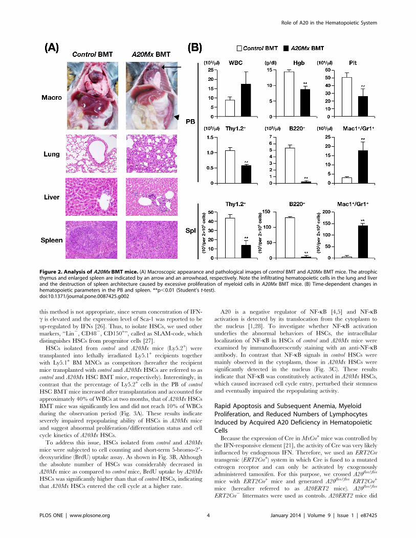
Macroscopically, A20Mx BMT mice exhibited thymic atrophy and
splenic enlargement (indicated by an arrow and an arrowhead,
respectively, in the right top panel of Fig. 2A). Pathological
analysis revealed marked infiltration of hematopoietic cells in the
lung and liver (right 2nd and 3rd panels of Fig. 2A) and massive
proliferation of the same cell types in the spleen (right bottom
panel of Fig. 2A) of A20 BMT mice. The proliferated cells in the
spleen were with segmented or multi-lobulated nuclei, indicating
that the cells were mature granulocytes, as observed in the A20Mx
spleen (indicated by arrowheads in the right panel of Fig. S2).
Hematopoietic analysis showed an elevated WBC count, reduced
Hgb concentration, and decreased Plt number in A20Mx BMT
mice (top panel of Fig. 2B). Lineage analysis of the PB and spleen
revealed a significant increase in the numbers of myeloid cells and
a significant decrease in the numbers of B and T lymphoid cells in
both tissues (middle and bottom panels of Fig. 2B). The abnormal
phenotypes of A20Mx BMT mice were similar to those of A20Mx
mice, indicating that the inflammatory changes detected in A20Mx
mice were caused by hematopoietic cells.
Impaired Repopulation Ability, Enhanced Cell CycleEntry, and Constitutive NF-kB Activation inHematopoietic Stem Cells of A20Mx MiceWe then investigated whether the abnormal phenotypes of
A20Mx and A20Mx BMT mice were induced by hematopoietic
stem cells (HSCs). HSCs are usually defined as ‘‘lineage marker
(Lin) 2, Sca-1+ and c-Kit+ (LSK)’’ cells. However, in A20Mx mice,
Figure 1. Analysis of A20Mx mice. (A) Kaplan–Meier survival curves. (B) Macroscopic appearance and pathological images of an A20Mx mouse sixmonths after birth. In the left panel, the enlarged liver and spleen are indicated by an arrowhead and an arrow, respectively. In the right panel,infiltrated myeloid cells in the lung and liver, granuloma formation in the liver, and destruction of spleen architecture are indicated by arrows, anarrowhead, and a white arrowhead, respectively. (C) Time-dependent changes in hematopoietic parameters in the PB and spleen. *p,0.05 and**p,0.01 (Student’s t-test). (D) Serum concentrations of pro-inflammatory cytokines. *p,0.05 and **p,0.01 (Student’s t-test). 1 below standardrange and out of invertable range.doi:10.1371/journal.pone.0087425.g001
Role of A20 in the Hematopoietic System
PLOS ONE | www.plosone.org 3 January 2014 | Volume 9 | Issue 1 | e87425
this method is not appropriate, since serum concentration of IFN-
c is elevated and the expression level of Sca-1 was reported to be
up-regulated by IFNs [26]. Thus, to isolate HSCs, we used other
markers, ‘‘Lin2, CD482, CD150+’’, called as SLAM-code, which
distinguishes HSCs from progenitor cells [27].
HSCs isolated from control and A20Mx mice (Ly5.2+) were
transplanted into lethally irradiated Ly5.1+ recipients together
with Ly5.1+ BM MNCs as competitors (hereafter the recipient
mice transplanted with control and A20Mx HSCs are referred to as
control and A20Mx HSC BMT mice, respectively). Interestingly, in
contrast that the percentage of Ly5.2+ cells in the PB of control
HSC BMT mice increased after transplantation and accounted for
approximately 40% of WBCs at two months, that of A20Mx HSCs
BMT mice was significantly less and did not reach 10% of WBCs
during the observation period (Fig. 3A). These results indicate
severely impaired repopulating ability of HSCs in A20Mx mice
and suggest abnormal proliferation/differentiation status and cell
cycle kinetics of A20Mx HSCs.
To address this issue, HSCs isolated from control and A20Mx
mice were subjected to cell counting and short-term 5-bromo-29-
deoxyuridine (BrdU) uptake assay. As shown in Fig. 3B, Although
the absolute number of HSCs was considerably decreased in
A20Mx mice as compared to control mice, BrdU uptake by A20Mx
HSCs was significantly higher than that of control HSCs, indicating
that A20Mx HSCs entered the cell cycle at a higher rate.
A20 is a negative regulator of NF-kB [4,5] and NF-kBactivation is detected by its translocation from the cytoplasm to
the nucleus [1,28]. To investigate whether NF-kB activation
underlies the abnormal behaviors of HSCs, the intracellular
localization of NF-kB in HSCs of control and A20Mx mice were
examined by immunofluorescently staining with an anti-NF-kBantibody. In contrast that NF-kB signals in control HSCs were
mainly observed in the cytoplasm, those in A20Mx HSCs were
significantly detected in the nucleus (Fig. 3C). These results
indicate that NF-kB was constitutively activated in A20Mx HSCs,
which caused increased cell cycle entry, perturbed their stemness
and eventually impaired the repopulating activity.
Rapid Apoptosis and Subsequent Anemia, MyeloidProliferation, and Reduced Numbers of LymphocytesInduced by Acquired A20 Deficiency in HematopoieticCellsBecause the expression of Cre in MxCre+ mice was controlled by
the IFN-responsive element [21], the activity of Cre was very likely
influenced by endogenous IFN. Therefore, we used an ERT2Cre
transgenic (ERT2Cre+) system in which Cre is fused to a mutated
estrogen receptor and can only be activated by exogenously
administered tamoxifen. For this purpose, we crossed A20flox/flox
mice with ERT2Cre+ mice and generated A20flox/flox ERT2Cre+
mice (hereafter referred to as A20ERT2 mice). A20flox/flox
ERT2Cre2 littermates were used as controls. A20ERT2 mice did
Figure 2. Analysis of A20Mx BMT mice. (A) Macroscopic appearance and pathological images of control BMT and A20Mx BMT mice. The atrophicthymus and enlarged spleen are indicated by an arrow and an arrowhead, respectively. Note the infiltrating hematopoietic cells in the lung and liverand the destruction of spleen architecture caused by excessive proliferation of myeloid cells in A20Mx BMT mice. (B) Time-dependent changes inhematopoietic parameters in the PB and spleen. **p,0.01 (Student’s t-test).doi:10.1371/journal.pone.0087425.g002
Role of A20 in the Hematopoietic System
PLOS ONE | www.plosone.org 4 January 2014 | Volume 9 | Issue 1 | e87425
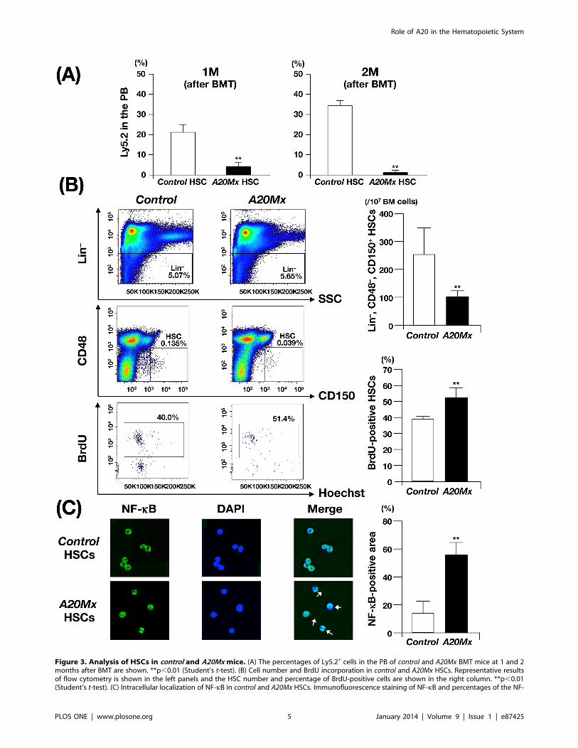
Figure 3. Analysis of HSCs in control and A20Mx mice. (A) The percentages of Ly5.2+ cells in the PB of control and A20Mx BMT mice at 1 and 2months after BMT are shown. **p,0.01 (Student’s t-test). (B) Cell number and BrdU incorporation in control and A20Mx HSCs. Representative resultsof flow cytometry is shown in the left panels and the HSC number and percentage of BrdU-positive cells are shown in the right column. **p,0.01(Student’s t-test). (C) Intracellular localization of NF-kB in control and A20Mx HSCs. Immunofluorescence staining of NF-kB and percentages of the NF-
Role of A20 in the Hematopoietic System
PLOS ONE | www.plosone.org 5 January 2014 | Volume 9 | Issue 1 | e87425
not spontaneously develop abnormal phenotypes in contrast to
A20Mx mice. However, after tamoxifen administration, A20ERT2
mice became rapidly moribund, exhibited a marked decrease in
the number of hematopoietic cells and died within several days
(not shown). Macroscopical examinations revealed that the thymus
was atrophic, the spleen was pale, and liver had white spots (not
shown). Pathological analysis revealed that massive apoptosis
occurred in major organs, including hematopoietic tissues such as
the thymus, spleen, liver, and BM (Fig. S4A). Microemboli and
necrotic areas were also observed in the liver (indicated by an
arrow and arrowheads in Fig. S4A), suggesting that the white spots
in the liver were caused by ischemic necrosis. The measurement of
pro-inflammatory cytokines revealed that the concentrations of
these cytokines were higher in A20ERT2 mice than in control mice,
and those of TNF-a, IFN-c, and IL-6 were significantly increased
(Fig. S4B). These results indicate that loss of A20 in adult mice
induced fulminant apoptosis, possibly via rapid elevation of pro-
inflammatory cytokines.
We next investigated the effect of deleting A20 from hemato-
poietic cells by transplanting control and A20ERT2 BM cells into
lethally irradiated syngeneic mice and then administrating
tamoxifen (mice transplanted with A20ERT2 BM cells are
hereafter referred to as A20ERT2 BMT mice). Before tamoxifen
stimulation, the number of transplanted Ly5.2+ cells was
comparably increased in control BMT and A20ERT2 BMT mice,
and there was no significant difference in the values of
hematopoietic parameters between them (not shown). Tamoxifen
was administered eight weeks after transplantation [29] (Fig. 4A),
and two days later, all A20ERT2 BMT mice became moribund
with marked decrease in WBC count and Plt number in the PB (‘‘2
days’’ at top panels of Fig. 4A).
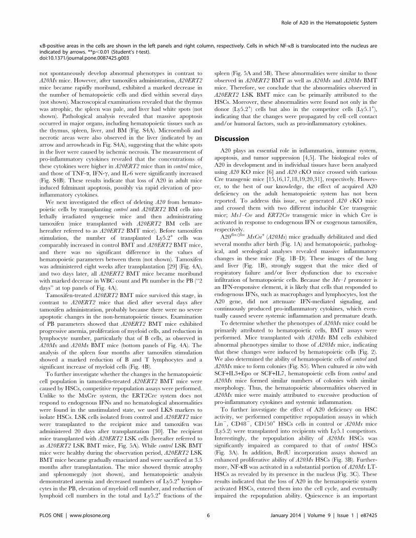
Tamoxifen-treated A20ERT2 BMT mice survived this stage, in
contrast to A20ERT2 mice that died after several days after
tamoxifen administration, probably because there were no severe
apoptotic changes in the non-hematopoietic tissues. Examination
of PB parameters showed that A20ERT2 BMT mice exhibited
progressive anemia, proliferation of myeloid cells, and reduction in
lymphocyte number, particularly that of B cells, as observed in
A20Mx and A20Mx BMT mice (bottom panels of Fig. 4A). The
analysis of the spleen four months after tamoxifen stimulation
showed a marked reduction of B and T lymphocytes and a
significant increase of myeloid cells (Fig. 4B).
To further investigate whether the changes in the hematopoietic
cell population in tamoxifen-treated A20ERT2 BMT mice were
caused by HSCs, competitive repopulation assays were performed.
Unlike to the MxCre system, the ERT2Cre system does not
respond to endogenous IFNs and no hematological abnormalities
were found in the unstimulated state, we used LKS markers to
isolate HSCs. LSK cells isolated from control and A20ERT2 mice
were transplanted to the recipient mice and tamoxifen was
administered 20 days after transplantation [30]. The recipient
mice transplanted with A20ERT2 LSK cells (hereafter referred to
as A20ERT2 LSK BMT mice, Fig. 5A). While control LSK BMT
mice were healthy during the observation period, A20ERT2 LSK
BMT mice became gradually emaciated and were sacrificed at 3.5
months after transplantation. The mice showed thymic atrophy
and splenomegaly (not shown), and hematopoietic analysis
demonstrated anemia and decreased numbers of Ly5.2+ lympho-
cytes in the PB, elevation of myeloid cell number, and reduction of
lymphoid cell numbers in the total and Ly5.2+ fractions of the
spleen (Fig. 5A and 5B). These abnormalities were similar to those
observed in A20ERT2 BMT as well as A20Mx and A20Mx BMT
mice. Therefore, we conclude that the abnormalities observed in
A20ERT2 LSK BMT mice can be primarily attributed to the
HSCs. Moreover, these abnormalities were found not only in the
donor (Ly5.2+) cells but also in the competitor cells (Ly5.1+),
indicating that the changes were propagated by cell–cell contact
and/or humoral factors, such as pro-inflammatory cytokines.
Discussion
A20 plays an essential role in inflammation, immune system,
apoptosis, and tumor suppression [4,5]. The biological roles of
A20 in development and in individual tissues have been analyzed
using A20 KO mice [6] and A20 cKO mice crossed with various
Cre transgenic mice [15,16,17,18,19,20,31], respectively. Howev-
er, to the best of our knowledge, the effect of acquired A20
deficiency on the adult hematopoietic system has not been
reported. To address this issue, we generated A20 cKO mice
and crossed them with two different inducible Cre transgenic
mice; Mx1–Cre and ERT2Cre transgenic mice in which Cre is
activated in response to endogenous IFN or exogenous tamoxifen,
respectively.
A20flox/flox MxCre+ (A20Mx) mice gradually debilitated and died
several months after birth (Fig. 1A) and hematopoietic, patholog-
ical, and serological analyses revealed massive inflammatory
changes in these mice (Fig. 1B–D). These images of the lung
and liver (Fig. 1B), strongly suggest that the mice died of
respiratory failure and/or liver dysfunction due to excessive
infiltration of hematopoietic cells. Because the Mx–1 promoter is
an IFN-responsive element, it is likely that cells that responded to
endogenous IFNs, such as macrophages and lymphocytes, lost the
A20 gene, did not attenuate IFN-mediated signaling, and
continuously produced pro-inflammatory cytokines, which even-
tually caused severe systemic inflammation and premature death.
To determine whether the phenotypes of A20Mx mice could be
primarily attributed to hematopoietic cells, BMT assays were
performed. Mice transplanted with A20Mx BM cells exhibited
abnormal phenotypes similar to those of A20Mx mice, indicating
that these changes were induced by hematopoietic cells (Fig. 2).
We also determined the ability of hematopoietic cells of control and
A20Mx mice to form colonies (Fig. S5). When cultured in vitro with
SCF+IL3+Epo or SCF+IL7, hematopoietic cells from control and
A20Mx mice formed similar numbers of colonies with similar
morphology. Thus, the hematopoietic abnormalities observed in
A20Mx mice were mainly attributed to excessive production of
pro-inflammatory cytokines and systemic inflammation.
To further investigate the effect of A20 deficiency on HSC
activity, we performed competitive repopulation assays in which
Lin2, CD482, CD150+ HSCs cells in control or A20Mx mice
(Ly5.2) were transplanted into recipients with Ly5.1 competitors.
Interestingly, the repopulation ability of A20Mx HSCs was
significantly impaired as compared to that of control HSCs
(Fig. 3A). In addition, BrdU incorporation assays showed an
enhanced proliferative ability of A20Mx HSCs (Fig. 3B). Further-
more, NF-kB was activated in a substantial portion of A20Mx LT-
HSCs as revealed by its presence in the nucleus (Fig. 3C). These
results indicated that the loss of A20 in the hematopoietic system
activated HSCs, entered them into the cell cycle, and eventually
impaired the repopulation ability. Quiescence is an important
kB-positive areas in the cells are shown in the left panels and right column, respectively. Cells in which NF-kB is translocated into the nucleus areindicated by arrows. **p,0.01 (Student’s t-test).doi:10.1371/journal.pone.0087425.g003
Role of A20 in the Hematopoietic System
PLOS ONE | www.plosone.org 6 January 2014 | Volume 9 | Issue 1 | e87425
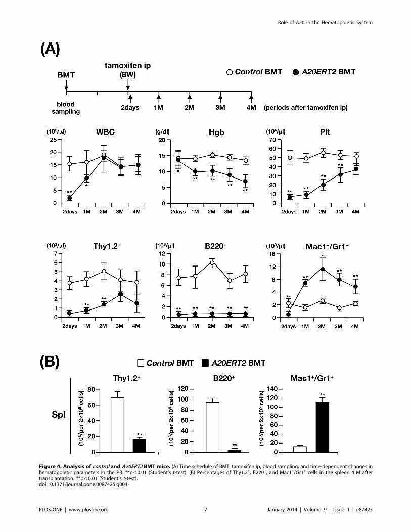
Figure 4. Analysis of control and A20ERT2 BMT mice. (A) Time schedule of BMT, tamoxifen ip, blood sampling, and time-dependent changes inhematopoietic parameters in the PB. **p,0.01 (Student’s t-test). (B) Percentages of Thy1.2+, B220+, and Mac1+/Gr1+ cells in the spleen 4 M aftertransplantation. **p,0.01 (Student’s t-test).doi:10.1371/journal.pone.0087425.g004
Role of A20 in the Hematopoietic System
PLOS ONE | www.plosone.org 7 January 2014 | Volume 9 | Issue 1 | e87425
Figure 5. Analysis of control and A20ERT2 LSK BMT mice. (A) Time schedule of BMT, tamoxifen ip, blood sampling, and time-dependentchanges of hematopoietic parameters and percentages of Ly5.2+ cells in the PB. *p,0.05 and **p,0.01 (Student’s t-test). (B) Percentages of differenthematopoietic compartments and the population of Ly5.2+ cells in the spleen 3.5 M after transplantation. **p,0.01 (Student’s t-test).doi:10.1371/journal.pone.0087425.g005
Role of A20 in the Hematopoietic System
PLOS ONE | www.plosone.org 8 January 2014 | Volume 9 | Issue 1 | e87425
characteristic for HSCs to maintain their stemness and engraft-
ment activity. Our findings in this regard are consistent with those
of previous studies demonstrating that cycling HSCs lose its
repopulating ability [32,33,34]. Whether HSCs sense and directly
respond to inflammation is controversial [35,36]. Our results
suggest that HSCs could be a direct target of systemic
inflammation and support the idea that TNF-a and IFN-c are
major regulators of inflammatory responses of HSCs, as
documented in previous studies [34,37].
Next, we used ERT2Cre mice that respond to exogenously
administered tamoxifen but not to endogenous estrogens. As
expected, unlike A20Mx mice, A20ERT2 mice did not exhibit an
abnormal phenotype before tamoxifen stimulation. However,
tamoxifen administration induced rapid death of the A20ERT2
mice, which we attribute to severe apoptosis (Fig. S4). Moreover,
there was a marked decrease in the numbers of hematopoietic cells
in A20ERT2 BMT mice following tamoxifen treatment (Fig. 4).
These results indicate that loss of A20 induces rapid systemic
apoptosis of hematopoietic cells.
The roles of A20 in apoptosis differ among published studies.
For example, certain A20 deficient cells such as fibroblasts,
splenocytes, and enterocytes exhibit enhanced sensitivity to TNF-
a-mediated apoptosis [6,20]. In contrast, re-expressing A20 in A20
deficient lymphoma cells promotes apoptosis [7,8]. These findings
indicate that A20 might play reciprocal roles in cell survival and
cell death, depending on the cell types and cell contexts.
Accordingly, our present results demonstrate that acquired A20
deficiency induces fulminant apoptosis in all cell types, including
hematopoietic cells. This suggests that A20 primarily prevents
apoptosis in adult tissues. There is evidence that the interaction of
A20 with caspase-8 might play a role in this process [38].
The features shared by A20ERT2 and A20ERT2 LSK BMT
included an increase in the number of myeloid cells, a decrease in
the number of lymphocytes, and anemia, and similar to those of
A20Mx and A20Mx BMT mice (Figs. 1, 2, 4, and 5). Thus,
cytokine synthesis by hematopoietic cells that survived the
apoptotic stage was uncontrolled and caused fulminant inflam-
mation. Proliferation of myeloid cells would be a direct effect of
increased production of pro-inflammatory cytokines, and anemia
would be attributed to inflammation-induced impaired erythro-
poiesis as previously described [39]. In contrast, the mechanism
responsible for reducing lymphocyte numbers, particularly those of
B cells, remains to be clarified. Pathological analysis revealed that
B lymphocytes underwent apoptosis in the spleen (Fig. S3), in
contrast to previous reports, which show that in B cell-specific A20
deficient mice, B lymphocytes were hyper-reactive to exogenous
stimuli and resistant to apoptosis [15,16,17]. The mechanism(s)
underlying this discrepancy is unknown. One possibility is that B
cell behaviors in response to A20 deficiency might differ,
depending on the developmental stages at which A20 is deleted
(namely, in B lineage-committed cells or in more primitive cells
including HSCs). Another possibility is that death factor(s) secreted
by systemic inflammation, such as TNF-a might affect lympho-
cytes and induce apoptosis.
The role of A20 in B cell development/differentiation is of
particular interest, because hematopoietic malignancies with A20
mutations are exclusively B lineage lymphomas [7,8]. Mice with
deletions of A20 in the B lineage cells exhibit autoimmune-like
diseases but fail to develop lymphoproliferative disorders
[15,16,17]. Moreover, MALT lymphoma, a subtype of B-cell
lymphoma in which A20 is frequently mutated, originates from
mutated hematopoietic stem–early progenitor cells [40]. There-
fore, A20 function must be abrogated in early progenitor cells to
mimic B-cell lymphomas harboring A20 mutations, as accom-
plished here. However, because apoptosis decreased the number of
B cells in A20Mx and A20ERT2 mice (Figs. S3 and S4), anti-
apoptotic signal(s) might be necessary to exhibit a fully malignant
phenotype. To address this possibility, we are crossing A20Mx
mice with p53 knockout mice to prevent apoptosis, and with CagA
transgenic mice, a model for Helicobactor pylori infection that plays a
pivotal role in the initiation of MALT lymphoma [41].
In summary, we generated mice in which A20 can be inducibly
and preferentially deleted from hematopoietic cells and found that
acquired loss of A20 induced fulminant apoptosis and subsequent
systemic inflammation. Our results demonstrate the essential role
of A20 in the maintenance of adult hematopoietic cell homeostasis
and provide insights into the mechanism responsible for A20
dysfunction and human diseases with A20 mutations.
Supporting Information
Figure S1 Generation of A20 conditional knockout mice.(A) Targeting strategy. Exon 3 of mouse A20 was floxed and the Frt-
flanked Neo-resistance gene was removed using Flp recombinase.
The positions of a 59 probe for Southern blotting and P1 and P2
primers for 39 genomic PCR analysis are shown. Restriction sites:
Ba, BamHI; Sac, SacI; Stu, StuI; Sal, SalI; Bg, BglII. (B) Southern
blotting and genomic PCR using a 59 probe and 39 primers,
respectively, to detect homologous recombination in two depen-
dent ES clones (#1 and #2). Germline (GL) and targeted allele-
derived bands are indicated by arrows (left panel), and the
recombination-specific PCR product is indicated by an arrowhead
(right panel). (C) Western blotting of A20 expression in A20flox/flox
MxCre+ mice. Proteins extracted from the spleens of LPS-
stimulated A20flox/flox MxCre2 and A20flox/flox MxCre+ mice were
blotted and probed with anti-A20- (upper panel) or anti-b-actinantibodies (lower panel). The position of A20 is indicated by an
arrow.
(PDF)
Figure S2 Proliferation of mature myeloid cells in thespleen of A20Mx and A20Mx BMT mice. Higher magnifi-
cation of HE-stained sections of the spleen of A20Mx and A20Mx
BMT mice. Mature myeloid cells with segmented or multi-
lobulated nuclei are indicated by arrowheads.
(PDF)
Figure S3 Apoptosis of B cells. Representative results of
double staining with an anti-B cell antibody and TUNEL in control
and A20Mx spleens (three weeks old). Blue and brown staining
patterns show B and apoptotic cells, respectively. The boxed areas
in the upper panels are magnified in the lower panels.
(PDF)
Figure S4 Analysis of control and A20ERT2 mice. (A) HE-
stained A20ERT2 tissues that show severe apoptosis. Microemboli
and necrotic areas in the liver are indicated by arrowheads and an
arrow, respectively. 1 below standard range and out of invertable
range. (B) Serum concentrations of pro-inflammatory cytokines.
*p,0.05 and **p,0.01 (Student’s t-test). 1 below standard range
and out of invertable range.
(PDF)
Figure S5 Colony formation assay. (A) The colony numbers
generated in the presence of SCF+IL3+Epo, and those generated
with SCF+IL7 are shown. No significant difference was observed
between control and A20Mx mice. (B) Representative images of
colonies. Colonies derived from both types of mice are similar in
size and shape.
(PDF)
Role of A20 in the Hematopoietic System
PLOS ONE | www.plosone.org 9 January 2014 | Volume 9 | Issue 1 | e87425
Table S1 Antibodies. All the antibodies used in this study are
listed.
(PDF)
Table S2 Characteristics of A20Mx mice. Hematopoietic
parameters of moribund A20Mx mice are described.
(PDF)
Text S1 Construction of a targeting vector and gener-ation of A20 knockout mice. Detailed procedure of Construc-
tion of a targeting vector and generation of A20 knockout mice is
described.
(PDF)
Acknowledgments
We thank Yuki Sakai, Yu Iwai, Sawako Ogata, and Rika Tai for animal
care, genotyping, and molecular experiments. We also thank Dr. Junji
Takeda of Osaka University and the RIKEN BioResource Center in Japan
for providing us with KY1.1 ES cells and B6-Tg(CAG-FLPe)36 mice
(RBRC01834), respectively. We express our sincere gratitude to Kalmia
Caravan for encouragement and emotional support.
Author Contributions
Conceived and designed the experiments: AN ZH TS TI HH. Performed
the experiments: AN TU NY YE K. Tsuji K. Takubo HO HH. Analyzed
the data: AN YN TU NY YE K. Tsuji HO HH. Wrote the paper: TU YE
K. Takubo HH.
References
1. Ghosh S, Hayden MS (2008) New regulators of NF-kappaB in inflammation.
Nat Rev Immunol 8: 837–848.
2. Shembade N, Harhaj EW (2012) Regulation of NF-kB signaling by the A20deubiquitinase. Cell Mol Immunol 9: 123–130.
3. DiDonato JA, Mercurio F, Karin M (2012) NF-kB and the link betweeninflammation and cancer. Immunol Rev 246: 379–400.
4. Hymowitz SG, Wertz IE (2010) A20: from ubiquitin editing to tumoursuppression. Nat Rev Cancer 10: 332–341.
5. Ma A, Malynn BA (2012) A20: linking a complex regulator of ubiquitylation to
immunity and human disease. Nat Rev Immunol 12: 774–785.6. Lee EG, Boone DL, Chai S, Libby SL, Chien M, et al. (2000) Failure to regulate
TNF-induced NF-kappaB and cell death responses in A20-deficient mice.Science 289: 2350–2354.
7. Kato M, Sanada M, Kato I, Sato Y, Takita J, et al. (2009) Frequent inactivation
of A20 in B-cell lymphomas. Nature 459: 712–716.8. Compagno M, Lim WK, Grunn A, Nandula SV, Brahmachary M, et al. (2009)
Mutations of multiple genes cause deregulation of NF-kappaB in diffuse large B-cell lymphoma. Nature 459: 717–721.
9. Graham RR, Cotsapas C, Davies L, Hackett R, Lessard CJ, et al. (2008) Geneticvariants near TNFAIP3 on 6q23 are associated with systemic lupus
erythematosus. Nat Genet 40: 1059–1061.
10. Musone SL, Taylor KE, Lu TT, Nititham J, Ferreira RC, et al. (2008) Multiplepolymorphisms in the TNFAIP3 region are independently associated with
systemic lupus erythematosus. Nat Genet 40: 1062–1064.11. Adrianto I, Wen F, Templeton A, Wiley G, King JB, et al. (2011) Association of
a functional variant downstream of TNFAIP3 with systemic lupus erythema-
tosus. Nat Genet 43: 253–258.12. Plenge RM, Cotsapas C, Davies L, Price AL, de Bakker PI, et al. (2007) Two
independent alleles at 6q23 associated with risk of rheumatoid arthritis. NatGenet 39: 1477–1482.
13. Thomson W, Barton A, Ke X, Eyre S, Hinks A, et al. (2007) Rheumatoidarthritis association at 6q23. Nat Genet 39: 1431–1433.
14. Consortium WTCC (2007) Genome-wide association study of 14,000 cases of
seven common diseases and 3,000 shared controls. Nature 447: 661–678.15. Tavares RM, Turer EE, Liu CL, Advincula R, Scapini P, et al. (2010) The
ubiquitin modifying enzyme A20 restricts B cell survival and preventsautoimmunity. Immunity 33: 181–191.
16. Chu Y, Vahl JC, Kumar D, Heger K, Bertossi A, et al. (2011) B cells lacking the
tumor suppressor TNFAIP3/A20 display impaired differentiation and hyper-activation and cause inflammation and autoimmunity in aged mice. Blood 117:
2227–2236.17. Hovelmeyer N, Reissig S, Xuan NT, Adams-Quack P, Lukas D, et al. (2011)
A20 deficiency in B cells enhances B-cell proliferation and results in the
development of autoantibodies. Eur J Immunol 41: 595–601.18. Kool M, van Loo G, Waelput W, De Prijck S, Muskens F, et al. (2011) The
ubiquitin-editing protein A20 prevents dendritic cell activation, recognition ofapoptotic cells, and systemic autoimmunity. Immunity 35: 82–96.
19. Matmati M, Jacques P, Maelfait J, Verheugen E, Kool M, et al. (2011) A20(TNFAIP3) deficiency in myeloid cells triggers erosive polyarthritis resembling
rheumatoid arthritis. Nat Genet 43: 908–912.
20. Vereecke L, Sze M, Mc Guire C, Rogiers B, Chu Y, et al. (2010) Enterocyte-specific A20 deficiency sensitizes to tumor necrosis factor-induced toxicity and
experimental colitis. J Exp Med 207: 1513–1523.21. Kuhn R, Schwenk F, Aguet M, Rajewsky K (1995) Inducible gene targeting in
mice. Science 269: 1427–1429.
22. Miyamoto K, Miyamoto T, Kato R, Yoshimura A, Motoyama N, et al. (2008)FoxO3a regulates hematopoietic homeostasis through a negative feedback
pathway in conditions of stress or aging. Blood 112: 4485–4493.
23. Yamasaki N, Miyazaki K, Nagamachi A, Koller R, Oda H, et al. (2010)
Identification of Zfp521/ZNF521 as a cooperative gene for E2A-HLF to
develop acute B-lineage leukemia. Oncogene 29: 1963–1975.
24. Honda H, Takubo K, Oda H, Kosaki K, Tazaki T, et al. (2011) Hemp, an mbt
domain-containing protein, plays essential roles in hematopoietic stem cell
function and skeletal formation. Proc Natl Acad Sci USA 108: 2468–2473.
25. Nagamachi A, Htun PW, Ma F, Miyazaki K, Yamasaki N, et al. (2010) A 59
untranslated region containing the IRES element in the Runx1 gene is required
for angiogenesis, hematopoiesis and leukemogenesis in a knock-in mouse model.
Dev Biol 345: 226–236.
26. Sinclair A, Daly B, Dzierzak E (1996) The Ly-6E.1 (Sca-1) gene requires a 39
chromatin-dependent region for high-level gamma-interferon-induced hemato-
poietic cell expression. Blood 87: 2750–2761.
27. Kiel MJ, Yilmaz OH, Iwashita T, Yilmaz OH, Terhorst C, et al. (2005) SLAM
family receptors distinguish hematopoietic stem and progenitor cells and reveal
endothelial niches for stem cells. Cell 7: 1109–1121.
28. Iwai K (2012) Diverse ubiquitin signaling in NF-kB activation. Trends Cell Biol
22: 355–364.
29. Mochizuki-Kashio M, Mishima Y, Miyagi S, Negishi M, Saraya A, et al. (3011)
Dependency on the polycomb gene Ezh2 distinguishes fetal from adult
hematopoietic stem cells. Blood 118: 6553–6561.
30. Tanaka S, Miyagi S, Sashida G, Chiba T, Yuan J, et al. (2012) Ezh2 augments
leukemogenicity by reinforcing differentiation blockage in acute myeloid
leukemia. Blood 120: 1107–1117.
31. Lippens S, Lefebvre S, Gilbert B, Sze M, Devos M, et al. (2011) Keratinocyte-
specific ablation of the NF-kB regulatory protein A20 (TNFAIP3) reveals a role
in the control of epidermal homeostasis. Cell Death Differ 18: 1845–1853.
32. Passegue E, Wagers AJ, Giuriato S, Anderson WC, Weissman IL (2005) Global
analysis of proliferation and cell cycle gene expression in the regulation of
hematopoietic stem and progenitor cell fates. J Exp Med 202: 1599–1611.
33. Rodriguez S, Chora A, Goumnerov B, Mumaw C, Goebel WS, et al. (2009)
Dysfunctional expansion of hematopoietic stem cells and block of myeloid
differentiation in lethal sepsis. Blood 114: 4064–4076.
34. Baldridge MT, King KY, Boles NC, Weksberg DC, Goodell MA (2010)
Quiescent haematopoietic stem cells are activated by IFN-gamma in response to
chronic infection. Nature 465: 793–797.
35. Baldridge MT, King KY, Goodell MA (2010) Inflammatory signals regulate
hematopoietic stem cells. Trends Immunol 32: 57–65.
36. King KY, Goodell MA (2011) Inflammatory modulation of HSCs: viewing the
HSC as a foundation for the immune response. Nat Rev Immunol 11: 685–692.
37. Rezzoug F, Huang Y, Tanner MK, Wysoczynski M, Schanie CL, et al. (2008)
TNF-alpha is critical to facilitate hemopoietic stem cell engraftment and
function. J Immunol 180: 49–57.
38. Jin Z, Li Y, Pitti R, Lawrence D, Pham VC, et al. (2009) Cullin3-based
polyubiquitination and p62-dependent aggregation of caspase-8 mediate
extrinsic apoptosis signaling Cell 137: 721–735.
39. Morceau F, Dicato M, Diederich M (2009) Pro-inflammatory cytokine-mediated
anemia: regarding molecular mechanisms of erythropoiesis. Mediators Inflamm
Volume 2009, Article ID 405016.
40. Vicente-Duenas C, Fontan L, Gonzalez-Herrero I, Romero-Camarero I, Segura
V, et al. (2012) Expression of MALT1 oncogene in hematopoietic stem/
progenitor cells recapitulates the pathogenesis of human lymphoma in mice.
Proc Natl Acad Sci USA 109: 10534–10539.
41. Ohnishi N, Yuasa H, Tanaka S, Sawa H, Miura M, et al. (2008) Transgenic
expression of Helicobacter pylori CagA induces gastrointestinal and hemato-
poietic neoplasms in mouse. Proc Natl Acad Sci USA 105: 1003–1008.
Role of A20 in the Hematopoietic System
PLOS ONE | www.plosone.org 10 January 2014 | Volume 9 | Issue 1 | e87425


![02 A20 [CORREGIDO]](https://img.pdfslide.net/doc/110x75/568c0ec41a28ab955a91b063/02-a20-corregido.jpg)