Embed Size (px)
Citation preview

any granules pass into the canal. When the opening is wide and the flagella point outward, there is a steady flow into the periphery in anesthetized as well as in unanesthetized animals. When the entrance is wide and the flagella point into the pouch, the flow is into the stomach. The entrance into and the exit from the gonadal cavities show very similar conditions (Figs. 5 and 6).
The stimuli affecting the muscles are not known. However, I to 2 hours after feeding, when the stomach is full of digestive material, two or three exits are usually wide open, and flow out of the stomach is almost continuous. At this time, the entrance into the gonads is usually open, but the exit is some- times open and sometimes closed. An hour or two later when the stomach is nearly empty, it is common to see digestive matter circulating within the gonadal cavity, with the "sphincters" on both sides of the cavity closed. The net effect of sphincter control on the gastric side appears to be such that coarse material such as the exoskeletons of brine shrimp never get into the circula- tion at all. Large cellular clumps are broken up and almost squeezed into the canals by the muscular action of the stomach. On the peripheral side, the marginal canal receives much less gran- ular material than the gonads do, and it begins to show a distinct increase in granules long after the gonadal cavi- ties become filled with them.
Observations and measurements on the living system and various support- ive findings give rise to the following conclusions.
1) The gastrovascular system of Phialidiun is truly circulatory in that material is circulated in it, but the sys- tem has a complex and highly variable pattern, partly because it is subdivided functionally into compartments and partly because the possibility for re- versal of flow exists in any part of the system.
2) The essential and main driving force is delivered by the action of long flagella (minimal length 55 /). This is demonstrated by the fact that flow continues in anesthetized animals and that it occurs in the canals of isolated immobile pieces of animals in the ab- sence of gastric or umbrellar muscular ture (6). The flow is always in the direc- tion of the flagella. Where the flagella are looped or disordered, the cur- rents are irregular and display whirl- pools. In addition, the motion pictures
76
show that peripheral granules common- ly overtake central ones, an indication that the currents are fastest at the periphery.
3) Muscular action plays a twofold role. Slow and seemingly erratic conw tractions in the walls of the gastric pouches create pressure and suction at their exits into the canals. The positive or negative pressure waves produced are usually weaker than the pressure continually generated by the flagella. Occasionally, however, they are stro-no enough to stop and reverse the flow, and when this happens the flagella flip, reversing their net effect. Unless muitiscu lar action interfers with the flow, the flagella remain in their orientation and the currents, once set in motion, conw tinue. Muscle also appears to widen or narrow the lumen in three well-defined locations, the exit from the gastric pouches and the proximal and distal ends of the gonads. The rhythmic col- tractions of the umbrella musculature, previously thought to play the major role in driving the circulation, appear to have little influence on it. They may cause very brief local interruptions of flow through bending and clamping the canals, and they may help in preventing large clumps of debris from clogging the passages. The functional sphincters described still. do not fully account tor the flexibility of the system. In addi- tion to them, the "whirlpools" arising anywhere in the canals in regions of fluctuating pressure will serve as tenm porary blocks to linear flow in either direction.
4) The system is under low prIes- sure. With the dimensions of the sys- tem (Fig. 1) and the speed of flow one can, by assuming the viscosity of the circulating fluid to be that of sea water, estimate the pressure by applying Poir seutille's law. At a speed of I 00 pJsec,
the pressure gradient must be of the order of 0. 12 mm-Hg.
Gray (7) has pointed out that the movement of fluids in tubes by means, of flagella is reasonably effective only if the diameter of the tube is not much more than twice the length of the flagella. In the radial canals of Phialid itlan, the flagella are twice as -ic lng as the tubes are wide, and they are very effective in driving the contents in either direction. Flagellar action in the wider compartments is bound to be less effective in promoting linear flow, but it creates circular currents well, suit- ed to the relatively slow digestive proc- esses occurring in the gastric and gonadal cavities. in most animals, the function of cilia is taken over in ci-rcuL] latory systems by muscular action (8). Even in hydromedusae the inherent inability of cilia to change their direc- tion actively must be overcome by muscle. It is the combination of flagel lar and muscular motive forces which makes this system unusual, if not tinique.
E. C. ROOSEN-R .NGF Department of Biological Structure, University of Washington School of Medicine, Seattle
Refereiceas anid Notes
1. L. Ilyan, The Ilnertebrates (McGraw- Iil, New York, 1940), vol. 1, pp. 366-67.
2. -B, Biol. Bull. 79, 282 (1940). 3, P. 1,. Kramp, Pacific Sci. 16, 1 (1962); 11. C.
Roosen-Runge, ibid., p. 15, 4. F. J. W. Roughton, Amer. J. Physiol. 143, 621
(1945) 5. R. E. Lee, Angiology 6, 369 (1955). 6. E. C. Roosen-Runge, Aunat. Rec. 142, 273
(1962). 7. J. Gray, Ciliary Movement (Cambridge Univ.
Press, Cambridge, 1928). 8. M. A. Sleigh, The Biology of Cilia and Flagella
(Macmillan, New York, 1962). 9. Supported by NSF grant GB-4886. 1 thank J.
W. Prothero for advice and U. L. Roosen- Runge for technical assistance. The work was performed at the Friday Harbor L.iahoratories of the University of Washington.
20 January 1967 N
Adenosine Triphosphate Usage by Flagella
Abstract. Comparison of beat frequiencies 'with rates of dephosphorylation of adenosine triphosphate by glycerinated sea urchin spermatozoa as functions of adenosine triphosphate concentration suggests that each molecule of the flagellar adenosine triphosphatase, dynein, depliosphorylates one adenosine triphosphate molecule during each heat cycle.
Dynein, the adenosine triphosphatase isolated from the cilia of Telralynena pyriforinis (I), has a molecular weight of approximately 600,000 and a specific activity -of 1.3 to 3.5 micromoles of
phosphorus per milligram of protein per minute at 20'-C, depending on the degree of depolymerization and the conditions of cation activation (2). This enzyme, which is believed to be ret
SCIENCE, VOL. 156
sponsible for the ATP-generated move- ments of cilia and flagella, will there- fore dephosphorylate 13 to 35 mole- cules of ATP (adeno-sine triphosphate) per second per enzyme molecule (2). This number is significant, because it is of the same order of magnitude as the beat frequencies of cilia and flagella (3-5). The passage of a bend along a cilium or flagellum may therefore be associated with the use of one ATP molecule by each enzyme molecule distributed along the appropriate half of the cilium. Such a model would be appropriate to the generation of flagel- lar bending by the propagation of abrupt transitions between bent and un- bent states, as recently suggested (5, 6), and some of its kinetic properties at the molecular level have already been considered (7).
This model predicts that the rate at which a flagellum uses ATP for move- ment should be proportional to its beat frequency. A test of this pre- diction is complicated by the dif- ficulty of measuring the use of ATP by flagella under conditions of normal movement and by the possibility that ATP will be used for other activi- ties besides movement, for example, for membrane transport, perhaps by the adenosine triphosphatase which Gib- bons found associated with the mem- brane fraction of Tetrahymena cilia (8).
By the use of glycerinated flagella, movement and adenosine triphosphatase activity can both be studied under simi- lar conditions, but it is not certain that the enzyme activity is completely coupled to movement under these con- ditions. Studies with glycerinated flagel- la have indicated that the beat fre- quency increases with ATP concen- trations from 10-3 to 1O-4M (9), but estimates of the Michaelis-Menten constant (Kin) for the adenosine tri- phosphatase activity of glycerinated flagella (10) or purified dynein (2) have given values of the order of 10- 5M, indicating that the enzyme activity is relatively independent of ATP concen- tration above 10-4M. A reexamina- tion of this problem under more fa- vorable conditions has- now given a dif- ferent result, which is more consistent with the model discussed above.
Spermatozoa of .a sea urchin, Lyt- echinus pictus, have been used for these experiments because of the high de- gree to which the movements of their flagella can be reactivated by ATP
7 APRIL 1967
20 _
15
10 _ X
Ca 5 _- /
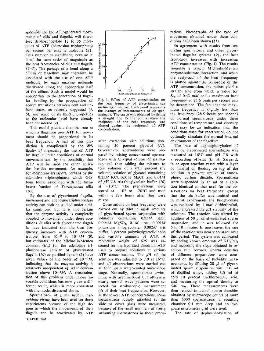
0.1 0.3 1.0 3.0 ATP concentration (mM/lit)
Fig. 1. Effect of ATP concentration on the beat frequency of glycerinated sea urchin spermatozoa. Each point represents the average of measurements of 20 sper- matozoa. The curve was obtained by fitting a straight line to the points when the reciprocal of the beat frequency was plotted against the reciprocal of ATP concentration.
after extraction with solutions con- taining 50 percent glycerol (11). Glycerinated spermatozoa were pre- pared by mixing concentrated sperma- tozoa with an equal volume of sea wa- ter, and then adding the mixture to four volumes of a 62.5 percent (by volume) solution of glycerol containing 0.25M KCI, 0.01M MgCl2 and 0.01M of pH 7.8 tris-thioglycolate buffer (10) at -100C. The preparations were stored at -10? to -20'C and used within a few hours after they were mixed.
Observations on beat frequency were carried out by diluting small amounts of glycerinated sperm suspension with solutions containing 0.25M KCl, 0.004M MgSO4, O.1M urea, 0.001M potassium thioglycolate, 0.002M tris buffer, 3 percent polyvinylpyrrolidinone and variable amounts of ATP. A molecular weight of 625 was as- sumed for the hydrated disodium ATP used to prepare solutions at various ATP concentrations. The pH of the solutions was adjusted to 7.8 at 16"C, and all observations were carried out at 160C on a water-cooled microscope stage. Normally, spermatozoa swim- ming with asymmetrical but otherwise nearly normal wave patterns were se- lected for stroboscopic measurement (4) of their beat frequencies. However, at the lowest ATP concentrations, some spermatozoa loosely attached to the slide or cover glass were measured, because of the small numbers of freely swimming spermatozoa in these prepa-
rations. Photographs of the type of movement obtained under these con- ditions have been shown (11).!
In agreement with results from sea urchin spermatozoa -and other glycer- inated fiagellar systems (9), the beat frequency increases with increasing ATP concentration (Fig. 1). The results resemble a typical Michaelis-Menten enzyme-substrate interaction, and when the reciprocal of the beat frequency is plotted against the reciprocal of the ATP concentration, the points yield a straight line from which a value for Km of 0.43 mM and a maximum beat frequency of 25.6 beats per second can be determined. The fact that the maxi- mum frequency is slightly less than the frequency (28.5 beats per second) of normal spermatozoa under these conditions of temperature and viscosity (11) may be an indication that the conditions used for reactivation do not optimally simulate the normal internal environment of the flagellum.
The rate of dephosphorylation of ATP by glycerinated spermatozoa was measured at 161C and pH 7.8 with a recording p1H-stat (E. H. Sargent), in an open reaction vessel with a layer of mineral oil floating on top of the solution to prevent uptake of atmos- pheric carbon dioxide. Spermatozoa were suspended in 15 ml of a solu- tion identical to that used for the ob- servations on beat frequency, except that the tris buffer was omitted and in most experiments the thioglycolate was replaced by 1 mM dithiothreitol, which increased the pH stability of the solutions. The reaction was started by addition of 50 Al of glycerinated sperm suspension, and it was followed for 5 to 10 minutes. In most cases, the rate of the reaction was nearly constant over this period. The system was calibrated by adding known amounts of KH2PO4 and recording the steps obtained in re- action rate records. Sperm densities of different preparations were com- pared on the basis of turbidity meas. urements, by diluting 20 l of glycer- inated sperm suspension with 1.0 ml of distilled water, adding 3.0 ml of cold 10 percent trichloroacetic acid, and measuring the optical density at 540 m1u. These measurements were then related to actual sperm densities obtained by microscope counts of more than 6000 spermatozoa; a counting chamber 0.1 mm deep and an eye- piece micrometer grid were used.
The rate of dephosphorylation of
77
0 S
B 1 .5-< <0
a:-
0 5 0 5 20
Beat frequency (beats/sec)
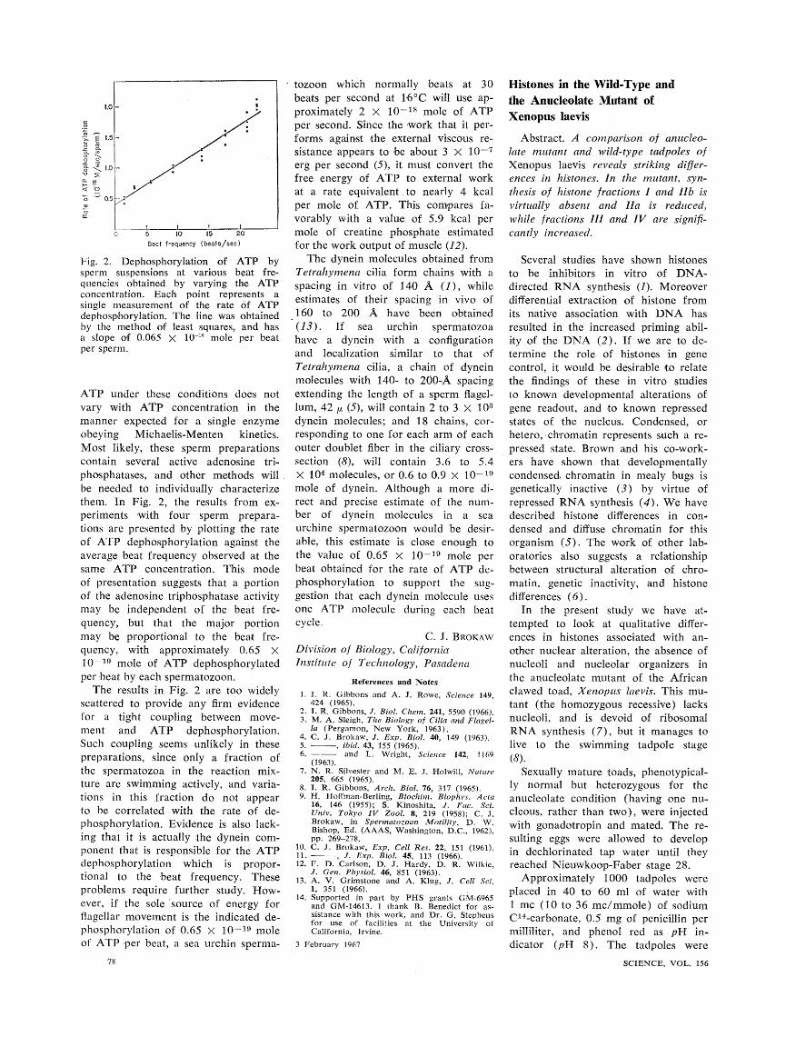
Fig. 2. Dephosphorylation of ATP by sperm suspensions at various beat fre- quencies obtained by varying the ATP concentration. Each point represents a single measurement of the rate of ATP dephosphorylation. The line was obtained by the method of least squares, and has a slope of 0.065 X 10" mole per beat per sperm.
ATP under these conditions does not vary with ATP concentration in the manner expected for a single enzyme obeying Michaelis-Menten kinetics. Most likely, these sperm preparations contain several active adenosine tri- phosphatases, and other methods will be needed to individually characterize them. In Fig. 2, the results from ex- periments with four sperm prepara- tions are presented by plotting the rate of ATP dephosphorylation against the average beat frequency observed at the same ATP concentration. This mode of presentation suggests that a portion of the adenosine triphosphatase activity may be independent of the beat fre- quency, but that the major portion may be proportional to the beat fre- quency, with approximately 0.65 X 10⁻¹⁹ mole of ATP dephosphorylated per beat by each spermatozoon.
The results in Fig. 2 are too widely scattered to provide any firm evidence for a tight coupling between move- ment and ATP dephosphorylation. Such coupling seems unlikely in these preparations, since only a fraction of the spermatozoa in the reaction mix- ture are swimming actively, and varia- tions in this fraction do not appear to be correlated with the rate of de- phosphorylation. Evidence is also lack- ing that it is actually the dynein com- ponent that is responsible for the ATP dephosphorylation which is propor- tional to the beat frequency. These problems require further study. How- ever, if the sole source of energy for flagellar movement is the indicated de- phosphorylation of 0.65 X 10⁻¹⁹ mole of ATP per beat, a sea urchin sperma-
78
tozoon which normally beats at 30 beats per second at 160C will use ap- proximately 2 X 10-18 mole of ATP per second. Since the work that it per- forms against the external viscous re- sistance appears to be about 3 X 10⁷ erg per second (5), it must convert the free energy of ATP to external work at a rate equivalent. to nearly 4 kcal per mole of ATP. This compares fa- vorably with a value of 5.9 kcal per mole of creatine phosphate estimated for the work output of muscle (12).
The dynein molecules obtained from Tetrahymena cilia form chains with a spacing in vitro of 1.40 A (1), while estimates of their spacing in vivo of 1 60 to 200 A have been obtained (13). If sea urchin spermatozoa have a dynein with a configuration and localization similar to that of Tetrahymena cilia, a chain of dynein molecules with 140- to 200-A spacing extending the length of a sperm flagel- lum, 42 tt (5), will contain 2 to 3 XI 03
dynein molecules; and 18 chains, cor- responding to one for each arm of each outer doublet fiber in the ciliary cross- section (8), will contain 3.6 to 5.4 X 10⁴ molecules, or 0.6 to 0.9 X 10-t'S mole of dynein. Although a more di- rect and precise estimate of the num- ber of dynein molecules in a sea urchine spermatozoon would be desir- able, this estimate is close enough to the value of 0.65 X 10⁻¹⁹ mole per beat obtained for the rate of ATP de- phosphorylation to support the sug- gestion that each dynein molecule uses one ATP molecule during each beat cycle.
C. J. BROKAW
Division of Biology, California Institute of Technology, Pasadena
References and Notes
]. I. R. Gibbons and A. J. Rowe, Science 149, 424 (1965).
2. I. R. Gibbons, J. Biol. Chemn. 241, 5590 (1966). 3. M. A. Sleigh, The Biology of Cilia and Flagel-
la (Pergamon, New York, 1963). 4. C. J. Brokaw, J. Exp. Biol. 40, 149 (1963). 5. , ibid. 43, 155 (1965). 6. and L. Wright, Science 142, 1169
(1963). 7. N. R. Silvester and M. E. J. Holwill, Nature
205, 665 (1965). 8. I. R. Gibbons, Arch. Biol. 76, 317 (1965). 9. H. Hoffman-Berling, Biochim. Biophvs. Acta
16, 146 (1955); S. Kinoshita, J. Fac. Sci. Univ. Tokyo IV Zool. 8, 219 (1958); C. J. Brokaw, in Spermatozoan Motility, D. W. Bishop, Ed. (AAAS, Washington, D.C., 1962), pp. 269-278.
10. C. J. Brokaw, Exp. Cell Res. 22, 151 (1961). 11. , J. Exp. Biol. 45, 113 (1966). 12. F. D. Carlson, D. J. Hardy, D. R. Wilkie,
J. Gen. Physiol. 46, 851 (1963). 13. A. V. Grimstone and A. Klug, J. Cell Sci.
1, 351 (1966). 14. Supported in part by PHS grants GM-6965
and GM-14613. I thank B. Benedict for as- sistance with this work, and Dr. G. Stephens for use of facilities at the University of California, Irvine.
3 February 1967
Histones in the Wild-Type and the Anucleolate Mutant of Xenopus laevis
Abstract. A comparison of anucleo- late mutant and wild-type tadpoles of Xenopus laevis reveals striking differ- ences in histones. In the mutant, syn- thesis of histone fractions I and Ilb is virtually absent and Ha is reduced, while fractions III and IV are signifi- cantly increased.
Several studies have shown histones to be inhibitors in vitro of DNA- directed RNA synthesis (1). Moreover differential extraction of histone from its native association with DNA has resulted in the increased priming abil- ity of the DNA (2). If we are to de- termine the role. of histones in gene control, it would be desirable to relate the findings of these in vitro studies to known developmental alterations of gene readout, and to known repressed states of the nucleus. Condensed, or hetero, chromatin represents such a re- pressed state. Brown and his co-work- ers have shown that developmentally condensed chromatin in mealy bugs is genetically inactive (3) by virtue of repressed RNA synthesis (4). We have described histone differences in con- densed and diffuse chromatin for this organism (5). The work of other lab- oratories also suggests a relationship between structural alteration of chro- matin, genetic inactivity, and histone differences (6).
In the present study we have at- tempted to look at qualitative differ- ences in histones associated with an- other nuclear alteration, the absence of nucleoli and nucleolar organizers in the anucleolate mutant of the African clawed toad, Xenopus laevis. This mu- tant (the homozygous recessive) lacks nucleoli, and is devoid of ribosomal RNA synthesis (7), but it manages to live to the swimming tadpole stage (8).
Sexually mature toads, phenotypical- ly normal but heterozygous for the anucleolate condition (having one nu- cleous, rather than two), were injected with gonadotropin and mated. The re- sulting eggs were allowed to develop in dechlorinated tap water until they reached Nieuwkoop-Faber stage 28.
Approximately 1000 tadpoles were placed in 40 to 60 ml of water with 1. mc ( 10 to 36 mc/mmole) of sodium C'4-carbonate, 0.5 mg of penicillin per milliliter, and phenol red as pH in- dicator (pH 8 ). The tadpoles were
SCIENCE, VOL. 156


![Increased Rate of Adenosine Triphosphate …...(CANCER RESEARCH 55, 4352-4360, October 1, 1995] Increased Rate of Adenosine Triphosphate-dependent Etoposide (VP-16) Efflux in a Murine](https://img.pdfslide.net/doc/110x75/5e7e8d68c5d0407f2447f2a9/increased-rate-of-adenosine-triphosphate-cancer-research-55-4352-4360-october.jpg)