Embed Size (px)
Citation preview

Adhesion of Leukocytes to Dermal Endothelial Cells Is Induced After Single-Dose, But Reduced After Repeated Doses of UVA
Marc Heckmann, M agdalena. Pirthauer, and Gerd Plewig Department of Dermato logy, Ludwig-Maximil.ians Uni versity, Munich, German y
Approxhnately 20-50% of ultraviolet A (UVA) irradiation delivered to the skin surface may reach the human derm.al microvascular endothelial cells (HDMEC) that play a pivotal role in cellular inflammatory tissue; however, the pathophysiologic role of HDMEC ·in UVA-induced skin changes is largely unknown. Based on previous itl vivo and in vitro studies revealing UVA-induced expression of endothelial adhesion molec!Jles, we studied isolated HDMEC under various conditions in order to further delineate the impact ofUVA on these cells . The expression of cell adhesion molecules was determined by flow cytometry and the resulting changes of stable adhesion of leukocytes to endothelial cells were quantitated for granulocytes, lymphocytes, and tnonocytes using a newly developed multicellular adhesion assay. Additionally, antibody blocking experin1ents were perfonned to delineate the role ofindividual cell adhesion molecules in UVAinduced leulcocyte adherence. High-dose polychromatic UVA (25 J per cm2
, maximal emission at 375 nm) induced intercellular adhesion mo!ecule-1 and E-selectin with
E xp ress io n o f ce ll ad hesion molecu les (CAM) on th e sur£:1ce of derma.! endothelial cells is a hallmark of inflammatory skin co!i diti o ns allowing circubting leukocytes . to adh ere to specifi c sites within th e cutaneo us vascular bed and subsequ entl y infiltrate th e peri vascular space (G riffiths ct a/,
1989; Swerli ck and Lawley, 1993). A three-step m odel has been pro posed fo r this p rocess consisting of roll ing of leukocytes along th e endothelial lining, firm leukocyte-endo theli al adh esion, and leukocyte extravasation (Springer, 1994; H ogg and Berlin , 1995) . In th.is context leulwqtcs refers to all whi te blood cells. All three steps of tb e adhesion cascade depend on the coordin ated expression of different C A.M types: P-selectin and E-selectin occur at th e sur£:1ce of endothelial cells within minutes to a few hours after stimulati on and parti cipate primaril y in wiling (Bcvi.lacqua, 1993; Sp ringer, 1994). In contrast, in te rcellular adhesion mo lecu le--1 (I C AM- 1) and vascular cell adh esio n molecule-
Manusc ript rece ived March 2 1, 1997; revised August "15, 1997; accepted fo r publi ca ti on August 29, 1997.
R epri ut requests to : D r. Marc Heckmann, Dermatologische Klini k und Po li kli n ik, Lud wig-Maximilians-Uni versitat, Fra uenJobstr. 9- .1 "1, D- 80337 MOnchen, Gem1a ny.
Abbreviations: H DMEC, hu man de rmal microvascular endothelial cells; VC AM-1, vascul ar cell ad hesion molec ule-1.
different kinetics but correlating the adhesion ofleukocyte subsets. This effect subsided, however, in the course of3-6 daily applied UVA doses. Moreover, pro-inflammatory cytokine challenge by tumor necrosis factor-a. and interleukin-1-a. resulted in significantly weaker induction of intercellular adhesion molecule-1 and E-selectin in repeatedly UVA-exposed HDMEC. Differential quantitation of peripheral blood derived granulocytes, lymphocytes, and monocytes revealed reduced adhesion p;trticularly of lymphocytes followed by monocytes and granulocytes compared with leukocyte adhesion to nonirradiated but cytokine-stimulated HDMEC. It is concluded that UVA substantially influences endothelial cell adhesion n1olecul~s expression and thus directly interferes with leukocyte adhesion to endothelial cells. Divergent UVA-induced effects in this respect can be attributed to the mode of UV exposure as well as to the condition of endothelial cells prior to UVA exposure. Key words: cellcell a.dhesiou/E-sclectin/ICAM-1. J Inpest Dermatol109:7to-715, 1997
1 (VCAM- l ) display maximal ~....:press ion at 16-24 h afte r stimulatio n and are dmninantly involved in stable leukocyte adh esion. It is noteworth y, however, that E-selec tin may also contribute to stable leukocyte-endotheli al adhesion, particularly of gran ul ocytes and Thelper- lymphocyte-subsets (P icker et a/, 199"1; Sporn et a/, 1993) .
We have previo usly shown that high doses of ultrav iolet A (UVA) irrad iati o n as a defi ned external physical stimulus ca n upregula te endothelial C AM expression in 11i11o and i11 11itm (Heckmann ct a/, 1994) . Although UVA can precipitate inflammatory skin diseases such as po lymorphous light eruptio n and subsets of lupus erythematosus (Le hmann eta/, 1990), it is also used to redu ce cutaneous inflamm ation as in atop ic dermatitis in which particu larly hi gh-dose UVA 1 has proved to be a potent anti - inA ammato ry regim en (Krutmann et a/, 1.992). T hllS the· sa me stimulus depending on difFe rent circumsta ntia l conditio ns can lead to inAamm.ati o n characterized by CAM upregulatio n or reduction of inflamn1atio n requiring the. opposite effect , i. e., do wnregulation of CAM expression. We th erefore wished to furth er characterize endothelial C AM exp ress io n and subsequent changes of leukocyte adherence in respo nse to UVA under va ri ous experimenta l co nditi ons. We used an i11 11itrr model, all owing us to quantitate C AM expression and multicellular adh esion on human derma.! microvascular endotheli"l cells (1-!DMEC) afte r reprod ucible UVA exposures defined by wavelength and energy fh.1x (UV dose) that are actually effective at the level of these cells.
0022-202X/97/$10.50 · Copyright © 1997 by T he Society fo r In vestigative Derma to logy, Inc.
710

VOL. 109, NO. 6 DE EM UER. 1997
MATERIALS AND METHODS
Materia ls lscovc's modified Dulbcco's medium , feta l calf se rum , and R.PMl
medium were purchas~d ti·o1n G ibco (Ne w York. NY). All other tissne culture
suppl ements and chemicals were o btained fi·om Sigma (St. Lo uis, MO). For
immunostaining procedures the fo ll owing antibod ies (clones) were used: anti
IC AM-1 (U13 IG- II ) and anti -E-sclcctin (13B IG-E6) ti ·omBritish Biotechno logy
(A bingdon, U.K .), ami-V AM ( I .G l lill ) fro m C:unon (Wiesbade n, Genmny) ,
anti-P-sclectin (AI<4) , ami-C D3 (U C H T I), a11ti-CD 13 (W M - 47). anti
C D15 (C3D- 1), an ti-C!) I \1 (C2 D-7), anti -CD3 1 UC / /7CA I), and ami von
Will ebrand- f.1 cto r fro m Dako (Glosrru p, Denmark), and ami-CI)'I-1 (2 M052)
fi·o m D iano vo (H amburg, Germany) .
Endotheljal cell culture HDMEC were isobted from neonatal fo reskins as
desc ribed (Ko rasek, 1989) and cultured in lscove's modifi ed Du lbecco's med ium
suppl em ented w ith thymidine (3 .6 ~g per rnl) , hypoxanthi ne ( 13.6 ~lg per ml),
d ibute ryl cyclic adenosine mo nophosphate (245 ~l g per ml) , isobutyl m eth yl
xa nthine (36.6 ~lg per ml), fe tal ca lf serum (8%), and human m ate rn a.! prepartum
serum (2%). All preparatio ns of se rum w ere used after heat inactivation (56°C
for 60 min). The cells were characteri zed by th eir typical cobble-sto ne
mo rpho logy, by d<!tecti o n of vo n W ill ebrand-f.1cto r and CD3 1, and by
uptake o f flu o rosccin iso thiocyanate-conjugated U lex Europaeus agglu tinin .
Fluorescence-ac tivated cd l so rter (FACS) anal ys is demo nstroted positi ve expres
sio n of th ese cell markers in >98'){, of viab le ce lls. Cells we re passaged at a spli t
rati o n o f I :3 afte r reaching confluence. All experiments we re carri ed out w ith
confluellt cells between rh e thi rd and sixth passage.
UVA irradiation conditions UVASUN 5000 (Mutzhas, Munich, Ge rm any),
e mitting in th e ran ge of 320-460 11111 w ith a lll :tximuin ;tt 375 11111 , was used
as the UVA so urce (Mutzhas eta/, 198 1). lrradi:m ce was 42 mW per cm2 at a
d istance of 40 cm. Exact and reprodu cible deli very of UVA energy was
m o nitored by the use o f an in tegratin g insrrunte nt equipped \·Vith t\vo
fi ltered photodiodcs w ith d ifferent spectral sensitiviti es (Centra-UV, Osr:un
M uni ch, Gen11a ny) . Cell culture dishes were placed at a consta llt distance of 40 cm to th e UV
so uret: and kept :1t a constallt temperature of 30° by using a water bath.
1 urin g th e ti1ne of i rr:~d iatjo n ce lls were submerged 0.5 cn1 into phosphate
bufFered saline that was exchan ged by fi·csh culture medium aftcn·va rds. Contro l
cells were treated in the sam e (t.; hion as irradiated cells; durin g irradiati o n they
we re placed in th e sam e wa ter bath as irradiated cells, although under an UV
imperm ea ble cover. Ce ll viabili ty befo re and after irradiation was determ ined
by excl usio n of trypan blue as well as 7-:unino-ac tin o mycin- D by usc of du al
colo r immunoflu o rescence in single laser Aow cytom cny as described (Schmid
et a/, 1992).
Preparation of p e ripheral blood leukocytes Blood was drawn fi·o n1
hea lth y volunteers with complete blood counts and d ifrtTent ial w hite blood
ce lls w ith in no rmal limi ts. H eparin-sodium (500 U pe r ml ) was added as
anti coagulant. Platelets were remo ved by cemrifugatio n and washing in phos
phate-bufrc red sali ne. Po lymo rpho nucl ea r granulocytes and mo nonuclea r
lymphocytes/ m o nocytes were separnted by cen trifuga tio n over " density gradi ent
using Mono- Po ly n .. eso lving M ediu m (I C N, Biom edi ca.ls, Costa Mesa, A.
wo rld w ide) according to th e supplier 's protocol. T he resulting cell prepa rati o ns
were exami ned by flow cytomeny afte r inununo phenotypic labeling of CD3,
C D I4, C D IS, o r C DI 9, respecti vel y.
Adhesion assay A novel multi cellular :1dhesio n :~ ssny was e rnploycd as
described in det<Ji l elsew here (H eckm:111n and Pirrh:~u er. I 996) . Briefly, HDM EC
we re ~eedcd in to .1 2-wel \ culture pbtes , grown to conflucncy. rinsed \vith
ph osph ate- bufle rcd s :~ lin e three ri mes, and th en ex posed to peri phcr:JI blood
leuk ocyte in a se ru m fi·ee cell suspensio n (1 00, 00 per well) with a s tand:~ rdi zed
I: I ratio of granul ocytes to mo no nucl ea r cells. After 30 min at 37°C under
stati c conditi o ns, no nadherem cells w ere removed by a standardi zed procedure
incl udi ng three consecu ti ve ge ntl e washin gs. All retna_i ni_ng: ce lls, incl udi ng
adh erent leukocytes as well as unde rl yin g 1-1 DMEC, were coll ected fo r A ow
cytometty after b ri ef incubatio n in phosphate- buflt:red saline containin g 0. I%
trypsin and 10 111 M e rh ylencdian1ine tetr:1acetic acid.
Flow cytomerry Cells we re kept o n ice at all times. Aliquo ts were subj ected
to imrnunosta inin g w ith the respec tive antibodies acco rdi ng to the protocols
o f th e suppli e rs. Appro priate n1 o noc lo nal mo use isotype lgG were used in all
cxpeL"inn:nts ro d ete n nin c b.-,c kgro ull d staining. Eva luation of Aow cyto n1c try
was assisted by Lysis- I l and Ceii Q ucsteTM softw:J re (13ecton-Dickenson, Erembas
tcgem- Aa lst, Belgium) fo r com putel; zed acqu isitio n and analysis of data.
Antibody blockin g experiments Blockin g anti bodies were used as desc ~:ibed
previo usly (H eckman n cl a/, 1993) accord ing to the specification of th e suppli e rs
listed under t\tlMcrials. Success fu l blockin g was confi rm ed by adding fluorescence-
I~EDUCED CELL AD HES ION AFT ER REPEATED UVA IR .. RADIAT10N 711
0 0 '<t
0 0 '<t
lgG
lgG
Control
4
UVA 25 J/cm2
4
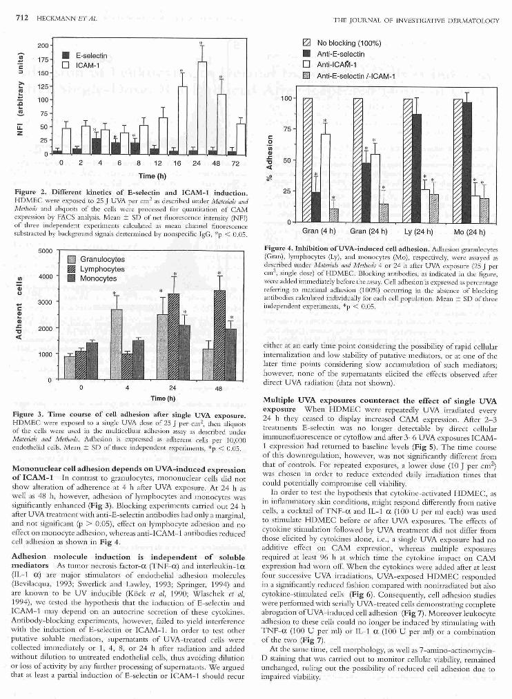
Figure 1. UVA induces E - selectin o n isolated endoth eHal cells. FACS
ana lysis o f HDM EC was carried out 4 h after 25 J UVA per cm2 as described
under Materials rwd J\1/cthods. Arbitrary uni ts of flu o resce nce (x a.xi s) fo r unspecifi c
immunoglo bu li n (lgG) and E-selectin (E) are plo tted aga inst ce ll numbers(]' a..xis) .
labeled antibodi es againsr th e same epi tope to pretrea ted ('' blocked") cells
yielding no Au o rcscence ovt!r basel ine levels obtained \Vlth no nspecifi c flu o res
cence IgG-isotypes.
Statistical a n alysis The Student's t tesr was used fo r statistica l analysis; p
values above 0.05 were considered not signifi c:mt.
R ES ULTS
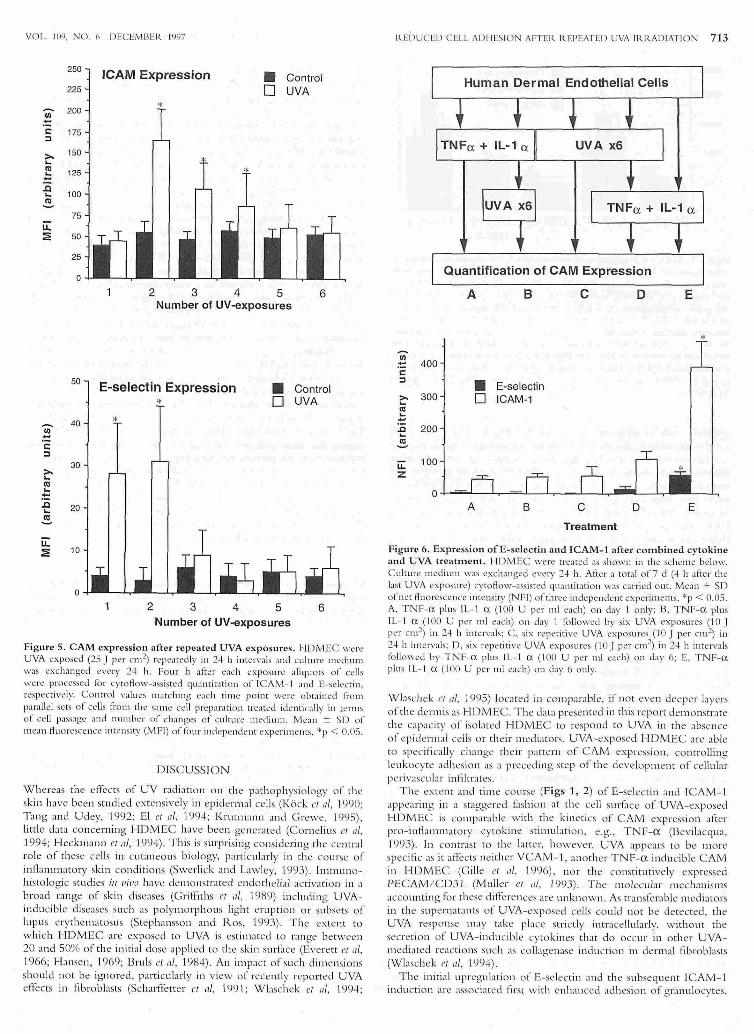
Single-dose UVA induces e ndoth e lial ICAM-1 and E - selectin
with different kinetics lsobted e ndo thelia l c~ll s expressed con stitu
tive levels of lC AM - '1, w h e reas n o express io n of V C AM- 1 or E
selectin was d e tecte d (d ata not sh own ). F o ur h o urs afte r , 25 J UVA
p e r c m 2 cell su rf.1ce expressio n of E-se lectin was sn·ongly induced
(Fig 1) follo wed b y a g rad ual d eclin e over the n ext 16 b , disappea rin g
co mplete ly 24 h after irradiation (Fig 2) . IC AM- 1 expressio n rose
from 12 h afte r radi a ti o n , reaching a plate au afte r 2 -J. b , th e n graduall y
declining a ti:e r 48 h to baseline: levels (F ig 2 ). In co ntrast, n o
ellect was n o ted for VCAM- 1 o r PEC AM / D 3 1 a t a ny of the
afo re m e ntio ned ti m e po in ts (data not sh ow 11) .
Granulocyte a dhesion depends o n UVA-induced expression of
E-selectin and ICAM-1 In o rd e r to delin ea te the conseq uen ces of
UVA-induced C AM expressio n in te rm s o f ac tual le ukocyte adhe re n ce
to UVA-trea ted HDM..EC, a re e ntl y d evelo ped cell adh esio n assay
was e mpl oyed th a t allows to q u an ti ta te g ranulo-, l)'mpho-, and
m o n ocyte ad h es io n simu lwneously. By this m eth od a time ly ditli:re nti
a te d pattern o fl e ukocytes adh esio n could b e demo nstra te d . Gra nu locyte
adh esion was significantl y e nhanced fro m4 to 24 h a fter UVA trea tme nt
of 1-:IDM.EC, th en at 48 h returning to b ase lin e levels (F i g 3 ). Fo r
b locking expe rime nts isolated g ranulo cytes were added to HDMEC
4 h after UVA e xposure of HDM..E in o rde r to yield m aximal cell
ad h esio n. In t h e presen ce o f anti-E-selectin antibodies g ranulocyte
adh esion de l in e d to 24')1, compare d with m aximal adhesio n (1 00%) . Anti- IC AM- ·1 a ntibodies redu ced ad h esio n to 71% w h e reas the com
binatio n of b o t h antibodies y ielded 'J.l 'i(, (Fig 4 ) .
712 HECKMANN ET AL
200
~ 175 • E-selectin
c D ICAM-1 :I 150
* ~ 125
~ 100 -e ~ 75
u:: 50 z
25
0 0 2 4 6 8 12 16 24 48 72
Time (h)
Figure 2. Different kinetics of E-selectin and ICAM-1 induction. HDMEC were exposed to 25 J UVA per cm2 as described under Materials a11d Methods and aliquots of the ceUs were processed fo r quantitation of AM expression by FACS analysis. Mean ± SJ of net Auorescence intensity (NFI) of three independent experiments calculated as mean channel Auoresccnce subsrracted by background signals dertem1ined by nonspecific lgG, *p < 0.05 .
5000
~ Granulocytes
~ Lymphocytes VI 4000 • Monocytes Gi u
.... 3000 c ~ Q) .:: 2000 "C <(
1000
0 0 4 24 48
Time(h)
Figure 3. Time course of cell adl1esion after single UVA exposure. HD MEC were exposed to a single UVA dose of 25 J per cm2, then aliquots of the ceUs were used in the multicelluar adhesion assay as described under Materials a11d Methods. Adhesion is expressed as adherent ceiJs per I 0,000 endothelial cells. Mean ± SD of three independent experim ents, *p < 0.05.
Mononuclear cell adhesion depends on UVA-induced expression of ICAM-1 ln contrast to granul ocytes, m ononudear cells did no t show alterati on of adherence at 4 h after UVA exposure. At 24 h as well as 48 h, ho wever, adhesion o f lymphocytes and monocytes was significantly enhanced (Fig 3) . Blocking experiments carri ed out 24 h after UVA trea tm ent with anti -E-selectin. antibodies had only a margi nal , and not significant (p > 0.05), effect on lymphocyte adh esio n and no effect o n monocyte adhesio n, whereas anti - IC AM-1 antibodi es redu ced cell adhesio n as shown in Fig 4 .
Ad11esion molecule induction is independent of soluble mediators As tumo r necrosis fac tor-a (TN F-a ) and interleuk.in- 1a (IL-1 a ) are majo r stimulators of endoth elial adh esio n molecul es (Bevilacqua, 1993; Swerlick and Lawley, 1993; Springer, 1994) and are known to be UV inducible (Kock et a/, 1990; W laschek et a/, 1994), we tested th e hypo thesis that the indu ction. of E-selectin and IC AM- 1 may depend on an autocrine secretio n of these cytokines . Antibody-blocking experiments, however, f:l.iled to yield interfe rence with th e inducti on of E-selectin or LCAM - 1. In order to test o ther putative solubl e mediators, supernatants of UVA-trea ted cells were collected immediately o r 1, 4, 8, or 24 h after radiation and added with out dilution to untrea ted endothelial cells, thus avoiding diluti on or loss o f ac tivity by any further processing of supernatants. W e argu ed that at least a partial induction o f E-seJectin or ICAM-1 should recur
100
75 r::: 0 'iii Q) .:: 50 "C < <f.
25
0
THE JOUR NAL OF INVESTIGATIVE DER.MATOLOGY
E:a No blocking (1 00%)
• Anti·E-selectin D Anti-ICArV1-1
~ Anti-E-selectin /- ICAM-1
Gran (4 h) Gran (24 h) Ly (24 h) Mo (24 h)
Figure 4. Inhibition ofUVA- induced cell adhesion . Adhesion granulocyws (G ran), lymphocytes (Ly), and monocytes (Mo), respecti vely, were assayed as described under Materials a11d Methods 4 or 24 h after UVA exposure (25 J per cm2, single dose) of 1-IDMEC. Blocking antibodies, as indicated in the figure, were added immediately before the assay. Cell adhesion is expressed as percentage refeJTing to maximal adhesion ('100%) occurring in the absence of blocking antibodies ca lculated individually for each ce ll population. Mean ± SD of three independent experiments, *p < 0.05 .
either at an early time point considering th e possibility of rapid cellular inte rn alizati on and low stabiliry o f pu tative mediators, or at one of the later time points considering slow accumulatio n of such mediators; however, no ne o f th e supernatants eli cited the effects observed after direc t UVA radiation (data not show n).
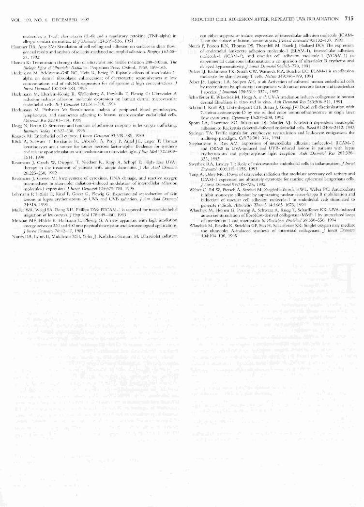
Multiple UVA exposures counteract the effect of single UVA exposure Wh en HDMEC w ere repeatedly UVA irradiated every 24 b they ceased to display increased C AM expression . After 2-3 treatments E-selec tin was no lo nger detectable by direct cellular inu11uno fluoresce nce or cytoflow and after 3-6 UVA exposm es ICAM-1 expression had return ed to baseline levels (Fig 5). The time course o f this dow nregulation, however, was not significantly different from that of controls. For repeated exposures, a lower dose (tO J per cm2) was chosen in order to reduce extended daily irradiation times that could potentiaJJ y compro mise cell viability.
In o rder to test th e hypoth esis that cytokin e-acti va ted HDMEC, as in inflanm1atory skin conditio ns, might respond differently from native celJs, a cocktail of TNF-a and IL-l a (100 U per ml each) was used to stimulate HDMEC before or after UVA exposures. The effe cts of cytokine stimulation followed by UVA treatment did not differ from those elicited by cyto kin es alone, i. e. , a single UVA exposure had no additi ve effec t o n CAM expression, whereas mu ltip le exposures required at least 96 h at which tim e th e cytokine impac t o n C AM expressi o n had wo rn off. Wh en the cytokin es were added after at least fom successive UVA irradiati ons, UVA-exposed HDMEC responded in a significantly ,-edu ced fashio n compared w ith nonirTadiated but also cytokine-stimulated cells (Fig 6) . Consequ ently, ce ll adh esion stu cU es were performed with serially UVA-trea ted cells demonstrating complete abroga ti.on ofUVA-indu ced cell adhesion (Fig 7). Moreo ver leukocyte adh esio n to th ese cells could no longer be indu ced by stimulating with TNF- a (100 U per m.l) or IL-1 a (100 U per nli) or a combination of th e two (Fig 7).
At th e same time, cell morphology, as w ell as 7-amino- ac tinonrycinD staining that was carried o ut to monitor cellular viability, remained unchanged, ruling o ut the possibili ty o f reduced cell adhesio n du e to impaired viabi li ty.
VOL. 109, NO. r, DECEM IJER 1997
250
225
u;- 200 -1: 175 :I
>. 150 ... Cll 125 .!:: .a
100 ... .e.
75 ii: :a; 50
25
0
ICAM Expression
*
2 3 4
• Control 0 UVA
5 6 Number of UV-exposures
Figure 5. CAM expression after repeated UVA exposures. l-IDM EC were UVA exposed (25 J per crn 2) repe:1redly in 2·1 h inrcrvals ;utd culrure mediunt was exchanged every 24 h. Four h after e:1ch exposure :~liquors of cells were processed for cyroAow-assisted quontitation of !CAM- I and E-sd ectin , respL'ctivd y. Contro l values m:~tching ~ach tim e po in t were ob tained ti·om para llel sets of cells ti·om the same cel l preparation treated identicall y in te rms of cell passage and number of changes of culture nteu ium. Mc:1n ± SD of mc:1 n Auorescence inrcnsity (M Fl) of fo ur independellt experim ems, *p < 0.05.
DISC USSION
W hereas the efiects of UV rad iatio n o n the patho physio logy of the skin have been studied extensively in ep idermal ce lls (K ock eta/ , 1990; Tang and Udey, 1992; El et a/ , 1994; Krut.n1 ann and G rewe , 1995), Li ttle data concerning HDMEC have been gene rJted (Corn elius et a/ , 1994; H eckmann eta/, 1994) . This is surpri sing considering the central ro le of these ce lls in cu taneous bi o logy, p:~rti c u la rl y in the co urse of inflammato ry skin conditi o ns (Swerli ck and Lawley, 1993). Immun ohistologic stud ies i11 t1irm ha1re de lllo nstrated endothelial activa tion in a b road range of skin diseases (G rilf 1ths rt a/, 'I 989) incl uding UVAinducib le d iseases such as po lym o rpho us li<>h t eruptio n o r subsets of lu pus erythematosus (Step hansson and R os, 1993). T he extent to w hich HDMEC are exposed to UVA is estimated to ran ge between 20 and 50% of the in itia l dose app lied to the skin surface (Everett ct nl, 1966; H ansen , 1969; Bruls et a/, 1984) . An impact of such di111ens.ions sho uld not be ignored , parti cularly in view of recently reported U VA efrects in fibrob lasts (Scharffetter ct nl, 'J 99 1; Wlaschek er nl, l 99.f;
REi)UCED CELL ADHESION AFTER. REPEATED UVA lll RADI ATION 713
Quantification of CAM Expression
A 8 c D E
* ~
rn 400 -c::
:I • E-selectin ;:- 300 0 ICAM-1 ('CJ ... -.c 200 ... .e. u::: 100 z
0 A B c D E
Treatment
Figure 6. Expression ofE-selectiu and ICAM-1 after combined cytokine and UVA treatment. 1-l DM.EC were treated as shown in the scheme below. C ul tu re medium was exchanged every 24 h. After a tor:li of 7 d (4 h after the last UVA exposure) cytoHow-:~ss i st.cd quamitation was carried ou r. Mean + SD of net flu orescence intensity (NFI) of three independent experiments, *p < 0.05. A, TN i=-a plus 1 L- 1 <X ( I 00 U per ml each) on day 1 only; B, TN F-a plus Il- l <X ( I 00 U per rnl each) on d:t y 'I fO ll owed by six UVA exposures ( I 0 J per cm2) in 2-1 h intervals; C. six repetitive UVA exposures ( I 0 J per crn2) in 1-l h intervals; I , six repetitiw UVA exposures ( I 0 J per cm2
) in 24 h interva ls to ll owcd by TNI'-<X plus IL- l <X (100 U per ml e:tch) on day 6; E, TNF-<X plus IL- l a ( I 00 U per ntl each) on day 6 only.
\Xflaschek e1 a/, 1995) located in comparable, if not even deeper layers of the dennis as H DMEC. The data presen ted in this report dem onstrate the capaciry of iso lated H DMEC to respo nd to UVA in th e absence of epiderm al cel ls or their m ediators. VVA-exposed HDMEC are :1ble to speci.fi call y clunge their pattem of CAM expression. co ntro lling leuk ocyte adhesio n as a preceding step of th e deve lo pment of d lula r perivascular infiltrates.
The ext<:nt and rime course (Figs 1 , 2) of E-selectin and !CAM-I appea ring in a staggered £1s hio n at the cell surf.1ce of UVA-exposed H DMEC is co mparab le ' ith the kinetics of C AM expression afi:e r pro- inflammatory cycokine stimulatio n , e.g .. TNF-a (Bevilacqua, 1993). J n contr;lst to the larter, however, UVA appea rs to be m ore specifi c as it affects neither VC AM-1 , another TN F-a inducible C AM in H DI\IIEC (G iLl e ct a/, 1996), no r the constitutive ly e:>.lJressed PECAM / CDJ I (Mulle r e1 a/, I. 9YJ) . T he mo lecular m ech:lll isms accoun tin g for these differe nces are unkn own . As transfe rable m ediato rs in the supem atams of UVA-exposed cells could not be de tected, th e UVA response m ay take pbce stri ctly in tracellu la rly, w itho ut the secretio n of UVA- in ducible cy tok ines that do occur in otl1er UVAmediated reactio ns such as colb genase iDdu ctio n in dermal fib ro blasts (Wlaschek ct a/ , I 994).
The init.ia l upregulatio n of E-selectin and the subsequent !C AM- I indu ctio n are associated f1rst w ith enhan ced adhesio n of gra nulocytes,

714 HECKMANN ET AL
5000
4000
3000
2000
1000
0
Control UVA x1
Granulocytes 1m Lymphocytes
Monocytes
UVAx6 UVA x6 TNF+IL-1
TNF+IL-1
Figure 7. Leukocyte adhesion after multiple UVA exposures. HI MEC we re rcpeotcd ly UVA trea ted ( I 0 J per cm 2) over a tota l o f 7 d w ith dail y changes o f culture medium. T hen the cells were used in the mu lticel lular ad hesio n assay as desc ribed un der Materials a11d i\1/ethor/s. Mean ::!:: SO o l fo ur independent experimen t., , *p < 0 .05. Contro l, no UVA irrad iatio n , no cyto kin c stimulation ; UVA X I , UVA exposures o n day 6; UVA X 6 , repeated UVA cJqJosures on da ys '1-6; UVA X 6 + TN F + IL- l , repeated UVA exposures o n days 1-6 plus cytokin e stimula tio n o n day 6 ; TN F + lL - 1, cytokin c stimulatio n o n day 6 o n ly. Note that Figs 6 and 7 are not designed congruentl y as cond itions A and 13 in Fig 6 revealing no signifi ca nt chan ges arc not depicted in Fig 7, w hereas contro l and UVA X I (co mpare w ith Fig 3) arc depicted for refe rence.
and then with lymphocytes and mo nocytes (Fig 3). This observation parall els previous iu vivo studi es de tec tin g granul ocyte emi gration prio r to monon u cle~r ce lls afte r UV irradiati o n (N o rTis ct a!, 1991) . ln other experiments in w hi ch poison ivy as a different extrinsic stimu lus bad been used to in du ce cutaneous in:fbmmation, there was no granu locyte infiltration, despite upregul ;1ted E-selectin and IC AM- 1 o nmi crovas ular endo thelial ce lls (Griffi ths et a/, 1991 ), suggesting that these tv.ro C AM ~re necessary but no t sufficient fo r successful emigration of leukocytes as they co ntrol predominantl y stable adhesion that is only one of a mu ltistep cascade of inte rcellular signaling in leukocyte emigrati o n (Springer, 1994; Hogg and Berlin , 1995). T he extent to whi ch E-selectin andi CAM - 1 contri bute to stable leukocyte adhesion to endothelial ce Ll s sho uld no t be es timated by mere ceLl surf::J ce expressio n quantitated in arbitraty units (Fig 1), but ha. to be investiga ted b y blocking experiments o f ce ll adh esion (H eckmann and Pirthauer, 1996) . Gran ulocyte adhesio n on UVA- rrea ted HDMEC could be redu ced by blocking E-selec tin and IC AM - 1 with add itive inhibitory en'ects usi ng antibo di es against both simu ltaneo usly (Fig 4). T he propo rtion of E -selectin-dependent ad hesion was signifi cantly hi gher at 4 h co mpared w ith that at 24 h after UVA exposure, sugges ting a dynamic rath er than a fi xed utiliza ti o n of d iffe rent endothe lial liga nds by granu locytes. In contrast, lymphocyte and m onocyte adh esio n could on ly be bl ocked by anti- IC AM - 1 antibod ies in this experimental setting.
Even tho ugh T lymph ocyte subsets use E-select in as a co un ter recepto r of the cuta neous lymphocytes antigen (Picker et a/, <1 991 ; Springer, 1994), this ad hesion mechanism appea rs to be irreleva nt in UVA-in duced lymphocyte adh esion because upn:gulation o f E-selectin 4 h after UVA exposure did no t indu ce lym phocyte ad hesio n, nor did the additio n of blockin g antibod ies against E-selec tin block an induced lymph ocyte adh esio n occuring 24 h aft<:r UVA ex posure (Fig 4). T wo exp.lanatio ns ma y be o ffered fo r this seeming paradigm. (i) As o nl y a subse t of aU pe1ipheral bl ood lymph ocytes express cutaneo us lymph ocytes antigen (Berg et a/, 1991.), enhanced ad hesion of thi s subset may not be sufficient to cause signifi cant chan ges of the overall o utco me of lymph ocyte adh esion. (i i) Under conditi ons of A ow, enhanced Esc lectin exl'ression is associated primarily w ith leukocyte roUing, a dynamic process (Hammer and Apte, 19';)2) that is not mimi cked by a static ad hesio n assay as used here .
It is also noteworth y that m o nocyte ad hesion was signifi ca ntl y enhanced afte r UVA exposure, despite the lack ofV C AM- 1 indu ctio n. VC AM - l that ca n func ti on as the co unter receptor of th e monocytic VLA- 4 has been esta blished as an important recepto r for monocytes
TH E JOU ilNA L O F INVESTIGATIVE DERMATOLOGY
(Sprin ger, 1994). Its absence does no t preclude effe ctive monocyte adhes io n, even th o ugh its presence can be suffi c ient fo r it (Weber ct a/, 1994), un de rscoring that a given patte rn o f ce ll adhesion may be the result of va riab le patterns of C AM <: xprcssio n.
R.epetiti vc UVA exposures rendered HDMEC signi fi cantly less responsive to subsequ ent UVA-radiation leve lling at baseline after 3-6 repe titions. Moreo~er, the ce ll s were also signifi ca ntl y re liactory ag:1inst pot<:nt pro- inAam matory stimu li, such as TN F-cx and I L- 1 ex, representin g two m;uor endogeno us mediato rs ca pable of.initiating ;111d propagating C AM upregulati on and subsequ<:nt pe ri vascular inA ammatio n (Bevilacqu a, 1993). As a consequ en ce of this inh_ibitory qual ity of UVA on <:ndothelial C AM express io n, cell adh esio n of all majo r periph eral blood leukocytes to HDMEC was severely hampered compared w ith nonirrad iated 1-JDMEC receivin g the same cytokin e stimula tion . Repeated stimula tio n with TN.F- cx am o un ted tO the sa me effect: exhausted indu cibi lity of leukocyti c adhesion by a mech~ nism that has not yet been expbined o n a mo lecular basis (Pober eta/, 1987) .
T he anti - inAammato1y cyto kin e IL- l 0 capable of inhibiting ICAM-1 expression is indu ced in UVA !-ex posed keratinocytes (G rewe et a/, J 995) . UVA- indu cib le seco nd messe ngers in d ude free oxygen radi cals (Krutman n and G rewe, 1995) th at in turn can activate transcrip tio n r.1 ctor binding to genes leadi ng to en haJJced DNA transcrip tio n (.Bae ue rl e and Henkel, 1994); in particular, th e nuclear facto r NF- KB that has been implica ted in the transcriptio nal induction of IC AM-1 and .E-seleclin (C hen ~ nd M anning, 1995).
Anti- inAammato ty effi cacy o f UVA ·1 exposm es may be explain ed in part by the ind uction of IL- l 0 in kerati nocytes (G rewe et a/, 1995). .Based on o ur o bservations with repeated ly irradiated HDMEC, we suggest that these ce lls th~t are criti c~ ll y situ ated between circulating leukocytes and th e peri vasc ular derma l space pla y a role in UVA- in duced anti - inA amma to ry mechanisms. UV- indu ciblc effec ts o n pa rticular ce ll fun cti ons are in creasingly sharpening the image of UV radia tio n as a highly ple iotrophi c stimu lus fo r opposing efrects w ithin the sa me experimental setting (Norris e/ a/, '1990; Krutmann and G rewe, '1995). T his is also va li d for the d iversity of leukocyte- endoth elial inter~ ction that is specifi caLl y <lm enabl e to UVA.
/111· tl/tlllk Mrs. Sciliill-1-lllpka for cxccllmt tcdllliml assisttiiJCC . This st11 d)' "'"s ki11dly s11pported by the Dc11tschc f'orsdiiiJJg.\~CIII Cillscllt!{t (1-le 1593 13- 2) a11rl the Blllldeslllill ist.critlllt _(l'ir Bildr 111g, Wissm sclll!/i , Forscllllllg 1111 d Ti•r/11wlo.~ir
(U?U f/1359 2).
IU FER.EN CES
Uacucrl c PA. Henkel T : Fun cti o n :md acti v:~t i on o f NF- kappa JJ in the imnnn1 c systc111 . 1'1111111 IICII IIIIIIIIIIIVI 12:141 - 179, 1994
IJcrg EL, Yoshino T, R.ott LS, el a/: T he cutaneous lymphocytt: nmigcn is a skin lyn1phocytc homi11 g: receptor for the v~JSC ttl:'lr lcc tiJJ CJJ dotltcli<J I ccll - lt:ukocytc ad!Jcsiot J JJlOicculc I..J Jixp Mcd 174:146 1- 1466, 199 1
Bcv il acquJ MP: Endothdi:ll - lcukocytc ad hesion lllolcculcs. 1 111 1111 Rev lmniiiiiOI II :767-80'1, 1993
Uru ls WA, V:lll Wcc lclcn H , van d~r Lcun JC : Trtii1Sil li $s ion or UV- rad i:ll ion through hum:111 cpidcnn ;Ji lnycrs ;~ s a f..1<:tor inf-luencing the rninimal L' ly th cn\ :1 dose. PIIIJfc'du:m Pltotc>hi<>l 39:63- 67. 19H4
hen CC, M:~rming AM : Tra nscri ptiona l regulati on ,,r c ncloth c\i:-~ 1 ce ll :-~cl h csion niOieculcs: a don1 inant role for N I'- kappa 13 . ll. ~clfls Ar1io11s 47 (S uppL): 135- 14 I. 1995
C ornelius LA. Scpp N , Li LJ , Dq;it'l. 1< . Swcrlick I ~A . l.awky TA, Caughman SW: Sch:cti vc.; upn.:gul:l tion of intcn.:L·IIu lar a clh~s i o n rnolcc ul ~ (I AM - 1) by ultr:l violct IJ in lwm~111 dcnn:d llli crovascul ;~ r endo theli al ce ll s. J lmJC:s t Dcrmatol I 03 :23-28 . \ 994
El GA. Picrik F, Norv;t1 M: ompa r:1 tiv~.: potency ol difl:Crcnt UV sources in reducing the density ~ nd antige n-present ing capacity or L:n1gerhans ct.:lls .i n C3 1-1 mi ce. Plwwchcm 1'/wtouio/ (>():256-2(, I , 1994
Everett MA , Vi:u·gers E. Sayre RM , lson R.L: Pcnctr:'tt ion of epidcn nis by u l tr~v io l c t
roys. 1'/wrorl<rw l'fwtoltiol 5:533- 542. 1%6 Gi lle J, Swerlick llA, Lawley TJ. Ca ughu mu SW: l)iOe rclllial regulati ou of vascular cel l
ad hesio n lllo lecule- 1 g~.: n c l ra nscription by tum or necrosis r.1 ctor ~lpha and imcrleukin - 1 alph;l in dcnnalmicrovascular cndoLhd b l cd ls. B/ctod H7:2 11- 2 17, \ 996
Grewe M, Gyufko K, Krutmil nn J: lntcl'l cukin- \0 produn ion by cultured lnn11an ker:ltinocytcs: regulatio n by u h.r~w i o l c l 13 :'IIICI ultrnviolct A 1 radiation. J lllllt'Sf
/Jcn nalol I 04:3-6. 1995 G rirliths CE, Voo rh ccs JJ , Nicko lofrJJj: Clwraclcri z:ai on ol intcrccllular ad ht.:sion nlolccuh.!-
1 and I-lLA- DR c>1 pn:ss io n in nonnal and irJ!l :1 111 C<.l skin : modul:1tion by r~.:co tnbinnnt gamma intcrrcron and tu mor necrosis l~lc to r . .J Aw 11c(I{/ /Jcmullvl 20:(> 17-629, 1989
Grilriths CE, Uarkcr JN , Kuuk el S. Nickolon· UJ : Modu lation of leucocyte ad lt csiou
VOL. 109 . NO. 6 D ECEM UER. 1997
1\JOk·nd l.'s , a T- cc ll chcmotaxin (11..-H) :111d :\ regulatory cytoki nc (TNF-alph:t) in
:Jll crg-ic co llt;lc l dcrmalilis. lJr.f Demratol 124:5 19- 526 . 199 1
1-l:.unnJcr 11/\, Aptt' SM: Si mulation of cdl ro lling :m el adhesion o n surf..1ees in she:1r Aow: ge neral rcsul t'i J lld an.1lysis of sclcctin- •ll cdi:. tcd ne u trophil :tcihcs i011 . Bi{)phys) 63:35-
57, 1'!<)2 1-la nsc n K: T r::tnsnJiss ion through skin o f uh raviokt and visibk r:1 di:-~t i on 2H0- 500nm . 711C'
JJivh!!,! i( l~'[li:ct ,y· Ultrr lllio!t:r R11difuhm. Pcrg,ul\t )ll Press, Oxford. 19()9 . 159- 103. 1-k~..: k nl a n•• M. Adch n;~n n-C rill 13 C, 1-k in R, l<rh:g T : 13iph:lsit.: L' ill:c ts or intcrlt:ukin- 1-
:llpha on dcnn:d fibrob l;1s ts: c nha n CI.! IliCilt of chcmor:tCLic n.:sponsivcncss :tt low co ll CL' lltt~ ti om :nlll of m lt.NA c.x pr~:o;s i o n fO r co \\ ;-~gc n asL' :tr hi gh ron c~ntrations . j III II(.\'( D l' fll/0(() / 10!1 :7:-10-78~. 1993
H eck1m nn M , Eberle in- Ko nig U. Wollenberg A. l' rzybi lb T, Pbvig G: Ultravio let A
r;-~ d i at i on i11duccs ad iJ csiun lliOicctll l' ..:xprcs~ i o 1 1 011 hun1att d..:nnal JlticrovasL' tlb r
endot he li al ce lls. Ur j Dcrllwt<•l 13 1:3 11- 3 1R. 1994
1-l cc km:llll l M. Pirth :~t~ ..: r M : Simu ltatl L'O liS :l tl :dysis or periphera l bl od gr:.Hlll lozyn:s.
l y n1ph ocytc~ . :mel tn onucytcs :-.dh crin g t.o lnun:m tnicrovasc ular c ndoth c li:~l cells.
1\/imwasr J<.c.~ 52: 10 1- 114 , 1996 H ob!; N. 13nlin C: StrU Cllii"C :'llld function or :-.dhL•sion I"L'Ceptors in \ ~._·ukocyrc tr:-.flicking.
lww111wl 7·~,dtl)' 16:327- 330, l l'JtJS
l<a r:~se k M : Endoth e li:~l cell culture. J i iii'C.' I Dcmcat"l 93:33S-38S. 1989
I(Qck A. Sch w;~rz T. l<irnb:w er It, U rbanski A. i>L'ITy P, t\ nsd J C . Luger T : Human
kcr,ttinoc.:ytcs arc :l sOtlret· l't1r tu 1no r necrosis [tcto r- alp ha: E.videnC{' fOr synth ~s i s
:md n;lc:tse up0 11 stimul:lt io n w ith cndowxit1 r ultrnvi let ligh t.) Exp !\It'd 172: I (,09-
10 1-t, 1990 l<ruttnann J. Czech W. Di epge n T , Ni edn er 1 ~. l<app A, Sc hopf E: Hi gh- ctose UVA I
d lt.: rapy in the tn:iltnt cnt of p:1ticm s w ith atopic dt:rm:ltitis . .J A111 Actld D cmu1ftl/
2(.:225-230, 1992 Knlllll :l llll J, Grewe M : lnvoiV\..' II ICllt o r cytokin cs. DNA damage. and reactive oxygen
illtl'rtli Cd i :~teS in u\t r:~vioh;t r:tdbrio n- induccd moduJ:ttiOil o f in tc..~rcd Jular :-.dhcsio n
nt o lcculc- 1 expressio n. J '"""" Dan~<tl, / 105:67S- 70S, I'J95 Lehmann P, H 6 lzlt:. E, l<ind P. Gocrz G. Plt.:wig G: Exp~rim t:! tH :'l ! reprodu ction or ski n
les ions in lupus t:. ryth cntatosus by UVA and UV U r:tdintio n . f .r\ 111 /Jauf D CI/IIIItol
24:5 15 . 19')0 Mu ll er WA, We igl SA. Deng X I-I , Phillips I M : PE AM - I is req 1>ircd fo r tr,.oscncto th cl i :~ l
ntigratio n oC le ukocytes. j Exp t\ trd 178: 449-160. 1993
Mulzhas Mi=. 1-IOizk E. H oflll :t llll C . Plcwig G: A new app:1r:l tuS with hi gh irr:u.l i:-. tio n
Cllcrgy be tween 3211 :111d 400 111 11 : physk:t l d t:.sc ription :t11d ch:nl lii lo lot,ri cal applications.
j fiiii ('S f Oemwro/ 7(':'12-17, l lJR I
N o rris DA , Lyons U, Middle-to" MI-l , Yo l111 JJ , K"shihar"-S" w" llli M : U ltravio let r:~ diation
RE DUCED CELL ADHESION AFTER REPEATED UVA IRRAD IATION 715
e m eith er suppress o r induce expression of lntercellu\:Jr :1dhcsio n mo lecule (I CA MI) o n rhc sur6ce o f hum:m kcr:-.rinocytcs. j .lllt't!SI D c.rmnro/95:1 32-'137 . 1990
Norris 1'. Poston I~N, Thomas OS, Tho rnhill M. H awk J, H askard DO: T ho expressio n
o f enclorhdi:~ l lt: ukocytc :tdhesio n molcclllc- 1 (ELA M - 1). inrerce.llu l:tr adhesion
molecule- 1 (IC AM- 1), and vascular cell adh esion mokcule- 1 0J AM- I) in
experimenta l curaneous inA:tnu nari o n: :1 comparison of ultraviolet B erythema and
clclny<'d hypersensitivity. J 111r•cst Dcmwtol 96:763-770. 199 1 Pi cker LJ, Ki~h im oto T IC Smith \V. Warnock l"tA. _Butcher EC: ELAM- 1 is :m adhesion
molecule fo r skin- ho min g T cell s. lotC/Ir 349:796- 799 , 199 1
Po inT J S. L1picn·c LA. Srolpen A H . l' f a!: Activation o f cul tu red h un1 :111 cndoth eli :t l cells hy n:coJnbin:ult ly1npho rox-i n: com pari SOil with tun lo r n ecrosis f:1ctor and intcrlcu kin
I species. j ' "'"'"""' 138:33 19- 3324 , 1987 ScharA-ette r K. \J.Il:tst:hck M. 1-logg A. t•r nl: UV-A irr.~di ation induces coUagcnasc in human
dcrm:1l fibrobl:tsts in vitro and in vivo . A1rh DenHmol Res 283:506-5 11. 199 1
Sch(llid I, l<r"ll W.J . Uittenbo!;J:Il't H. ll r.lll n J, Giorgi JV: Dood cd l d iscri(llination with
7-:nnin o-:~cti nomycin-D by usc o f du:t l color inu nun oA uo_n::sccnce in single .lase r
tlow cytomerry. C ytomcrry 13:204-208. 1992 Spo m LA, La wrence SO, Silvcr(llan I 0. Marder VJ: E-selectin-dependent neutro phil
adhesio n to R..i cket<Sia ri ckctt<ii - infccted endo th elial cells. Blood 8 1 :2406-2412, 1993
Springer TA : Traflic s i gn ~1l s fo r lymph ocyte rcci rc ul :~ti on and le ukocyte cmigr.~tiou : the
lllllltistep par.~di gm. Cell 76:30 1-3 14. 1994 Stcphansson E, R os AM: Expression of intercellular adhesio n molecul e- 1 (ICAM - 1)
aud O KMS in UVA-ind ucL·d and UVD- lnduced lesio ns in p3ticnts w ith lupus
crytll e lll :lrosus 3 11d polynl orpllm ts tight eruption . .r1n:h Dcnuarol Res 285:328-
333, 1993 Swerlick R.A. L1\vh.:y TJ: R o le o f rnicrov:t!'cul:lr endorhclia\ ce lls in inthmmation . J l11 vcst
Dcm~<~tc•l 100: Ill S- 11 5 , 1993
T3 ng t\, Udcy M C: Doses of ultr3violc t radh1tion th:ll" mo dularc accessory cell activity :tnd
I C AM - I expressio n an~ ultinl;-~t.c l y cytotoxic for mu rinc epidermal L:m gerhans cells.
J )u !lcSt Ocnu./lc>l 99:7 1S-73S, 1992 Weber C . Erl W, Pietsch A, Stro bel M , Z ieglerhcitbrock 1-I WL, Weber PC: AntioxidantS
inhibit 1\lOilOCytc adhesi o n by supprcsi'ing uu clc3r f.1cm r- kappa B mo biliz:t tio n and
indu ctio n of v:.tscula r cell adhesio n mo lecult:- 1 in e ndo thelial cdl :o; stimubted to
generot.e " 'dica ls. Artcritosrlcr 7711UIIIb 14: 1665- 1673 . 1994 \Vlas.chck M. H einen G, Poswig A. Scln va rz A , Krieg T . Sch:trfl'Cttcr KK : UVA-induccd
:1utocrinc stim ulation offtbrobbst-ckri ved col bg~u:tsl· /MMP- 1 by tntcrrcbtcd loops
of in tcrkukiu- 1 and imcrlcukin-6. P/rotarltc/11 Phnll•bio/ 59:550-556, 1994
\V 1 3 sch ~k N\ , Brivib:t 1<, Stricklin G P, Sics l-1 , Scharfrcttc r KK: Singlet oxygcn may mediate
the ulrr:wiokt A-indu ced synthesis of in terstitial co l.l agc.n:lsc. J llwcst Dermatdl 104: 19-t- 198 . 1995