Embed Size (px)
Citation preview
materials
Review
Advances in 3D Printing for Tissue Engineering
Angelika Zaszczynska , Maryla Moczulska-Heljak, Arkadiusz Gradys and Paweł Sajkiewicz *
�����������������
Citation: Zaszczynska, A.;
Moczulska-Heljak, M.; Gradys, A.;
Sajkiewicz, P. Advances in 3D
Printing for Tissue Engineering.
Materials 2021, 14, 3149. https://
doi.org/10.3390/ma14123149
Academic Editor: Brunella Grigolo
Received: 15 May 2021
Accepted: 4 June 2021
Published: 8 June 2021
Publisher’s Note: MDPI stays neutral
with regard to jurisdictional claims in
published maps and institutional affil-
iations.
Copyright: © 2021 by the authors.
Licensee MDPI, Basel, Switzerland.
This article is an open access article
distributed under the terms and
conditions of the Creative Commons
Attribution (CC BY) license (https://
creativecommons.org/licenses/by/
4.0/).
Institute of Fundamental Technological Research, Polish Academy of Sciences, Pawinskiego 5b St.,02-106 Warsaw, Poland; [email protected] (A.Z.); [email protected] (M.M.-H.); [email protected] (A.G.)* Correspondence: [email protected]
Abstract: Tissue engineering (TE) scaffolds have enormous significance for the possibility of regener-ation of complex tissue structures or even whole organs. Three-dimensional (3D) printing techniquesallow fabricating TE scaffolds, having an extremely complex structure, in a repeatable and precisemanner. Moreover, they enable the easy application of computer-assisted methods to TE scaffolddesign. The latest additive manufacturing techniques open up opportunities not otherwise available.This study aimed to summarize the state-of-art field of 3D printing techniques in applications fortissue engineering with a focus on the latest advancements. The following topics are discussed:systematics of the available 3D printing techniques applied for TE scaffold fabrication; overview of3D printable biomaterials and advancements in 3D-printing-assisted tissue engineering.
Keywords: tissue engineering; 3D printing; biomaterials
1. Introduction
Recent progress in the 3D printing method stems from the regenerative ability of thehuman body. It was reported that there were about 31 million Americans who sufferedfrom body defects [1]. Every year, there is globally an increasing number of patientssuffering from various types of body defects caused by injuries and degenerative processesof various origin [2,3]. Critical defects require support for the growth of the cells [4]. Nativeregeneration of the human body is limited by multiple elements such as availability of thegrowth hormones or by functionality of the defected tissue. For many years, the standardmedical treatment in such cases has been autologous transplantation (less frequently,allologous) or implantation of an endoprosthesis imitating the lost organ. The above-mentioned methods allow to restore the full or partial function of the lost organ (tissuedefect); however, it should be noted that they are characterized by many disadvantagesaffecting the comfort of the patient’s life. Hence, the idea of developing methods supportingthe full regeneration of tissue defects was born, which are based on laboratory cell culturescollectively referred to as tissue engineering (TE).
Tissue engineering belongs to a group of relatively new fields of human activity. Itcombines elements of biology, medicine, material engineering, and mechanics. The basicaim of tissue engineering is to develop methods supporting the regeneration of damagedtissues and organs, especially those so far considered to be non-regenerative. Examplesof such tissue and organ damage are provided by everyday clinical practice. These areusually critical defects of bone, skin, or nerve tissue. The most common cause of suchdefects is various types of trauma, with the second most common being those resultingdirectly from tumor activity or those resulting from resection of tumor sites. Typically, theregenerated tissue (cell culture) is initially cultured in vitro (in a bioreactor), and then thepartially regenerated tissue is implanted in situ at the site of the defect. To ensure an evendistribution of the cells in the defect space, so-called TE scaffolds are used, which are porousstructures that provide an appropriate substrate for the cultured cells and, at the sametime, allow free access to nutrients and drainage of cell metabolism products. An equallyimportant task of tissue scaffolds is to take over the mechanical function of the damaged
Materials 2021, 14, 3149. https://doi.org/10.3390/ma14123149 https://www.mdpi.com/journal/materials
Materials 2021, 14, 3149 2 of 28
tissue (organ). For this reason, they should be characterized by appropriate stiffness. Itis also expected that the implanted scaffold will be fully resorbed by the time the tissuedefect is fully regenerated. To meet this requirement, scaffolds are most often fabricatedfrom biodegradable polymers, either natural, such as chitosan or cellulose, or synthetic(polycaprolactone (PCL), polylactide (PLA), etc.). It is not uncommon to use ceramicmaterials (β-TCP, hydroxyapatite) in a polymeric matrix to improve the biocompatibilityof the material used. The designed TE scaffolds must meet many different requirements (inpractice, often contradictory). It also turns out that how the scaffold performs its functionis determined by factors of various nature, ranging from purely biological to mechanical.
There are numerous methods of TE scaffolds’ fabrication. Amongst them, one canmention a few conventional methods, such as the solvent casting method, phase separation,or electrospinning, which enable limited control over the scaffold geometry. Additionally,they are characterized by poor repeatability. The above limitations do not apply to theadditive manufacturing (AM) methods, commonly known as 3D printing methods. Ad-ditionally, 3D printing methods enable easy application of computer-assisted methods ofTE scaffold design. Presently, there are a multitude of 3D printing techniques applied forTE purposes. They enable fabrication of TE scaffolds made of different types of materialsincluding polyesters, ceramics, metals, or hydrogels.
Generally, an incredible advantage of 3D printing is the possibility of the fabricationof complex structures, unprofitable to manufacture using injection molding methods [5].Furthermore, 3D printers have been improved for extremely high resolution, which fosterstheir use in tissue engineering. There are documented attempts of the adaptation of indus-trial printers to make them usable for printing scaffolds for tissue engineering. Nowadays,3D printing methods enable fabrication of TE constructs used for the regeneration of dif-ferent types of tissues, such as skin [6], cartilage [7], and vascular networks [8], as well aswhole organs [9].
This review summarizes limitations and general principles of the most extensivelyused additive manufacturing technologies, including extrusion-based as well as jettingsystems. Thus, current methods of printing and printable materials will be discussed.Additionally, the article highlights advanced scaffold fabrication methods for tissue engi-neering applications.
2. Scaffolds for Tissue Engineering
Daily, by average, 13 people die due to a long waiting time for organ transplanta-tion [10]. There exists also a problematic issue related to tissue compatibility. In sucha situation, tissue engineering may offer various unique methods of scaffold formation,where the tissue compatibility issue may be easily overcome. The idea and the goal isto deliver a functional compatible organ using the patient’s own cells. However, sucha process may be a highly complex task as there exist numerous factors related to theorganism’s physiology, such as culturing many cell types [11]. In general, scaffolds areessential for the creation of graft structures. TE scaffolds are a substratum for cells’ migra-tion/differentiation and the creation of new regenerated tissue. Thus, properties of thematerials, especially chemical and physical, as well as the architecture and morphology, arecrucial for cell proliferation and viability [12,13]. Moreover, successful repair of the defectssometimes requires reconstruction of different types of coexisting tissues, such as bones,glands, muscles, vessels, ligaments, nerves, and cartilage. The scaffolds’ morphology andarchitecture are crucial at various levels: macro, micro, and nano. At the macro level, thearchitecture is related to the scaffold size and shape from the perspective of the size andshape of the defect, which are essential for the contact and interactions between the scaffoldand the native tissues, matrix-cell interactions, and nutrients’ transport [14]. At the microlevel, it is characterized by scaffold porosity, pore shape, or pore spatial distribution, eachof which is responsible for general scaffold permeability. At the nano level, the morphologyis related to the fiber surface characteristics, which are supposed to be responsible for cells’differentiation and proliferation [15].
Materials 2021, 14, 3149 3 of 28
The most critical factors in 3D printing scaffolds are the type of fabrication methodand the choice of a biomaterial. Biomaterials interact with biological systems and can beclassified by various criteria such as biodegradability, physical and chemical composition,or the application of certain modifications [16]. The choice of a biomaterial is connectedwith the character of the damaged tissue. Favored materials are usually biodegradableand piezoelectric biomaterials. The main groups of these materials consist of polymers(synthetic and natural), ceramics, and composites. Ceramic scaffolds are preferred in or-thodontic applications; composite scaffolds have applications in dental tissue engineering,whereas polymers are used in soft tissue engineering [17].
2.1. Different TE Strategies
Generally, two distinct strategies are used in TE to treat tissue defects using tissuescaffolds [18]. In each, the fabricated scaffold is seeded with cells (sometimes cells areembedded in a scaffold matrix), followed by cell culture in a bioreactor, after which thescaffold filled with the newly formed tissue is implanted into the defect site. The differencelies in the choice of the moment of implantation. In the first of the strategies, fully maturedand remodeled tissue is implanted in the defect site. In this case, the scaffold should becompletely degraded and metabolized before the moment of implantation. In the secondstrategy, a scaffold filled with not fully matured tissue is implanted. Depending on thestrategy chosen, the implanted scaffold should be characterized by different degradation(erosion) kinetics.
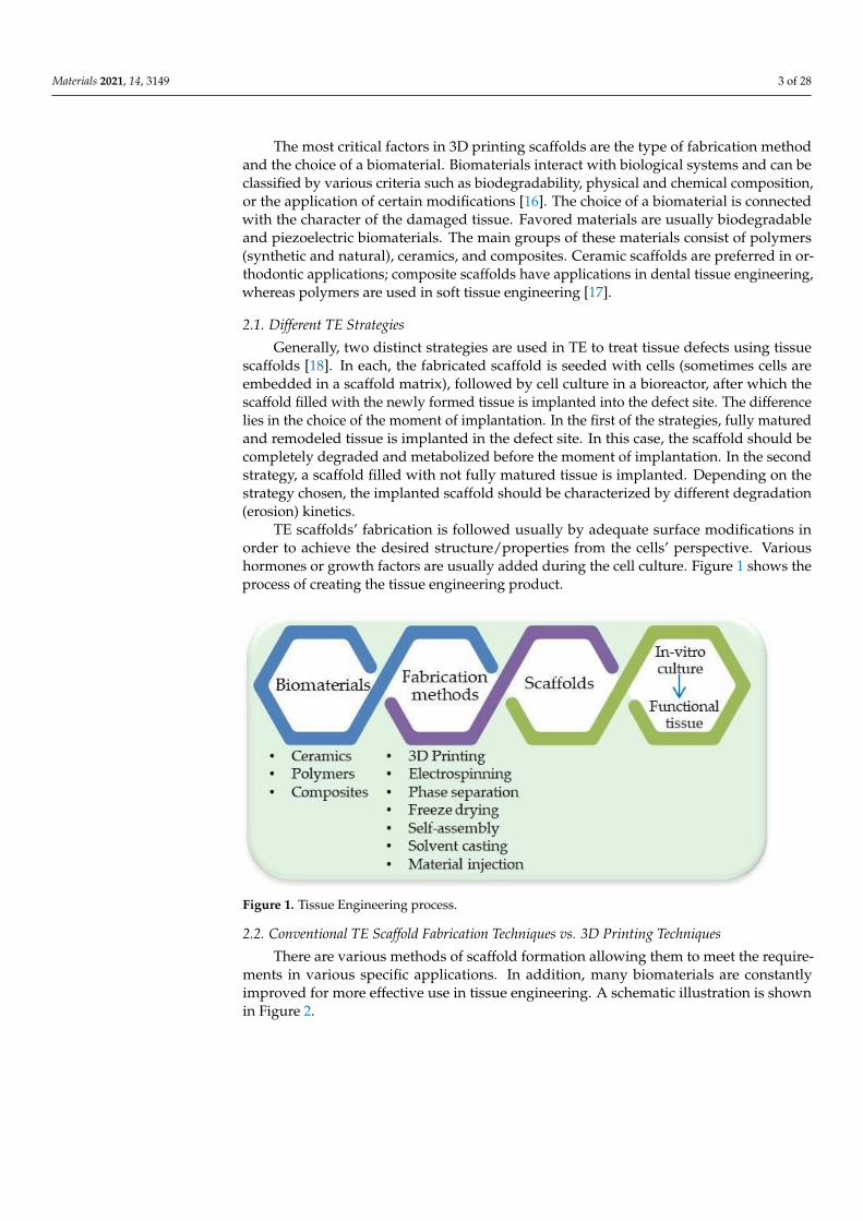
TE scaffolds’ fabrication is followed usually by adequate surface modifications inorder to achieve the desired structure/properties from the cells’ perspective. Varioushormones or growth factors are usually added during the cell culture. Figure 1 shows theprocess of creating the tissue engineering product.
Figure 1. Tissue Engineering process.
2.2. Conventional TE Scaffold Fabrication Techniques vs. 3D Printing Techniques
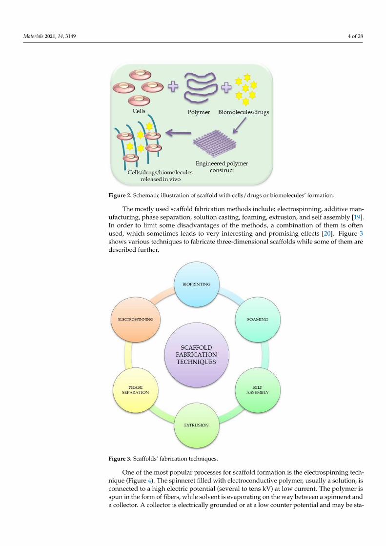
There are various methods of scaffold formation allowing them to meet the require-ments in various specific applications. In addition, many biomaterials are constantlyimproved for more effective use in tissue engineering. A schematic illustration is shownin Figure 2.
Materials 2021, 14, 3149 4 of 28
Figure 2. Schematic illustration of scaffold with cells/drugs or biomolecules’ formation.
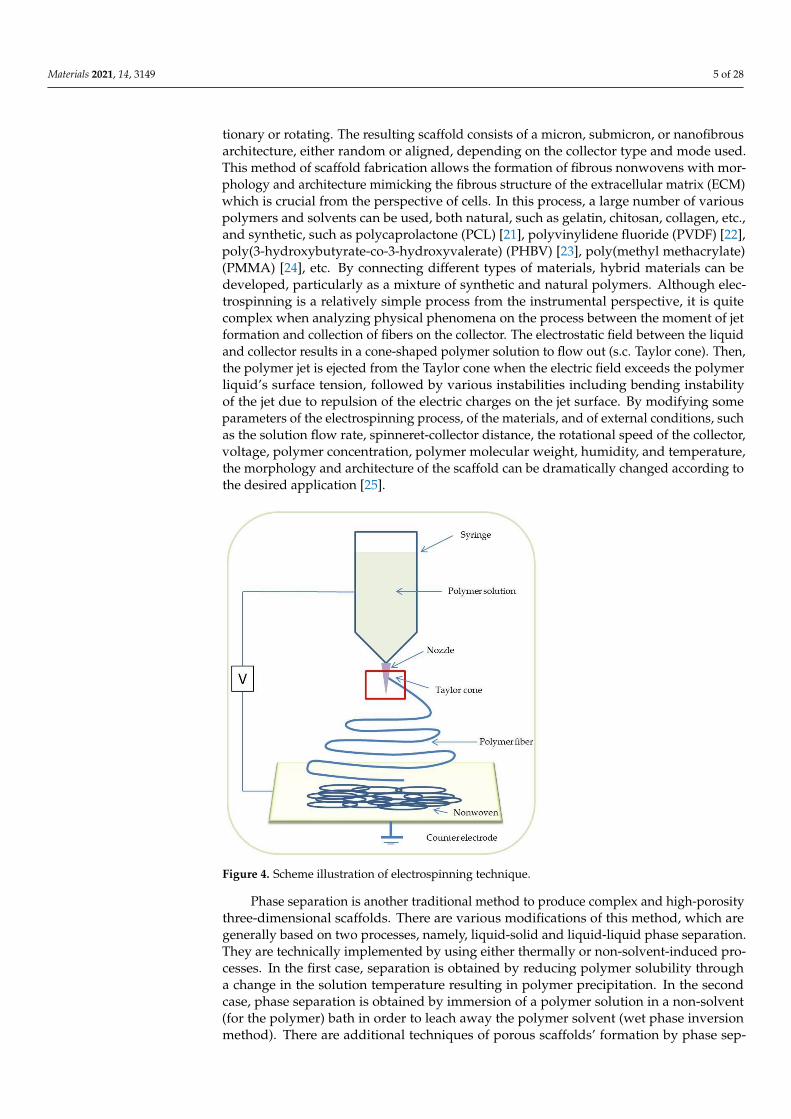
The mostly used scaffold fabrication methods include: electrospinning, additive man-ufacturing, phase separation, solution casting, foaming, extrusion, and self assembly [19].In order to limit some disadvantages of the methods, a combination of them is oftenused, which sometimes leads to very interesting and promising effects [20]. Figure 3shows various techniques to fabricate three-dimensional scaffolds while some of them aredescribed further.
Figure 3. Scaffolds’ fabrication techniques.
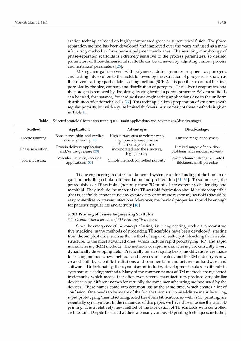
One of the most popular processes for scaffold formation is the electrospinning tech-nique (Figure 4). The spinneret filled with electroconductive polymer, usually a solution, isconnected to a high electric potential (several to tens kV) at low current. The polymer isspun in the form of fibers, while solvent is evaporating on the way between a spinneret anda collector. A collector is electrically grounded or at a low counter potential and may be sta-
Materials 2021, 14, 3149 5 of 28
tionary or rotating. The resulting scaffold consists of a micron, submicron, or nanofibrousarchitecture, either random or aligned, depending on the collector type and mode used.This method of scaffold fabrication allows the formation of fibrous nonwovens with mor-phology and architecture mimicking the fibrous structure of the extracellular matrix (ECM)which is crucial from the perspective of cells. In this process, a large number of variouspolymers and solvents can be used, both natural, such as gelatin, chitosan, collagen, etc.,and synthetic, such as polycaprolactone (PCL) [21], polyvinylidene fluoride (PVDF) [22],poly(3-hydroxybutyrate-co-3-hydroxyvalerate) (PHBV) [23], poly(methyl methacrylate)(PMMA) [24], etc. By connecting different types of materials, hybrid materials can bedeveloped, particularly as a mixture of synthetic and natural polymers. Although elec-trospinning is a relatively simple process from the instrumental perspective, it is quitecomplex when analyzing physical phenomena on the process between the moment of jetformation and collection of fibers on the collector. The electrostatic field between the liquidand collector results in a cone-shaped polymer solution to flow out (s.c. Taylor cone). Then,the polymer jet is ejected from the Taylor cone when the electric field exceeds the polymerliquid’s surface tension, followed by various instabilities including bending instabilityof the jet due to repulsion of the electric charges on the jet surface. By modifying someparameters of the electrospinning process, of the materials, and of external conditions, suchas the solution flow rate, spinneret-collector distance, the rotational speed of the collector,voltage, polymer concentration, polymer molecular weight, humidity, and temperature,the morphology and architecture of the scaffold can be dramatically changed according tothe desired application [25].
Figure 4. Scheme illustration of electrospinning technique.
Phase separation is another traditional method to produce complex and high-porositythree-dimensional scaffolds. There are various modifications of this method, which aregenerally based on two processes, namely, liquid-solid and liquid-liquid phase separation.They are technically implemented by using either thermally or non-solvent-induced pro-cesses. In the first case, separation is obtained by reducing polymer solubility througha change in the solution temperature resulting in polymer precipitation. In the secondcase, phase separation is obtained by immersion of a polymer solution in a non-solvent(for the polymer) bath in order to leach away the polymer solvent (wet phase inversionmethod). There are additional techniques of porous scaffolds’ formation by phase sep-
Materials 2021, 14, 3149 6 of 28
aration techniques based on highly compressed gases or supercritical fluids. The phaseseparation method has been developed and improved over the years and used as a man-ufacturing method to form porous polymer membranes. The resulting morphology ofphase-separated scaffolds is extremely sensitive to the process parameters, so desiredparameters of three-dimensional scaffolds can be achieved by adjusting various processand materials’ parameters [26].
Mixing an organic solvent with polymers, adding granules or spheres as porogens,and casting this solution to the mold, followed by the extraction of porogens, is known asthe solvent casting/particulate leaching method (SCPL). It is possible to control the finalpore size by the size, content, and distribution of porogens. The solvent evaporates, andthe porogen is removed by dissolving, leaving behind a porous structure. Solvent scaffoldscan be used, for instance, for cardiac tissue engineering applications due to the uniformdistribution of endothelial cells [27]. This technique allows preparation of structures withregular porosity, but with a quite limited thickness. A summary of these methods is givenin Table 1.
Table 1. Selected scaffolds’ formation techniques—main applications and advantages/disadvantages.
Method Applications Advantages Disadvantages
Electrospinning Bone, nerve, skin, and cardiactissue engineering [28]
High surface area to volume ratio,high porosity, easy process Limited range of polymers
Phase separation Protein delivery applicationsand/or drug release [29]
Bioactive agents can beincorporated into the structure,
high porosity
Limited ranges of pore size,problems with residual solvents
Solvent casting Vascular tissue engineeringapplications [30] Simple method, controlled porosity Low mechanical strength, limited
thickness, small pore size
Tissue engineering requires fundamental systemic understanding of the human or-ganism including cellular differentiation and proliferation [31–34]. To summarize, theprerequisites of TE scaffolds (not only those 3D printed) are extremely challenging andmanifold. They include: he material for TE scaffold fabrication should be biocompatible(that is, scaffolds cannot cause any cytotoxicity or immune response); scaffolds should beeasy to sterilize to prevent infections. Moreover, mechanical properties should be enoughfor patients’ regular life and activity [18].
3. 3D Printing of Tissue Engineering Scaffolds3.1. Overall Characteristics of 3D Printing Techniques
Since the emergence of the concept of using tissue engineering products in reconstruc-tive medicine, many methods of producing TE scaffolds have been developed, startingfrom the simplest ones, such as the method of sugar- or salt-crystal-leaching from a solidstructure, to the most advanced ones, which include rapid prototyping (RP) and rapidmanufacturing (RM) methods. The methods of rapid manufacturing are currently a verydynamically developing field. Practically on an ongoing basis, modifications are madeto existing methods; new methods and devices are created, and the RM industry is nowcreated both by scientific institutions and commercial manufacturers of hardware andsoftware. Unfortunately, the dynamism of industry development makes it difficult tosystematize existing methods. Many of the common names of RM methods are registeredtrademarks, which means that often even several manufacturers produce very similardevices using different names for virtually the same manufacturing method used by thedevices. These names come into common use at the same time, which creates a lot ofconfusion. One needs to be aware of the fact that terms such as additive manufacturing,rapid prototyping/manufacturing, solid free-form fabrication, as well as 3D printing, areessentially synonymous. In the remainder of this paper, we have chosen to use the term 3Dprinting. It is a relatively new method of the fabrication of TE scaffolds with controlledarchitecture. Despite the fact that there are many various 3D printing techniques, including
Materials 2021, 14, 3149 7 of 28
stereolithography, bioprinting, inkjet printing, fused deposition modelling (FDM), PED(Precision Extruding Deposition), laser beam melting, polyjet, electron beam melting, digi-tal laser printing (DLP), and selective laser sintering (SLS) [35], the common feature of allmentioned methods is the general principle of material deposition layer-by-layer until thefinal product is created [36].
Thus, the 3D TE scaffold is fabricated by the successive addition of consecutive 2Dlayers of a material. Additive manufacturing has numerous advantages, such as the abilityto create complex structures and the possibility of the application of the Computer-AidedDesign (CAD) methods. It enables the use of various types of biomaterials [37]. Usingliving cells and biodegradable polymers allows for the development of methods and novelstrategies to create complex tissues and, possibly in the future, whole organs [38]. A3D-printed TE scaffold can be designed using patient-specific data. The CAD methodallows for the precise designing of the 3D organ or its missing part. Selected features ofliving organs, such as porosity or vasculature, may be taken into account in the CAD 3Dmodel. Due to these remarkable advantages, 3D printing is gaining significant interest inregenerative medicine and tissue engineering [39].
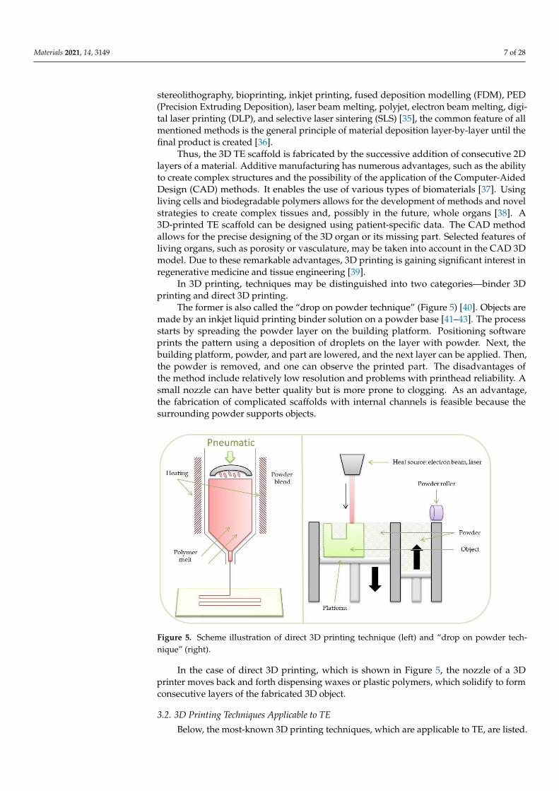
In 3D printing, techniques may be distinguished into two categories—binder 3Dprinting and direct 3D printing.
The former is also called the “drop on powder technique” (Figure 5) [40]. Objects aremade by an inkjet liquid printing binder solution on a powder base [41–43]. The processstarts by spreading the powder layer on the building platform. Positioning softwareprints the pattern using a deposition of droplets on the layer with powder. Next, thebuilding platform, powder, and part are lowered, and the next layer can be applied. Then,the powder is removed, and one can observe the printed part. The disadvantages ofthe method include relatively low resolution and problems with printhead reliability. Asmall nozzle can have better quality but is more prone to clogging. As an advantage,the fabrication of complicated scaffolds with internal channels is feasible because thesurrounding powder supports objects.
Figure 5. Scheme illustration of direct 3D printing technique (left) and “drop on powder tech-nique” (right).
In the case of direct 3D printing, which is shown in Figure 5, the nozzle of a 3Dprinter moves back and forth dispensing waxes or plastic polymers, which solidify to formconsecutive layers of the fabricated 3D object.
3.2. 3D Printing Techniques Applicable to TE
Below, the most-known 3D printing techniques, which are applicable to TE, are listed.
Materials 2021, 14, 3149 8 of 28
3.2.1. Bioprinting
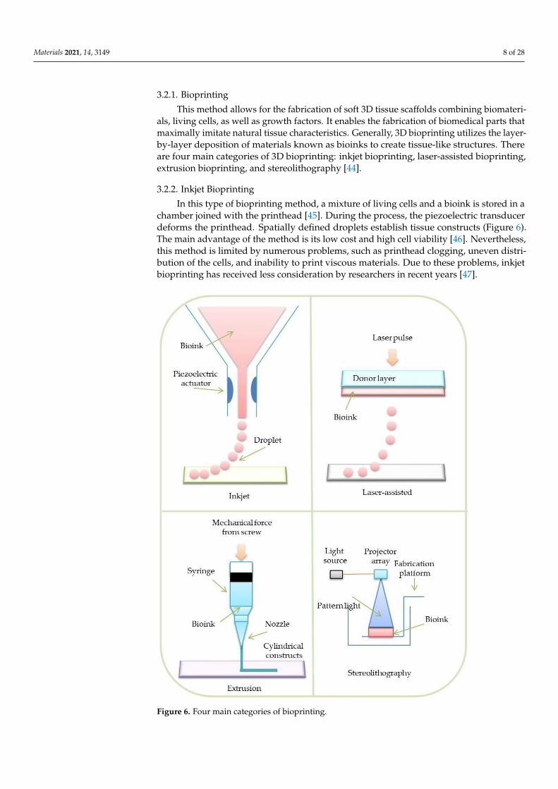
This method allows for the fabrication of soft 3D tissue scaffolds combining biomateri-als, living cells, as well as growth factors. It enables the fabrication of biomedical parts thatmaximally imitate natural tissue characteristics. Generally, 3D bioprinting utilizes the layer-by-layer deposition of materials known as bioinks to create tissue-like structures. Thereare four main categories of 3D bioprinting: inkjet bioprinting, laser-assisted bioprinting,extrusion bioprinting, and stereolithography [44].
3.2.2. Inkjet Bioprinting
In this type of bioprinting method, a mixture of living cells and a bioink is stored in achamber joined with the printhead [45]. During the process, the piezoelectric transducerdeforms the printhead. Spatially defined droplets establish tissue constructs (Figure 6).The main advantage of the method is its low cost and high cell viability [46]. Nevertheless,this method is limited by numerous problems, such as printhead clogging, uneven distri-bution of the cells, and inability to print viscous materials. Due to these problems, inkjetbioprinting has received less consideration by researchers in recent years [47].
Figure 6. Four main categories of bioprinting.
Materials 2021, 14, 3149 9 of 28
3.2.3. Laser-Assisted Bioprinting
Typical laser-assisted bioprinting (LAB) involves specialized layers, such as a bioinklayer, an energy-absorbing layer, a donor (quartz/glass), and a collecting layer, to formstructures [48]. During the process, a laser beam is focused on the energy-absorbing layer.Next, this layer vaporizes and creates an air bubble between the bioink and donor layers.The formation of a bubble ejects the desired amount of the bioink on the collecting layer. Atissue structure is created in a droplet-by-droplet manner (Figure 6) [49]. LAB is feasiblefor use with high cell density and viscous materials. Additionally, it has been reportedthat the method is characterized by high cell viability (95%) and resolves the cloggingissues. Nevertheless, LAB is an expensive process, which generates a very high cost withlarge-scale projects. Therefore, only a few printer prototypes were created. [50,51].
3.2.4. Extrusion Bioprinting
The extrusion bioprinting technique is based on liquid extrusion (paste, solution)from a pressurized syringe through a needle to a solution with controlled density. Thematerials are extruded in a form of long strands or dots to create complex structures [52].The printing process can be conducted at room temperature and used to print naturalbiomaterials, especially hydrogels (Figure 6) [53].
3.2.5. Stereolithography
Stereolithography (SLA) is the first developed method of rapid prototyping expandedin the late 1980s [54]. Stereolithography rasters use a laser beam to control the polymeriza-tion process of bioinks in a 2D layer. After the deposition of each layer of a material, curingfollows. During the curing process, a photosensitive hydrogel is subjected to UV or visiblelight. When a given layer is polymerized, the process is repeated, overlapping the previouslayer, up to the moment when the whole scaffold is completed. This method allows theuse of the following hydrogel materials (Figure 6) [55]: Polyethylene glycol diacrylate(PEGDA) and gelatin methacryloyl (GelMA) [56]; photo-initiators can be also added [57,58].The adjustment of various polymerization process parameters, including light energy andintensity, speed of printing, layer thickness, and exposure time, enables the achievement ofa high-quality (including resolution) product [59–64]. Nevertheless, compared to the othermethods, the SLA process is relatively time consuming, which makes the process feasiblefor small-detailed objects.
3.3. Fused Deposition Modeling and the Other Microfiber Extrusion Methods
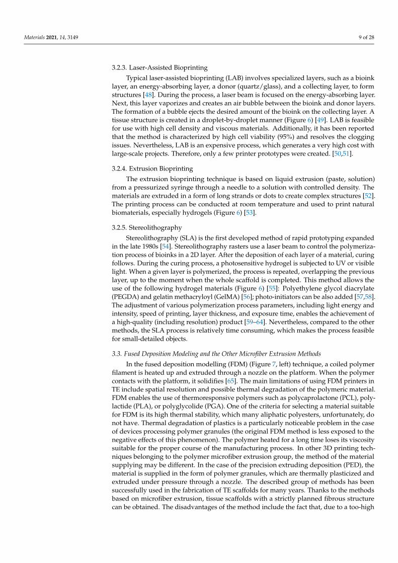
In the fused deposition modelling (FDM) (Figure 7, left) technique, a coiled polymerfilament is heated up and extruded through a nozzle on the platform. When the polymercontacts with the platform, it solidifies [65]. The main limitations of using FDM printers inTE include spatial resolution and possible thermal degradation of the polymeric material.FDM enables the use of thermoresponsive polymers such as polycaprolactone (PCL), poly-lactide (PLA), or polyglycolide (PGA). One of the criteria for selecting a material suitablefor FDM is its high thermal stability, which many aliphatic polyesters, unfortunately, donot have. Thermal degradation of plastics is a particularly noticeable problem in the caseof devices processing polymer granules (the original FDM method is less exposed to thenegative effects of this phenomenon). The polymer heated for a long time loses its viscositysuitable for the proper course of the manufacturing process. In other 3D printing tech-niques belonging to the polymer microfiber extrusion group, the method of the materialsupplying may be different. In the case of the precision extruding deposition (PED), thematerial is supplied in the form of polymer granules, which are thermally plasticized andextruded under pressure through a nozzle. The described group of methods has beensuccessfully used in the fabrication of TE scaffolds for many years. Thanks to the methodsbased on microfiber extrusion, tissue scaffolds with a strictly planned fibrous structurecan be obtained. The disadvantages of the method include the fact that, due to a too-high
Materials 2021, 14, 3149 10 of 28
polymer processing temperature, it is not possible to produce scaffolds with biomoleculesor living cells incorporated into the fiber structure.
Figure 7. Scheme illustration of FDM (left) and SLS (right) process.
3.4. Selective Laser Sintering
In the method, the polymeric powder particles are heated up slightly above thepolymer glass transition temperature by a laser beam [66]. This leads to partial melting ofthe particles [67], during which molecular diffusion on the particles’ surface takes place,which leads to particles’ fusion. After fabrication of each object layer, the building platformis lowered, a new layer of powder particles is spread on the top and connected with theprevious layer (Figure 7, right).
3.5. Melt-Spinning
Melt electrospinning (MES) is a relatively new 3D TE scaffold fabrication technique,being the alternative to conventional solution electrospinning (SES) known for disadvan-tages related to toxic polymeric solutions [68]. Residues of solvents, e.g., chloroform,DMSO (dimethyl sulfoxide), DMF (dimethyloformamid), that can be used by SES maybe harmful to living cells seeded on the scaffold. SES limitations were overcome by theuse of the molten polymer instead of the polymer solution. To be jetted in an electricfield, the molten polymer should be characterized by a suitable viscosity. The moltenpolymer would be collected by a rotating drum; however, implementation of the numericalcontrol (NC) enables the precise deposition of fiber in X, Y axes. The mentioned approachmakes the MES another class of 3D printing techniques [69]. Recent works on the meltelectrospinning report that this technique allows for depositing continuous fibers charac-terized by a diameter less than 1 micrometer, which is comparable to the classic solution ofelectrospinning [70].
Summarizing information about 3D printing methods are listed in Table 2.
Materials 2021, 14, 3149 11 of 28
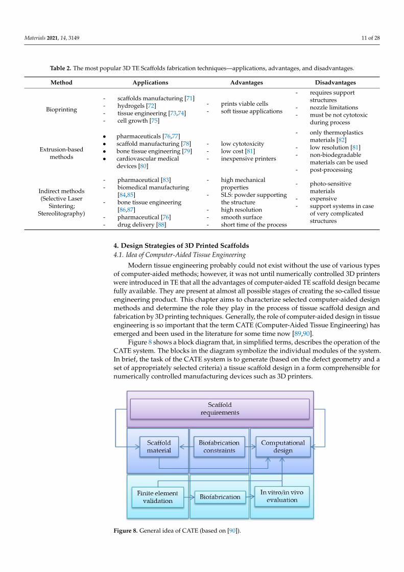
Table 2. The most popular 3D TE Scaffolds fabrication techniques—applications, advantages, and disadvantages.
Method Applications Advantages Disadvantages
Bioprinting
- scaffolds manufacturing [71]- hydrogels [72]- tissue engineering [73,74]- cell growth [75]
- prints viable cells- soft tissue applications
- requires supportstructures
- nozzle limitations- must be not cytotoxic
during process
Extrusion-basedmethods
• pharmaceuticals [76,77]• scaffold manufacturing [78]• bone tissue engineering [79]• cardiovascular medical
devices [80]
- low cytotoxicity- low cost [81]- inexpensive printers
- only thermoplasticsmaterials [82]
- low resolution [81]- non-biodegradable
materials can be used- post-processing
Indirect methods(Selective Laser
Sintering;Stereolitography)
- pharmaceutical [83]- biomedical manufacturing
[84,85]- bone tissue engineering
[86,87]- pharmaceutical [76]- drug delivery [88]
- high mechanicalproperties
- SLS: powder supportingthe structure
- high resolution- smooth surface- short time of the process
- photo-sensitivematerials
- expensive- support systems in case
of very complicatedstructures
4. Design Strategies of 3D Printed Scaffolds4.1. Idea of Computer-Aided Tissue Engineering
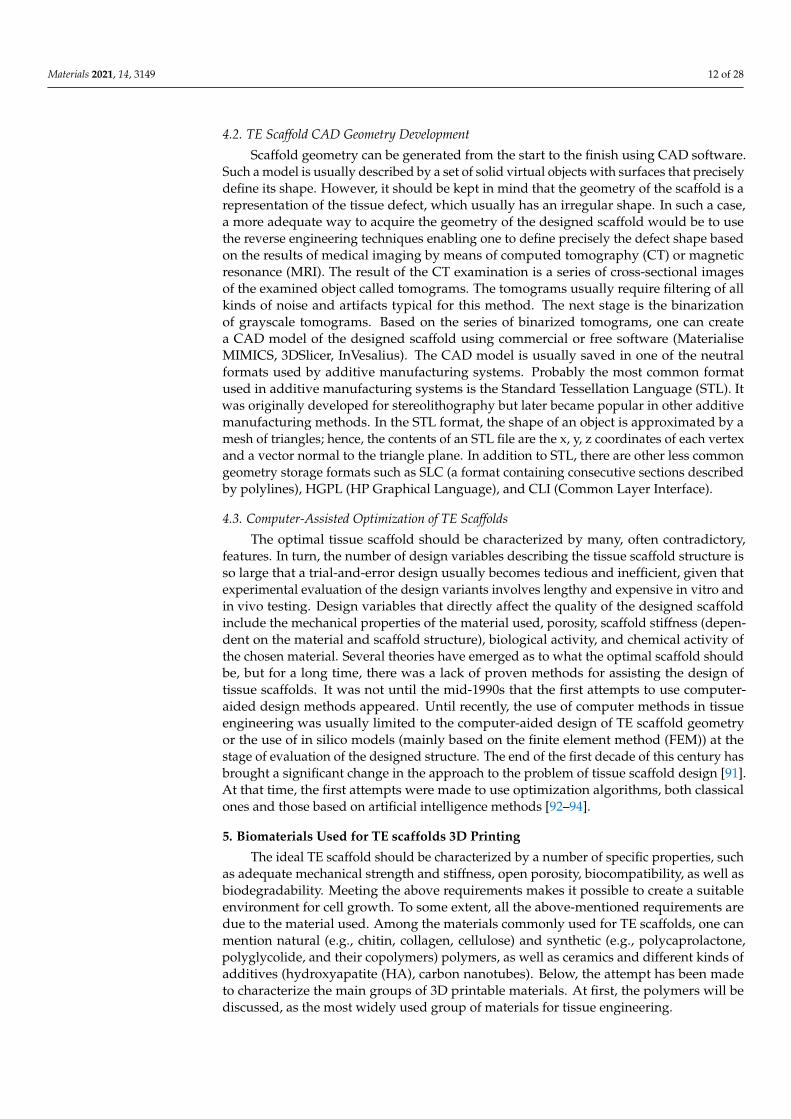
Modern tissue engineering probably could not exist without the use of various typesof computer-aided methods; however, it was not until numerically controlled 3D printerswere introduced in TE that all the advantages of computer-aided TE scaffold design becamefully available. They are present at almost all possible stages of creating the so-called tissueengineering product. This chapter aims to characterize selected computer-aided designmethods and determine the role they play in the process of tissue scaffold design andfabrication by 3D printing techniques. Generally, the role of computer-aided design in tissueengineering is so important that the term CATE (Computer-Aided Tissue Engineering) hasemerged and been used in the literature for some time now [89,90].
Figure 8 shows a block diagram that, in simplified terms, describes the operation of theCATE system. The blocks in the diagram symbolize the individual modules of the system.In brief, the task of the CATE system is to generate (based on the defect geometry and aset of appropriately selected criteria) a tissue scaffold design in a form comprehensible fornumerically controlled manufacturing devices such as 3D printers.
1
Figure 8. General idea of CATE (based on [90]).
Materials 2021, 14, 3149 12 of 28
4.2. TE Scaffold CAD Geometry Development
Scaffold geometry can be generated from the start to the finish using CAD software.Such a model is usually described by a set of solid virtual objects with surfaces that preciselydefine its shape. However, it should be kept in mind that the geometry of the scaffold is arepresentation of the tissue defect, which usually has an irregular shape. In such a case,a more adequate way to acquire the geometry of the designed scaffold would be to usethe reverse engineering techniques enabling one to define precisely the defect shape basedon the results of medical imaging by means of computed tomography (CT) or magneticresonance (MRI). The result of the CT examination is a series of cross-sectional imagesof the examined object called tomograms. The tomograms usually require filtering of allkinds of noise and artifacts typical for this method. The next stage is the binarizationof grayscale tomograms. Based on the series of binarized tomograms, one can createa CAD model of the designed scaffold using commercial or free software (MaterialiseMIMICS, 3DSlicer, InVesalius). The CAD model is usually saved in one of the neutralformats used by additive manufacturing systems. Probably the most common formatused in additive manufacturing systems is the Standard Tessellation Language (STL). Itwas originally developed for stereolithography but later became popular in other additivemanufacturing methods. In the STL format, the shape of an object is approximated by amesh of triangles; hence, the contents of an STL file are the x, y, z coordinates of each vertexand a vector normal to the triangle plane. In addition to STL, there are other less commongeometry storage formats such as SLC (a format containing consecutive sections describedby polylines), HGPL (HP Graphical Language), and CLI (Common Layer Interface).
4.3. Computer-Assisted Optimization of TE Scaffolds
The optimal tissue scaffold should be characterized by many, often contradictory,features. In turn, the number of design variables describing the tissue scaffold structure isso large that a trial-and-error design usually becomes tedious and inefficient, given thatexperimental evaluation of the design variants involves lengthy and expensive in vitro andin vivo testing. Design variables that directly affect the quality of the designed scaffoldinclude the mechanical properties of the material used, porosity, scaffold stiffness (depen-dent on the material and scaffold structure), biological activity, and chemical activity ofthe chosen material. Several theories have emerged as to what the optimal scaffold shouldbe, but for a long time, there was a lack of proven methods for assisting the design oftissue scaffolds. It was not until the mid-1990s that the first attempts to use computer-aided design methods appeared. Until recently, the use of computer methods in tissueengineering was usually limited to the computer-aided design of TE scaffold geometryor the use of in silico models (mainly based on the finite element method (FEM)) at thestage of evaluation of the designed structure. The end of the first decade of this century hasbrought a significant change in the approach to the problem of tissue scaffold design [91].At that time, the first attempts were made to use optimization algorithms, both classicalones and those based on artificial intelligence methods [92–94].
5. Biomaterials Used for TE scaffolds 3D Printing
The ideal TE scaffold should be characterized by a number of specific properties, suchas adequate mechanical strength and stiffness, open porosity, biocompatibility, as well asbiodegradability. Meeting the above requirements makes it possible to create a suitableenvironment for cell growth. To some extent, all the above-mentioned requirements aredue to the material used. Among the materials commonly used for TE scaffolds, one canmention natural (e.g., chitin, collagen, cellulose) and synthetic (e.g., polycaprolactone,polyglycolide, and their copolymers) polymers, as well as ceramics and different kinds ofadditives (hydroxyapatite (HA), carbon nanotubes). Below, the attempt has been madeto characterize the main groups of 3D printable materials. At first, the polymers will bediscussed, as the most widely used group of materials for tissue engineering.
Materials 2021, 14, 3149 13 of 28
5.1. Polymers
Polymers represent the main category of materials with high potential for use in 3Dprinting of TE scaffolds and can be widely used for various tissues’ imitation. TE scaffoldsmay be fabricated from non-biodegradable as well as biodegradable polymers. In thecontext of tissue engineering, biodegradable polymers generally have more advantages ascompared to the non-biodegradable.
5.1.1. Natural Polymers
Natural polymers are known to be the right candidates for TE scaffold fabrication,mostly due to their bioactivity, biocompatibility, minimal immune response, as well asnatural biodegradability of most of them [95]. As an example of the natural polymers’application in TE, one can mention the work of [96] reporting fabrication of TE scaffolds forcartilage regeneration made of bacterial cellulose. Another study confirms that cellulosefrom Acetobacter xylinum can be used in the cartilage regeneration [97]. Collagen andchitosan also belong to the polymers widely investigated and applied in TE [98]. Allof the above-mentioned materials are known for supporting the cell proliferation andviability [99].
Another biocompatible and easily accessible natural material is gelatine, being anirreversible hydrolyzed form of collagen [100]. There are numerous attempts of usinggelatine as biomaterial for 3D printing of TE scaffolds. In the work by [101], the gela-tine/hydroxyapatite composite was investigated as material for 3D printing scaffolds forstem cells’ chondrogenic differentiation. Pure gelatine 3D scaffolds were proven to be agood environment for the proliferation and viability of hepatocyte cells [99].
In the work by [102], high proliferation and viability of mesenchymal stem cellscultured on/in collagen/agarose scaffolds wer observed.
5.1.2. Synthetic Polymers
The usefulness of biodegradable synthetic polymers (mainly aliphatic polyesters suchas PCL or PLGA) in TE has already been investigated for many years [103,104]. Thebiodegradable aliphatic polyesters are characterized by relatively low toxicity [105]; how-ever, the acidic oligomeric release, being the effect of polymer hydrolytic degradation,can initiate the inflammatory reaction [106], negatively affecting the tissue regenerationprocess [107]. Other research works on the degradation kinetics of 3D-printed TE scaffoldsmade from various aliphatic polyesters, have shown the differences in the degree of thedegradation for PLGA (40,000–75,000 Da) and PCL (Mw = 114,000 Da) as 18% and 56%on day 14 and day 28 for PLGA, and 33% on day 21 and 39% on day 28 for PCL, respec-tively [108]. TE scaffolds made of aliphatic polyesters are known to be successfully appliedin the tissue loss treatment [109,110] including bone regeneration [111,112]. The degrada-tion time of TE scaffolds made of aliphatic polymers can be thoroughly controlled [113].The predominant degradation mechanism for all bioresorbable polyesters used in bioengi-neering is hydrolysis occurring in enzymatic conditions. From the moment an implant (e.g.,TE scaffold) is placed in the living organism, water, which is one of the main components ofthe physiological environment, penetrates the polymer matrix at various speeds [114]. Thispenetration speed depends on many factors, including the hydrophilicity of the implantmaterial. Water molecules cause weakening and consequently breaking of ester bonds,which are responsible for the cohesion of polymer chains. It was found that the degradationof some objects made from aliphatic polymers proceeds heterogeneously in such a waythat the central part of the object degrades faster than the areas in direct contact with theenvironment. One can find numerous examples of aliphatic polyesters’ application intissue engineering [115].
Copolymerization is another way of effectively controlling the final properties of 3D-printed TE scaffolds. Copolymers such as PCL with a PEG (Mn = 1000) addition [116] orPCL (Mw = 2000) with a PLGA addition [117] were synthesized for controlled degradationdedicated to drug-release applications. Different types of printable copolymers, such as
Materials 2021, 14, 3149 14 of 28
poly(hydroxybutyrate) (PHB) [118], poly(propylene fumarate) [119] (PPF), and polyglycolicacid (PGA) [120], were also tested.
Systematic studies concerning bone tissue engineering have been carried out for years.A multitude of 3D-printed scaffolds made of different polyesters and their copolymerswere tested under in vivo and in vitro conditions to investigate their abilities for the neo-vascularization and the bone ingrowth [121]. Many works concern the 3D printing ofpolymeric scaffolds filled with growth factors such as TGF-β and BMP-2 which enableobtaining specifically vascularized bone constructs [122,123].
5.1.3. Hydrogels
Hydrogels belong to crosslinked polymers having the property of binding relativelylarge amounts of water. They can be made of synthetic or natural polymers such ascollagen or alginate [124,125]. Due to their relatively high water content, hydrogels arequite biocompatible and have relatively low mechanical properties. Because of theirmechanical similarity to the native tissue, their transport/diffusion properties and highbiocompatibility, hydrogels are among the most promising materials from which tissuescaffolds can be fabricated. Moreover, they allow relatively easy and safe immobilizationof biologically active molecules. So far, various bioink biomaterials, such as gelatin-methacrylates, agarose, alginate, collagen, chitin, silk, hyaluronic acid, cellulose, andtheir mixtures have been used together with various crosslinking methods such as clickchemistry, ionic/hydrogen bonding, or chemical bonding via radical initiators. Amongthem, alginates are the most attractive for bioprinting, mainly due to their ability toform a soft gel matrix in a low-aggressive environment for living cells and encapsulatedbiomolecules. One of the important properties of alginate is its ability to form gels by ioniccrosslinking with calcium cations. However, environmental factors such as buffer acidityor temperature can easily affect the condition of the hydrogel material and its degradation,leading to the consequent loss of the biomolecules contained in the hydrogel matrix.Polymers such as poly(ethylene glycol) diacrylate (PEGDA) or natural gelatin methacrylate(GelMA) can also be used for the preparation of hydrogels [126,127]. Hydrogels oftenare used as a component of hybrid TE scaffolds mimicking the soft tissues (e.g., musclestissue) [128].
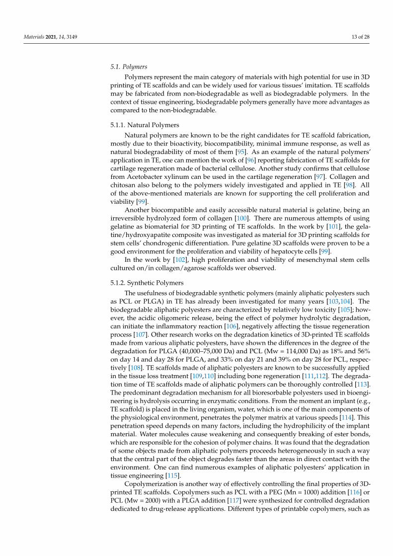
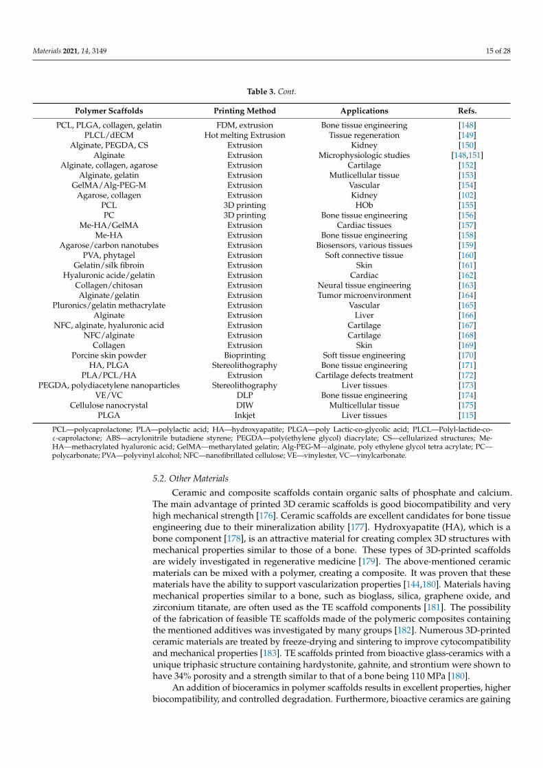
In Table 3, polymer scaffolds with applications and printing methods are summarized.
Table 3. Polymer scaffolds with applications and printing methods.
Polymer Scaffolds Printing Method Applications Refs.
Chitosan/Rhizopus mycelia/Fungi - Bone regeneration [129]PCL Direct Printing Heart and cartilage tissue [130]PCL FDM Tissue engineering [131]
PCL/alginate-based hydrogel Extrusion Bone tissue engineering [132]PCL/PLA Bioextrusion Tissue engineering [133]
PCL, chitosan FDM Bone tissue engineering [134]PCL/HA FDM Tissue engineering [135]PCL/silk Extrusion Tissue engineering [136]
PCL/castor oil FDM Bone tissue engineering [137]PCL FDM Bone tissue engineering [138]
PCL/HA Indirect printing Tissue engineering [139]PCL/diamond Extrusion Tissue engineering [140]
PLA, PLGA, collagen FDM Tendon-bone [141]PLA, collagen FDM Bone tissue engineering [142]
PLA FDM Bone tissue engineering [143]PLCL FDM Tissue engineering [144]
PLA/ABS FDM Bone tissue engineering [145]PLA FDM Bone tissue engineering [146]
PLA/cellulose Extrusion Tissue engineering [147]
Materials 2021, 14, 3149 15 of 28
Table 3. Cont.
Polymer Scaffolds Printing Method Applications Refs.
PCL, PLGA, collagen, gelatin FDM, extrusion Bone tissue engineering [148]PLCL/dECM Hot melting Extrusion Tissue regeneration [149]
Alginate, PEGDA, CS Extrusion Kidney [150]Alginate Extrusion Microphysiologic studies [148,151]
Alginate, collagen, agarose Extrusion Cartilage [152]Alginate, gelatin Extrusion Mutlicellular tissue [153]
GelMA/Alg-PEG-M Extrusion Vascular [154]Agarose, collagen Extrusion Kidney [102]
PCL 3D printing HOb [155]PC 3D printing Bone tissue engineering [156]
Me-HA/GelMA Extrusion Cardiac tissues [157]Me-HA Extrusion Bone tissue engineering [158]
Agarose/carbon nanotubes Extrusion Biosensors, various tissues [159]PVA, phytagel Extrusion Soft connective tissue [160]
Gelatin/silk fibroin Extrusion Skin [161]Hyaluronic acide/gelatin Extrusion Cardiac [162]
Collagen/chitosan Extrusion Neural tissue engineering [163]Alginate/gelatin Extrusion Tumor microenvironment [164]
Pluronics/gelatin methacrylate Extrusion Vascular [165]Alginate Extrusion Liver [166]
NFC, alginate, hyaluronic acid Extrusion Cartilage [167]NFC/alginate Extrusion Cartilage [168]
Collagen Extrusion Skin [169]Porcine skin powder Bioprinting Soft tissue engineering [170]
HA, PLGA Stereolithography Bone tissue engineering [171]PLA/PCL/HA Extrusion Cartilage defects treatment [172]
PEGDA, polydiacetylene nanoparticles Stereolithography Liver tissues [173]VE/VC DLP Bone tissue engineering [174]
Cellulose nanocrystal DIW Multicellular tissue [175]PLGA Inkjet Liver tissues [115]
PCL—polycaprolactone; PLA—polylactic acid; HA—hydroxyapatite; PLGA—poly Lactic-co-glycolic acid; PLCL—Polyl-lactide-co-ε-caprolactone; ABS—acrylonitrile butadiene styrene; PEGDA—poly(ethylene glycol) diacrylate; CS—cellularized structures; Me-HA—methacrylated hyaluronic acid; GelMA—metharylated gelatin; Alg-PEG-M—alginate, poly ethylene glycol tetra acrylate; PC—polycarbonate; PVA—polyvinyl alcohol; NFC—nanofibrillated cellulose; VE—vinylester, VC—vinylcarbonate.
5.2. Other Materials
Ceramic and composite scaffolds contain organic salts of phosphate and calcium.The main advantage of printed 3D ceramic scaffolds is good biocompatibility and veryhigh mechanical strength [176]. Ceramic scaffolds are excellent candidates for bone tissueengineering due to their mineralization ability [177]. Hydroxyapatite (HA), which is abone component [178], is an attractive material for creating complex 3D structures withmechanical properties similar to those of a bone. These types of 3D-printed scaffoldsare widely investigated in regenerative medicine [179]. The above-mentioned ceramicmaterials can be mixed with a polymer, creating a composite. It was proven that thesematerials have the ability to support vascularization properties [144,180]. Materials havingmechanical properties similar to a bone, such as bioglass, silica, graphene oxide, andzirconium titanate, are often used as the TE scaffold components [181]. The possibilityof the fabrication of feasible TE scaffolds made of the polymeric composites containingthe mentioned additives was investigated by many groups [182]. Numerous 3D-printedceramic materials are treated by freeze-drying and sintering to improve cytocompatibilityand mechanical properties [183]. TE scaffolds printed from bioactive glass-ceramics with aunique triphasic structure containing hardystonite, gahnite, and strontium were shown tohave 34% porosity and a strength similar to that of a bone being 110 MPa [180].
An addition of bioceramics in polymer scaffolds results in excellent properties, higherbiocompatibility, and controlled degradation. Furthermore, bioactive ceramics are gaining
Materials 2021, 14, 3149 16 of 28
more and more attention due to their excellent osteogenic properties [184]. Calcium phos-phates (CaPs) are the most frequently used bioceramics in tissue engineering applications,due to their similarity to the chemical structure of a bone.
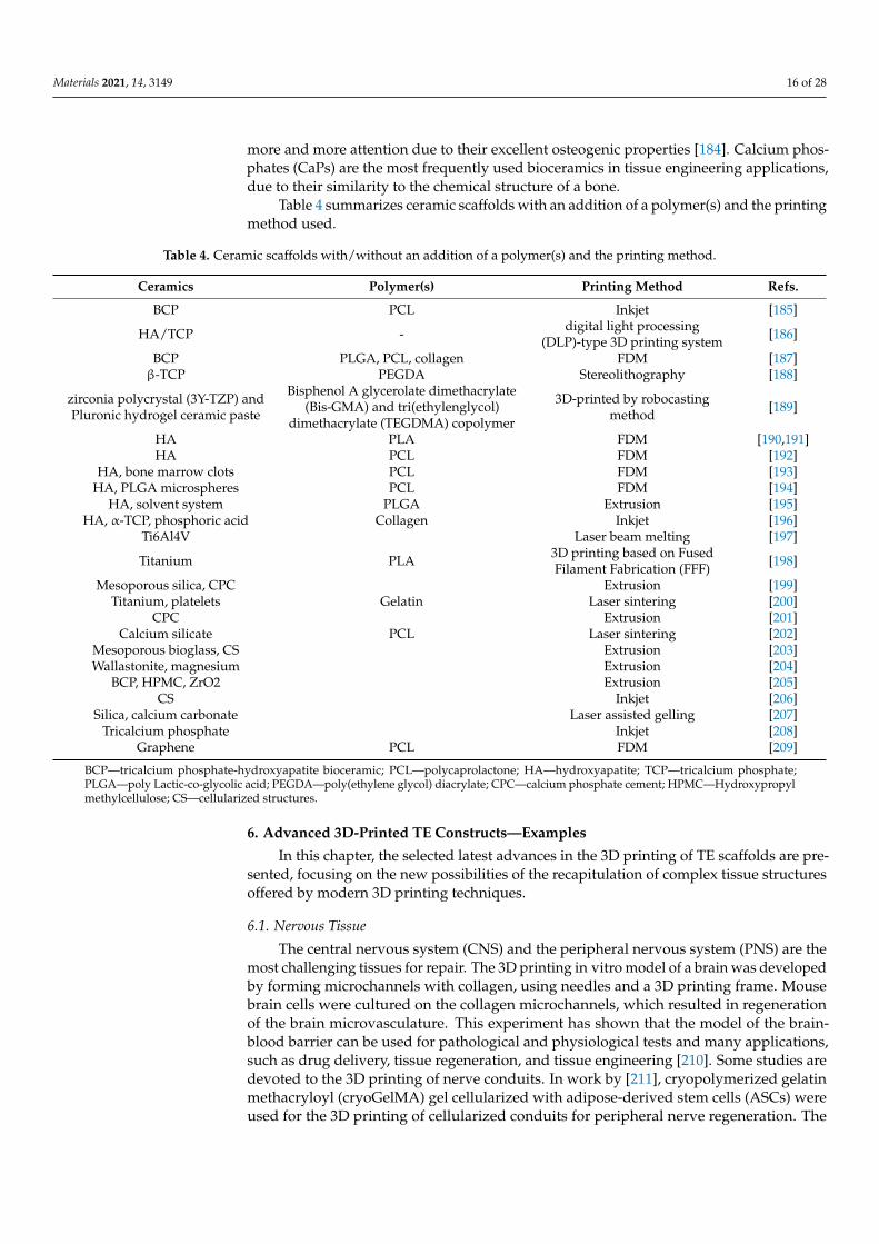
Table 4 summarizes ceramic scaffolds with an addition of a polymer(s) and the printingmethod used.
Table 4. Ceramic scaffolds with/without an addition of a polymer(s) and the printing method.
Ceramics Polymer(s) Printing Method Refs.
BCP PCL Inkjet [185]
HA/TCP - digital light processing(DLP)-type 3D printing system [186]
BCP PLGA, PCL, collagen FDM [187]β-TCP PEGDA Stereolithography [188]
zirconia polycrystal (3Y-TZP) andPluronic hydrogel ceramic paste
Bisphenol A glycerolate dimethacrylate(Bis-GMA) and tri(ethylenglycol)
dimethacrylate (TEGDMA) copolymer
3D-printed by robocastingmethod [189]
HA PLA FDM [190,191]HA PCL FDM [192]
HA, bone marrow clots PCL FDM [193]HA, PLGA microspheres PCL FDM [194]
HA, solvent system PLGA Extrusion [195]HA, α-TCP, phosphoric acid Collagen Inkjet [196]
Ti6Al4V Laser beam melting [197]
Titanium PLA 3D printing based on FusedFilament Fabrication (FFF) [198]
Mesoporous silica, CPC Extrusion [199]Titanium, platelets Gelatin Laser sintering [200]
CPC Extrusion [201]Calcium silicate PCL Laser sintering [202]
Mesoporous bioglass, CS Extrusion [203]Wallastonite, magnesium Extrusion [204]
BCP, HPMC, ZrO2 Extrusion [205]CS Inkjet [206]
Silica, calcium carbonate Laser assisted gelling [207]Tricalcium phosphate Inkjet [208]
Graphene PCL FDM [209]
BCP—tricalcium phosphate-hydroxyapatite bioceramic; PCL—polycaprolactone; HA—hydroxyapatite; TCP—tricalcium phosphate;PLGA—poly Lactic-co-glycolic acid; PEGDA—poly(ethylene glycol) diacrylate; CPC—calcium phosphate cement; HPMC—Hydroxypropylmethylcellulose; CS—cellularized structures.
6. Advanced 3D-Printed TE Constructs—Examples
In this chapter, the selected latest advances in the 3D printing of TE scaffolds are pre-sented, focusing on the new possibilities of the recapitulation of complex tissue structuresoffered by modern 3D printing techniques.
6.1. Nervous Tissue
The central nervous system (CNS) and the peripheral nervous system (PNS) are themost challenging tissues for repair. The 3D printing in vitro model of a brain was developedby forming microchannels with collagen, using needles and a 3D printing frame. Mousebrain cells were cultured on the collagen microchannels, which resulted in regenerationof the brain microvasculature. This experiment has shown that the model of the brain-blood barrier can be used for pathological and physiological tests and many applications,such as drug delivery, tissue regeneration, and tissue engineering [210]. Some studies aredevoted to the 3D printing of nerve conduits. In work by [211], cryopolymerized gelatinmethacryloyl (cryoGelMA) gel cellularized with adipose-derived stem cells (ASCs) wereused for the 3D printing of cellularized conduits for peripheral nerve regeneration. The
Materials 2021, 14, 3149 17 of 28
re-innervation ability of the fabricated conduits was proven in vivo. It is worth mentioningthat 3D printing was used for the fabrication of patient-specific casting molds.
6.2. Ocular Tissues
Interest in 3D printing techniques in ophthalmology is still growing; however, themajority of 3D printing applications does not concern tissue engineering. Here are examplesof works on using 3D printing for ocular tissue regeneration: In the work by [212], anattempt of the reconstruction of a 3D retina is reported. The retina-like structure containingadult rat retinal ganglion cells and glia were 3D printed. It was proven that these types ofretinal cells can be successfully printed without loss of viability and certain phenotypicfeatures. Another example of the application of 3D printing in ocular tissue engineeringwould be the work by [213] concerning the fabrication of the TE corneal scaffold made ofcollagen-based bio-ink containing encapsulated corneal keratocytes.
6.3. Ear
The computer-aided design has been used to create the bionic human ear. A hydrogelmatrix containing cells and a conductive polymer with the addition of silver nanoparticleswere used during printing—bioprinted in the shape of a human ear. The studies allowedcontrol of the signals from the cochlea-shaped electrodes. The in vitro culture was providedon the cartilage tissues on every side of the inductive coil. The printed ear was found toenhance the auditory sensing. Another study showed that the printed ear can be formedby 3D bioprinting with the subject’s lipid tissue and an auricular cartilage. Adipocytesand chondrocytes differentiated from the adipose-derived stromal cells were enclosed inhydrogels and then placed at the lipid and cartilage tissue [214–216].
6.4. Kidney
Scaffolds from PEGDA with the addition of sodium alginate and calcium sulfate weretested [150]. After fabrication, scaffolds were crosslinked using UVclight, and subsequenthuman embryonic kidney cells (HEK) were cultured. It was shown that the mentionedcomposite materials are characterized by properties supporting the proliferation andviability of the cells. In the work of Lawlor et al. [217], extrusion-based 3D bioprintingwas applied for the generation of human kidney organoids (the organoid is a simplifiedversion of a living organ produced in vitro). The used fabrication method enables forprecise manipulation of organoid size and cell number and conformation. The developedin vitro model of kidney organoids could be used for drug testing or disease modeling.
6.5. Skin
Using a laser-assisted method, a 3D-printed skin was developed. Collagen type Iand Matriderm (for matrix stabilization) were mixed and cultured with fibroblasts andkeratinocytes. The experiment was also performed at in-vivo conditions by placing abioprinted construct on the murine skin. In the effect, mainly an epidermis formingwas observed [218]. In [219], the method of biofabrication of skin equivalents (SE) thatare bioprinted using open-market bioprinter, made with fibroblasts and keratinocytessuspended in the gelatin-based hydrogel, was discussed. SE construct layers were extrudeddirectly onto the multi-well plate. Three levels comprise the developed structure: dermis,laminin/entactin basal layer, and epidermis. The developed SE may be used for in vitroskin disease modeling.
6.6. Cancer Models
Recent progress in bioprinting enables the development of 3D in vitro models ofvarious kinds of cancerous tissue [220]. Such models enable the design of patient-specifictherapies as well as for the investigation of the processes related to carcinogenesis, suchas tumor extravasation [221]. Bioprinted cancer models usually are composed of multiplelayers containing different cell types including tumor cells (usually patient-derived cells),
Materials 2021, 14, 3149 18 of 28
the extracellular matrix, growth factors, and vasculature [222]. Bioprinted tumor modelsshould recapitulate the actual tumor heterogeneity. They enable anti-cancer therapy screen-ing as well as the investigation of cell-cell and cell-matrix interactions. Bioprinted cancermodels are characterized by great advantages over 2D in vitro models, which cannot mimicthe structural complexity of tumors.
6.7. Bone and Cartilage Tissue Engineering
Bone and cartilage defects repair is one of the most common regenerative procedures.The principal part of bone and cartilage tissue engineering is to replace a damaged bone.Therefore, 3D printing techniques try to print a structure of artificial bone with requiredproperties, such as appropriate mechanical properties, shape, and size [223]. The majorcauses of bone and cartilage defects are trauma, congenital anomalies, and tissue resectiondue to cancer. Such treatments such as autogenous bone grafting are characterized byseveral disadvantages, such as unsuitable donor tissue availability or donor site morbidity.On the other hand, allogeneic bone grafts are avoided mainly due to the risk of diseasetransmission. Over the past several years, the importance of therapies using the 3D-printedTE scaffolds has been growing gradually. TE scaffolds enable seeded cells to adhere,migrate, grow, and differentiate into chondrogenesis and osteogenesis.
Here are examples of recently published works on the application of 3D printingin bone and cartilage regeneration: Most of the proposed solutions are based on thecombination of several different materials—ceramic, polyesters, and hydrogels [224–226]Quite often, to improve the cell-seeding efficiency and osteoinductivity, an injectablehydrogel is incorporated into a 3D-printed porous structure to form a hybrid scaffold [227].Despite the fact that multiple types of materials are used to fabricate 3D-printed bonescaffolds, biodegradable aliphatic polyesters remain the gold standard [228,229]. On theother hand, hydrogels are the most popular group of materials for the cartilage TE [230].Osteochondral scaffolds remain a particular challenge for tissue engineering. Typically, thefabrication of osteochondral scaffolds requires a combination of several printing techniquesand materials [231], as it should be remembered that osteochondral scaffolds are usuallybi- or even tri-phasic.
7. Future Directions and Conclusions
Various approaches in scaffolds’ formation for use in tissue engineering applicationsare experiencing rapid advances. Regarding the development of 3D-printed scaffolds,the most important goal is to mimic the complexity of a natural living tissue truly. Itsstructure should have appropriate mechanical properties, pore size distribution, andpores’ arrangement (allowing cell migration and diffusion). While numerous tissues weresuccessfully cultured as proof of the principle, the development of a fully functionalcomplex human-size organ is still pending.
The types of fabrication methods and the materials provided in this review serve toimprove current TE procedures.
Drawbacks and Future Directions of the 3D Printing of TE Scaffolds
Even though 3D printing is extremely promising from a TE point of view, it is character-ized by several limitations related mainly to the lack of legal regulations and standardizedprocedures. Moreover, the fabrication of any TE product requires advanced and costlyinfrastructure that may include software, robust computer workstations, 3D printers, andcell culture laboratory facilities. Nowadays, most TE product implantation attempts wererealized in cooperation between hospitals and research institutes. Usually, such cooperationwas of an ad hoc nature and did not go beyond the research study. It is clear that to increasethe availability of 3D printing in TE applications, the current collaboration model betweenengineers and doctors needs to be modified. One of the ideas is to establish regional 3Dprinting centers, adequately equipped and staffed [232]. Such centers would contribute to
Materials 2021, 14, 3149 19 of 28
more efficient use of the equipment and human resources. The idea is that such centerscould serve many medical facilities in a given region.
A major problem facing researchers, doctors, and engineers is the lack of an establishedlegal framework and procedures for validating tissue-engineered products. It is a well-known fact that existing legal provisions hinder the scale-up from the laboratory to a largerscale. Attesting of patient-specific TE products is also problematic from the point of viewof current standards.
Another drawback related to the 3D printing of TE scaffolds is a lack of standardizedterminology used to systematize the field, which is a characteristic of new and rapidlydeveloping fields of knowledge.
Still, the actual issue and the most challenging step involve a translation of the tech-nology to the next level—the availability to the patients, giving a chance to improve thequality of their lives. Not so long ago, the 3D-printing-assisted cultivation of TE constructshas been started using the patient’s cells [233,234], and now the symbol of the cutting-edgetechnology for TE is 4D printing [235]. This advanced 3D-printed technology adds a fourthdimension—time—to currently-used 3D printing. It enables the fabrication of TE scaffoldshaving a self-assembly ability. This technique assumes the use of smart materials character-ized by the ability to change their properties under the influence of an applied stimulus(e.g., thermoresponsive shape memory polymers). Furthermore, 4D printing can be usedfor the fabrication of TE scaffolds enabling the mechanical stimulation of living cells bythe external signal (e.g., magnetic field) [236]. Vascularization is another challenging goal,and the new generation of bioprinters with multiple print-heads seems very promising.Loaded with various cell types, they are expected to reconstruct and recapitulate the factualcomplexity of a multi-tissue organ.
Future activities should include testing materials for medical-oriented 3D printingmethods, creating new printers to provide high precision of TE scaffolds, making unifiedstandards for scaffolds, strengthening market supervision to optimize implants for clinicaluse, and establishing a 3D printing platform to enhance communication among researchinstitutes, hospitals, and companies. These advancements should further promote thedevelopment of 3D-printed tissue engineering technology.
Author Contributions: Conceptualization, A.Z., M.M.-H., A.G. and P.S.; validation, A.G., P.S.;investigation, A.Z., M.M.-H.; writing—original draft preparation, A.Z., M.M.-H.; writing—reviewand editing, A.G., P.S.; visualization, A.Z., M.M.-H., A.G. and P.S.; supervision, A.G., P.S. All authorshave read and agreed to the published version of the manuscript.
Funding: The Article Processing Charge (APC) was covered by the Project no. POWR.03.02.00-00-1028/17-00 implemented under the Operational Program Knowledge Education Development2014–2020, co-financed by the European Social Fund.
Institutional Review Board Statement: Not applicable.
Informed Consent Statement: Not applicable.
Data Availability Statement: No new data were created or analyzed in this study.
Conflicts of Interest: The authors declare no conflict of interest.
References1. Deng, C.; Chang, J.; Wu, C. Bioactive Scaffolds for Osteochondral Regeneration. Degener. Musculoskelet. Dis. Pathol. Treat. 2019, 17,
15–25. [CrossRef] [PubMed]2. Zaszczynska, A.; Sajkiewicz, P.; Gradys, A. Piezoelectric Scaffolds as Smart Materials for Neural Tissue Engineering. Polymers
2020, 12, 161. [CrossRef] [PubMed]3. Zaszczynska, A.; Gradys, A.; Sajkiewicz, P. Progress in the Applications of Smart Piezoelectric Materials for Medical Devices.
Polymers 2020, 12, 2754. [CrossRef] [PubMed]4. Kowalewicz, K.; Vorndran, E.; Feichtner, F.; Waselau, A.-C.; Brueckner, M.; Meyer-Lindenberg, A. In-Vivo Degradation Behavior
and Osseointegration of 3D Powder-Printed Calcium Magnesium Phosphate Cement Scaffolds. Materials 2021, 14, 946. [CrossRef]
Materials 2021, 14, 3149 20 of 28
5. Seoane-Viaño, I.; Januskaite, P.; Alvarez-Lorenzo, C.; Basit, A.W.; Goyanes, A. Semi-Solid Extrusion 3D Printing in Drug Deliveryand Biomedicine: Personalised Solutions for Healthcare Challenges. J. Control. Release Off. J. Control. Release Soc. 2021, 332,367–389. [CrossRef]
6. Lee, V.; Singh, G.; Trasatti, J.P.; Bjornsson, C.; Xu, X.; Tran, T.N.; Yoo, S.-S.; Dai, G.; Karande, P. Design and Fabrication of HumanSkin by Three-Dimensional Bioprinting. Tissue Eng. Part C Methods 2014, 20, 473–484. [CrossRef]
7. Markstedt, K.; Mantas, A.; Tournier, I.; Martínez Ávila, H.; Hägg, D.; Gatenholm, P. 3D Bioprinting Human Chondrocytes withNanocellulose-Alginate Bioink for Cartilage Tissue Engineering Applications. Biomacromolecules 2015, 16, 1489–1496. [CrossRef]
8. Cui, H.; Zhu, W.; Nowicki, M.; Zhou, X.; Khademhosseini, A.; Zhang, L.G. Hierarchical Fabrication of Engineered Vascu-larized Bone Biphasic Constructs via Dual 3D Bioprinting: Integrating Regional Bioactive Factors into Architectural Design.Adv. Healthc. Mater. 2016, 5, 2174–2181. [CrossRef]
9. Laronda, M.M.; Rutz, A.L.; Xiao, S.; Whelan, K.A.; Duncan, F.E.; Roth, E.W.; Woodruff, T.K.; Shah, R.N. A Bioprosthetic OvaryCreated Using 3D Printed Microporous Scaffolds Restores Ovarian Function in Sterilized Mice. Nat. Commun. 2017, 8, 15261.[CrossRef]
10. OPTN: Organ Procurement and Transplantation Network—OPTN. Available online: https://optn.transplant.hrsa.gov/ (accessedon 5 May 2021).
11. Singh, D.; Thomas, D.J.; Motamarry, A. 13—3D printing future perspective in medicine. In 3D Printing in Medicine and Surgery;Woodhead Publishing Series in Biomaterials; Thomas, D.J., Singh, D., Eds.; Woodhead Publishing: Cambridge, UK, 2021;pp. 265–270. ISBN 978-0-08-102542-0.
12. Wibowo, A.; Vyas, C.; Cooper, G.; Qulub, F.; Suratman, R.; Mahyuddin, A.I.; Dirgantara, T.; Bartolo, P. 3D Printing ofPolycaprolactone–Polyaniline Electroactive Scaffolds for Bone Tissue Engineering. Materials 2020, 13, 512. [CrossRef]
13. Moczulska, M.; Bitar, M.; Swieszkowski, W.; Bruinink, A. Biological Characterization of Woven Fabric Using Two- and Three-Dimensional Cell Cultures. J. Biomed. Mater. Res. A 2012, 100A, 882–893. [CrossRef]
14. Stevens, M.M.; George, J.H. Exploring and Engineering the Cell Surface Interface. Science 2005, 310, 1135–1138. [CrossRef]15. Hollister, S.J. Porous Scaffold Design for Tissue Engineering. Nat. Mater. 2005, 4, 518–524. [CrossRef]16. Navarro, M.; Michiardi, A.; Castaño, O.; Planell, J. A Biomaterials in Orthopaedics. J. R. Soc. Interface 2008, 5, 1137–1158.
[CrossRef]17. Nair, L.S.; Laurencin, C.T. Polymers as Biomaterials for Tissue Engineering and Controlled Drug Delivery. Adv. Biochem.
Eng. Biotechnol. 2006, 102, 47–90. [CrossRef]18. Abdollahiyan, P.; Oroojalian, F.; Mokhtarzadeh, A. The Triad of Nanotechnology, Cell Signalling, and Scaffold Implantation
for the Successful Repair of Damaged Organs: An Overview on Soft-Tissue Engineering. J. Control. Release 2021, 332, 460–492.[CrossRef]
19. Badekila, A.K.; Kini, S.; Jaiswal, A.K. Fabrication Techniques of Biomimetic Scaffolds in Three-Dimensional Cell Culture: AReview. J. Cell. Physiol. 2021, 236, 741–762. [CrossRef]
20. Bongiovanni Abel, S.; Montini Ballarin, F.; Abraham, G.A. Combination of Electrospinning with Other Techniques for theFabrication of 3D Polymeric and Composite Nanofibrous Scaffolds with Improved Cellular Interactions. Nanotechnology 2020, 31,172002. [CrossRef]
21. Sajkiewicz, P.; Heljak, M.K.; Gradys, A.; Choinska, E.; Ruminski, S.; Jaroszewicz, T.; Bissenik, I.; Swieszkowski, W. Degradationand Related Changes in Supermolecular Structure of Poly(Caprolactone) in Vivo Conditions. Polym. Degrad. Stab. 2018, 157,70–79. [CrossRef]
22. Szewczyk, P.K.; Gradys, A.; Kim, S.K.; Persano, L.; Marzec, M.; Kryshtal, A.; Busolo, T.; Toncelli, A.; Pisignano, D.; Bernasik,A.; et al. Enhanced Piezoelectricity of Electrospun Polyvinylidene Fluoride Fibers for Energy Harvesting. ACS Appl. Mater.Interfaces 2020, 12, 13575–13583. [CrossRef]
23. Kaniuk, Ł.; Ferraris, S.; Spriano, S.; Luxbacher, T.; Krysiak, Z.; Berniak, K.; Zaszczynska, A.; Marzec, M.M.; Bernasik, A.;Sajkiewicz, P.; et al. Time-Dependent Effects on Physicochemical and Surface Properties of PHBV Fibers and Films in Relation toTheir Interactions with Fibroblasts. Appl. Surf. Sci. 2021, 545, 148983. [CrossRef]
24. Ura, D.P.; Rosell-Llompart, J.; Zaszczynska, A.; Vasilyev, G.; Gradys, A.; Szewczyk, P.K.; Knapczyk-Korczak, J.; Avrahami, R.;Šišková, A.O.; Arinstein, A.; et al. The Role of Electrical Polarity in Electrospinning and on the Mechanical and StructuralProperties of As-Spun Fibers. Materials 2020, 13, 4169. [CrossRef] [PubMed]
25. Ghosal, K.; Augustine, R.; Zaszczynska, A.; Barman, M.; Jain, A.; Hasan, A.; Kalarikkal, N.; Sajkiewicz, P.; Thomas, S. Novel DrugDelivery Systems Based on Triaxial Electrospinning Based Nanofibers. React. Funct. Polym. 2021, 163, 104895. [CrossRef]
26. Ulbricht, M. Design and Synthesis of Organic Polymers for Molecular Separation Membranes. Curr. Opin. Chem. Eng. 2020, 28,60–65. [CrossRef]
27. Sin, D.; Miao, S.; Liu, G.; Fan, W.; Chadwick, G.; Yan, C.; Friis, T. Polyurethane (PU) Scaffolds Prepared by Solvent Cast-ing/Particulate Leaching (SCPL) Combined with Centrifugation. Mater. Sci. Eng. C Mater. Biol. Appl. 2010, 30, 78–85. [CrossRef]
28. Parham, S.; Kharazi, A.Z.; Bakhsheshi-Rad, H.R.; Ghayour, H.; Ismail, A.F.; Nur, H.; Berto, F. Electrospun Nano-Fibers forBiomedical and Tissue Engineering Applications: A Comprehensive Review. Materials 2020, 13, 2153. [CrossRef]
29. Nyamweya, N.N. Applications of Polymer Blends in Drug Delivery. Future J. Pharm. Sci. 2021, 7, 18. [CrossRef]30. Subramaniyan, M.; Eswaran, P.; Appusamy, A.; Srimannarayana Raju, P.; Rahini, V.; Madhumitha, T.R.; Thisha, R. A Survey on
Applications of Additive Manufacturing Techniques in Tissue Engineering. Mater. Today Proc. 2021. [CrossRef]
Materials 2021, 14, 3149 21 of 28
31. Yang, S.; Leong, K.F.; Du, Z.; Chua, C.K. The Design of Scaffolds for Use in Tissue Engineering. Part I. Traditional Factors.Tissue Eng. 2001, 7, 679–689. [CrossRef]
32. Ikeda, T.; Ikeda, K.; Yamamoto, K.; Ishizaki, H.; Yoshizawa, Y.; Yanagiguchi, K.; Yamada, S.; Hayashi, Y. Fabrication andCharacteristics of Chitosan Sponge as a Tissue Engineering Scaffold. BioMed. Res. Int. 2014, 2014, e786892. [CrossRef]
33. Drury, J.L.; Mooney, D.J. Hydrogels for Tissue Engineering: Scaffold Design Variables and Applications. Biomaterials 2003, 24,4337–4351. [CrossRef]
34. Zaszczynska, A.; Sajkiewicz, P.Ł.; Gradys, A.; Tymkiewicz, R.; Urbanek, O.; Kołbuk, D. Influence of Process-Material Conditionson the Structure and Biological Properties of Electrospun Polyvinylidene Fluoride Fibers. Bull. Pol. Acad. Sci. Tech. Sci. 2020, 68.[CrossRef]
35. Gibson, I.; Rosen, D.; Stucker, B.; Khorasani, M. Development of Additive Manufacturing Technology. In Additive ManufacturingTechnologies; Gibson, I., Rosen, D., Stucker, B., Khorasani, M., Eds.; Springer International Publishing: Cham, Switzerland, 2021;pp. 23–51. ISBN 978-3-030-56127-7.
36. Jain, K.; Shukla, R.; Yadav, A.; Ujjwal, R.R.; Flora, S.J.S. 3D Printing in Development of Nanomedicines. Nanomaterials 2021, 11,420. [CrossRef]
37. Wang, C.; Huang, W.; Zhou, Y.; He, L.; He, Z.; Chen, Z.; He, X.; Tian, S.; Liao, J.; Lu, B.; et al. 3D Printing of Bone TissueEngineering Scaffolds. Bioact. Mater. 2020, 5, 82–91. [CrossRef]
38. Ong, C.S.; Yesantharao, P.; Huang, C.Y.; Mattson, G.; Boktor, J.; Fukunishi, T.; Zhang, H.; Hibino, N. 3D Bioprinting Using StemCells. Pediatr. Res. 2018, 83, 223–231. [CrossRef]
39. Egan, P.F. Integrated Design Approaches for 3D Printed Tissue Scaffolds: Review and Outlook. Materials 2019, 12, 2355. [CrossRef]40. Sachs, E.M.; Haggerty, J.S.; Cima, M.J.; Williams, P.A. Three-Dimensional Printing Techniques. CIRP Ann. 1993, 42, 257–260.
[CrossRef]41. Cima, M.J.; Sachs, E.; Cima, L.G.; Yoo, J.; Khanuja, S.; Borland, S.W.; Wu, B.; Giordano, R.A. Computer-Derived Microstructures
by 3D Printing: Sio- and Structural Materials. Mater. Sci. 1994. [CrossRef]42. Griffith, L.G.; Wu, B.; Cima, M.J.; Powers, M.J.; Chaignaud, B.; Vacanti, J.P. In Vitro Organogenesis of Liver Tissuea. Ann. N. Y.
Acad. Sci. 1997, 831, 382–397. [CrossRef]43. Wu, B.M.; Borland, S.W.; Giordano, R.A.; Cima, L.G.; Sachs, E.M.; Cima, M.J. Solid Free-Form Fabrication of Drug Delivery
Devices. J. Control. Release 1996, 40, 77–87. [CrossRef]44. Mandrycky, C.; Wang, Z.; Kim, K.; Kim, D.-H. 3D Bioprinting for Engineering Complex Tissues. Biotechnol. Adv. 2016, 34, 422–434.
[CrossRef] [PubMed]45. Xu, T.; Jin, J.; Gregory, C.; Hickman, J.J.J.J.; Boland, T. Inkjet Printing of Viable Mammalian Cells. Biomaterials 2005, 26, 93–99.
[CrossRef] [PubMed]46. Pepper, M.E.; Seshadri, V.; Burg, T.C.; Burg, K.J.L.; Groff, R.E. Characterizing the Effects of Cell Settling on Bioprinter Output.
Biofabrication 2012, 4, 011001. [CrossRef] [PubMed]47. Li, X.; Liu, B.; Pei, B.; Chen, J.; Zhou, D.; Peng, J.; Zhang, X.; Jia, W.; Xu, T. Inkjet Bioprinting of Biomaterials. Chem. Rev. 2020, 120,
10793–10833. [CrossRef]48. Guillemot, F.; Souquet, A.; Catros, S.; Guillotin, B.; Lopez, J.; Faucon, M.; Pippenger, B.; Bareille, R.; Rémy, M.; Bellance, S.; et al.
High-Throughput Laser Printing of Cells and Biomaterials for Tissue Engineering. Acta Biomater. 2010, 6, 2494–2500. [CrossRef]49. Hakobyan, D.; Kerouredan, O.; Remy, M.; Dusserre, N.; Medina, C.; Devillard, R.; Fricain, J.-C.; Oliveira, H. Laser-Assisted
Bioprinting for Bone Repair. In 3D Bioprinting: Principles and Protocols; Crook, J.M., Ed.; Springer US: New York, NY, USA, 2020;pp. 135–144. ISBN 978-1-07-160520-2.
50. Salah, M.; Tayebi, L.; Moharamzadeh, K.; Naini, F.B. Three-Dimensional Bio-Printing and Bone Tissue Engineering: TechnicalInnovations and Potential Applications in Maxillofacial Reconstructive Surgery. Maxillofac. Plast. Reconstr. Surg. 2020, 42.[CrossRef]
51. Osidak, E.O.; Kozhukhov, V.I.; Osidak, M.S.; Domogatsky, S.P. Collagen as Bioink for Bioprinting: A Comprehensive Review.Int. J. Bioprinting 2020, 6. [CrossRef]
52. Landers, R.; Mülhaupt, R. Desktop Manufacturing of Complex Objects, Prototypes and Biomedical Scaffolds by Meansof Computer-Assisted Design Combined with Computer-Guided 3D Plotting of Polymers and Reactive Oligomers.Macromol. Mater. Eng. 2000, 282, 17–21. [CrossRef]
53. Maher, P.S.; Keatch, R.P.; Donnelly, K.; Paxton, J.Z. Formed 3D Bio-Scaffolds via Rapid Prototyping Technology. In Proceedingsof the 4th European Conference of the International Federation for Medical and Biological Engineering, Antwerp, Belgium,23–27 November 2008; Vander Sloten, J., Verdonck, P., Nyssen, M., Haueisen, J., Eds.; Springer: Berlin/Heidelberg, Germany,2009; pp. 2200–2204.
54. Dowler, C. Automatic Model Building Cuts Design Time. Costs. Plast. Eng. 1989, 45, 43–45.55. Arcaute, K.; Mann, B.K.; Wicker, R.B. Practical Use of Hydrogels in Stereolithography for Tissue Engineering Applications.
In Stereolithography: Materials, Processes and Applications; Bártolo, P.J., Ed.; Springer US: Boston, MA, USA, 2011; pp. 299–331.ISBN 978-0-387-92904-0.
56. Magalhães, L.S.S.; Santos, F.E.P.; Elias, C.D.M.V.; Afewerki, S.; Sousa, G.F.; Furtado, A.S.; Marciano, F.R.; Lobo, A.O. Printing 3DHydrogel Structures Employing Low-Cost Stereolithography Technology. J. Funct. Biomater. 2020, 11, 12. [CrossRef]
Materials 2021, 14, 3149 22 of 28
57. Jiao, Z.; Wang, C.; Yang, Q.; Wang, X. Preparation and Characterization of UV-Curable Urethane Acrylate Oligomers Modifiedwith Cycloaliphatic Epoxide Resin. J. Coat. Technol. Res. 2018, 15, 251–258. [CrossRef]
58. Sangermano, M.; Carbonaro, W.; Malucelli, G.; Priola, A. UV-Cured Interpenetrating Acrylic-Epoxy Polymer Networks: Prepara-tion and Characterization. Macromol. Mater. Eng. 2008, 293, 515–520. [CrossRef]
59. Hong, B.T.; Shin, K.S.; Kim, D.S. Ultraviolet-Curing Behavior of an Epoxy Acrylate Resin System. J. Appl. Polym. Sci. 2005, 98,1180–1185. [CrossRef]
60. Lovell, L.G.; Lu, H.; Elliott, J.E.; Stansbury, J.W.; Bowman, C.N. The Effect of Cure Rate on the Mechanical Properties of DentalResins. Dent. Mater. 2001, 17, 504–511. [CrossRef]
61. Chen, K.; Kuang, X.; Li, V.; Kang, G.; Jerry Qi, H. Fabrication of Tough Epoxy with Shape Memory Effects by UV-AssistedDirect-Ink Write Printing. Soft Matter. 2018, 14, 1879–1886. [CrossRef] [PubMed]
62. Zhang, Z.; Li, P.; Chu, F.; Shen, G. Influence of the Three-Dimensional Printing Technique and Printing Layer Thickness on ModelAccuracy. J. Orofac. Orthop. Fortschritte Kieferorthopädie 2019, 80, 194–204. [CrossRef]
63. Watters, M.P.; Bernhardt, M.L. Curing Parameters to Improve the Mechanical Properties of Stereolithographic Printed Specimens.Rapid Prototyp. J. 2018, 24, 46–51. [CrossRef]
64. Yankov, E.; Nikolova, M.P. Comparison of the Accuracy of 3D Printed Prototypes Using the Stereolithography (SLA) Methodwith the Digital CAD Models. In Proceedings of the MATEC Web of Conferences, Online, 9 August 2017; Volume 137, p. 02014.[CrossRef]
65. Van Noort, R. The Future of Dental Devices Is Digital. Dent. Mater. 2012, 28, 3–12. [CrossRef]66. Pattanayak, D.K.; Fukuda, A.; Matsushita, T.; Takemoto, M.; Fujibayashi, S.; Sasaki, K.; Nishida, N.; Nakamura, T.; Kokubo, T.
Bioactive Ti Metal Analogous to Human Cancellous Bone: Fabrication by Selective Laser Melting and Chemical Treatments.Acta Biomater. 2011, 7, 1398–1406. [CrossRef]
67. Lohfeld, S.; Tyndyk, M.A.; Cahill, S.; Flaherty, N.; Barron, V.; McHugh, P.E. A Method to Fabricate Small Features on Scaffolds forTissue Engineering via Selective Laser Sintering. J. Biomed. Sci. Eng. 2010, 3, 138. [CrossRef]
68. Wunner, F.M.; Florczak, S.; Mieszczanek, P.; Bas, O.; De-Juan-Pardo, E.M.; Hutmacher, D.W. 5.13 Electrospinning with PolymerMelts—State of the Art and Future Perspectives. In Comprehensive Biomaterials II; Ducheyne, P., Ed.; Elsevier: Oxford, UK, 2017;pp. 217–235. ISBN 978-0-08-100692-4.
69. Dalton, P.D.; Grafahrend, D.; Klinkhammer, K.; Klee, D.; Möller, M. Electrospinning of Polymer Melts: PhenomenologicalObservations. Polymer 2007, 48, 6823–6833. [CrossRef]
70. Hochleitner, G.; Jüngst, T.; Brown, T.D.; Hahn, K.; Moseke, C.; Jakob, F.; Dalton, P.D.; Groll, J. Additive Manufacturing of Scaffoldswith Sub-Micron Filaments via Melt Electrospinning Writing. Biofabrication 2015, 7, 035002. [CrossRef]
71. Narayanan, L.K.; Shirwaiker, R.A. Experimental Characterization and Finite Element Modeling of the Effects of 3D BioplottingProcess Parameters on Structural and Tensile Properties of Polycaprolactone (PCL) Scaffolds. Appl. Sci. 2020, 10, 5289. [CrossRef]
72. Rajabi, M.; McConnell, M.; Cabral, J.; Ali, M.A. Chitosan Hydrogels in 3D Printing for Biomedical Applications. Carbohydr. Polym.2021, 260, 117768. [CrossRef]
73. Su, X.; Wang, T.; Guo, S. Applications of 3D Printed Bone Tissue Engineering Scaffolds in the Stem Cell Field. Regen. Ther. 2021,16, 63–72. [CrossRef]
74. Guzzi, E.A.; Tibbitt, M.W. Additive Manufacturing of Precision Biomaterials. Adv. Mater. 2020, 32, 1901994. [CrossRef]75. Zhang, H.; Cong, Y.; Osi, A.R.; Zhou, Y.; Huang, F.; Zaccaria, R.P.; Chen, J.; Wang, R.; Fu, J. Direct 3D Printed Biomimetic Scaffolds
Based on Hydrogel Microparticles for Cell Spheroid Growth. Adv. Funct. Mater. 2020, 30, 1910573. [CrossRef]76. Melocchi, A.; Briatico-Vangosa, F.; Uboldi, M.; Parietti, F.; Turchi, M.; von Zeppelin, D.; Maroni, A.; Zema, L.; Gazzaniga, A.;
Zidan, A. Quality Considerations on the Pharmaceutical Applications of Fused Deposition Modeling 3D Printing. Int. J. Pharm.2021, 592, 119901. [CrossRef]
77. Melocchi, A.; Uboldi, M.; Cerea, M.; Foppoli, A.; Maroni, A.; Moutaharrik, S.; Palugan, L.; Zema, L.; Gazzaniga, A. A GraphicalReview on the Escalation of Fused Deposition Modeling (FDM) 3D Printing in the Pharmaceutical Field. J. Pharm. Sci. 2020, 109,2943–2957. [CrossRef]
78. Choi, W.J.; Hwang, K.S.; Kwon, H.J.; Lee, C.; Kim, C.H.; Kim, T.H.; Heo, S.W.; Kim, J.-H.; Lee, J.-Y. Rapid Development of DualPorous Poly(Lactic Acid) Foam Using Fused Deposition Modeling (FDM) 3D Printing for Medical Scaffold Application. Mater. Sci.Eng. C Mater. Biol. Appl. 2020, 110, 110693. [CrossRef]
79. Xiong, Z.; Liu, W.; Qian, H.; Lei, T.; He, X.; Hu, Y.; Lei, P. Tantalum Nanoparticles Reinforced PCL Scaffolds Using Direct 3DPrinting for Bone Tissue Engineering. Front. Mater. 2021, 8. [CrossRef]
80. Casanova-Batlle, E.; Guerra, A.J.; Ciurana, J. Continuous Based Direct Ink Write for Tubular Cardiovascular Medical Devices.Polymers 2021, 13, 77. [CrossRef] [PubMed]
81. Gross, B.C.; Erkal, J.L.; Lockwood, S.Y.; Chen, C.; Spence, D.M. Evaluation of 3D Printing and Its Potential Impact on Biotechnologyand the Chemical Sciences. Anal. Chem. 2014, 86, 3240–3253. [CrossRef] [PubMed]
82. Wong, K.V.; Hernandez, A. A Review of Additive Manufacturing. ISRN Mech. Eng. 2012, 2012, e208760. [CrossRef]83. Awad, A.; Fina, F.; Goyanes, A.; Gaisford, S.; Basit, A.W. 3D Printing: Principles and Pharmaceutical Applications of Selective
Laser Sintering. Int. J. Pharm. 2020, 586. [CrossRef]
Materials 2021, 14, 3149 23 of 28
84. Riza, S.H.; Masood, S.H.; Rashid, R.A.R.; Chandra, S. 6—Selective laser sintering in biomedical manufacturing. In MetallicBiomaterials Processing and Medical Device Manufacturing; Woodhead Publishing Series in Biomaterials; Wen, C., Ed.; WoodheadPublishing: Cambridge, UK, 2020; pp. 193–233. ISBN 978-0-08-102965-7.
85. Salmi, M. Additive Manufacturing Processes in Medical Applications. Materials 2021, 14, 191. [CrossRef]86. Lin, K.; Liu, J.; Wu, J.-M.; Sun, Y.; Li, F.; Zhou, Y.; Shi, Y. Selective Laser Sintered Nano-HA/PDLLA Composite Microspheres for
Bone Scaffolds Applications. Rapid Prototyp. J. 2020, 26, 1131–1143. [CrossRef]87. Qin, T.; Li, X.; Long, H.; Bin, S.; Xu, Y. Bioactive Tetracalcium Phosphate Scaffolds Fabricated by Selective Laser Sintering for Bone
Regeneration Applications. Materials 2020, 13, 2268. [CrossRef]88. Xu, X.; Goyanes, A.; Trenfield, S.J.; Diaz-Gomez, L.; Alvarez-Lorenzo, C.; Gaisford, S.; Basit, A.W. Stereolithography (SLA) 3D
Printing of a Bladder Device for Intravesical Drug Delivery. Mater. Sci. Eng. C 2021, 120, 111773. [CrossRef]89. Sun, W.; Starly, B.; Darling, A.; Gomez, C. Computer-aided Tissue Engineering: Application to Biomimetic Modelling and Design
of Tissue Scaffolds. Biotechnol. Appl. Biochem. 2004, 39, 49–58. [CrossRef]90. Giannitelli, S.M.; Accoto, D.; Trombetta, M.; Rainer, A. Current Trends in the Design of Scaffolds for Computer-Aided Tissue
Engineering. Acta Biomater. 2014, 10, 580–594. [CrossRef]91. Heljak, M.K.; Kurzydlowski, K.J.; Swieszkowski, W. Computer Aided Design of Architecture of Degradable Tissue Engineering
Scaffolds. Comput. Methods Biomech. Biomed. Engin. 2017, 20, 1623–1632. [CrossRef]92. Mahmood, M.A.; Visan, A.I.; Ristoscu, C.; Mihailescu, I.N. Artificial Neural Network Algorithms for 3D Printing. Materials 2021,
14, 163. [CrossRef]93. Conev, A.; Litsa, E.E.; Perez, M.R.; Diba, M.; Mikos, A.G.; Kavraki, L.E. Machine Learning-Guided Three-Dimensional Printing of
Tissue Engineering Scaffolds. Tissue Eng. Part A 2020, 26, 1359–1368. [CrossRef]94. Ng, W.L.; Chan, A.; Ong, Y.S.; Chua, C.K. Deep Learning for Fabrication and Maturation of 3D Bioprinted Tissues and Organs.
Virtual Phys. Prototyp. 2020, 15, 340–358. [CrossRef]95. Dhandayuthapani, B.; Yoshida, Y.; Maekawa, T.; Kumar, D.S. Polymeric Scaffolds in Tissue Engineering Application: A Review.
Int. J. Polym. Sci. 2011, 2011, e290602. [CrossRef]96. Hölzl, K.; Lin, S.; Tytgat, L.; Vlierberghe, S.V.; Gu, L.; Ovsianikov, A. Bioink Properties before, during and after 3D Bioprinting.
Biofabrication 2016, 8, 032002. [CrossRef]97. Andersson, J.; Stenhamre, H.; Bäckdahl, H.; Gatenholm, P. Behavior of Human Chondrocytes in Engineered Porous Bacterial
Cellulose Scaffolds. J. Biomed. Mater. Res. A 2010, 94A, 1124–1132. [CrossRef]98. Jia, M.A.; Zhao, Y.; Liu, L.; Qin, H.; Li, M. A Study on the Chondrogenesis of Collagen Gel Containing Rapid Prototyping PLGA
and Chon-Drocytes in Vivo. J. Pract. Stomatol. 2009, 25, 9–12.99. Yan, Q.; Dong, H.; Su, J.; Han, J.; Song, B.; Wei, Q.; Shi, Y. A Review of 3D Printing Technology for Medical Applications.
Engineering 2018, 4, 729–742. [CrossRef]100. Kołbuk, D.; Heljak, M.; Choinska, E.; Urbanek, O. Novel 3D Hybrid Nanofiber Scaffolds for Bone Regeneration. Polymers 2020, 12,
544. [CrossRef] [PubMed]101. Huang, J.; Huang, Z.; Liang, Y.; Yuan, W.; Bian, L.; Duan, L.; Rong, Z.; Xiong, J.; Wang, D.; Xia, J. 3D Printed
Gelatin/Hydroxyapatite Scaffolds for Stem Cell Chondrogenic Differentiation and Articular Cartilage Repair. Biomater. Sci. 2021,9, 2620–2630. [CrossRef]
102. Graham, A.D.; Olof, S.N.; Burke, M.J.; Armstrong, J.P.K.; Mikhailova, E.A.; Nicholson, J.G.; Box, S.J.; Szele, F.G.; Perriman, A.W.;Bayley, H. High-Resolution Patterned Cellular Constructs by Droplet-Based 3D Printing. Sci. Rep. 2017, 7, 7004. [CrossRef]
103. Zwawi, M. A Review on Natural Fiber Bio-Composites, Surface Modifications and Applications. Molecules 2021, 26, 404.[CrossRef] [PubMed]
104. Garot, C.; Bettega, G.; Picart, C. Additive Manufacturing of Material Scaffolds for Bone Regeneration: Toward Application in theClinics. Adv. Funct. Mater. 2020, 31. [CrossRef] [PubMed]
105. Top, N.; Sahin, I.; Gökçe, H.; Gökçe, H. Computer-Aided Design and Additive Manufacturing of Bone Scaffolds for TissueEngineering: State of the Art. J. Mater. Res. 2021, 2044–5326. [CrossRef]
106. Intra, J.; Salem, A.K. Characterization of the Transgene Expression Generated by Branched and Linear Polyethylenimine-PlasmidDNA Nanoparticles in Vitro and after Intraperitoneal Injection in Vivo. J. Control. Release Off. J. Control. Release Soc. 2008, 130,129–138. [CrossRef] [PubMed]
107. Rajan, V.; Murray, R. The Duplicitous Nature of Inflammation in Wound Repair. Wound Pract. Res. 2008, 16, 122–129.108. Sung, H.-J.; Meredith, C.; Johnson, C.; Galis, Z.S. The Effect of Scaffold Degradation Rate on Three-Dimensional Cell Growth and
Angiogenesis. Biomaterials 2004, 25, 5735–5742. [CrossRef] [PubMed]109. Park, S.H.; Park, D.S.; Shin, J.W.; Kang, Y.G.; Kim, H.K.; Yoon, T.R.; Shin, J.-W. Scaffolds for Bone Tissue Engineering Fabricated
from Two Different Materials by the Rapid Prototyping Technique: PCL versus PLGA. J. Mater. Sci. Mater. Med. 2012, 23,2671–2678. [CrossRef]
110. Ma, P.X. Scaffolds for Tissue Fabrication. Mater. Today 2004, 7, 30–40. [CrossRef]111. Oryan, A.; Kamali, A.; Moshiri, A.; Baghaban Eslaminejad, M. Role of Mesenchymal Stem Cells in Bone Regenerative Medicine:
What Is the Evidence? Cells Tissues Organs 2017, 204, 59–83. [CrossRef]112. Aydin, A.; Memisoglu, K.; Cengiz, A.; Atmaca, H.; Muezzinoglu, B.; Muezzinoglu, U.S. Effects of Botulinum Toxin A on Fracture
Healing in Rats: An Experimental Study. J. Orthop. Sci. Off. J. Jpn. Orthop. Assoc. 2012, 17, 796–801. [CrossRef]
Materials 2021, 14, 3149 24 of 28
113. Xu, T.; Binder, K.W.; Albanna, M.Z.; Dice, D.; Zhao, W.; Yoo, J.J.; Atala, A. Hybrid Printing of Mechanically and BiologicallyImproved Constructs for Cartilage Tissue Engineering Applications. Biofabrication 2013, 5, 015001. [CrossRef]
114. Heljak, M.K.; Moczulska-Heljak, M.; Choinska, E.; Chlanda, A.; Kosik-Kozioł, A.; Jaroszewicz, T.; Jaroszewicz, J.; Swieszkowski,W. Micro and Nanoscale Characterization of Poly(DL-Lactic-Co-Glycolic Acid) Films Subjected to the L929 Cells and the CyclicMechanical Load. Micron 2018, 115, 64–72. [CrossRef]
115. Kim, S.S.; Utsunomiya, H.; Koski, J.A.; Wu, B.M.; Cima, M.J.; Sohn, J.; Mukai, K.; Griffith, L.G.; Vacanti, J.P. Survival and Functionof Hepatocytes on a Novel Three-Dimensional Synthetic Biodegradable Polymer Scaffold with an Intrinsic Network of Channels.Ann. Surg. 1998, 228, 8–13. [CrossRef]
116. Gong, C.; Shi, S.; Dong, P.; Kan, B.; Gou, M.; Wang, X.; Li, X.; Luo, F.; Zhao, X.; Wei, Y.; et al. Synthesis and Characterization ofPEG-PCL-PEG Thermosensitive Hydrogel. Int. J. Pharm. 2009, 365, 89–99. [CrossRef]
117. Choi, S.H.; Park, T.G. Synthesis and Characterization of Elastic PLGA/PCL/PLGA Tri-Block Copolymers. J. Biomater. Sci.Polym. Ed. 2002, 13, 1163–1173. [CrossRef]
118. Frone, A.N.; Batalu, D.; Chiulan, I.; Oprea, M.; Gabor, A.R.; Nicolae, C.-A.; Raditoiu, V.; Trusca, R.; Panaitescu, D.M. Morpho-Structural, Thermal and Mechanical Properties of PLA/PHB/Cellulose Biodegradable Nanocomposites Obtained by CompressionMolding, Extrusion, and 3D Printing. Nanomaterials 2020, 10, 51. [CrossRef]
119. Koons, G.L.; Kontoyiannis, P.D.; Diba, M.; Chim, L.K.; Scott, D.W.; Mikos, A.G. Effect of 3D Printing Temperature on Bioactivityof Bone Morphogenetic Protein-2 Released from Polymeric Constructs. Ann. Biomed. Eng. 2021, 1–12. [CrossRef]
120. Suo, H.; Zhang, J.; Xu, M.; Wang, L. Low-Temperature 3D Printing of Collagen and Chitosan Composite for Tissue Engineering.Mater. Sci. Eng. C 2021, 123, 111963. [CrossRef] [PubMed]
121. Daly, A.C.; Cunniffe, G.M.; Sathy, B.N.; Jeon, O.; Alsberg, E.; Kelly, D.J. 3D Bioprinting of Developmentally Inspired Templates forWhole Bone Organ Engineering. Adv. Healthc. Mater. 2016, 5, 2353–2362. [CrossRef]
122. Park, J.Y.; Shim, J.-H.; Choi, S.-A.; Jang, J.; Kim, M.; Lee, S.H.; Cho, D.-W. 3D Printing Technology to Control BMP-2 and VEGFDelivery Spatially and Temporally to Promote Large-Volume Bone Regeneration. J. Mater. Chem. B 2015, 3, 5415–5425. [CrossRef]
123. Shim, J.-H.; Jang, K.-M.; Hahn, S.K.; Park, J.Y.; Jung, H.; Oh, K.; Park, K.M.; Yeom, J.; Park, S.H.; Kim, S.W.; et al. Three-Dimensional Bioprinting of Multilayered Constructs Containing Human Mesenchymal Stromal Cells for Osteochondral TissueRegeneration in the Rabbit Knee Joint. Biofabrication 2016, 8, 014102. [CrossRef]
124. Niemczyk, B.; Sajkiewicz, P.; Kolbuk, D. Injectable Hydrogels as Novel Materials for Central Nervous System Regeneration.J. Neural Eng. 2018, 15, 051002. [CrossRef]
125. Jeznach, O.; Kołbuk, D.; Sajkiewicz, P. Injectable Hydrogels and Nanocomposite Hydrogels for Cartilage Regeneration. J. Biomed.Mater. Res. A 2018, 106, 2762–2776. [CrossRef]
126. Billiet, T.; Gevaert, E.; De Schryver, T.; Cornelissen, M.; Dubruel, P. The 3D Printing of Gelatin Methacrylamide Cell-LadenTissue-Engineered Constructs with High Cell Viability. Biomaterials 2014, 35, 49–62. [CrossRef]
127. Hribar, K.C.; Soman, P.; Warner, J.; Chung, P.; Chen, S. Light-Assisted Direct-Write of 3D Functional Biomaterials. Lab Chip 2013,14, 268–275. [CrossRef]
128. Hosseini, V.; Ahadian, S.; Ostrovidov, S.; Camci-Unal, G.; Chen, S.; Kaji, H.; Ramalingam, M.; Khademhosseini, A. EngineeredContractile Skeletal Muscle Tissue on a Microgrooved Methacrylated Gelatin Substrate. Tissue Eng. Part A 2012, 18, 2453–2465.[CrossRef]
129. Perez, D.; Silva, J.; Lunz, C.; Fernandes, E.R.; Oliveira, G.E.; Francisco, E.; Jesus, O. Poly (Butylene Succinate) Scaffolds Preparedby Leaching. Polym. Sci. 2017, 1, 210–212. [CrossRef]
130. Pati, F.; Jang, J.; Ha, D.-H.; Won Kim, S.; Rhie, J.-W.; Shim, J.-H.; Kim, D.-H.; Cho, D.-W. Printing Three-Dimensional TissueAnalogues with Decellularized Extracellular Matrix Bioink. Nat. Commun. 2014, 5, 3935. [CrossRef] [PubMed]
131. Hutmacher, D.W.; Schantz, T.; Zein, I.; Ng, K.W.; Teoh, S.H.; Tan, K.C. Mechanical Properties and Cell Cultural Response ofPolycaprolactone Scaffolds Designed and Fabricated via Fused Deposition Modeling. J. Biomed. Mater. Res. 2001, 55, 203–216.[CrossRef]
132. Bari, E.; Scocozza, F.; Perteghella, S.; Sorlini, M.; Auricchio, F.; Torre, M.L.; Conti, M. 3D Bioprinted Scaffolds ContainingMesenchymal Stem/Stromal Lyosecretome: Next Generation Controlled Release Device for Bone Regenerative Medicine.Pharmaceutics 2021, 13, 515. [CrossRef]
133. Patrício, T.; Domingos, M.; Gloria, A.; Bártolo, P. Characterisation of PCL and PCL/PLA Scaffolds for Tissue Engineering.Procedia CIRP 2013, 5, 110–114. [CrossRef]
134. Dong, L.; Wang, S.-J.; Zhao, X.-R.; Zhu, Y.-F.; Yu, J.-K. 3D- Printed Poly(ε-Caprolactone) Scaffold Integrated with Cell-LadenChitosan Hydrogels for Bone Tissue Engineering. Sci. Rep. 2017, 7, 13412. [CrossRef]
135. Jiao, Z.; Luo, B.; Xiang, S.; Ma, H.; Yu, Y.; Yang, W. 3D Printing of HA/PCL Composite Tissue Engineering Scaffolds. Adv. Ind.Eng. Polym. Res. 2019, 2, 196–202. [CrossRef]
136. Vyas, C.; Zhang, J.; Øvrebø, Ø.; Huang, B.; Roberts, I.; Setty, M.; Allardyce, B.; Haugen, H.; Rajkhowa, R.; Bartolo, P. 3D Printingof Silk Microparticle Reinforced Polycaprolactone Scaffolds for Tissue Engineering Applications. Mater. Sci. Eng. C 2021, 118,111433. [CrossRef] [PubMed]
137. Miao, S.; Zhu, W.; Castro, N.J.; Leng, J.; Zhang, L.G. Four-Dimensional Printing Hierarchy Scaffolds with Highly BiocompatibleSmart Polymers for Tissue Engineering Applications. Tissue Eng. Part C Methods 2016, 22, 952–963. [CrossRef]
Materials 2021, 14, 3149 25 of 28
138. Mellor, L.F.; Huebner, P.; Cai, S.; Mohiti-Asli, M.; Taylor, M.A.; Spang, J.; Shirwaiker, R.A.; Loboa, E.G. Fabrication and Evaluationof Electrospun, 3D-Bioplotted, and Combination of Electrospun/3D-Bioplotted Scaffolds for Tissue Engineering Applications.BioMed Res. Int. 2017, 2017, 6956794. [CrossRef]
139. Doyle, S.E.; Henry, L.; McGennisken, E.; Onofrillo, C.; Bella, C.D.; Duchi, S.; O’Connell, C.D.; Pirogova, E. Characterization ofPolycaprolactone Nanohydroxyapatite Composites with Tunable Degradability Suitable for Indirect Printing. Polymers 2021, 13,295. [CrossRef]
140. Fox, K.; Ratwatte, R.; Booth, M.A.; Tran, H.M.; Tran, P.A. High Nanodiamond Content-PCL Composite for Tissue EngineeringScaffolds. Nanomaterials 2020, 10, 948. [CrossRef] [PubMed]
141. Chou, Y.-C.; Yeh, W.-L.; Chao, C.-L.; Hsu, Y.-H.; Yu, Y.-H.; Chen, J.-K.; Liu, S.-J. Enhancement of Tendon–Bone Healing via theCombination of Biodegradable Collagen-Loaded Nanofibrous Membranes and a Three-Dimensional Printed Bone-AnchoringBolt. Int. J. Nanomed. 2016, 11, 4173–4186. [CrossRef]
142. Ritz, U.; Gerke, R.; Götz, H.; Stein, S.; Rommens, P.M. A New Bone Substitute Developed from 3D-Prints of Polylactide (PLA)Loaded with Collagen I: An In Vitro Study. Int. J. Mol. Sci. 2017, 18, 2569. [CrossRef]
143. Holmes, B.; Zhu, W.; Li, J.; Lee, J.D.; Zhang, L.G. Development of Novel Three-Dimensional Printed Scaffolds for OsteochondralRegeneration. Tissue Eng. Part A 2015, 21, 403–415. [CrossRef]
144. Tan, Y.J.; Tan, X.; Yeong, W.Y.; Tor, S.B. Additive Manufacturing of Patient-Customizable Scaffolds for Tubular Tissues Using theMelt-Drawing Method. Materials 2016, 9, 893. [CrossRef]
145. Rosenzweig, D.H.; Carelli, E.; Steffen, T.; Jarzem, P.; Haglund, L. 3D-Printed ABS and PLA Scaffolds for Cartilage and NucleusPulposus Tissue Regeneration. Int. J. Mol. Sci. 2015, 16, 15118–15135. [CrossRef]
146. Gregor, A.; Filová, E.; Novák, M.; Kronek, J.; Chlup, H.; Buzgo, M.; Blahnová, V.; Lukášová, V.; Bartoš, M.; Necas, A.; et al.Designing of PLA Scaffolds for Bone Tissue Replacement Fabricated by Ordinary Commercial 3D Printer. J. Biol. Eng. 2017, 11, 31.[CrossRef]
147. Feng, C.; Zhou, J.; Xu, X.; Jiang, Y.; Shi, H.; Zhao, G. Performance Study of Grass-Derived Nano-Cellulose and PolycaprolactoneComposites for 3D Printing. Appl. Sci. 2021, 11, 1273. [CrossRef]
148. Shim, J.-H.; Kim, S.E.; Park, J.Y.; Kundu, J.; Kim, S.W.; Kang, S.S.; Cho, D.-W. Three-Dimensional Printing of RhBMP-2-LoadedScaffolds with Long-Term Delivery for Enhanced Bone Regeneration in a Rabbit Diaphyseal Defect. Tissue Eng. Part A 2014, 20,1980–1992. [CrossRef]
149. Lee, S.; Lee, H.S.; Chung, J.J.; Kim, S.H.; Park, J.W.; Lee, K.; Jung, Y. Enhanced Regeneration of Vascularized Adipose Tissue withDual 3D-Printed Elastic Polymer/DECM Hydrogel Complex. Int. J. Mol. Sci. 2021, 22, 2886. [CrossRef]
150. Hong, S.; Sycks, D.; Chan, H.F.; Lin, S.; Lopez, G.P.; Guilak, F.; Leong, K.W.; Zhao, X. 3D Printing of Highly Stretchable and ToughHydrogels into Complex, Cellularized Structures. Adv. Mater. 2015, 27, 4035–4040. [CrossRef] [PubMed]
151. Williams, S.K.; Touroo, J.S.; Church, K.H.; Hoying, J.B. Encapsulation of Adipose Stromal Vascular Fraction Cells in AlginateHydrogel Spheroids Using a Direct-Write Three-Dimensional Printing System. BioResearch 2013, 2, 448–454. [CrossRef]
152. Yang, X.; Lu, Z.; Wu, H.; Li, W.; Zheng, L.; Zhao, J. Collagen-Alginate as Bioink for Three-Dimensional (3D) Cell Printing BasedCartilage Tissue Engineering. Mater. Sci. Eng. C Mater. Biol. Appl. 2018, 83, 195–201. [CrossRef] [PubMed]
153. Jiang, T.; Munguia-Lopez, J.G.; Flores-Torres, S.; Grant, J.; Vijayakumar, S.; Leon-Rodriguez, A.D.; Kinsella, J.M. Directing theSelf-Assembly of Tumour Spheroids by Bioprinting Cellular Heterogeneous Models within Alginate/Gelatin Hydrogels. Sci. Rep.2017, 7, 4575. [CrossRef] [PubMed]
154. Jia, W.; Gungor-Ozkerim, P.S.; Zhang, Y.S.; Yue, K.; Zhu, K.; Liu, W.; Pi, Q.; Byambaa, B.; Dokmeci, M.R.; Shin, S.R.; et al. Direct3D Bioprinting of Perfusable Vascular Constructs Using a Blend Bioink. Biomaterials 2016, 106, 58–68. [CrossRef] [PubMed]
155. Maia-Pinto, M.O.C.; Brochado, A.C.B.; Teixeira, B.N.; Sartoretto, S.C.; Uzeda, M.J.; Alves, A.T.N.N.; Alves, G.G.; Calasans-Maia,M.D.; Thiré, R.M.S.M. Biomimetic Mineralization on 3d Printed Pla Scaffolds: On the Response of Human Primary OsteoblastsSpheroids and in Vivo Implantation. Polymers 2021, 13, 74. [CrossRef]
156. Posa, F.; Di Benedetto, A.; Ravagnan, G.; Cavalcanti-Adam, E.A.; Lo Muzio, L.; Percoco, G.; Mori, G. Bioengineering Bone Tissuewith 3D Printed Scaffolds in the Presence of Oligostilbenes. Materials 2020, 13, 4471. [CrossRef]
157. Duan, B.; Kapetanovic, E.; Hockaday, L.A.; Butcher, J.T. Three-Dimensional Printed Trileaflet Valve Conduits Using BiologicalHydrogels and Human Valve Interstitial Cells. Acta Biomater. 2014, 10, 1836–1846. [CrossRef]
158. Poldervaart, M.T.; Goversen, B.; de Ruijter, M.; Abbadessa, A.; Melchels, F.P.W.; Öner, F.C.; Dhert, W.J.A.; Vermonden, T.; Alblas, J.3D Bioprinting of Methacrylated Hyaluronic Acid (MeHA) Hydrogel with Intrinsic Osteogenicity. PLoS ONE 2017, 12, e0177628.[CrossRef]
159. Nadernezhad, A.; Khani, N.; Skvortsov, G.A.; Toprakhisar, B.; Bakirci, E.; Menceloglu, Y.; Unal, S.; Koc, B. Multifunctional 3DPrinting of Heterogeneous Hydrogel Structures. Sci. Rep. 2016, 6, 33178. [CrossRef]
160. Tan, Z.; Parisi, C.; Di Silvio, L.; Dini, D.; Forte, A.E. Cryogenic 3D Printing of Super Soft Hydrogels. Sci. Rep. 2017, 7, 16293.[CrossRef]
161. Xia, Y.; Xue, B.; Qin, M.; Cao, Y.; Li, Y.; Wang, W. Printable Fluorescent Hydrogels Based on Self-Assembling Peptides. Sci. Rep.2017, 7, 9691. [CrossRef]
162. Gaetani, R.; Feyen, D.A.M.; Verhage, V.; Slaats, R.; Messina, E.; Christman, K.L.; Giacomello, A.; Doevendans, P.A.F.M.; Sluijter,J.P.G. Epicardial Application of Cardiac Progenitor Cells in a 3D-Printed Gelatin/Hyaluronic Acid Patch Preserves CardiacFunction after Myocardial Infarction. Biomaterials 2015, 61, 339–348. [CrossRef]
Materials 2021, 14, 3149 26 of 28
163. Fu, F.; Qin, Z.; Xu, C.; Chen, X.; Li, R.; Wang, L.; Peng, D.; Sun, H.; Tu, Y.; Chen, C.; et al. Magnetic Resonance Imaging-Three-Dimensional Printing Technology Fabricates Customized Scaffolds for Brain Tissue Engineering. Neural Regen. Res. 2017, 12,614–622. [CrossRef]
164. Dai, X.; Liu, L.; Ouyang, J.; Li, X.; Zhang, X.; Lan, Q.; Xu, T. Coaxial 3D Bioprinting of Self-Assembled Multicellular HeterogeneousTumor Fibers. Sci. Rep. 2017, 7, 1457. [CrossRef]
165. Suntornnond, R.; Tan, E.Y.S.; An, J.; Chua, C.K. A Highly Printable and Biocompatible Hydrogel Composite for Direct Printing ofSoft and Perfusable Vasculature-like Structures. Sci. Rep. 2017, 7, 16902. [CrossRef]
166. Kim, Y.; Kang, K.; Jeong, J.; Paik, S.S.; Kim, J.S.; Park, S.A.; Kim, W.D.; Park, J.; Choi, D. Three-Dimensional (3D) Printing ofMouse Primary Hepatocytes to Generate 3D Hepatic Structure. Ann. Surg. Treat. Res. 2017, 92, 67–72. [CrossRef]
167. Nguyen, D.; Hägg, D.A.; Forsman, A.; Ekholm, J.; Nimkingratana, P.; Brantsing, C.; Kalogeropoulos, T.; Zaunz, S.; Concaro, S.;Brittberg, M.; et al. Cartilage Tissue Engineering by the 3D Bioprinting of IPS Cells in a Nanocellulose/Alginate Bioink. Sci. Rep.2017, 7. [CrossRef]
168. Möller, T.; Amoroso, M.; Hägg, D.; Brantsing, C.; Rotter, N.; Apelgren, P.; Lindahl, A.; Kölby, L.; Gatenholm, P. In VivoChondrogenesis in 3D Bioprinted Human Cell-Laden Hydrogel Constructs. Plast. Reconstr. Surg. Glob. Open 2017, 5. [CrossRef]
169. Zhang, X.; Zhang, Y. Tissue engineering applications of three-dimensional bioprinting. Cell Biochem. Biophys. 2015, 72, 777–782.[CrossRef]
170. Lee, S.J.; Lee, J.H.; Park, J.; Kim, W.D.; Park, S.A. Fabrication of 3D Printing Scaffold with Porcine Skin Decellularized Bio-Ink forSoft Tissue Engineering. Materials 2020, 13, 3522. [CrossRef] [PubMed]
171. Castro, N.J.; O’Brien, J.; Zhang, L.G. Integrating Biologically Inspired Nanomaterials and Table-Top Stereolithography for 3DPrinted Biomimetic Osteochondral Scaffolds. Nanoscale 2015, 7, 14010–14022. [CrossRef]
172. Thunsiri, K.; Pitjamit, S.; Pothacharoen, P.; Pruksakorn, D.; Nakkiew, W.; Wattanutchariya, W. The 3D-Printed Bilayer’s Bioactive-Biomaterials Scaffold for Full-Thickness Articular Cartilage Defects Treatment. Materials 2020, 13, 3417. [CrossRef]
173. Gou, M.; Qu, X.; Zhu, W.; Xiang, M.; Yang, J.; Zhang, K.; Wei, Y.; Chen, S. Bio-Inspired Detoxification Using 3D-Printed HydrogelNanocomposites. Nat. Commun. 2014, 5, 3774. [CrossRef] [PubMed]
174. Russmueller, G.; Liska, R.; Stampfl, J.; Heller, C.; Mautner, A.; Macfelda, K.; Kapeller, B.; Lieber, R.; Haider, A.; Mika, K.; et al. 3DPrintable Biophotopolymers for in Vivo Bone Regeneration. Materials 2015, 8, 3685–3700. [CrossRef]
175. Li, V.C.-F.; Dunn, C.K.; Zhang, Z.; Deng, Y.; Qi, H.J. Direct Ink Write (DIW) 3D Printed Cellulose Nanocrystal Aerogel Structures.Sci. Rep. 2017, 7, 8018. [CrossRef] [PubMed]
176. Seitz, H.; Rieder, W.; Irsen, S.; Leukers, B.; Tille, C. Three-Dimensional Printing of Porous Ceramic Scaffolds for Bone TissueEngineering. J. Biomed. Mater. Res. B Appl. Biomater. 2005, 74, 782–788. [CrossRef] [PubMed]
177. Wu, C.; Fan, W.; Zhou, Y.; Luo, Y.; Gelinsky, M.; Chang, J.; Xiao, Y. 3D-Printing of Highly Uniform CaSiO3 Ceramic Scaffolds:Preparation, Characterization and in Vivo Osteogenesis. J. Mater. Chem. 2012, 22, 12288–12295. [CrossRef]
178. Roy, D.M.; Linnehan, S.K. Hydroxyapatite Formed from Coral Skeletal Carbonate by Hydrothermal Exchange. Nature 1974, 247,220–222. [CrossRef]
179. Do, A.-V.; Khorsand, B.; Geary, S.M.; Salem, A.K. 3D Printing of Scaffolds for Tissue Regeneration Applications.Adv. Healthc. Mater. 2015, 4, 1742–1762. [CrossRef]
180. Roohani-Esfahani, S.-I.; Newman, P.; Zreiqat, H. Design and Fabrication of 3D Printed Scaffolds with a Mechanical StrengthComparable to Cortical Bone to Repair Large Bone Defects. Sci. Rep. 2016, 6, 19468. [CrossRef]
181. Özcan, M.; Hotza, D.; Fredel, M.C.; Cruz, A.; Volpato, C.A.M. Materials and Manufacturing Techniques for Polymeric andCeramic Scaffolds Used in Implant Dentistry. J. Compos. Sci. 2021, 5, 78. [CrossRef]
182. Hua, L.; Lei, T.; Qian, H.; Zhang, Y.; Hu, Y.; Lei, P. 3D-Printed Porous Tantalum: Recent Application in Various Drug DeliverySystems to Repair Hard Tissue Defects. Expert Opin. Drug Deliv. 2021, 1–10. [CrossRef]
183. Ma, H.; Feng, C.; Chang, J.; Wu, C. 3D-Printed Bioceramic Scaffolds: From Bone Tissue Engineering to Tumor Therapy.Acta Biomater. 2018, 79, 37–59. [CrossRef]
184. Gao, C.; Deng, Y.; Feng, P.; Mao, Z.; Li, P.; Yang, B.; Deng, J.; Cao, Y.; Shuai, C.; Peng, S. Current Progress in Bioactive CeramicScaffolds for Bone Repair and Regeneration. Int. J. Mol. Sci. 2014, 15, 4714–4732. [CrossRef]
185. Tarafder, S.; Bose, S. Polycaprolactone-Coated 3D Printed Tricalcium Phosphate Scaffolds for Bone Tissue Engineering: In VitroAlendronate Release Behavior and Local Delivery Effect on in Vivo Osteogenesis. ACS Appl. Mater. Interfaces 2014, 6, 9955–9965.[CrossRef]
186. Kim, J.-W.; Yang, B.-E.; Hong, S.-J.; Choi, H.-G.; Byeon, S.-J.; Lim, H.-K.; Chung, S.-M.; Lee, J.-H.; Byun, S.-H. Bone RegenerationCapability of 3D Printed Ceramic Scaffolds. Int. J. Mol. Sci. 2020, 21, 4837. [CrossRef]
187. Hwang, K.-S.; Choi, J.-W.; Kim, J.-H.; Chung, H.Y.; Jin, S.; Shim, J.-H.; Yun, W.-S.; Jeong, C.-M.; Huh, J.-B. Comparative Efficaciesof Collagen-Based 3D Printed PCL/PLGA/β-TCP Composite Block Bone Grafts and Biphasic Calcium Phosphate Bone Substitutefor Bone Regeneration. Materials 2017, 10, 421. [CrossRef] [PubMed]
188. Zhang, W.; Lian, Q.; Li, D.; Wang, K.; Hao, D.; Bian, W.; He, J.; Jin, Z. Cartilage Repair and Subchondral Bone Migration Using3D Printing Osteochondral Composites: A One-Year-Period Study in Rabbit Trochlea. BioMed Res. Int. 2014, 2014, e746138.[CrossRef]
189. Hodásová, L.; Sans, J.; Molina, B.G.; Alemán, C.; Llanes, L.; Fargas, G.; Armelin, E. Polymer Infiltrated Ceramic Networks withBiocompatible Adhesive and 3D-Printed Highly Porous Scaffolds. Addit. Manuf. 2021, 39, 101850. [CrossRef]
Materials 2021, 14, 3149 27 of 28
190. Zhang, H.; Mao, X.; Du, Z.; Jiang, W.; Han, X.; Zhao, D.; Han, D.; Li, Q. Three Dimensional Printed Macroporous Polylac-tic Acid/Hydroxyapatite Composite Scaffolds for Promoting Bone Formation in a Critical-Size Rat Calvarial Defect Model.Sci. Technol. Adv. Mater. 2016, 17, 136–148. [CrossRef] [PubMed]
191. Zhang, H.; Mao, X.; Zhao, D.; Jiang, W.; Du, Z.; Li, Q.; Jiang, C.; Han, D. Three Dimensional Printed Polylactic Acid-Hydroxyapatite Composite Scaffolds for Prefabricating Vascularized Tissue Engineered Bone: An in Vivo Bioreactor Model.Sci. Rep. 2017, 7, 15255. [CrossRef] [PubMed]
192. Shor, L.; Güçeri, S.; Wen, X.; Gandhi, M.; Sun, W. Fabrication of Three-Dimensional Polycaprolactone/Hydroxyapatite TissueScaffolds and Osteoblast-Scaffold Interactions in Vitro. Biomaterials 2007, 28, 5291–5297. [CrossRef] [PubMed]
193. Zheng, P.; Yao, Q.; Mao, F.; Liu, N.; Xu, Y.; Wei, B.; Wang, L. Adhesion, Proliferation and Osteogenic Differentiation ofMesenchymal Stem Cells in 3D Printed Poly-ε-Caprolactone/Hydroxyapatite Scaffolds Combined with Bone Marrow Clots.Mol. Med. Rep. 2017, 16, 5078–5084. [CrossRef]
194. Lee, C.H.; Hajibandeh, J.; Suzuki, T.; Fan, A.; Shang, P.; Mao, J.J. Three-Dimensional Printed Multiphase Scaffolds for Regenerationof Periodontium Complex. Tissue Eng. Part A 2014, 20, 1342–1351. [CrossRef]
195. Alluri, R.; Jakus, A.; Bougioukli, S.; Pannell, W.; Sugiyama, O.; Tang, A.; Shah, R.; Lieberman, J.R. 3D Printed Hyperelastic“Bone” Scaffolds and Regional Gene Therapy: A Novel Approach to Bone Healing. J. Biomed. Mater. Res. A 2018, 106, 1104–1110.[CrossRef]
196. Annis, D.S.; Mosher, D.F.; Roberts, D.D. 3D Printing of Composite Calcium Phospahte and Collagen Scaffolds for Bone Regenera-tion. Biomaterials 2014, 35. [CrossRef]
197. Yang, F.; Chen, C.; Zhou, Q.; Gong, Y.; Li, R.; Li, C.; Klämpfl, F.; Freund, S.; Wu, X.; Sun, Y.; et al. Laser Beam Melting 3D Printingof Ti6Al4V Based Porous Structured Dental Implants: Fabrication, Biocompatibility Analysis and Photoelastic Study. Sci. Rep.2017, 7, 45360. [CrossRef]
198. Portan, D.V.; Ntoulias, C.; Mantzouranis, G.; Fortis, A.P.; Deligianni, D.D.; Polyzos, D.; Kostopoulos, V. Gradient 3D Printed PLAScaffolds on Biomedical Titanium: Mechanical Evaluation and Biocompatibility. Polymers 2021, 13, 682. [CrossRef]
199. Li, C.; Jiang, C.; Deng, Y.; Li, T.; Li, N.; Peng, M.; Wang, J. RhBMP-2 Loaded 3D-Printed Mesoporous Silica/Calcium PhosphateCement Porous Scaffolds with Enhanced Vascularization and Osteogenesis Properties. Sci. Rep. 2017, 7. [CrossRef]
200. Zhu, W.; Xu, C.; Ma, B.-P.; Zheng, Z.-B.; Li, Y.-L.; Ma, Q.; Wu, G.-L.; Weng, X.-S. Three-Dimensional Printed Scaffolds with Gelatinand Platelets Enhance In Vitro Preosteoblast Growth Behavior and the Sustained-Release Effect of Growth Factors. Chin. Med. J.2016, 129, 2576–2581. [CrossRef]
201. Moussa, M.; Carrel, J.-P.; Scherrer, S.; Cattani-Lorente, M.; Wiskott, A.; Durual, S. Medium-Term Function of a 3D PrintedTCP/HA Structure as a New Osteoconductive Scaffold for Vertical Bone Augmentation: A Simulation by BMP-2 Activation.Materials 2015, 8, 2174–2190. [CrossRef]
202. Tsai, K.-Y.; Lin, H.-Y.; Chen, Y.-W.; Lin, C.-Y.; Hsu, T.-T.; Kao, C.-T. Laser Sintered Magnesium-Calcium Silicate/Poly-ε-Caprolactone Scaffold for Bone Tissue Engineering. Materials 2017, 10, 65. [CrossRef]
203. Qi, X.; Pei, P.; Zhu, M.; Du, X.; Xin, C.; Zhao, S.; Li, X.; Zhu, Y. Three Dimensional Printing of Calcium Sulfate and MesoporousBioactive Glass Scaffolds for Improving Bone Regeneration in Vitro and in Vivo. Sci. Rep. 2017, 7, 42556. [CrossRef]
204. Sun, M.; Liu, A.; Shao, H.; Yang, X.; Ma, C.; Yan, S.; Liu, Y.; He, Y.; Gou, Z. Systematical Evaluation of Mechanically Strong 3DPrinted Diluted Magnesium Doping Wollastonite Scaffolds on Osteogenic Capacity in Rabbit Calvarial Defects. Sci. Rep. 2016, 6,34029. [CrossRef]
205. Sa, M.-W.; Nguyen, B.-N.B.; Moriarty, R.A.; Kamalitdinov, T.; Fisher, J.P.; Kim, J.Y. Fabrication and Evaluation of 3D Printed BCPScaffolds Reinforced with ZrO2 for Bone Tissue Applications. Biotechnol. Bioeng. 2018, 115, 989–999. [CrossRef]
206. Asadi-Eydivand, M.; Solati-Hashjin, M.; Shafiei, S.S.; Mohammadi, S.; Hafezi, M.; Abu Osman, N.A. Structure, Properties, and InVitro Behavior of Heat-Treated Calcium Sulfate Scaffolds Fabricated by 3D Printing. PLoS ONE 2016, 11. [CrossRef]
207. Chang, C.-H.; Lin, C.-Y.; Liu, F.-H.; Chen, M.H.-C.; Lin, C.-P.; Ho, H.-N.; Liao, Y.-S. 3D Printing Bioceramic Porous Scaffolds withGood Mechanical Property and Cell Affinity. PLoS ONE 2015, 10, e0143713. [CrossRef]
208. Komlev, V.S.; Popov, V.K.; Mironov, A.V.; Fedotov, A.Y.; Teterina, A.Y.; Smirnov, I.V.; Bozo, I.Y.; Rybko, V.A.; Deev, R.V. 3D Printingof Octacalcium Phosphate Bone Substitutes. Front. Bioeng. Biotechnol. 2015, 3. [CrossRef]
209. Wang, W.; Caetano, G.; Ambler, W.S.; Blaker, J.J.; Frade, M.A.; Mandal, P.; Diver, C.; Bártolo, P. Enhancing the Hydrophilicity andCell Attachment of 3D Printed PCL/Graphene Scaffolds for Bone Tissue Engineering. Materials 2016, 9, 992. [CrossRef]
210. Kim, J.A.; Kim, H.N.; Im, S.-K.; Chung, S.; Kang, J.Y.; Choi, N. Collagen-Based Brain Microvasculature Model in Vitro UsingThree-Dimensional Printed Template. Biomicrofluidics 2015, 9, 024115. [CrossRef]
211. Hu, Y.; Wu, Y.; Gou, Z.; Tao, J.; Zhang, J.; Liu, Q.; Kang, T.; Jiang, S.; Huang, S.; He, J.; et al. 3D-Engineering of CellularizedConduits for Peripheral Nerve Regeneration. Sci. Rep. 2016, 6, 32184. [CrossRef]
212. Lorber, B.; Hsiao, W.-K.; Martin, K.R. Three-Dimensional Printing of the Retina. Curr. Opin. Ophthalmol. 2016, 27, 262–267.[CrossRef]
213. Isaacson, A.; Swioklo, S.; Connon, C.J. 3D Bioprinting of a Corneal Stroma Equivalent. Exp. Eye Res. 2018, 173, 188–193. [CrossRef][PubMed]
214. Mannoor, M.S.; Jiang, Z.; James, T.; Kong, Y.L.; Malatesta, K.A.; Soboyejo, W.O.; Verma, N.; Gracias, D.H.; McAlpine, M.C. 3DPrinted Bionic Ears. Nano Lett. 2013, 13, 2634–2639. [CrossRef] [PubMed]
Materials 2021, 14, 3149 28 of 28
215. Lee, J.-S.; Hong, J.M.; Jung, J.W.; Shim, J.-H.; Oh, J.-H.; Cho, D.-W. 3D Printing of Composite Tissue with Complex Shape Appliedto Ear Regeneration. Biofabrication 2014, 6, 024103. [CrossRef] [PubMed]
216. Pati, F.; Shim, J.-H.; Lee, J.-S.; Cho, D.-W. 3D Printing of Cell-Laden Constructs for Heterogeneous Tissue Regeneration. Manuf. Lett.2013, 1, 49–53. [CrossRef]
217. Lawlor, K.T.; Vanslambrouck, J.M.; Higgins, J.W.; Chambon, A.; Bishard, K.; Arndt, D.; Er, P.X.; Wilson, S.B.; Howden, S.E.; Tan,K.S.; et al. Cellular Extrusion Bioprinting Improves Kidney Organoid Reproducibility and Conformation. Nat. Mater. 2021, 20,260–271. [CrossRef]
218. Lee, K.; Silva, E.A.; Mooney, D.J. Growth Factor Delivery-Based Tissue Engineering: General Approaches and a Review of RecentDevelopments. J. R. Soc. Interface 2011, 8, 153–170. [CrossRef]
219. Derr, K.; Zou, J.; Luo, K.; Song, M.J.; Sittampalam, G.S.; Zhou, C.; Michael, S.; Ferrer, M.; Derr, P. Fully Three-DimensionalBioprinted Skin Equivalent Constructs with Validated Morphology and Barrier Function. Tissue Eng. Part C Methods 2019, 25,334–343. [CrossRef]
220. Augustine, R.; Kalva, S.N.; Ahmad, R.; Zahid, A.A.; Hasan, S.; Nayeem, A.; McClements, L.; Hasan, A. 3D Bioprinted CancerModels: Revolutionizing Personalized Cancer Therapy. Transl. Oncol. 2021, 14, 101015. [CrossRef]
221. Albritton, J.L.; Miller, J.S. 3D Bioprinting: Improving in Vitro Models of Metastasis with Heterogeneous Tumor Microenvironments.Dis. Model. Mech. 2017, 10, 3–14. [CrossRef]
222. Datta, P.; Dey, M.; Ataie, Z.; Unutmaz, D.; Ozbolat, I.T. 3D Bioprinting for Reconstituting the Cancer Microenvironment.NPJ Precis. Oncol. 2020, 4, 1–13. [CrossRef]
223. Murphy, S.V.; Atala, A. 3D bioprinting of tissues and organs. Nat. Biotechnol. 2014, 32, 773–785. [CrossRef]224. Kankala, R.K.; Xu, X.M.; Liu, C.G.; Chen, A.Z.; Wang, S.B. 3D-Printing of Microfibrous Porous Scaffolds Based on Hybrid
Approaches for Bone Tissue Engineering. Polymers 2018, 10, 807. [CrossRef]225. Neufurth, M.; Wang, X.; Wang, S.; Steffen, R.; Ackermann, M.; Haep, N.D.; Müller, W.E. 3D printing of hybrid biomaterials
for bone tissue engineering: Calcium-polyphosphate microparticles encapsulated by polycaprolactone. Acta Biomater. 2017, 64,377–388. [CrossRef]
226. Oladapo, B.I.; Zahedi, S.A.; Adeoye, A.O.M. 3D printing of bone scaffolds with hybrid biomaterials. Compos. Part B Eng. 2019,158, 428–436. [CrossRef]
227. Bahcecioglu, G.; Hasirci, N.; Bilgen, B.; Hasirci, V. 3D printed PCL/hydrogel construct with zone-specific biochemical compositionmimicking that of the meniscus. Biofabrication 2019, 11, 025002. [CrossRef]
228. Dávila, J.L.; Freitas, M.S.; Inforçatti Neto, P.; Silveira, Z.C.; Silva, J.V.L.; d’Ávila, M.A. Fabrication of PCL/β-TCP scaffolds by 3Dmini-screw extrusion printing. J. Appl. Polym. Sci. 2016, 133, 43031. [CrossRef]
229. Nyberg, E.; Rindone, A.; Dorafshar, A.; Grayson, W.L. Comparison of 3D-Printed Poly-ε-Caprolactone Scaffolds Functionalizedwith Tricalcium Phosphate, Hydroxyapatite, Bio-Oss, or Decellularized Bone Matrix. Tissue Eng. Part A 2017, 23, 503–514.[CrossRef]
230. Axpe, E.; Oyen, M.L. Applications of Alginate-Based Bioinks in 3D Bioprinting. Int. J. Mol. Sci. 2016, 17, 1976. [CrossRef][PubMed]
231. Kilian, D.; Ahlfeld, T.; Akkineni, A.R.; Bernhardt, A.; Gelinsky, M.; Lode, A. 3D Bioprinting of osteochondral tissue substitutes—in vitro—chondrogenesis in multi-layered mineralized constructs. Sci. Rep. 2020, 10, 1–17. [CrossRef] [PubMed]
232. Placone, J.K.; Mahadik, B.; Fisher, J.P. Addressing present pitfalls in 3D printing for tissue engineering to enhance future potential.APL Bioeng. 2020, 4, 010901. [CrossRef] [PubMed]
233. Raya-Rivera, A.; Esquiliano, D.R.; Yoo, J.J.; Lopez-Bayghen, E.; Soker, S.; Atala, A. Tissue-Engineered Autologous Urethras forPatients Who Need Reconstruction: An Observational Study. Lancet 2011, 377, 1175–1182. [CrossRef]
234. Raya-Rivera, A.M.; Esquiliano, D.; Fierro-Pastrana, R.; López-Bayghen, E.; Valencia, P.; Ordorica-Flores, R.; Soker, S.; Yoo, J.J.;Atala, A. Tissue-Engineered Autologous Vaginal Organs in Patients: A Pilot Cohort Study. Lancet 2014, 384, 329–336. [CrossRef]
235. Alshahrani, H.A. Review of 4D printing materials and reinforced composites: Behaviors, applications and challenges. J. Sci. Adv.Mater. Devices 2021, 6, 167–185. [CrossRef]
236. Tamay, D.G.; Dursun Usal, T.; Alagoz, A.S.; Yucel, D.; Hasirci, N.; Hasirci, V. 3D and 4D Printing of Polymers for TissueEngineering Applications. Front. Bioeng. Biotechnol. 2019, 7, 164. [CrossRef]