Embed Size (px)
Citation preview
Review ArticleTheme: Celebrating Women in the Pharmaceutical SciencesGuest Editors: Diane Burgess, Marilyn Morris and Meena Subramanyam
Recent Advances in 3D Printing for Parenteral Applications
Ryan Ivone,1 Yan Yang,2,4 and Jie Shen1,3,4
Received 9 February 2021; accepted 17 May 2021
Abstract. 3D printing has emerged as an advanced manufacturing technology in the fieldof pharmaceutical sciences. Despite much focus on enteral applications, there has been a lackof research focused on potential benefits of 3D printing for parenteral applications such aswound dressings, biomedical devices, and regenerative medicines. 3D printing technologies,including fused deposition modeling, vat polymerization, and powder bed printing, allow forrapid prototyping of personalized medications, capable of producing dosage forms withflexible dimensions based on patient anatomy as well as dosage form properties such asporosity. Considerations such as printing properties and material selection play a key role indetermining overall printability of the constructs. These parameters also impact drug releasekinetics, and mechanical properties of final printed constructs, which play a role inmodulating immune response upon insertion in the body. Despite challenges in sterilizationof printed constructs, additional post-printing processing procedures, and lack of regulatoryguidance, 3D printing will continue to evolve to meet the needs of developing effective,personalized medicines for parenteral applications.
KEY WORDS: 3D printing; parenteral; personalized medicines; printability.
INTRODUCTION
3D printing has revolutionized the way researchersapproach developing treatments for patients in recent years.3D printing is an additive manufacturing technology in whichobjects are constructed in a layer-by-layer fashion. Layeradhesion can be achieved via heat fusion, ultraviolet light(UV), and through chemical bonding depending upon thetype of 3D printing technology used. Common techniquesinclude fused deposition modeling (FDM), vat polymeriza-tion (VP), and powder bed printing. Although its history canbe traced back to the 1980s, 3D printing was not well studiedfor pharmaceutical applications until the mid-2000s. In 2015,the US Food and Drug Administration (FDA) approved thefirst 3D printed drug product Spritam®, a fast disintegratingorodispersible tablet containing levetiracetam for epilepsy
treatment [1]. Spritam® is produced using ZipDose®technology, which is a proprietary powder bed-based 3Dprinting technology capable of producing highly poroustablets. The FDA approval of this product was pivotal as itdemonstrated the commercial success of 3D printed drugproducts. There have been numerous research articles andreviews highlighting applications of 3D printing technologyin oral dosage forms. However, there is a lack of literatureand research centered on 3D printing for parenteralapplications.
3D printing allows for quick and flexible design andproduction of patient-personalized parenteral medicines, withprecise control over size and shape, porosity, and mechanicalproperties of printed constructs [2]. For example, 3D printingtechnology enables researchers to produce scaffolds withtunable drug release kinetics by modulating pore size/architecture as well as shape of printed constructs [3].Through optimization of materials and printing parameters,3D printed constructs can be fabricated exhibiting porousarchitecture with mechanical properties that more closelymimic native tissue, resulting in more biocompatible con-structs favoring cell adhesion and proliferation, suitable forregenerative applications [4]. In addition, 3D printing tech-nology has evolved to allow for multi-material printing, whichallows scientists to harness the benefits of each material in asingle dosage form. This enhanced design flexibility has pavedthe way for the development of complex constructs, such asfabricating prints with a core/shell structure to enhance either
Guest Editors: Diane Burgess, Marilyn Morris and MeenaSubramanyam
1Department of Biomedical and Pharmaceutical Sciences, Universityof Rhode Island, 7 Greenhouse Road, Kingston, Rhode Island02881, USA.
2 College of Pharmaceutical Science, Zhejiang University of Technol-ogy, Hangzhou, 310014, China.
3 Department of Chemical Engineering, University of Rhode Island, 7Greenhouse Road, Kingston, Rhode Island 02881, USA.
4 To whom co r r e s p o n d e n c e s h o u l d b e a d d r e s s e d .([email protected]; [email protected])
DOI: 10.1208/s12248-021-00610-zThe AAPS Journal (2021) 23: 87
1550-7416/21/0400-0001/0 # 2021 American Association of Pharmaceutical Scientists
; published online 18 June 2021
stent patency or improve vascularization for bone regenera-tion [5,6].
The present review highlights recent parenteral applica-tions aided by 3D printing, current challenges, and futureperspectives of this emerging manufacturing technology.
KEY ASPECTS OF FABRICATING PARENTERALDOSAGE FORMS VIA 3D PRINTING
Types of 3D Printing Technology
Extrusion-Based 3D Printing
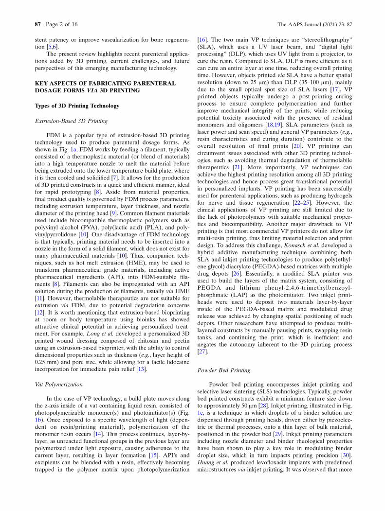
FDM is a popular type of extrusion-based 3D printingtechnology used to produce parenteral dosage forms. Asshown in Fig. 1a, FDM works by feeding a filament, typicallyconsisted of a thermoplastic material (or blend of materials)into a high temperature nozzle to melt the material beforebeing extruded onto the lower temperature build plate, whereit is then cooled and solidified [7]. It allows for the productionof 3D printed constructs in a quick and efficient manner, idealfor rapid prototyping [8]. Aside from material properties,final product quality is governed by FDM process parameters,including extrusion temperature, layer thickness, and nozzlediameter of the printing head [9]. Common filament materialsused include biocompatible thermoplastic polymers such aspolyvinyl alcohol (PVA), poly(lactic acid) (PLA), and poly-vinylpyrrolidone [10]. One disadvantage of FDM technologyis that typically, printing material needs to be inserted into anozzle in the form of a solid filament, which does not exist formany pharmaceutical materials [10]. Thus, companion tech-niques, such as hot melt extrusion (HME), may be used totransform pharmaceutical grade materials, including activepharmaceutical ingredients (API), into FDM-suitable fila-ments [8]. Filaments can also be impregnated with an APIsolution during the production of filaments, usually via HME[11]. However, thermolabile therapeutics are not suitable forextrusion via FDM, due to potential degradation concerns[12]. It is worth mentioning that extrusion-based bioprintingat room or body temperature using bioinks has showedattractive clinical potential in achieving personalized treat-ment. For example, Long et al. developed a personalized 3Dprinted wound dressing composed of chitosan and pectinusing an extrusion-based bioprinter, with the ability to controldimensional properties such as thickness (e.g., layer height of0.25 mm) and pore size, while allowing for a facile lidocaineincorporation for immediate pain relief [13].
Vat Polymerization
In the case of VP technology, a build plate moves alongthe z-axis inside of a vat containing liquid resin, consisted ofphotopolymerizable monomer(s) and photoinitiator(s) (Fig.1b). Once exposed to a specific wavelength of light (depen-dent on resin/printing material), polymerization of themonomer resin occurs [14]. This process continues, layer-by-layer, as unreacted functional groups in the previous layer arepolymerized under light exposure, causing adherence to thecurrent layer, resulting in layer formation [15]. API’s andexcipients can be blended with a resin, effectively becomingtrapped in the polymer matrix upon photopolymerization
[16]. The two main VP techniques are “stereolithography”(SLA), which uses a UV laser beam, and “digital lightprocessing” (DLP), which uses UV light from a projector, tocure the resin. Compared to SLA, DLP is more efficient as itcan cure an entire layer at one time, reducing overall printingtime. However, objects printed via SLA have a better spatialresolution (down to 25 μm) than DLP (35–100 μm), mainlydue to the small optical spot size of SLA lasers [17]. VPprinted objects typically undergo a post-printing curingprocess to ensure complete polymerization and furtherimprove mechanical integrity of the prints, while reducingpotential toxicity associated with the presence of residualmonomers and oligomers [18,19]. SLA parameters (such aslaser power and scan speed) and general VP parameters (e.g.,resin characteristics and curing duration) contribute to theoverall resolution of final prints [20]. VP printing cancircumvent issues associated with other 3D printing technol-ogies, such as avoiding thermal degradation of thermolabiletherapeutics [21]. More importantly, VP techniques canachieve the highest printing resolution among all 3D printingtechnologies and hence process great translational potentialin personalized implants. VP printing has been successfullyused for parenteral applications, such as producing hydrogelsfor nerve and tissue regeneration [22–25]. However, theclinical applications of VP printing are still limited due tothe lack of photopolymers with suitable mechanical proper-ties and biocompatibility. Another major drawback to VPprinting is that most commercial VP printers do not allow formulti-resin printing, thus limiting material selection and printdesign. To address this challenge, Konasch et al. developed ahybrid additive manufacturing technique combining bothSLA and inkjet printing technologies to produce poly(ethyl-ene glycol) diacrylate (PEGDA)-based matrices with multipledrug depots [26]. Essentially, a modified SLA printer wasused to build the layers of the matrix system, consisting ofPEGDA and lithium phenyl-2,4,6-trimethylbenzoyl-phosphinate (LAP) as the photoinitiator. Two inkjet print-heads were used to deposit two materials layer-by-layerinside of the PEGDA-based matrix and modulated drugrelease was achieved by changing spatial positioning of suchdepots. Other researchers have attempted to produce multi-layered constructs by manually pausing prints, swapping resintanks, and continuing the print, which is inefficient andnegates the autonomy inherent to the 3D printing process[27].
Powder Bed Printing
Powder bed printing encompasses inkjet printing andselective laser sintering (SLS) technologies. Typically, powderbed printed constructs exhibit a minimum feature size downto approximately 50 μm [28]. Inkjet printing, illustrated in Fig.1c, is a technique in which droplets of a binder solution aredispensed through printing heads, driven either by piezoelec-tric or thermal processes, onto a thin layer of bulk material,positioned in the powder bed [29]. Inkjet printing parametersincluding nozzle diameter and binder rheological propertieshave been shown to play a key role in modulating binderdroplet size, which in turn impacts printing precision [30].Huang et al. produced levofloxacin implants with predefinedmicrostructures via inkjet printing. It was observed that more
The AAPS Journal (2021) 23: 8787 Page 2 of 16
complex drug release (e.g., bimodal and pulsatile) can beachieved via this method in comparison to the traditionalcompression method, as 3D printing allows for the flexibilityto incorporate multiple types of structures such as reservoirand matrix architectures in one dosage form [31]. Adisadvantage associated with inkjet printing is caused by thebinder hitting the powder bed and displacing powder, leadingto sub-surface depletion zones. SLS is very similar to inkjetprinting, instead using a laser to bind particles/powdertogether to form layers, as opposed to depositing a bindingsolution (Fig. 1d) [32]. Xia et al. developed SLS printed nano-hydroxyapatite/poly-ε-caprolactone (PCL) scaffolds with ahighly porous architecture (150 μm in layer thickness and 70–79% porosity) and sustained rhBMP-2 release for improvedbone defect repair [33]. One main drawback associated withSLS is potential degradation of payload when exposed to highenergy lasers used in the printing process, which has limitedits pharmaceutical applications [14].
Design and Personalization
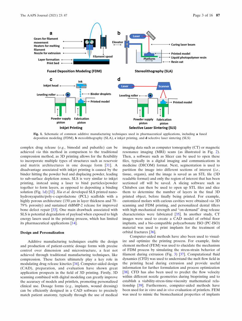
Additive manufacturing techniques enable the designand production of patient-centric dosage forms with precisecontrol over dimension and microstructure, a feat notachieved through traditional manufacturing techniques, likecompression. These factors ultimately play a key role inmodulating drug release kinetics [34]. Computer-aided design(CAD), preparation, and evaluation have shown greatapplication prospects in the field of 3D printing. Firstly, 3Dscanning combined with digital modeling can greatly improvethe accuracy of models and printlets, promoting personalizedclinical use. Dosage forms (e.g., implants, wound dressing)can be efficiently designed in a CAD software to preciselymatch patient anatomy, typically through the use of medical
imaging data such as computer tomography (CT) or magneticresonance imaging (MRI) scans (as illustrated in Fig. 2).Then, a software such as Slicer can be used to open thesefiles, typically in a digital imaging and communications inmedicine (DICOM) format. Next, segmentation is used topartition the image into different sections of interest (i.e.,tissue, organs), and the image is saved as an STL file (3Dreadable format) and only the region of interest that has beensectioned off will be saved. A slicing software such asChitubox can then be used to open up STL files and slicethem to determine the number of layers in the final 3Dprinted object, before finally being printed. For example,customized molars with carious cavities were obtained via 3Dscanning and FDM printing, and personalized dental fillerswith high mechanical strength and “on-demand” drug releasecharacteristics were fabricated [35]. In another study, CTimages were used to create a CAD model of orbital floorimplants, and a bio-compatible polycarbonate ISO (PC-ISO)material was used to print implants for the treatment oforbital fractures [36].
Computer-aided methods have also been used to visual-ize and optimize the printing process. For example, finiteelement method (FEM) was used to elucidate the mechanismof FDM process by simulating the stress-strain behavior offilament during extrusion (Fig. 3) [37]. Computational fluiddynamics (CFD) was used to understand the melt flow field inthe printing head during extrusion and provide usefulinformation for further formulation and process optimization[38]. CFD has also been used to predict the flow velocitywithin different nozzle geometries during bioprinting and toestablish a viability-stress-time-viscosity mathematical rela-tionship [39]. Furthermore, computer-aided methods havebeen used for in vitro and in vivo evaluation of printlets. FEMwas used to mimic the biomechanical properties of implants
Fig. 1. Schematic of common additive manufacturing techniques used in pharmaceutical applications, including a fuseddeposition modeling (FDM), b stereolithography (SLA), c inkjet printing, and d selective laser sintering (SLS)
The AAPS Journal (2021) 23: 87 Page 3 of 16 87
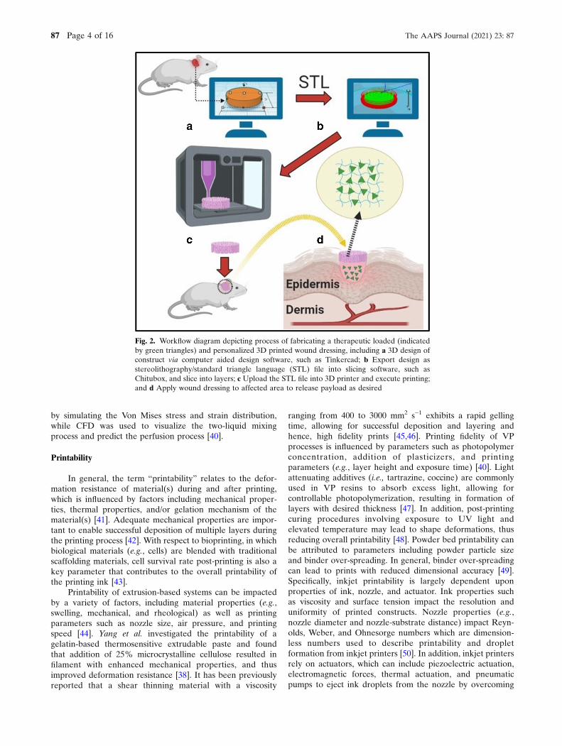
by simulating the Von Mises stress and strain distribution,while CFD was used to visualize the two-liquid mixingprocess and predict the perfusion process [40].
Printability
In general, the term “printability” relates to the defor-mation resistance of material(s) during and after printing,which is influenced by factors including mechanical proper-ties, thermal properties, and/or gelation mechanism of thematerial(s) [41]. Adequate mechanical properties are impor-tant to enable successful deposition of multiple layers duringthe printing process [42]. With respect to bioprinting, in whichbiological materials (e.g., cells) are blended with traditionalscaffolding materials, cell survival rate post-printing is also akey parameter that contributes to the overall printability ofthe printing ink [43].
Printability of extrusion-based systems can be impactedby a variety of factors, including material properties (e.g.,swelling, mechanical, and rheological) as well as printingparameters such as nozzle size, air pressure, and printingspeed [44]. Yang et al. investigated the printability of agelatin-based thermosensitive extrudable paste and foundthat addition of 25% microcrystalline cellulose resulted infilament with enhanced mechanical properties, and thusimproved deformation resistance [38]. It has been previouslyreported that a shear thinning material with a viscosity
ranging from 400 to 3000 mm2 s−1 exhibits a rapid gellingtime, allowing for successful deposition and layering andhence, high fidelity prints [45,46]. Printing fidelity of VPprocesses is influenced by parameters such as photopolymerconcentration, addition of plasticizers, and printingparameters (e.g., layer height and exposure time) [40]. Lightattenuating additives (i.e., tartrazine, coccine) are commonlyused in VP resins to absorb excess light, allowing forcontrollable photopolymerization, resulting in formation oflayers with desired thickness [47]. In addition, post-printingcuring procedures involving exposure to UV light andelevated temperature may lead to shape deformations, thusreducing overall printability [48]. Powder bed printability canbe attributed to parameters including powder particle sizeand binder over-spreading. In general, binder over-spreadingcan lead to prints with reduced dimensional accuracy [49].Specifically, inkjet printability is largely dependent uponproperties of ink, nozzle, and actuator. Ink properties suchas viscosity and surface tension impact the resolution anduniformity of printed constructs. Nozzle properties (e.g.,nozzle diameter and nozzle-substrate distance) impact Reyn-olds, Weber, and Ohnesorge numbers which are dimension-less numbers used to describe printability and dropletformation from inkjet printers [50]. In addition, inkjet printersrely on actuators, which can include piezoelectric actuation,electromagnetic forces, thermal actuation, and pneumaticpumps to eject ink droplets from the nozzle by overcoming
Fig. 2. Workflow diagram depicting process of fabricating a therapeutic loaded (indicatedby green triangles) and personalized 3D printed wound dressing, including a 3D design ofconstruct via computer aided design software, such as Tinkercad; b Export design asstereolithography/standard triangle language (STL) file into slicing software, such asChitubox, and slice into layers; c Upload the STL file into 3D printer and execute printing;and d Apply wound dressing to affected area to release payload as desired
The AAPS Journal (2021) 23: 8787 Page 4 of 16
ink surface tension [51]. Actuator type and its parameters canimpact droplet size and overall print quality of constructs.Similarly, powder composition and properties such as particlesize and polymer molecular weight (MW) impact printabilityand drug release behavior of SLS printed constructs [52].Finer powder results in structures exhibiting enhanced greenstrength, smoother surface, quicker drug release, and reducedporosity, as well as an overall improvement in mechanicalproperties [53–55]. Laser properties (e.g., laser energydensity) can also have an impact on SLS printability byaltering powder bed temperature [32]. Lastly, high laser scanspeeds have resulted in constructs exhibiting increasedporosity, leading to rapid drug release and reduced mechan-ical properties due to shortened contact time between laserand powder [56,57].
Printing Materials
Materials used in 3D printed parenteral constructs needto be biocompatible to minimize immune response in thebody, in addition to demonstrating suitable mechanicalproperties to ensure sufficient printability. Considerationssuch as ability to promote cell adhesion and proliferationshould also be taken into account for parenteral applicationsincluding bone and tissue scaffolds [58]. Some materialscommonly used in 3D printed parenteral constructs are listedin Tables I and II.
Synthetic Materials
Synthetic polymers such as polyesters, PVA, andpolyurethane (PU) typically have more reproducible poly-mer characteristics and desirable mechanical properties(e.g., tensile strength and elastic modulus) compared tonatural materials, which makes them more suitable for 3D
printing applications [61]. Polyesters. Biodegradablepolyester-based synthetic polymers, such as PLA,poly(lactic-co-glycolic acid) (PLGA), and PCL, are rela-tively hydrophobic and inherently biologically inert. Owingto their excellent biocompatibility and tunable mechanicalproperties, PLA and PLGA, a class of aliphatic polyesters,are suitable for 3D printing applications [62,63]. Tappa et al.developed FDM printed PLA-based osseous fixation de-vices, including surgical screws, pins, and bone plates [64].The 3D printed PLA devices exhibited compressivestrengths between 20 and 500 MPa, demonstrating feasibil-ity in orthopedic applications. In another study, Wang et al.developed a 3D printed bilayer membrane, consisting of aPLGA nanofiber outer layer (layer height: 0.05 μm) andalginate hydrogel inner layer (layer height: 100 μm),designed to mimic the epidermal and dermal layers of theskin for use as a wound dressing with demonstratedaccelerated wound healing ability in vivo [65]. Combiningpolyester materials with other polymers such as PU hasbeen used to further enhance mechanical properties ofpolyesters [66]. However, the hydrophobicity of polyestersresults in inadequate cell adhesion and poor osteogenesis,as well as potential bacterial adhesion and biofilm formation[67]. Thus, 3D printed polyester-based constructs have beenfunctionalized with biomolecules such as collagen,minocycline, and hydroxyapatite to improve cell adhesionand promote bone regeneration [68,69] In addition, chem-ical structure modifications have been used to improve cellbinding and hydrophilicity of polyesters [70].
Photopolymers. Biocompatible photopolymers such asPEGDA, PEG dimethacrylate (PEGDMA), and gelatinmethacrylate (GelMA) are commonly used in VP technology(Table II). PEGDMA hydrogels exhibit similar compressivemodulus to musculoskeletal tissue, making them a suitable
Fig. 3. Radial stress-stain simulation of filament: a a 2D mesh model; b typical nonlinear material properties; c von Misesstress distribution, MPa; and d von Mises strain distribution, % (37)
The AAPS Journal (2021) 23: 87 Page 5 of 16 87
choice for bone regeneration applications. A bioprintingsetup, consisting of a Hewlett-Packard (HP) Deskjet thermalinkjet printer modified with an overhead UV lamp, was usedto produce 3D printed PEGDMA bone constructs withIrgacure I-2959 as the photoinitiator [71]. In another study,Zhou et al. developed a GelMA-based bioink suitable forDLP printing containing LAP photoinitiator and a hyaluronicacid (HA) derivative to create functional living skin for skinregeneration applications [59]. Another photopolymer, poly(-propylene fumarate) (PPF), has been used in variousbiomedical applications including bone tissue engineeringdue to its similar compressive modulus values as humantrabecular bone [72]. Buyuksungur et al. developed PCL-based nanohydroxyapatite (HAp) and PPF-modified implants(PCL/HAp/PPF) produced via FDM printing for bone defecttreatment [60]. The 3D printed PCL/HAp/PPF implantsdemonstrated improved compressive tension stiffness values(394 and 463 N/mm) when compared to healthy rabbit femur(316 and 392 N/mm) after 8 weeks of implantation.
Other Synthetic Materials. PVA is a highly water-solublesynthetic polymer produced via the hydrolysis of polyvinylacetate. It is an attractive biomaterial due to its biocompat-ibility and unique ability to resist protein adsorption, as well
as favorable mechanical properties (e.g., high tensile strengthand elongation before breaking) [73]. Recently, Qamar et al.developed FDM printed PVA-based hernial meshes using aMakerBot FDM printer. It was observed that tensile strengthof the meshes increased with an increase in thread diameterand decrease in pore size [74]. Boyer et al. developed FDMprinted, cross-linked PVA-based porous hepatobiliary stentsfor the treatment of biliary obstruction [75].
PU is generally non-biodegradable but biocompatibleand has been extensively used in various medical applica-tions including vascular grafts, catheters, heart valves, andwound dressings. Recently, researchers have synthesizedbiodegradable PU to expand their biomedical applications[76,77]. Interestingly, PU are comprised of alternating hardand soft segments, the former owing to long-chain diols,which leads to enhanced elasticity, and the latter responsiblefor overall material strength due to the presence of crystal-line regions [78]. Jung et al. developed 3D printed thermo-plastic PU-based tracheal prostheses with higher tensilestrength and enhanced flexibility compared to native tracheatissue. The microporous architecture of the 3D printedprostheses promoted biological interactions by allowing forcellular infiltration and facilitating ingrowth of connectivetissue [79].
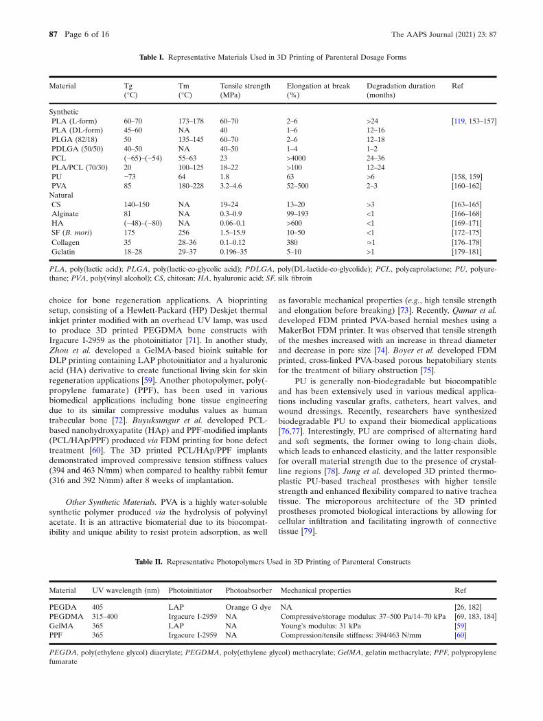
Table I. Representative Materials Used in 3D Printing of Parenteral Dosage Forms
Material Tg(°C)
Tm(°C)
Tensile strength(MPa)
Elongation at break(%)
Degradation duration(months)
Ref
SyntheticPLA (L-form) 60–70 173–178 60–70 2–6 >24 [119, 153–157]PLA (DL-form) 45–60 NA 40 1–6 12–16PLGA (82/18) 50 135–145 60–70 2–6 12–18PDLGA (50/50) 40–50 NA 40–50 1–4 1–2PCL (−65)–(−54) 55–63 23 >4000 24–36PLA/PCL (70/30) 20 100–125 18–22 >100 12–24PUPVA
−7385
64180–228
1.83.2–4.6
6352–500
>62–3
[158, 159][160–162]
NaturalCS 140–150 NA 19–24 13–20 >3 [163–165]Alginate 81 NA 0.3–0.9 99–193 <1 [166–168]HA (−48)–(−80) NA 0.06–0.1 >600 <1 [169–171]SF (B. mori) 175 256 1.5–15.9 10–50 <1 [172–175]Collagen 35 28–36 0.1–0.12 380 ≈1 [176–178]Gelatin 18–28 29–37 0.196–35 5–10 >1 [179–181]
PLA, poly(lactic acid); PLGA, poly(lactic-co-glycolic acid); PDLGA, poly(DL-lactide-co-glycolide); PCL, polycaprolactone; PU, polyure-thane; PVA, poly(vinyl alcohol); CS, chitosan; HA, hyaluronic acid; SF, silk fibroin
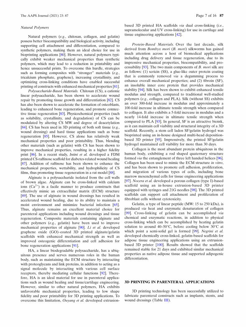
Table II. Representative Photopolymers Used in 3D Printing of Parenteral Constructs
Material UV wavelength (nm) Photoinitiator Photoabsorber Mechanical properties Ref
PEGDA 405 LAP Orange G dye NA [26, 182]PEGDMA 315–400 Irgacure I-2959 NA Compressive/storage modulus: 37–500 Pa/14–70 kPa [69, 183, 184]GelMA 365 LAP NA Young’s modulus: 31 kPa [59]PPF 365 Irgacure I-2959 NA Compression/tensile stiffness: 394/463 N/mm [60]
PEGDA, poly(ethylene glycol) diacrylate; PEGDMA, poly(ethylene glycol) methacrylate; GelMA, gelatin methacrylate; PPF, polypropylenefumarate
The AAPS Journal (2021) 23: 8787 Page 6 of 16
Natural Polymers
Natural polymers (e.g., chitosan, collagen, and gelatin)possess better biocompatibility and biological activity, includingsupporting cell attachment and differentiation, compared tosynthetic polymers, making them an ideal choice for use inbioprinting applications [80]. However, natural polymers typi-cally exhibit weaker mechanical properties than syntheticpolymers, which may lead to a reduction in printability andhence unsuccessful prints. To overcome this hurdle, strategiessuch as forming composites with “stronger” materials (e.g.,tricalcium phosphate, graphene), increasing crystallinity, andoptimizing cross-linking conditions have enabled successfulprinting of constructs with enhanced mechanical properties [81].
Polysaccharide-Based Materials. Chitosan (CS), a cationiclinear polysaccharide, has been shown to accelerate woundrepair by promoting tissue growth and differentiation [82]. CShas also been shown to accelerate the formation of osteoblasts,leading to enhanced bone regeneration, and promote connec-tive tissue regeneration [83]. Physicochemical properties (suchas solubility, crystallinity, and degradation) of CS can bemodulated by altering CS MW and degree of deacetylation[84]. CS has been used in 3D printing of both soft tissue (e.g.,wound dressing) and hard tissue applications such as boneregeneration [85]. However, CS alone has relatively weakmechanical properties and poor printability. Thus, blendingother materials (such as gelatin) with CS has been shown toimprove mechanical properties, resulting in a higher fidelityprint [86]. In a recent study, Intini et al. developed a FDMprintedCS-raffinose scaffold for diabetes-related wound healing[87]. Addition of raffinose has been shown to enhance themechanical properties, wettability, and hydrophilicity of CSfilms, thus promoting tissue regeneration in a rat model [88].
Alginate is a polysaccharide isolated from the cell wallsof brown algae. Alginate can be cross-linked with calciumions (Ca2+) in a facile manner to produce constructs thateffectively mimic an extracellular matrix (ECM) structure[89]. The use of alginate in wound dressings has resulted inaccelerated wound healing, due to its ability to maintain amoist environment and minimize bacterial infection [65].Thus, alginate remains an excellent material choice forparenteral applications including wound dressings and tissueregeneration. Composite materials containing alginate andother polymers (e.g., gelatin) have been used to improvemechanical properties of alginate [90]. Li et al. developedgraphene oxide (GO)–coated 3D printed alginate/gelatinscaffolds with enhanced mechanical strength as well asimproved osteogenic differentiation and cell adhesion forbone regeneration applications [91].
HA, a linear biodegradable polysaccharide, has a ubiq-uitous presence and serves numerous roles in the humanbody, such as maintaining the ECM structure by interactingwith proteoglycans and link proteins, in addition to acting as asignal molecule by interacting with various cell surfacereceptors, thereby mediating cellular functions [92]. There-fore, HA is an ideal material for use in parenteral applica-tions such as wound healing and tissue/cartilage engineering.However, similar to other natural polymers, HA exhibitsunfavorable mechanical properties, leading to low shapefidelity and poor printability for 3D printing applications. Toovercome this limitation, Ouyang et al. developed extrusion-
based 3D printed HA scaffolds via dual cross-linking (i.e.,supramolecular and UV cross-linking) for use in cartilage andtissue engineering applications [42].
Protein-Based Materials. Over the last decade, silkderived from Bombyx mori (B. mori) silkworms has gainedmuch attention across a host of biomedical applicationsincluding drug delivery and tissue regeneration, due to itsimpressive mechanical properties, biocompatibility, and pro-cessability [93]. The two main components of B. mori silk areas follows: (1) sericin (SS), a glue-like outer protein coatingthat is commonly removed via a degumming process toenhance overall mechanical properties; and (2) fibroin (SF),an insoluble inner core protein that provides mechanicalstability [94]. Silk has been shown to exhibit enhanced tensilemodulus and strength, compared to traditional well-studiedpolymers (e.g., collagen and PLA). Silk (without SS) exhibitsan over 300-fold increase in modulus and approximately a100-fold increase in ultimate tensile strength when comparedto collagen. It also exhibits a 5-fold increase in modulus and anearly 14-fold increase in ultimate tensile strength whencompared to PLA [93]. In general, SF is an attractive bioink,as it can maintain cell viability and structural integrity of a 3Dscaffold. Recently, a stem cell laden SF/gelatin hydrogel wasbioprinted using an in-house designed multi-head deposition-based 3D printer [95]. Impressively, the printed SF/gelatinhydrogel maintained cell viability for more than 30 days.
Collagen is the most abundant protein ubiquitous in thehuman body, exhibiting a rod shaped quaternary structureformed via the entanglement of three left handed helices [96].Collagen has been used to mimic the ECM structure in vitro,and has been shown to promote cell adhesion, proliferation,and migration of various types of cells, including bonemarrow mesenchymal cells for tissue engineering applications[97]. Nocera et al. developed a porous collagen (type I)-basedscaffold using an in-house extrusion-based 3D printerequipped with syringes and 21G needles [98]. The 3D printedscaffolds can support cell attachment and proliferation offibroblast cells without cytotoxicity.
Gelatin, a type of linear peptide (MW: 15 to 250 kDa), isproduced via heat and enzymatic denaturation of collagen[99]. Cross-linking of gelatin can be accomplished viachemical and enzymatic reactions, in addition to physicalcross-linking which can be accomplished by heating gelatinsolution to around 40–50°C, before cooling below 30°C atwhich point a semi-solid gel is formed [99]. Negrini et al.developed chemically cross-linked, gelatin-based scaffolds foradipose tissue engineering applications using an extrusion-based 3D printer [100]. Results showed that the scaffoldsremained stable for 21 days and exhibited similar mechanicalproperties as native adipose tissue and supported adipogenicdifferentiation.
3D PRINTING IN PARENTERAL APPLICATIONS
3D printing technology has been successfully utilized tofabricate parenteral constructs such as implants, stents, andwound dressings (Table III).
The AAPS Journal (2021) 23: 87 Page 7 of 16 87
TableIII.
Examples
of3D
PrintingTechn
olog
yforParen
teralApp
lications
Materials
3Dprinter
Design
App
lications
Ref
Implan
tsPLLA
SLA
Ana
tomically
relevant
sphe
ricalor
cylin
drical
shap
eSu
staine
dreleaseof
multiplechem
othe
rape
uticsfor12
wee
ksforosteosarcomatherap
y.[185
]PLA,P
VA,P
CL
FDM
Rod-shap
edim
plants
containing
differe
nt
size
d“w
indo
ws”
Sustaine
dpa
yloa
dreleasefrom
implan
tsmod
ulated
viathe“w
indo
ws.”
[101]
PCL,P
LGA
Extrusion
-ba
sed
Patches
withdifferen
tshap
edpo
res
Sustaine
d5-flou
racilreleaseov
er4wee
ksforpa
ncreatic
cancer
therap
y.[186
]
Calcium
phosph
ate
Inkjet
printer
Implan
tsCo-de
liveryof
multiplean
tibioticsforthetreatm
entof
bone
osteom
yelitis.
[102]
PLA
,collagen,
hydrox
yapa
tite
FDM
Scaffoldswith
uniform
macropo
rous
architecture
Combination
of
the
mac
roporo
us
arch
itec
ture
and
antibiotic
release
allowing
vascularizationwhile
againstbo
neinfection.
[103]
PLA,P
CL
FDM
O-,Y-,an
dM-sha
pedva
gina
lring
sSh
ape-de
pend
entprog
esterone
releaseforcontraceptivepu
rposes.
[104]
Biomed
ical
devices
PCL,P
LA
FDM
Bioresorbab
lesten
tsSten
tswithmod
ulab
lemecha
nicalprop
erties
forcardiova
scular
diseaseman
agem
ent.
[6]
PCL,
sulfate
dCS
(26S
CS)
Extrusion
-ba
sed
Bioresorbab
lesten
ts26
SCS-mod
ified
PCLsten
tallo
wed
foren
hanced
biocom
patib
ility
forcardiova
scular
disease
man
agem
ent.
[187
]
PLA,po
lydo
pamine,
PEI,he
parin
Extrusion
-ba
sed
Biode
grad
able
polymer–coa
tedsten
tsSten
tsex
hibited
excelle
ntan
ti-coa
gulant
activ
ityan
dbiocom
patib
ility
forcardiova
scular
diseaseman
agem
ent.
[188
]
PVA,co
llage
n,PCL,
cholan
giocyte
FDM
Stem
cell–
coated
bilia
rysten
tPVA-based
bilia
rysten
tswithresisted
biofi
lmform
ationan
den
hanced
sten
tpa
tencyfor
bilia
ryob
struction.
[75]
PCL,g
raph
ene
FDM
Multi-drugelutingsten
tSu
staine
dde
livery
ofmultiple
therap
eutics
with
simila
rmecha
nicalprop
erties
asconv
ention
alcorona
rysten
ts(elastic
mod
ulus
400MPa)
[189
]
PLA,T
PU
FDM
PLA/TPU
sten
twith
spiral
patterns
with
controlla
ble
spiral
angle,
thickn
ess,an
dpitch
Flexible,
self-exp
anding
sten
tswith
redu
ced
sten
tmigration
forcardiova
scular
disease
man
agem
ent.
[190
]
Wou
nddressing
Pectin
,CS
Extrusion
-ba
sed
Hyd
roge
lscaffold
Wou
nddressing
sex
hibitedgo
odbioa
dhesionstreng
th(86.5–12
6.9g),
while
maintaining
amoist
environm
entforskin
wou
ndhe
aling.
[13]
PCL,FPLA,PEGDA,
PEG
FDM/SLA
Persona
lized
anti-acne
patche
s/masks
Persona
lized
acne
treatm
entwithsalicylic
acid
basedon
patie
ntscan
s.[191
]
Chitosan,
genipin,
PEG
Extrusion
-ba
sed
Film
sMucoa
dhesivean
dsw
ellablefilm
sforpa
yloa
dreleaseto
prom
oteskin
wou
ndhe
aling.
[192
]
CS,
raffino
seFDM
Wou
nddressing
withcontrolla
blemicroarchitecture
CSscaffoldsprom
oted
tissue
regene
ration
inadiab
etes-related
skin
wou
ndratmod
el.
[87]
SS,G
elMA
Extrusion
-ba
sed
Transpa
rent
hydrog
elscaffold
withcontrolla
blepo
resizes
Wou
nddressing
design
edforreal-tim
emon
itoringof
wou
ndhe
alingprocess.
[193
]
PU,H
AFDM
Scaffoldsde
sign
edto
releasetw
obiom
olecules
Sustaine
dreleaseof
multipletherap
eutics
toaccelerate
wou
ndhe
alingprocessforcartila
gede
fect.
[194
]
PLGA,a
lginate
Extrusion
-ba
sed
Bila
yermem
bran
ede
sign
edto
mim
ictheskin
derm
isan
dep
idermis
Porou
sbilaye
rwou
nddressing
toen
hanced
wou
ndrepa
iror
beused
asaskin
substitute.
[65]
PLLA,po
ly(L
-lactide
);PEI,
polyethy
lenimine;
TPU,thermop
lastic
polyuretha
ne;FPLA,FlexEcoPLA™
;SS
,silk
sericin;
PEG,po
lyethy
lene
glycol;FDM,fusedde
position
mod
eling;
SLA,
stereo
litho
grap
hy
The AAPS Journal (2021) 23: 8787 Page 8 of 16
Long-Acting Implants/Inserts
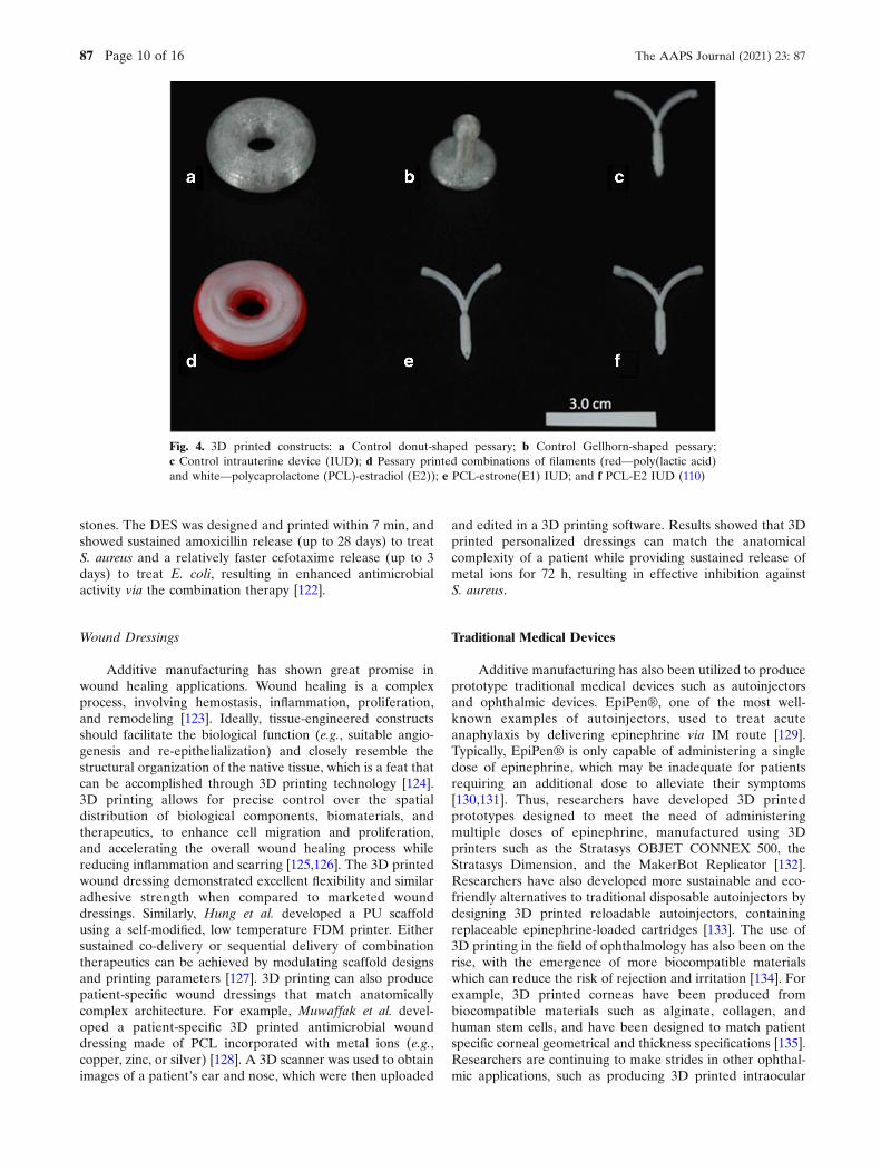
Long-acting implants/inserts have been widely utilizedfor various clinical applications, including contraception,cancer treatment, and localized delivery of anesthetics andantibiotics [105–108]. 3D printed implants can be designed toachieve tunable, sustained drug release through precisecontrol over implant shape, size, and microstructure. In arecent study, Stewart et al. produced 3D printed rod-shapedPVA/PLA implants via FDM, with designed “windows” tomodulate drug release [101]. Implants with smaller “win-dows” and a decreased number of total “windows” resulted inslower payload release. Impressively, these implants, dip-coated with a PCL polymer mixture, can retard payloadrelease for up to 300 days. In order to provide personalizedvaginal rings to avoid pelvic inflammatory disease and uterineperforations [109], Fu et al. developed 3D printed PLA/PCLcomposite vaginal rings with customized shapes (i.e., “O,”“Y,” and “M”) to accurately mimic the structure of femaleanatomy [104]. All printed vaginal rings exhibited shape-dependent progesterone release for 7 days. Similarly, Tappaet al. developed 3D printed vaginal inserts containing eitherestrogen or progesterone [110]. The inserts were fabricated tomimic clinically relevant surgical meshes, intrauterine devices,and pessaries, and demonstrated excellent biocompatibilityand sustained payload release (Fig. 4).
3D printing technology also allows for seamless integra-tion of multiple API’s in a single dosage form for combinationtherapy. Qiao et al. developed 3D printed PLGA scaffolds forthe combination therapy of doxorubicin and cisplatin againstbreast cancer [111]. The 3D printed scaffolds (pore sizes > 200μm) were produced using a customized E-jet printer and werecapable of delivering both drugs in a controlled releasemanner for up to 30 days, demonstrating synergistic antitu-mor effect of the combination therapy. Won et al. developed a3D printed core (alginate and dexamethasone)/shell (PCLand bevacizumab) structured rod using a multi-headbioprinter for the treatment of retinal vascular diseases[112]. The rods exhibited sustained bevacizumab release over60 days and dexamethasone release over 7 days, leading tosuppressed angiogenesis over a 4-week period in a rat model.
Regenerative Applications
3D printing technology has shown promising in regener-ative applications, particularly for treating bone defect, as itcan accurately and quickly produce customizable scaffoldswith defined microstructures and precise control over factors(such as shape, porosity, and mechanical properties), all ofwhich impact magnitude of osteogenesis and angiogenesis[5,113–116]. For example, Zhang et al. developed a multi-functional bioceramic scaffold capable of promotingvascularized bone regeneration to treat large segmental bonedefects [5]. Hollow-pipe-packed silicate bioceramic (BRT-H)scaffolds with a core/shell structure were produced via amodified extrusion-based 3D printer. The synergistic effect ofthe hollow channel structures produced via 3D printing andionic components (e.g., silicon, magnesium, and calcium) ofthe alginate-based scaffold led to enhanced tissue growth andvascularization [117]. Similarly, Martin et al. engineered amultifunctional 3D printed PLA scaffold via FDM for bone
regeneration [103]. The printed PLA scaffold exhibited alattice-shaped structure with a controllable pore size of1000 μm and a porosity around 55%. Multifunctionalizationvia a combination of collagen, minocycline, and hydroxyap-atite, aided by scaffold porosity, resulted in improvedantibacterial/antibiofilm properties while promoting osteo-genesis. Calcium phosphate scaffolds (CPS) containing anti-biotics (i.e., rifampin and vancomycin) have been developedvia inkjet 3D printing for osteomyelitis therapy [102]. 3Dprinted porous CPS implants allowed for a 6-fold increase invancomycin release compared to manually moldedpoly(methyl methacrylate) spacers, resulting in a reductionin mean bacterial load.
Implantable Biomedical Devices
Stents/Drug-Eluting Stents
Stents have been widely used to widen the affected bloodvessels and restore blood flow for treating cardiovasculardiseases, a leading cause of death around the world. Factorsincluding stent strut thickness and structure (i.e., shape,geometry) have been shown to have a substantial impact onoverall mechanical properties (e.g., radial force, radial recoil,and flexibility) of stents and hence stent effectiveness [118].3D printing technology allows for precise control over stentshape and dimensions using either a single material orcombinations of multiple materials, to achieve desiredmechanical and physical properties depending on applicationsite, which is crucial to stent effectiveness [119]. Moreover,conventional methods (e.g., laser cutting) to produce metallicand polymeric stents can negatively impact overall stentmicrostructure, leading to microcracks [120]. 3D printingcan minimize damage to stent microstructure by avoidingthe use of high temperatures inherent to conventional lasercutting manufacturing. Recently, 3D printing technology hasbeen implemented to produce biodegradable polymer-basedstents with structure flexibility, ideal for ease of insertion,while maintaining a rigid structure to support the bloodvessel. Guerra et al. developed biodegradable stentsconsisting of either PLA filament, PCL filament, or acombination of both via FDM printing [6]. PLA and PCLexhibit vastly different mechanical properties and degrada-tion profiles, which when used alone, are insufficient for usein stent applications. However, when used together, compos-ite stents can achieve more desirable mechanical properties.Composite stents composed of a PLA core and PCL shellexhibited a Young’s modulus around 1400 MPa and about 3%degradation over the span of 6 weeks, suitable for stentapplications.
Drug-eluting stents (DES) can not only physicallyprovide structure to keep the blood vessel open, but alsorelease multiple therapeutics designed to treat post-surgicalside effects such as inflammation. Therapeutics can beblended with polymers to create a drug-loaded filament for3D printing, or coated on the surface of printed stents [121].Kim et al developed a 3D printed PCL DES, using adeposition-based 3D printer, to treat recurrent obstructivesalivary gland disease, commonly caused by the buildup ofsalivary stones [121]. The stent shape was derived from CTimages to mimic salivary ducts after removal of salivary
The AAPS Journal (2021) 23: 87 Page 9 of 16 87
stones. The DES was designed and printed within 7 min, andshowed sustained amoxicillin release (up to 28 days) to treatS. aureus and a relatively faster cefotaxime release (up to 3days) to treat E. coli, resulting in enhanced antimicrobialactivity via the combination therapy [122].
Wound Dressings
Additive manufacturing has shown great promise inwound healing applications. Wound healing is a complexprocess, involving hemostasis, inflammation, proliferation,and remodeling [123]. Ideally, tissue-engineered constructsshould facilitate the biological function (e.g., suitable angio-genesis and re-epithelialization) and closely resemble thestructural organization of the native tissue, which is a feat thatcan be accomplished through 3D printing technology [124].3D printing allows for precise control over the spatialdistribution of biological components, biomaterials, andtherapeutics, to enhance cell migration and proliferation,and accelerating the overall wound healing process whilereducing inflammation and scarring [125,126]. The 3D printedwound dressing demonstrated excellent flexibility and similaradhesive strength when compared to marketed wounddressings. Similarly, Hung et al. developed a PU scaffoldusing a self-modified, low temperature FDM printer. Eithersustained co-delivery or sequential delivery of combinationtherapeutics can be achieved by modulating scaffold designsand printing parameters [127]. 3D printing can also producepatient-specific wound dressings that match anatomicallycomplex architecture. For example, Muwaffak et al. devel-oped a patient-specific 3D printed antimicrobial wounddressing made of PCL incorporated with metal ions (e.g.,copper, zinc, or silver) [128]. A 3D scanner was used to obtainimages of a patient’s ear and nose, which were then uploaded
and edited in a 3D printing software. Results showed that 3Dprinted personalized dressings can match the anatomicalcomplexity of a patient while providing sustained release ofmetal ions for 72 h, resulting in effective inhibition againstS. aureus.
Traditional Medical Devices
Additive manufacturing has also been utilized to produceprototype traditional medical devices such as autoinjectorsand ophthalmic devices. EpiPen®, one of the most well-known examples of autoinjectors, used to treat acuteanaphylaxis by delivering epinephrine via IM route [129].Typically, EpiPen® is only capable of administering a singledose of epinephrine, which may be inadequate for patientsrequiring an additional dose to alleviate their symptoms[130,131]. Thus, researchers have developed 3D printedprototypes designed to meet the need of administeringmultiple doses of epinephrine, manufactured using 3Dprinters such as the Stratasys OBJET CONNEX 500, theStratasys Dimension, and the MakerBot Replicator [132].Researchers have also developed more sustainable and eco-friendly alternatives to traditional disposable autoinjectors bydesigning 3D printed reloadable autoinjectors, containingreplaceable epinephrine-loaded cartridges [133]. The use of3D printing in the field of ophthalmology has also been on therise, with the emergence of more biocompatible materialswhich can reduce the risk of rejection and irritation [134]. Forexample, 3D printed corneas have been produced frombiocompatible materials such as alginate, collagen, andhuman stem cells, and have been designed to match patientspecific corneal geometrical and thickness specifications [135].Researchers are continuing to make strides in other ophthal-mic applications, such as producing 3D printed intraocular
Fig. 4. 3D printed constructs: a Control donut-shaped pessary; b Control Gellhorn-shaped pessary;c Control intrauterine device (IUD); d Pessary printed combinations of filaments (red—poly(lactic acid)and white—polycaprolactone (PCL)-estradiol (E2)); e PCL-estrone(E1) IUD; and f PCL-E2 IUD (110)
The AAPS Journal (2021) 23: 8787 Page 10 of 16
lenses (IOL), which require careful consideration of otherparameters, such as refractive index (RI) of 3D printedlayers, and 3D printed retinas, the success of which dependson the ability to successfully print multiple retinal cell types[134].
FUTURE PERSPECTIVES AND CHALLENGES
3D printing has tremendous potential in personalizedmedicines via parenteral routes. It has been successfullyutilized to print cells in specific and predetermined spatialarrangements, which closely mimic the cellular organizationof native tissue for tissue regeneration applications. Re-searchers have now turned their attention to using thisadditive manufacturing technology to print entire organs tosolve organ donor shortage and immune rejection issues[136]. In the future, this technology may become morepopular in hospital and emergency room settings, as 3Dprinting allows for rapid fabrication of clinically relevant andon-demand constructs. Another intriguing avenue that re-searchers have begun exploring is the combination of 3Dprinted constructs and biomedical electronics. 3D printedimplants/inserts offer an array of advantages over implantsfabricated via traditional molding/extrusion methods, such asthe ability to achieve patient-specific characteristics, loadmultiple therapeutics in one dosage form while avoidingincompatibilities between drugs, in addition to maintainingprecise control over microstructure and mechanical proper-ties, and drug release kinetics [137]. On the other hand,biomedical electronics have been used to achieve externallycontrolled drug delivery, including hormone-releasing micro-chips and miniaturized neural drug delivery systems [138,139].Recently, Kong et al. developed an FDM printed, Bluetooth-enabled gastric resident electronic device capable of on-demand release of antimicrobial and hormonal agents [140].In this application, 3D printing allows for the fabrication of aninsert with precise dimensions and the seamless integration ofmultiple materials, including PLA and PU to amplify theadhesion strength between the materials, to achieve gastricretention over 36 days.
Despite the tremendous potential that 3D printingtechnology offers, there are some challenges that need to beaddressed before this technology becomes mainstream inmanufacturing of parenteral constructs. General 3D printingconsiderations, such as material selection, printing parame-ters, post-printing treatment, and material toxicity concerns,need to be addressed prior to achieving a successful print[141]. The development of new materials or printing inks isthe key to the success in propelling 3D printing-basedparenteral applications. Scientists often need to modifycommercially available materials to satisfy certain printingrequirements, such as improving mechanical properties foradequate printability [38]. In addition to impacting printabil-ity, mechanical properties of 3D printed constructs play a keyrole in modulating cell-scaffold interactions, especially interms of cell adhesion ability and stent patency [142].Products manufactured via certain 3D printing techniques,such as VP, require an additional post-printing curing step toenhance mechanical integrity of the product, adding to thecomplexity of the overall 3D printing process [18]. This stepmay also have a negative impact on loaded therapeutics,
which can lead to compromised biocompatibility. Anotherimportant factor to consider is the sterilization of parenteralprints. Traditional sterilization techniques include exposure togamma-irradiation, ethylene oxide gas, UV irradiation, etha-nol washing, and autoclaving [143]. A recent study investi-gated the efficacy of sterilization techniques, including plasmairradiation and autoclave steam sterilization (121°C and134°C), for surgical guides and implants [144]. It wasconcluded that both plasma irradiation and autoclave steamsterilization are suitable sterilization methods, and that hightemperature steam sterilization caused no significant defor-mation of 3D printed implants. While these sterilizationmethods are promising, they are not suitable for all 3Dprinted constructs, as high temperature sterilization methodscan potentially compromise the integrity and efficacy of printscontaining temperature sensitive polymers and/or thermola-bile therapeutics. In addition, while personalized constructscan be produced, the time it takes to 3D print individualprints far exceeds the time it takes to commercially manufac-ture them as a result of the layer-by-layer addition processinherent to 3D printing. Furthermore, there is a need todevelop multi-material 3D printers to enable flexible con-struct design and combination therapy. Last but not the least,toxicological effects of materials used in the 3D printingprocess remain a paramount concern for researchers. Addi-tives such as photoinitiators and cross-linking agents aretypically required in the 3D printing process, to achievesuccessful prints. However, unreacted resin componentsfound in VP type 3D printing techniques have been shownto present cytotoxicity concerns, and are a key reason many3D printing procedures involve wash steps to removeunreacted material [145]. Two photoinitiators commonly usedin the 3D printing process of parenteral application includingLAP and Irgacure I-2959 have demonstrated increasedcytotoxicity levels at elevated concentrations [145,146]. Al-ternative photoinitiators, such as riboflavin, have been shownto exhibit UV cross-linking ability while remaining non-cytotoxic, despite exhibiting lower print resolution due tolonger reaction times [147]. Other cross-linking agents used inthe development of parenteral delivery systems, such asglutaraldehyde, which has been used in combination withchitosan to produce hydrogels, has been shown to demon-strate mutagenic and neurotoxic properties [148]. Thus,materials used in the 3D printing process of parenteralapplications must be carefully selected to ensure that finalprinted products remain biocompatible and non-toxic.
Despite the tremendous potential 3D printing has tooffer for parenteral applications, regulatory guidance oncharacterization and assessment methods as well as processvalidation methodology remains scarce. While dozens of 3Dprinted medical devices have received FDA approval such asdental crowns and bone plates, only one pharmaceutical drugproduct, Spritam® medication, has been approved by theFDA [149,150]. Clinical trials are underway for other 3Dprinted medical devices, such as 3D printed patient-specificintramedullary guide and 3D printed denture framework.Most recently, the FDA held a public workshop in 2014 andissued a guidance in 2017 covering technical considerationsfor additive manufactured medical devices, such as informa-tion regarding design and manufacturing considerations for3D printed medical devices [151]. This guidance recommends
The AAPS Journal (2021) 23: 87 Page 11 of 16 87
material controls, describing specifications for raw materialsincluding particle size, viscosity, and filament dimensions,should be well controlled. The guidance also recommends tounderstand and document the impact of post-processing stepsinvolved in residue removal and sterilization, including heator chemical treatments, on final product performance andproperties. Furthermore, process validation, including assess-ments on device dimensions, feature geometry, and materialproperties, must be performed on final prints to ensurequality is maintained for parts produced in a single buildcycle and between multiple build cycles. Lastly, final productmechanical properties such as modulus, yield strength, andcreep should be investigated once all post-processing,cleaning, and sterilization steps have been performed. Whilethis guidance provides insightful information, numerousregulatory concerns remain unaddressed, including regulationof 3D printed on-demand personalized products at hospitalsand pharmacies and the regulation of printer ink and 3Dprinter manufacturing [152]. There is a growing interest touse 3D printing to produce parenteral dosage forms, and withthis increased appeal, more specific and defined regulationswill need to be established.
CONCLUSION
Although still in its infancy, 3D printing has alreadydemonstrated tremendous potential for producing parenteralconstructs. The importance of producing on-demand, person-alized medications tailored to patient anatomy and diseaseconditions cannot be overstated. In addition, the ability toprogress from design to prototype in a matter of hours allowsscientist and physicians to quickly and efficiently test outvarious designs and therapeutic regimens until a desirabletreatment is obtained. Additive manufacturing techniquesalso allow for the flexibility to combine multiple therapeuticsin a single dosage form in a controllable and organizedfashion. Biomedical devices and implantable scaffolds can beprinted with controllable dimensions and microstructures,leading to tunable degradation and drug release characteris-tics, in addition to playing a key role in modulating cellproliferation and migration abilities. Thus, despite not beingan optimal solution for large-scale manufacturing, the use of3D printing for parenteral applications will continue to rise,to meet the growing demand for patient-centric medications.
DECLARATIONS
Conflict of Interest The authors declare that they have no conflictof interest.
REFERENCES
1. CENTER FOR DRUG EVALUATION AND RESEARCH.Approval package for SPRITAM. 2015; Available from:https://www.accessdata.fda.gov/drugsatfda_docs/nda/2015/207958Orig1s000Approv.pdf
2. Afsana JV, Haider N, Jain K. 3D printing in personalized drugdelivery. Curr Pharm Des. 2019;24(42):5062–71.
3. Do A, Worthington K, Tucker B, Salem AK, Therapeutics T,Engineering B, et al. Controlled drug delivery from 3D printedtwo-photon polymerized poly(ethylene glycol) dimethacrylatedevices. Int J Pharm. 2019;552:217–24.
4. Buj-Corral I, Bagheri A, Petit-Rojo O. 3D printing of porousscaffolds with controlled porosity and pore size values.Materials (Basel). 2018;11(9):1–18.
5. Zhang W, Feng C, Yang G, Li G, Ding X, Wang S, et al. 3D-printed scaffolds with synergistic effect of hollow-pipe struc-ture and bioactive ions for vascularized bone regeneration.Biomaterials [Internet]. 2017;135:85–95. Available from.https://doi.org/10.1016/j.biomaterials.2017.05.005.
6. Guerra AJ, Cano P, Rabionet M, Puig T, Ciurana J. 3D-printed PCL/PLA composite stents: towards a new solution tocardiovascular problems. Materials (Basel). 2018;11(9):1–13.
7. Long J, Gholizadeh H, Lu J, Seyfoddin A. Application offused deposition modelling (FDM) method of 3D printing indrug delivery. 2017 433–439.
8. Melocchi A, Parietti F, Maroni A, Foppoli A, Gazzaniga A,Zema L. Hot-melt extruded filaments based on pharmaceuticalgrade polymers for 3D printing by fused deposition modeling.Int J Pharm [Internet]. 2016;509(1–2):255–63. Available from.https://doi.org/10.1016/j.ijpharm.2016.05.036.
9. Popescu D, Zapciu A, Amza C, Baciu F, Marinescu R. FDMprocess parameters influence over the mechanical propertieso f po l ymer spec imens : a rev i ew. Po l ym Tes t .2018;69(May):157–66.
10. Azad MA, Olawuni D, Kimbell G, Badruddoza AZM, HossainMS, Sultana T. Polymers for extrusion-based 3D printing ofpharmaceuticals: a holistic materials–process perspective.Pharmaceutics. 2020;12:1–34.
11. Algahtani MS, Mohammed AA, Ahmad J. Extrusion-based3D printing for pharmaceuticals: contemporary research andapplications. Curr Pharm Des. 2019;24(42):4991–5008.
12. Araújo MRP, Sa-Barreto LL, Gratieri T, Gelfuso GM, Cunha-Filho M. The digital pharmacies era: how 3D printingtechnology using fused deposition modeling can become areality. Pharmaceutics. 2019;11:3.
13. Long J, Etxeberria AE, Nand AV, Bunt CR, Ray S, SeyfoddinA. A 3D printed chitosan-pectin hydrogel wound dressing forlidocaine hydrochloride delivery. Mater Sci Eng C [Internet].2019;104:109873. Available from. https://doi.org/10.1016/j.msec.2019.109873.
14. Alhnan MA, Okwuosa TC, Sadia M, Wan KW, Ahmed W,Arafat B. Emergence of 3D printed dosage forms: opportuni-ties and challenges. Pharm Res. 2016;33(8):1817–32.
15. McMains S. Layered manufacturing technologies. CommunACM. 2005;48(6):50–6.
16. Martinez PR, Goyanes A, Basit AW, Gaisford S. Fabricationof drug-loaded hydrogels with stereolithographic 3D printing.Int J Pharm [Internet]. 2017;532(1):313–7. Available from.https://doi.org/10.1016/j.ijpharm.2017.09.003.
17. Skoog SA, Goering PL, Narayan RJ. Stereolithography intissue engineering. J Mater Sci Mater Med. 2014;25(3):845–56.
18. Melchels FPW, Feijen J, Grijpma DW. A review onstereolithography and its applications in biomedical engineer-ing. Biomaterials. 2010;31(24):6121–30.
19. Gittard S, Narayan R. Laser direct writing of micro- andnano-scale medical devices. Expert Rev Med Devices.2010;7(3):343–56.
20. Lee ED, Sim JH, Kweon HJ, Paik IH. Determination ofprocess parameters in stereolithography using neural network.KSME Int J. 2004;18(3):443–52.
21. Kadry H, Wadnap S, Xu C, Ahsan F. Digital light processing(DLP)3D-printing technology and photoreactive polymers infabrication of modified-release tablets. Eur J Pharm Sci[Internet]. 2019;135:60–7. Available from. https://doi.org/10.1016/j.ejps.2019.05.008.
22. Lee S-J. Development of 3D printed hydrogel scaffold withcore-shell nanoparticles for nerve regeneration. IEEE TransBiomed Eng. 2015;64(2):408–18.
23. Kim JH, Lee JW, Yun WS. Fabrication and tissue engineeringapplication of a 3D PPF/DEF scaffold using Blu-ray based 3Dprinting system. J Mech Sci Technol. 2017;31(5):2581–7.
24. Arcaute K, Mann BK, Wicker RB. Stereolithography of three-dimensional bioactive poly(ethylene glycol) constructs withencapsulated cells. Ann Biomed Eng. 2006;34(9):1429–41.
The AAPS Journal (2021) 23: 8787 Page 12 of 16
25. Arcaute K, Mann B, Wicker R. Stereolithography of spatiallycontrolled multi-material bioactive poly(ethylene glycol) scaf-folds. Acta Biomater. 2010;6(3):1047–54.
26. Konasch J, Riess A, Mau R, Teske M, Rekowska N, Eickner T,et al. A novel hybrid additive manufacturing process for drugdelivery systems with locally incorporated drug depots.Pharmaceutics. 2019;11(12):1–14.
27. Robles-Martinez P, Xu X, Trenfield SJ, Awad A, Goyanes A,Telford R, et al. 3D printing of a multi-layered polypillcontaining six drugs using a novel stereolithographic method.Pharmaceutics. 2019;11:6.
28. Fayazfar H, Salarian M, Rogalsky A, Sarker D, Russo P,Paserin V, et al. A critical review of powder-based additivemanufacturing of ferrous alloys: process parameters, micro-structure and mechanical properties. Mater Des [Internet].2018;144:98–128. Available from. https://doi.org/10.1016/j.matdes.2018.02.018.
29. Shirazi SFS, Gharehkhani S, Mehrali M, Yarmand H,Metselaar HSC, Adib Kadri N, et al. A review on powder-based additive manufacturing for tissue engineering: Selectivelaser sintering and inkjet 3D printing. Sci Technol Adv Mater[Internet]. 2015;16(3):1–20. Available from. https://doi.org/10.1088/1468-6996/16/3/033502.
30. Rahmati S, Shirazi SF, Baghayeri H. Piezo-electric headapplication in a new 3D printing design. Rapid Prototyp J.2009;15(3):187–91.
31. Huang W, Zheng Q, Sun W, Xu H, Yang X. Levofloxacinimplants with predefined microstructure fabricated by three-dimensional printing technique. Int J Pharm. 2007;339(1–2):33–8.
32. Fina F, Goyanes A, Gaisford S, Basit AW. Selective lasersintering (SLS) 3D printing of medicines. Int J Pharm[Internet]. 2017;529(1–2):285–93. Available from. https://doi.org/10.1016/j.ijpharm.2017.06.082.
33. Xia Y, Zhou PY, Cheng XS, Xie Y, Liang C, Li C, et al.Selective laser sintering fabrication of nano-hydroxyapatite/poly-ε-caprolactone scaffolds for bone tissue engineeringapplications. Int J Nanomedicine. 2013;8:4197–213.
34. Mathew E, Pitzanti G, Larrañeta E, Lamprou DA. Three-dimensional printing of pharmaceuticals and drug deliverydevices. Pharmaceutics. 2020;12(3):1–9.
35. Yang Y, Li H, Xu Y, Dong Y, Shan W, Shen J. Fabrication andevaluation of dental fillers using customized molds via 3Dprinting technology. Int J Pharm [Internet]. 2019;562:66–75.Available from. https://doi.org/10.1016/j.ijpharm.2019.03.024.
36. Mohan AM, MH AAR. Manufacturing of customized implantsfor orbital fractures using 3D printing. Bioprinting [Internet].2021;21:e00118. Available from. https://doi.org/10.1016/j.bprint.2020.e00118.
37. Yang Y. Strategies and mechanisms to improve the printabilityof pharmaceutical polymers. International Journal ofPharmaceutics Int J Pharm. 2021.
38. Yang Y, Wang X, Lin X, Xie L, Ivone R, Shen J, et al. Atunable extruded 3D printing platform using thermo-sensitivepastes. Int J Pharm. 2020;583(April).
39. Lucas L, Aravind A, Emma P, Christophe M, Edwin-Joffrey C.Rheology, simulation and data analysis toward bioprinting cellviability awareness. Bioprinting. 2021;21.
40. Yang Y, Zhou Y, Lin X, Yang Q, Yang G. Printability ofexternal and internal structures based on digital light process-ing 3D printing technique. Pharmaceutics. 2020;12(3):1–16.
41. Godoi FC, Prakash S, Bhandari BR. 3d printing technologiesapplied for food design: status and prospects. J Food Eng.2016;179:44–54.
42. Ouyang L, Highley CB, Rodell CB, Sun W, Burdick JA. 3Dprinting of shear-thinning hyaluronic acid hydrogels withsecondary cross- l inking. ACS Biomater Sci Eng.2016;2(10):1743–51.
43. Ouyang L, Yao R, Zhao Y, Sun W. Effect of bioink propertieson printability and cell viability for 3D bioplotting of embry-onic stem cells. Biofabrication. 2016;8:3.
44. Naghieh S, Sarker MD, Sharma NK, Barhoumi Z, Chen X.Printability of 3D printed hydrogel scaffolds: influence ofhydrogel composition and printing parameters. Appl Sci.2020;10:1.
45. Wang S, Lee JM, Yeong WY. Smart hydrogels for 3Dbioprinting. Int J Bioprinting. 2015;1(1):3–14.
46. Zhao Y, Li Y, Mao S, Sun W, Yao R. The influence of printingparameters on cell survival rate and printability inmicroextrusion-based 3D cell printing technology.Biofabrication. 2015;7:4.
47. Grigoryan B, Paulsen SJ, Corbett DC, Sazer DW, Fortin CL,Zaita AJ, et al. Multivascular networks and functionalintravascular topologies within biocompatible hydrogels. Sci-ence. 2019;80(3646439):458–64.
48. Wu D, et al. Mechanics of shape distortion of DLP 3D printedstructures during UV post-curing. Soft Matter. 2019;30.
49. Chin SY, Dikshit V, Priyadarshini BM, Zhang Y. Powder-based3D printing for the fabrication of device with micro andmesoscale features. Micromachines. 2020;11(7):29–40.
50. Delrot P, Modestino MA, Gallaire F, Psaltis D, Moser C. Inkjetprinting of viscous monodisperse microdroplets by laser-induced flow focusing. Phys Rev Appl. 2016;6(2):1–8.
51. Azizi Machekposhti S, Movahed S, Narayan RJ. Physicochem-ical parameters that underlie inkjet printing for medicalapplications. Biophys Rev [Internet]. 2020;1(1):011301. Avail-able from. https://doi.org/10.1063/5.0011924.
52. Yang Y, Xu Y, Wei S, Shan W. Oral preparations with tunabledissolution behavior based on selective laser sintering tech-nique. Int J Pharm [Internet]. 2021;(593, November2020):120127. Available from. https://doi.org/10.1016/j.ijpharm.2020.120127.
53. Lu K, Hiser M, Wu W. Effect of particle size on threedimensional printed mesh structures. Powder Technol.2009;192(2):178–83.
54. Salmoria GV, Klauss P, Zepon KM, Kanis LA. The effects oflaser energy density and particle size in the selective lasersintering of polycaprolactone/progesterone specimens: Mor-phology and drug release. Int J Adv Manuf Technol.2013;66(5–8):1113–8.
55. Gayer C, Abert J, Bullemer M, Grom S, Jauer L, Meiners W,et al. Influence of the material properties of a poly(D,L-lactide)/β-tricalcium phosphate composite on the processabil-ity by selective laser sintering. J Mech Behav Biomed Mater.2018;87(July):267–78.
56. Fina F, Madla CM, Goyanes A, Zhang J, Gaisford S, BasitAW. Fabricating 3D printed orally disintegrating printletsusing selective laser sintering. Int J Pharm [Internet].2018;541(1–2):101–7. Available from. https://doi.org/10.1016/j.ijpharm.2018.02.015.
57. Barakh Ali SF, Mohamed EM, Ozkan T, Kuttolamadom MA,Khan MA, Asadi A, et al. Understanding the effects offormulation and process variables on the printlets qualitymanufactured by selective laser sintering 3D printing. Int JPharm [Internet]. 2019;570(August):118651. Available from.https://doi.org/10.1016/j.ijpharm.2019.118651.
58. Ahmed KK, Tamer MA, Ghareeb MM, Salem AK. Recentadvances in polymeric implants. AAPS PharmSciTech.2019;20:7.
59. Zhou F, Hong Y, Liang R, Zhang X, Liao Y, Jiang D, et al.Rapid printing of bio-inspired 3D tissue constructs for skinregeneration. Biomaterials [Internet]. 2020;258(July):120287.A v a i l a b l e f r o m . h t t p s : / / d o i . o r g / 1 0 . 1 0 1 6 /j.biomaterials.2020.120287.
60. Buyuksungur S, Endogan Tanir T, Buyuksungur A, Bektas EI,Torun Kose G, Yucel D, et al. 3D printed poly(ϵ-caprolactone)scaffolds modified with hydroxyapatite and poly(propylenefumarate) and their effects on the healing of rabbit femurdefects. Biomater Sci. 2017;5(10):2144–58.
61. Seal BL, Otero TC, Panitch A. Polymeric biomaterials fortissue and organ regeneration. Mater Sci Eng R Rep.2001;34(4–5):147–230.
62. Ritz U, Gerke R, Götz H, Stein S, Rommens PM. A new bonesubstitute developed from 3D-prints of polylactide (PLA)loaded with collagen i: an in vitro study. Int J Mol Sci.2017;18:12.
63. Farah S, Anderson DG, Langer R. Physical and mechanicalproperties of PLA, and their functions in widespread applica-tions — a comprehensive review. Adv Drug Deliv Rev
The AAPS Journal (2021) 23: 87 Page 13 of 16 87
[Internet]. 2016;107:367–92. Available from. https://doi.org/10.1016/j.addr.2016.06.012.
64. Tappa K, Jammalamadaka U, Weisman JA, Ballard DH,Wolford DD, Pascual-Garrido C, et al. 3D printing custombioactive and absorbable surgical screws, pins, and bone platesfor localized drug delivery. J Funct Biomater. 2019;10:2.
65. Wang S, Xiong Y, Chen J, Ghanem A, Wang Y, Yang J, et al.Three dimensional printing bilayer membrane scaffold pro-motes wound healing. Front Bioeng Biotechnol. 2019;7(No-vember):1–11.
66. Mi H-Y, Salick MR, Jing X, Jacques BR, Crone WC, Peng X-F,et al. Characterization of thermoplastic polyurethane/polylacticacid (TPU/PLA) tissue engineering scaffolds fabricated bymicrocellular injection molding. Mater Sci Eng C.2013;33(8):4767–76.
67. Krasowska A, Sigler K. How microorganisms use hydropho-bicity and what does this mean for human needs? Front CellInfect Microbiol. 2014;4(AUG):1–7.
68. Xu HHK, Wang P, Wang L, Bao C, Chen Q, Weir MD, et al.Calcium phosphate cements for bone engineering and theirbiological properties. Bone Res [Internet]. 2017;5(April):1–19.Available from. https://doi.org/10.1038/boneres.2017.56.
69. Saniei H, Mousavi S. Surface modification of PLA 3D-printedimplants by electrospinning with enhanced bioactivity and cellaffinity. Polymer (Guildf) [Internet]. 2020;196(March):122467.Available from. https://doi.org/10.1016/j.polymer.2020.122467.
70. Manavitehrani I, Fathi A, Badr H, Daly S, Shirazi AN,Dehghani F. Biomedical applications of biodegradable polyes-ters. Polymers (Basel). 2016;8:1.
71. Cui X, Breitenkamp K, Finn MG, Lotz M, D’Lima DD. Directhuman cartilage repair using three-dimensional bioprintingtechnology. Tissue Eng - Part A. 2012;18(11–12):1304–12.
72. Luo Y, Le Fer G, Dean D, Becker ML. 3D printing ofpoly(propylene fumarate) oligomers: evaluation of resin vis-cosity, printing characteristics and mechanical properties.Biomacromolecules. 2019;20(4):1699–708.
73. Baker MI, Walsh SP, Schwartz Z, Boyan BD. A review ofpolyvinyl alcohol and its uses in cartilage and orthopedicapplications. J Biomed Mater Res - Part B Appl Biomater.2012;100(B5):1451–7.
74. Qamar N, Abbas N, Irfan M, Hussain A, Arshad MS, Latif S,et al. Personalized 3D printed ciprofloxacin impregnatedmeshes for the management of hernia. J Drug Deliv SciTechnol [Internet]. 2019;53(July):101164. Available from.https://doi.org/10.1016/j.jddst.2019.101164.
75. Boyer CJ, Boktor M, Samant H, White LA, Wang Y, BallardDH, et al. 3D printing for bio-synthetic biliary stents.Bioengineering. 2019;6:1.
76. Wang C, Xie J, Xiao X, Chen S, Wang Y. Development ofnontoxic biodegradable polyurethanes based onpolyhydroxyalkanoate and l-lysine diisocyanate with improvedmechanical properties as new elastomers scaffolds. Polymers(Basel). 2019;11:12.
77. Zdrahala R. Biomedical applications of polyurethanes: areview of past promises, present realities, and a vibrant future.Polyurethanes as Specialty Chemicals. 1999:67–90.
78. Joseph J, Patel RM, Wenham A, Smith JR. Biomedicalapplications of polyurethane materials and coatings. Trans InstMet Finish. 2018;96(3):121–9.
79. Jung SY, Lee SJ, Kim HY, Park HS, Wang Z, Kim HJ, et al. 3Dprinted polyurethane prosthesis for partial tracheal reconstruc-tion: a pilot animal study. Biofabrication. 2016;8:4.
80. Liu F, Chen Q, Liu C, Ao Q, Tian X, Fan J, et al. Naturalpolymers for organ 3D bioprinting. Polymers (Basel).2018;10(11):1–26.
81. Balani K, Verma V, Agarwal A, Narayan R. Physical,thermal, and mechanical properties of polymers. Biosurfaces.2015:329–44.
82. Ramya R, Venkatesan J, Kim SK, Sudha PN. Biomedicalapplications of chitosan: an overview. J Biomater Tissue Eng.2012;2(2):100–11.
83. Vunain E, Mishra AK, Mamba BB. Fundamentals of chitosanfor biomedical applications [Internet] 1 Chitosan based bio-materials. Elsevier. 2017:3–30 Available from. https://doi.org/10.1016/B978-0-08-100230-8.00001-7.
84. Zhou HY. Effect of molecular weight and degree of chitosandeacetylation on the preparation and characteristics of chito-san thermosensitive hydrogel as a delivery system. CarbohydrPolym. 2008;73(2):265–73.
85. Pahlevanzadeh F, Emadi R, Valiani A, Kharaziha M,Poursamar SA, Bakhsheshi-Rad HR, et al. Three-dimensional printing constructs based on the chitosan fortissue regeneration: State of the art, developing directions andprospect trends. Materials. 2020;13.
86. Fischetti T, Celikkin N, Contessi Negrini N, Farè S,Swieszkowski W. Tripolyphosphate-crosslinked chitosan/gelatin biocomposite ink for 3D printing of uniaxial scaffolds.Front Bioeng Biotechnol. 2020;8(April):1–15.
87. Intini C, Elviri L, Cabral J, Mros S, Bergonzi C, Bianchera A,et al. 3D-printed chitosan-based scaffolds: an in vitro study ofhuman skin cell growth and an in-vivo wound healingevaluation in experimental diabetes in rats. Carbohydr Polym[Internet]. 2018;199(July):593–602. Available from. https://doi.org/10.1016/j.carbpol.2018.07.057.
88. Bettini R, Romani AA, Morganti MM, Borghetti AF. Physi-cochemical and cell adhesion properties of chitosan filmsprepared from sugar and phosphate-containing solutions. EurJ Pharm Biopharm. 2008;68(1):74–81.
89. Lee KY, Mooney DJ. Alginate: properties and biomedicalapplications. Prog Polym Sci. 2012;37(1):106–26.
90. He Y, Yang F, Zhao H, Gao Q, Xia B, Fu J. Research on theprintability of hydrogels in 3D bioprinting. Sci Rep [Internet].2016;6:1–13. Available from. https://doi.org/10.1038/srep29977.
91. Li J, Liu X, Crook JM, Wallace GG. 3D printing ofcytocompatible graphene/alginate scaffolds for mimetic tissueconstructs. Front Bioeng Biotechnol. 2020;8(July):1–11.
92. Dicker K. Hyaluronan: a simple polysaccharide with diverseb io log i ca l func t ions . Acta Biomater [ In terne t ] .2 0 1 4 ; 1 0 ( 4 ) : 1 5 5 8 – 7 0 Ava i l a b l e f r om : 1 0 . 1 0 1 6 /j . e a r l h u m d e v . 2 0 1 5 . 0 9 . 0 0 3 % 5 C n 1 0 . 1 0 1 6 /j.earlhumdev.2014.01.002%5Cn 10.1016/S0378-3782(12)70006-3%5Cnhttp://www.sciencedirect.com/science/article/pii/S2341287914000763%5Cn 10.1016/.
93. Vepari C, Kaplan DL. Silk as biomaterial. Prog Polym Sci.2007;100(2):130–4.
94. Wang Q, Han G, Yan S, Zhang Q. 3D printing of silk fibroinfor biomedical applications. Materials (Basel). 2019;12:3.
95. Das S, Pati F, Choi YJ, Rijal G, Shim JH, Kim SW, et al.Bioprintable, cell-laden silk fibroin-gelatin hydrogel supportingmultilineage differentiation of stem cells for fabrication ofthree-dimensional tissue constructs. Acta Biomater [Internet].2015;11(1):233–46. Available from. https://doi.org/10.1016/j.actbio.2014.09.023.
96. Shoulders MD, Raines RT. Collagen structure and stability.Annu Rev Biochem. 2009;78:929–58.
97. Somaiah C, Kumar A, Mawrie D, Sharma A. Collagenpromotes higher adhesion. Survival and proliferation ofmesenchymal stem cells. 2015:1–15.
98. Nocera AD, Comín R, Salvatierra NA, Cid MP. Developmentof 3D printed fibrillar collagen scaffold for tissue engineering.Biomed Microdevices. 2018;20(2):1–13.
99. Bello AB, Kim D, Kim D, Park H, Lee S. Engineering andfunctionalization of gelatin biomaterials. From cell culture tomedical applications. 2020;26(2):164–80.
100. Negrini N. 3D printing of chemically crosslinked gelatinhydrogels for adipose tissue engineering. Biofabrication. 2019.
101. Stewart SA, Domínguez-Robles J, McIlorum VJ, Mancuso E,Lamprou DA, Donnelly RF, et al. Development of a biode-gradable subcutaneous implant for prolonged drug deliveryusing 3D printing. Pharmaceutics. 2020;12:2.
102. Inzana JA, Trombetta RP, Schwarz EM, Kates SL, Awad HA.3D printed bioceramics for dual antibiotic delivery to treatimplant-associated bone infection. Eur Cells Mater.2015;30:232–47.
103. Martin V, Ribeiro IA, Alves MM, Gonçalves L, Claudio RA,Grenho L, et al. Engineering a multifunctional 3D-printedPLA-collagen-minocycline-nanohydroxyapatite scaffold withcombined antimicrobial and osteogenic effects for boner e g e n e r a t i o n . M a t e r S c i E n g C [ I n t e r n e t ] .
The AAPS Journal (2021) 23: 8787 Page 14 of 16
2019;101(March):15–26. Available from. https://doi.org/10.1016/j.msec.2019.03.056.
104. Fu J, Yu X, Jin Y. 3D printing of vaginal rings withpersonalized shapes for controlled release of progesterone.Int J Pharm [Internet]. 2018;539(1–2):75–82. Available fromhttps://doi.org/10.1016/j.ijpharm.2018.01.036, 2018
105. Bagshaw KR, Hanenbaum CL, Carbone EJ, Lo KWH,Laurencin CT, Walker J, et al. Pain management via localanesthetics and responsive hydrogels. Ther Deliv.2015;6(2):165–76.
106. Dash AK, Cudworth GC. Therapeutic applications of implant-able drug delivery systems. J Pharmacol Toxicol Methods.1998;40(1):1–12.
107. Gimeno M, Pinczowski P, Pérez M, Giorello A, Martínez MÁ,Santamaría J, et al. A controlled antibiotic release system toprevent orthopedic-implant associated infections: an in vitrostudy. Eur J Pharm Biopharm. 2015;96:264–71.
108. Stewart SA, Domínguez-Robles J, Donnelly RF, Larrañeta E.Implantable polymeric drug delivery devices: classification,manufacture, materials, and clinical applications. Polymers(Basel). 2018;10:12.
109. Espey E, Ogburn T. Long-acting reversible contraceptives:intrauterine devices and the contraceptive implant. ObstetGynecol. 2011;117(3):705–19.
110. Tappa K, Jammalamadaka U, Ballard DH, Bruno T, IsraelMR, Vemula H, et al. Medication eluting devices for the fieldof OBGYN (MEDOBGYN): 3D printed biodegradable hor-mone eluting constructs, a proof of concept study. PLoS One.2017;12(8):1–17.
111. Qiao X, Yang Y, Huang R, Shi X, Chen H, Wang J, et al. E-jet3D-printed scaffolds as sustained multi-drug delivery vehiclesin breast cancer therapy. Pharm Res. 2019;36:12.
112. Won JY, Kim J, Gao G, Kim J, Jang J, Park YH, et al. 3Dprinting of drug-loaded multi-shell rods for local delivery ofbevacizumab and dexamethasone: a synergetic therapy forretinal vascular diseases. Acta Biomater [Internet].2020;116:174–85. Available from. https://doi.org/10.1016/j.actbio.2020.09.015.
113. Lim SH, Chia SMY, Kang L, Yap KYL. Three-dimensionalprinting of carbamazepine sustained-release scaffold. J PharmSci [Internet]. 2016;105(7):2155–63. Available from. https://doi.org/10.1016/j.xphs.2016.04.031.
114. Wang M, Favi P, Cheng X, Golshan NH, Ziemer KS, KeidarM, et al. Cold atmospheric plasma (CAP) surfacenanomodified 3D printed polylactic acid (PLA) scaffolds forbone regeneration. Acta Biomater [Internet]. 2016;46:256–65.Available from. https://doi.org/10.1016/j.actbio.2016.09.030.
115. Roh HS, Lee CM, Hwang YH, Kook MS, Yang SW, Lee D,et al. Addition of MgO nanoparticles and plasma surfacetreatment of three-dimensional printed polycaprolactone/hydroxyapatite scaffolds for improving bone regeneration.Mater Sci Eng C [Internet]. 2017;74:525–35. Available from.https://doi.org/10.1016/j.msec.2016.12.054.
116. Yang S, Leong KF. The design of scaffolds for use in tissueengineering. Part II Rapid Prototyping Techniques. 2002;8:1.
117. He D, Zhuang C, Chen C, Xu S, Yang X, Yao C, et al. Rationaldesign and fabrication of porous calcium-magnesium silicateconstructs that enhance angiogenesis and improve orbitalimplantation. ACS Biomater Sci Eng. 2016;2(9):1519–27.
118. Ho MY, Chen CC, Wang CY, Chang SH, Hsieh MJ, Lee CH,et al. The development of coronary artery stents: from bare-metal to bio-resorbable types. Metals (Basel). 2016;6:7.
119. Ang HY, Huang YY, Lim ST, Wong P, Joner M, Foin N.Mechanical behavior of polymer-based vs. metallic-basedbioresorbable stents. J Thorac Dis. 2017;9(Suppl 9):S923–34.
120. J Guerra A. 3D-printed bioabsordable polycaprolactone stent:the effect of process parameters on its physical features. MaterDes. 2018;137:430–7.
121. Kim TH, Lee JH, Ahn CB, Hong JH, Son KH, Lee JW.Development of a 3D-printed drug-eluting stent for treatingobstructive salivary gland disease. ACS Biomater Sci Eng.2019;5(7):3572–81.
122. Wilson K, Meier J, Ward D. Salivary gland disorders -American family physician. Am Acad Dermatology.2014;89(11):882–8.
123. Guo S, DiPietro LA. Critical review in oral biology &medicine: factors affecting wound healing. J Dent Res.2010;89(3):219–29.
124. Malda J. 25th anniversary article: engineering hydrogels forbiofabrication. Adv Mater. 2013;25(36):5011–28.
125. Van Kogelenberg S, Yue Z, Dinoro JN, Baker CS, WallaceGG. Three-dimensional printing and cell therapy for woundrepair. Adv Wound Care. 2018;7(5):145–55.
126. Mironov V, Trusk T, Kasyanov V, Little S, Swaja R, MarkwaldR. Biofabrication: a 21st century manufacturing paradigm.Biofabrication. 2009;1:–2.
127. Wen YT, Dai NT, Hui HS. Biodegradable water-basedpolyurethane scaffolds with a sequential release function forcell-free cartilage tissue engineering. Acta Biomater [Internet].2019;88:301–13. Available from. https://doi.org/10.1016/j.actbio.2019.02.044.
128. Muwaffak Z, Goyanes A, Clark V, Basit AW, Hilton ST,Gaisford S. Patient-specific 3D scanned and 3D printedantimicrobial polycaprolactone wound dressings. Int J Pharm[Internet]. 2017;527(1–2):161–70. Available from. https://doi.org/10.1016/j.ijpharm.2017.04.077.
129. Worm M, Nguyen D, Rackley R, Muraro A, Du Toit G,Lawrence T, et al. Epinephrine delivery via EpiPen®Auto-Injector or manual syringe across participants with a widerange of skin-to-muscle distances. Clin Transl Allergy [Inter-net]. 2020;10(1):1–13. Available from. https://doi.org/10.1186/s13601-020-00326-x.
130. Uguz A, Lack G, Pumphrey R, Ewan P, Warner J, Dick J, et al.Allergic reactions in the community: a questionnaire survey ofmembers of the anaphylaxis campaign. Clin Exp Allergy.2005;35(6):746–50.
131. Korenblat P, Lundie MJ, Dankner RE, Day JH. A retro-spective study of epinephrine administration for anaphylaxis:how many doses are needed? Allergy Asthma Proc.1999;20(6):383–6.
132. Sheehan TC. Design of a double-dose epinephrine auto-injector using 3D-printing. 2015;
133. Nair G, Levin M, Sivarasu S. Design and verification of areloadable adrenaline auto-injector for intramuscular injec-tions. Front Biomed Devices, BIOMED Des Med DevicesConf DMD 2018 2–4.
134. Sommer AC, Blumenthal EZ. Implementations of 3D printingin ophthalmology. Graefes Arch Clin Exp Ophthalmol.2019;257(9):1815–22.
135. Isaacson A, Swioklo S, Connon CJ. 3D bioprinting of a corneals t r oma equ i v a l e n t . E x p Ey e Re s [ I n t e r n e t ] .2018;173(March):188–93. Available from. https://doi.org/10.1016/j.exer.2018.05.010.
136. Moniaux N, Faivre J. A reengineered liver for transplantation.J Hepatol. 2011;54(2):386–7.
137. Srinivas L, Jaswitha M, Manikanta V, Bhavya B, HimavantBD. 3D printing in pharmaceutical technology: a review. IntRes J Pharm. 2019;10(2):8–17.
138. Farra R, Sheppard NF, McCabe L, Neer RM, Anderson JM,Santini JT, et al. First-in-human testing of a wirelesslycontrolled drug delivery microchip. Sci Transl Med. 2012;4:122.
139. Dagdeviren C, Ramadi KB, Joe P, Spencer K, Helen N,Shimazu H, et al. Miniaturized neural system for chronic, localintracerebral drug delivery. 2019;10(425).
140. Kong YL, Zou X, McCandler CA, Kirtane AR, Ning S, ZhouJ, et al. 3D-printed gastric resident electronics. Adv MaterTechnol. 2019;4:3.
141. Gujrati A, Sharma A, Mahajan SC. Review on applications of3D printing in pharmaceuticals. Int J Pharm Sci Rev Res[Internet]. 2019;59(1):148–54 Available from: http://w w w . e m b a s e . c o m / s e a r c h / r e s u l t s ? s u b a c t i o n =viewrecord&from=export&id=L2003257894.
142. Mandrycky C, Wang Z, Kim K, Kim DH. 3D bioprinting foreng inee r i ng comp lex t i s s ue s . B io t e chno l Adv.2016;34(4):422–34.
143. Stoppel WL, White JC, Horava SD, Henry AC, Roberts SC,Bhatia SR. Terminal sterilization of alginate hydrogels: efficacyand impact on mechanical properties. J Biomed Mater Res -Part B Appl Biomater. 2014;102(4):877–84.
The AAPS Journal (2021) 23: 87 Page 15 of 16 87
144. Török G, Gombocz P, Bognár E, Nagy P, Dinya E, Kispélyi B,et al. Effects of disinfection and sterilization on the dimen-sional changes and mechanical properties of 3D printedsurgical guides for implant therapy - pilot study. BMC OralHealth. 2020;20(1):1–12.
145. Nguyen A, Goering P, Reipa V, Narayan R. Toxicity andphotosensitizing assessment of gelatin methacryloyl-basedhydrogels photoinitiated with human primary renal proximaltubule epithelial cells:1–22.
146. Sabnis A. Cytocompatibility Studies of an in situphotopolymerized thermoresponsive hydrogel nanoparticlesystem using human aortic smooth muscle cells .2010;91(1):52–9.
147. Choi G, Cha HJ. Recent advances in the development ofnature-derived photocrosslinkable biomaterials for 3D printingin tissue engineering. 2019;1–7.
148. Bergonzi C, Natale A Di, Zimetti F, Marchi C, Bianchera A,Bernini F, et al. Study of 3D-printed chitosan scaffold featuresafter different post-printing gelation processes. 2019;(Febru-ary):1–11.
149. 510(k) Premarket notification. US Food and Drug Adminis-tration [Internet]. Available from. www.accessdata.fda.gov/scripts/cdrh/cfdocs/cfPMN/pmn.cfm.
150. Vaz VM, Kumar L. 3D printing as a promising tool inpersonalized medicine. AAPS PharmSciTech. 2021;22:1.
151. FDA. Technical considerations for additive manufactureddevices draft guidance for industry and [Internet]. 2016. 28.Ava i l ab l e f r om : h t t p : / /www. fda . gov / down load s /Med i c a lDev i c e s /Dev i c eRegu l a t i onandGu idan ce /GuidanceDocuments/UCM499809.pdf
152. Di Prima M, Coburn J, Hwang D, Kelly J, Khairuzzaman A,Ricles L. Additively manufactured medical products – theFDA perspective. 3D Print Med [Internet]. 2016;2(1):4–9.Available from. https://doi.org/10.1186/s41205-016-0005-9.
Publisher’s Note Springer Nature remains neutral with regardto jurisdictional claims in published maps and institutionalaffiliations.
The AAPS Journal (2021) 23: 8787 Page 16 of 16


![Recent Advances in Microencapsulation Technology · Sustained or controlled drug delivery.[6] ... advantages over conventional parenteral formulations: ... The local delivery system](https://img.pdfslide.net/doc/110x75/5b8341167f8b9a934f8cdfc2/recent-advances-in-microencapsulation-technology-sustained-or-controlled-drug.jpg)


![3D printing of hydrogel composite systems: Recent advances ... · printing systems for hydrogel composites with improved mechanical performance and biofunctionality[1,16,17]. Herein,](https://img.pdfslide.net/doc/110x75/5fc3dd2b51a3ec072f59ccb6/3d-printing-of-hydrogel-composite-systems-recent-advances-printing-systems.jpg)