Embed Size (px)
Citation preview
8/13/2019 Af Ale Cailor Respiratorii
http://slidepdf.com/reader/full/af-ale-cailor-respiratorii 1/24
87
3
Diseases of theairways
Atelectasis and pulmonary collapseCollateral ventilationMiddle lobe syndrome
Obstruction of the upper airways
Obstructive sleep apnoeaTracheobronchopathia osteochondroplasticaRelapsing polychondritis
Acute tracheobronchitis and bronchiolitisDiphtheriaWhooping cough (pertussis)Necrotising sialometaplasia
Chronic bronchitis and emphysema, chronic obstructive lung diseaseChronic bronchitisSmall airway disease (chronic obstructive bronchiolitis)Emphysema
Plastic bronchitis
Bronchial asthma
Eosinophilic bronchitisBronchiectasis
Broncholithiasis
Chronic bronchiolitisObliterative bronchiolitis
Diffuse panbronchiolitis
References
The function of the airways is to conduct gas in and out of the
lungs and all airway diseases are liable to impede this, result-
ing in ‘obstructive lung disease’, as opposed to the ‘restrictive
lung disease’ caused by many diseases of the lung paren-
chyma. Airway obstruction has important effects on the lung
parenchyma and this chapter first considers one of these:
pulmonary collapse. Another important consequence of air-
way obstruction is obstructive pneumonia, which is dealt with
in Chapter 5.2.
ATELECTASIS AND PULMONARY COLLAPSE
The term atelectasis literally means imperfect expansion and isapplied specifically to failure of the lungs to expand fully at
birth. This may be due to congenital airway obstruction or pul-
monary compression and is of course found in stillbirths. Once
the lungs have expanded, return to the airless state is sometimes
referred to as secondary atelectasis, but is more widely known
as pulmonary collapse. Two types of pulmonary collapse are
recognised, one due to pressure changes and the other to
absorbed alveolar gas not being replenished.
Pressure collapse may result from external forces exerted by
air or fluid in the pleural cavity, enlargement of the heart or
mediastinum, or a thoracic tumour. Alternatively, pressure col-
lapse may be due to a rise in alveolar surface tension from
depletion of pulmonary surfactant, as in the infantile and adultrespiratory distress syndromes (see pp. 42, 131).
Absorption collapse is likely when bronchial obstruction pre-
vents free entry of air into the lungs. The causes are listed in
Box 3.1. Mucus frequently collects during anaesthesia, when
respiratory movements are reduced and the cough reflex sup-
pressed, while the inhalation of a foreign body is especially
common in children. The narrow, pliable bronchi of infants are
particularly liable to be compressed by distended pulmonary
arteries at points where they are in close anatomical proximity
(see Fig. 10.10, p. 481) or by abnormally located systemic arter-
ies. In time, the alveolar air – first the oxygen and later the nitro-
gen – is removed by the blood that passes through the affected
area and the alveoli then progressively collapse.
8/13/2019 Af Ale Cailor Respiratorii
http://slidepdf.com/reader/full/af-ale-cailor-respiratorii 2/24
Pulmonary collapse has been seen quite commonly in the crew
of high performance aircraft. An important cause is breathing
pure oxygen, which washes nitrogen from the alveoli and is
more rapidly absorbed into the blood. Parts of the lung filled
with oxygen but temporarily closed off by increased gravita-
tional forces distorting airways are liable to absorption collapse.
These forces operate whenever the pilot makes a tight turn at
high speed or pulls out of a steep dive. Clothing designed to
protect the aviator from a burst lung (see p. 370) increases the
adverse effect on the basal parts of the lungs by raising the
diaphragm and reducing lung volume.
Pathological findings
Whatever the cause, collapsed lungs are small and firm and
have a deeply wrinkled pleural surface. Portions of collapsed
lungs tend to sink when dropped into water but this is not an
infallible test of airlessness. Where part of a lung has recently
collapsed, the immediately adjacent, pale pink, aerated lobules
are sharply separated from the dark red depressed areas of col-
lapse by zigzag lines that correspond to the interlobular septa
(see Fig. 4.3, p. 133). The collapse involves alveoli and bronchi-
oles but bronchial cartilage maintains the patency of these larger
airways. Subsequent changes differ according to whether thecollapse is due to absorption or compression. With absorption
collapse the affected lung resembles splenic tissue, both grossly
and microscopically.1 Alveolar walls are in apposition and their
capillaries are greatly dilated so that the bulk of the collapsed
lung no longer consists of air space but of sinusoidal vessels
engorged with blood, although the circulation may be reduced.
The interlobular septa are thickened but there is little fibrosis of
the alveolar tissue. The changes are irreversible, suggesting that
there is fusion of the apposed alveolar walls. With pressure
collapse congestion is less marked and fibrosis of both the
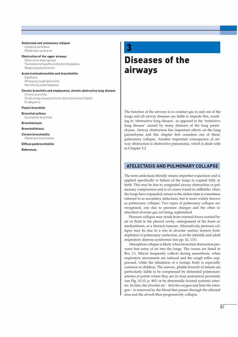
alveolar tissue and overlying pleura is more marked (Fig. 3.1).
In either case, re-inflation is prevented.
3 D I S E A S ES O F T H E A I R WA YS
88
Collateral ventilation
When obstruction to an airway is only partial, permitting inspi-
ration but hindering expiration, air is retained in the affected
area and there is full inflation rather than collapse (see ‘infantile
lobar emphysema’, p. 70). Absorption collapse may also be pre-
vented by collateral ventilation, a process by which one portion
of lung is ventilated through another via the pores of Kohn,Lambert’s canals and other peripheral communications (see
pp. 5, 13). Collateral ventilation is best developed at the acinar
level, being hindered by the interlobular septa and prevented by
interlobar fissures, but because interlobular septa are incom-
plete, it may prevent whole segments undergoing absorption
collapse. However, airflow through the tortuous bypass chan-
nels afforded by many pores of Kohn is poor and collateral
ventilation plays little part in gas exchange.2 Its function is to
maintain inflation of alveoli when their supplying airways are
obstructed by secretions or a foreign body. This is essential to the
cough mechanism, dependent upon which are the expulsion of
the obstructive material and the restoration of bronchial patency.
Box 3.1 Causes of absorption collapse
Intralumenal lesionsMucusForeign body
BroncholithEndobronchial tumour a
Mural lesionsBronchogenic carcinomaSarcoid
Extrinsic lesionsLymph nodes enlarged by metastatic tumour or tuberculosisDistended or aneurysmally dilated arteries
aTumours particularly prone to grow preferentially into the lumen ofan airway include carcinosarcoma, carcinoid, bronchial glandneoplasms, metastases, lymphoma, chondroid hamartoma, papillaryneoplasms, granular cell tumour and amyloid tumour.
Figure 3.1 Chronic pulmonary collapse due to long-standing pleuraleffusion. On the right of the picture pleuropulmonary fibrosis hasdeveloped, preventing the lung from ever expanding.
8/13/2019 Af Ale Cailor Respiratorii
http://slidepdf.com/reader/full/af-ale-cailor-respiratorii 3/24
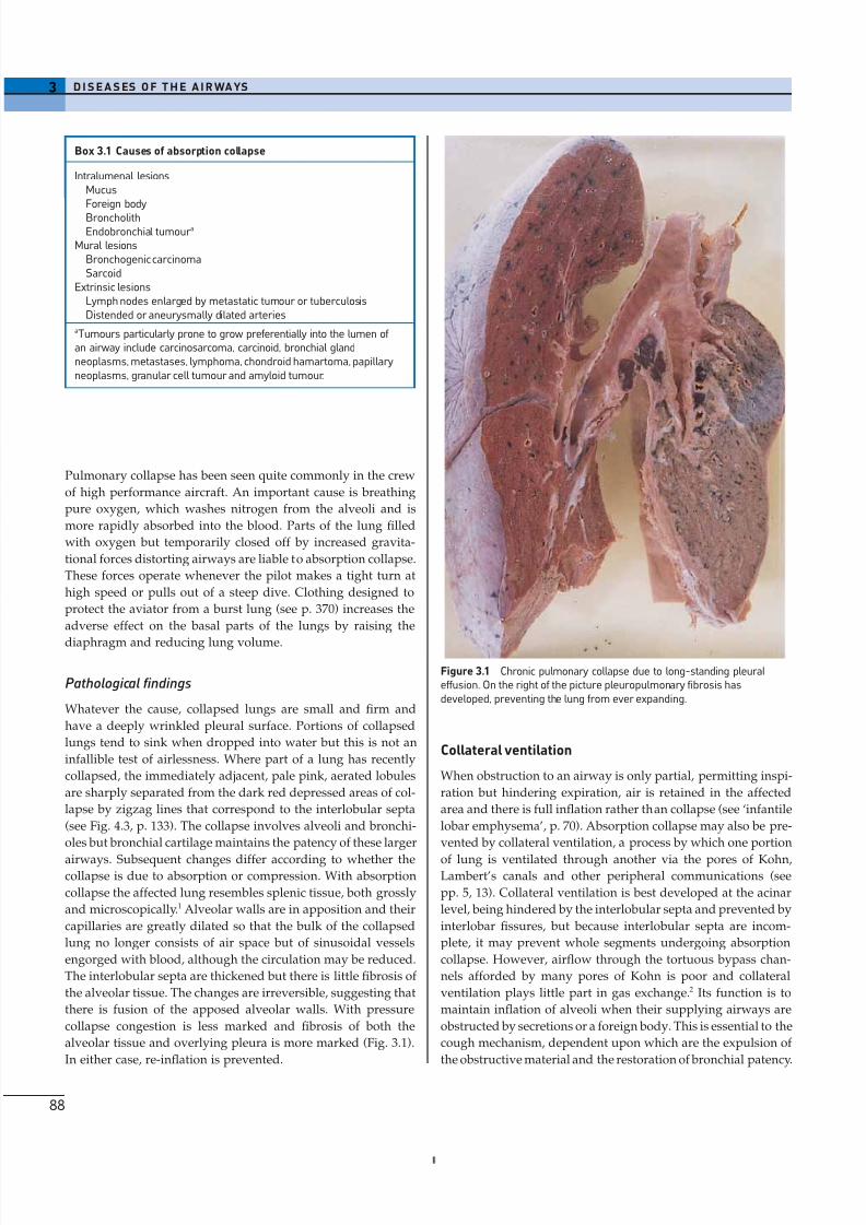
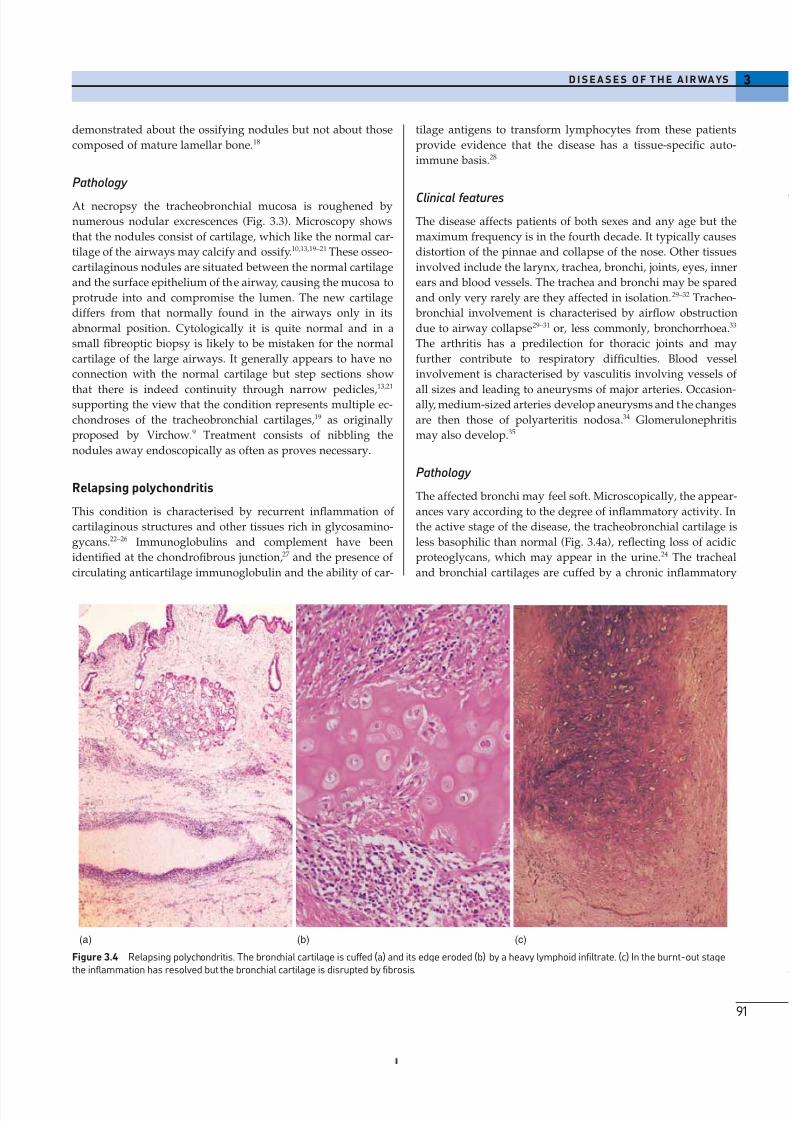
Middle lobe syndrome
The term ‘middle lobe syndrome’ was introduced in 1937 to
describe a condition of chronic or recurrent absorption collapse
of the right middle lobe.3 The collapse was most frequently
caused by tuberculous involvement of lymph nodes compress-ing the right middle lobe bronchus. Although tuberculosis is
less common today, any disease enlarging these lymph nodes
may have the same effect. Predilection for involvement of the
middle lobe was considered to be the result of a combination of
factors: the prominent collar of nodes about its bronchus, the
lymphatic drainage of these nodes being from much of the right
lung and parts of the left and the relatively narrow calibre and
possibly undue compressibility of the middle lobe bronchus. A
further possible factor is the limited capacity for collateral ven-
tilation (see above) within the middle lobe. This stems from the
fact that its two segments have relatively large proportions of
their surfaces covered by pleura and, together with the inferior
segment of the lingula, are the only ones that abut no more than
one other segment.4
Patients with the middle lobe syndrome complain of chronic
cough, haemoptysis, chest pain and dyspnoea, to relieve which,
the diseased lobe may be removed. Pathological changes in the
resected lobe include bronchiectasis, chronic bronchitis and
bronchiolitis, lymphoid hyperplasia, organising pneumonia
and abscess formation, in addition to collapse (Fig. 3.2).5 A
similar syndrome may affect the lingula.
OBSTRUCTION OF THE UPPER AIRWAYS
Obstruction of the upper airways may be complete and cause
rapid asphyxial death, or incomplete, when there is stridor or
wheezing, or distal complications such as obstructive pneumo-
nia may ensue. Foreign bodies are an important cause, espe-
cially in children and edentulous adults. Another important
cause, tumours, is dealt with in Chapter 13. Rare causes include
amyloid tumours (see p. 684) and tracheobronchomalacia (see
pp. 46, 50). In infancy, the airways are unduly pliable and may
be compressed by distended arteries, particularly where the two
are in close contact (see Fig. 10.10). Anomalous arteries may also
compress airways in infancy, as in the vascular sling and ring
syndromes (see Figs 10.11, 10.12, p. 482).
Obstructive sleep apnoea
Obstructive sleep apnoea is characterised by repeated periods
during which the patient stops breathing for 10s or more while
asleep. The patient may not waken, but is repeatedly aroused
so that the quality of the sleep is poor and daytime sleepiness
is consequently excessive.6 Snoring is a common accompani-
ment. The patient is generally obese and it is postulated that
cervical fat pads obstruct the upper airway. Often the patient is
male and excessively fond of alcohol. Other family members
are often similarly affected, possibly because of similarities in
cervicofacial structure. Obstructive sleep apnoea is to be
3D I S E A S E S O F T H E A I R WA YS
89
(b)
Figure 3.2 Middle lobe syndrome. (a) The middle lobe is collapsed andits bronchi are dilated. (b) In this patient, the syndrome was caused by a
broncholith (arrow) blocking the lobar bronchus.
distinguished from a central variety of apnoea known as
Ondine’s curse. (In German legend, the water nymph Ondine,
having been jilted by her mortal lover, took from him all auto-
matic functions, requiring him to remember to breathe. When
he finally fell asleep, he died.) Central apnoea has been encoun-
tered with bulbar poliomyelitis. It is likely that the central syn-
drome results from damage to the medullary CO2 receptor in
which airway patency is maintained but respiratory drive is
weak, especially during sleep.
(a)
8/13/2019 Af Ale Cailor Respiratorii
http://slidepdf.com/reader/full/af-ale-cailor-respiratorii 4/24
The principal problem in obstructive sleep apnoea is the
daytime tiredness, which leads to poor performance at work and a
tendency to fall sleep at inappropriate moments. The consequences
of this can be very serious if, for example, the patient drives.
Charles Dickens was evidently familiar with such individuals,portraying one in his novel ‘The Pickwick Papers’. Such patients
have therefore been termed ‘Pickwickian’, although the Dickensian
character was the ‘fat boy’ rather than Mr Pickwick himself. Defects
in the secretion of testosterone and growth hormone may also be
identified. These are reversible and are probably due to the central
effects of sleep fragmentation and hypoxaemia.
The periods of apnoea result in hypoxaemia, which in turn
causes pulmonary hypertension. The apnoeic episodes are also
accompanied by systemic hypertension and death may be
caused by biventricular cardiac failure. The pulmonary blood
vessels show the usual changes found with hypoxia, principally
hypertrophy of the arterial media (see p. 425). Pulmonary haem-
orrhage and haemosiderosis are further features, possiblyattributable to the left ventricular failure. Pronounced capillary
proliferation resembling capillary haemangiomatosis (see
p. 429) is also described.7
3 D I S E A S ES O F T H E A I R WA YS
90
(a)
(b)
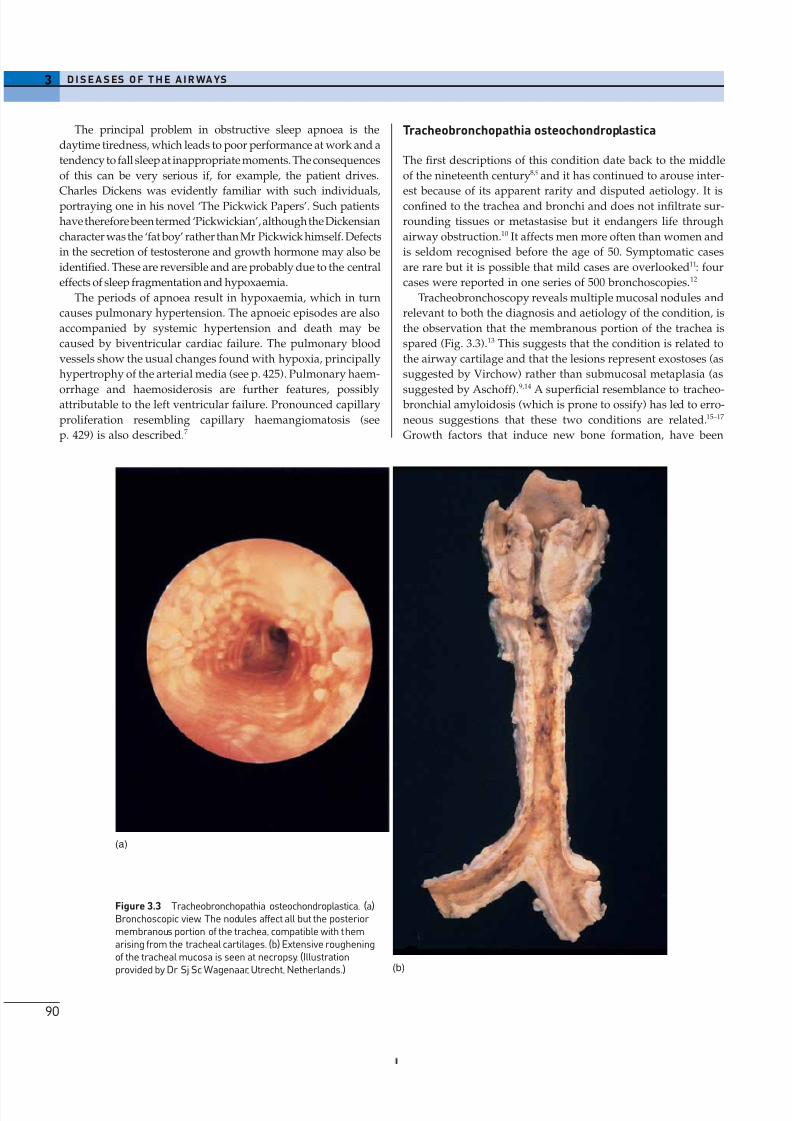
Figure 3.3 Tracheobronchopathia osteochondroplastica. (a)Bronchoscopic view. The nodules affect all but the posterior membranous portion of the trachea, compatible with themarising from the tracheal cartilages. (b) Extensive rougheningof the tracheal mucosa is seen at necropsy. (Illustrationprovided by Dr Sj Sc Wagenaar, Utrecht, Netherlands.)
Tracheobronchopathia osteochondroplastica
The first descriptions of this condition date back to the middle
of the nineteenth century8,9 and it has continued to arouse inter-
est because of its apparent rarity and disputed aetiology. It isconfined to the trachea and bronchi and does not infiltrate sur-
rounding tissues or metastasise but it endangers life through
airway obstruction.10 It affects men more often than women and
is seldom recognised before the age of 50. Symptomatic cases
are rare but it is possible that mild cases are overlooked11: four
cases were reported in one series of 500 bronchoscopies.12
Tracheobronchoscopy reveals multiple mucosal nodules and
relevant to both the diagnosis and aetiology of the condition, is
the observation that the membranous portion of the trachea is
spared (Fig. 3.3).13 This suggests that the condition is related to
the airway cartilage and that the lesions represent exostoses (as
suggested by Virchow) rather than submucosal metaplasia (as
suggested by Aschoff).9,14
A superficial resemblance to tracheo- bronchial amyloidosis (which is prone to ossify) has led to erro-
neous suggestions that these two conditions are related.15–17
Growth factors that induce new bone formation, have been
8/13/2019 Af Ale Cailor Respiratorii
http://slidepdf.com/reader/full/af-ale-cailor-respiratorii 5/24
demonstrated about the ossifying nodules but not about those
composed of mature lamellar bone.18
Pathology
At necropsy the tracheobronchial mucosa is roughened by
numerous nodular excrescences (Fig. 3.3). Microscopy shows
that the nodules consist of cartilage, which like the normal car-
tilage of the airways may calcify and ossify.10,13,19–21 These osseo-
cartilaginous nodules are situated between the normal cartilage
and the surface epithelium of the airway, causing the mucosa to
protrude into and compromise the lumen. The new cartilage
differs from that normally found in the airways only in its
abnormal position. Cytologically it is quite normal and in a
small fibreoptic biopsy is likely to be mistaken for the normal
cartilage of the large airways. It generally appears to have no
connection with the normal cartilage but step sections show
that there is indeed continuity through narrow pedicles,13,21
supporting the view that the condition represents multiple ec-
chondroses of the tracheobronchial cartilages,19 as originally
proposed by Virchow.9 Treatment consists of nibbling the
nodules away endoscopically as often as proves necessary.
Relapsing polychondritis
This condition is characterised by recurrent inflammation of
cartilaginous structures and other tissues rich in glycosamino-
gycans.22–26 Immunoglobulins and complement have been
identified at the chondrofibrous junction,27 and the presence of
circulating anticartilage immunoglobulin and the ability of car-
3D I S E A S E S O F T H E A I R WA YS
91
tilage antigens to transform lymphocytes from these patients
provide evidence that the disease has a tissue-specific auto-
immune basis.28
Clinical features
The disease affects patients of both sexes and any age but the
maximum frequency is in the fourth decade. It typically causes
distortion of the pinnae and collapse of the nose. Other tissues
involved include the larynx, trachea, bronchi, joints, eyes, inner
ears and blood vessels. The trachea and bronchi may be spared
and only very rarely are they affected in isolation. 29–32 Tracheo-
bronchial involvement is characterised by airflow obstruction
due to airway collapse29–31 or, less commonly, bronchorrhoea.33
The arthritis has a predilection for thoracic joints and may
further contribute to respiratory difficulties. Blood vessel
involvement is characterised by vasculitis involving vessels of
all sizes and leading to aneurysms of major arteries. Occasion-ally, medium-sized arteries develop aneurysms and the changes
are then those of polyarteritis nodosa.34 Glomerulonephritis
may also develop.35
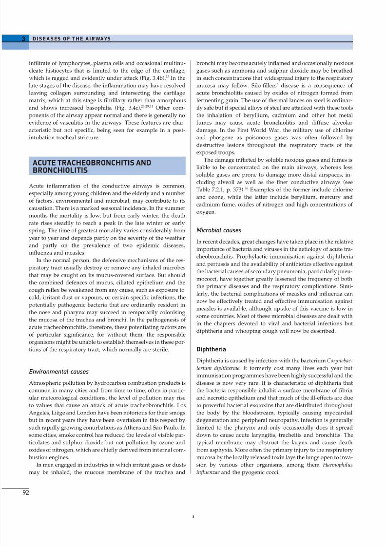
Pathology
The affected bronchi may feel soft. Microscopically, the appear-
ances vary according to the degree of inflammatory activity. In
the active stage of the disease, the tracheobronchial cartilage is
less basophilic than normal (Fig. 3.4a), reflecting loss of acidic
proteoglycans, which may appear in the urine.24 The tracheal
and bronchial cartilages are cuffed by a chronic inflammatory
(a) (b) (c)
Figure 3.4 Relapsing polychondritis. The bronchial cartilage is cuffed (a) and its edge eroded (b) by a heavy lymphoid infiltrate. (c) In the burnt-out stagethe inflammation has resolved but the bronchial cartilage is disrupted by fibrosis.
8/13/2019 Af Ale Cailor Respiratorii
http://slidepdf.com/reader/full/af-ale-cailor-respiratorii 6/24
infiltrate of lymphocytes, plasma cells and occasional multinu-
cleate histiocytes that is limited to the edge of the cartilage,
which is ragged and evidently under attack (Fig. 3.4b).23 In the
late stages of the disease, the inflammation may have resolved
leaving collagen surrounding and intersecting the cartilagematrix, which at this stage is fibrillary rather than amorphous
and shows increased basophilia (Fig. 3.4c).24,29,31 Other com-
ponents of the airway appear normal and there is generally no
evidence of vasculitis in the airways. These features are char-
acteristic but not specific, being seen for example in a post-
intubation tracheal stricture.
ACUTE TRACHEOBRONCHITIS ANDBRONCHIOLITIS
Acute inflammation of the conductive airways is common,
especially among young children and the elderly and a number
of factors, environmental and microbial, may contribute to its
causation. There is a marked seasonal incidence. In the summer
months the mortality is low, but from early winter, the death
rate rises steadily to reach a peak in the late winter or early
spring. The time of greatest mortality varies considerably from
year to year and depends partly on the severity of the weather
and partly on the prevalence of two epidemic diseases,
influenza and measles.
In the normal person, the defensive mechanisms of the res-
piratory tract usually destroy or remove any inhaled microbes
that may be caught on its mucus-covered surface. But should
the combined defences of mucus, ciliated epithelium and the
cough reflex be weakened from any cause, such as exposure to
cold, irritant dust or vapours, or certain specific infections, the
potentially pathogenic bacteria that are ordinarily resident in
the nose and pharynx may succeed in temporarily colonising
the mucosa of the trachea and bronchi. In the pathogenesis of
acute tracheobronchitis, therefore, these potentiating factors are
of particular significance, for without them, the responsible
organisms might be unable to establish themselves in these por-
tions of the respiratory tract, which normally are sterile.
Environmental causes
Atmospheric pollution by hydrocarbon combustion products iscommon in many cities and from time to time, often in partic-
ular meteorological conditions, the level of pollution may rise
to values that cause an attack of acute tracheobronchitis. Los
Angeles, Liège and London have been notorious for their smogs
but in recent years they have been overtaken in this respect by
such rapidly growing conurbations as Athens and Sao Paulo. In
some cities, smoke control has reduced the levels of visible par-
ticulates and sulphur dioxide but not pollution by ozone and
oxides of nitrogen, which are chiefly derived from internal com-
bustion engines.
In men engaged in industries in which irritant gases or dusts
may be inhaled, the mucous membrane of the trachea and
3 D I S E A S ES O F T H E A I R WA YS
92
bronchi may become acutely inflamed and occasionally noxious
gases such as ammonia and sulphur dioxide may be breathed
in such concentrations that widespread injury to the respiratory
mucosa may follow. Silo-fillers’ disease is a consequence of
acute bronchiolitis caused by oxides of nitrogen formed fromfermenting grain. The use of thermal lances on steel is ordinar-
ily safe but if special alloys of steel are attacked with these tools
the inhalation of beryllium, cadmium and other hot metal
fumes may cause acute bronchiolitis and diffuse alveolar
damage. In the First World War, the military use of chlorine
and phosgene as poisonous gases was often followed by
destructive lesions throughout the respiratory tracts of the
exposed troops.
The damage inflicted by soluble noxious gases and fumes is
liable to be concentrated on the main airways, whereas less
soluble gases are prone to damage more distal airspaces, in-
cluding alveoli as well as the finer conductive airways (see
Table 7.2.1, p. 373).36
Examples of the former include chlorineand ozone, while the latter include beryllium, mercury and
cadmium fume, oxides of nitrogen and high concentrations of
oxygen.
Microbial causes
In recent decades, great changes have taken place in the relative
importance of bacteria and viruses in the aetiology of acute tra-
cheobronchitis. Prophylactic immunisation against diphtheria
and pertussis and the availability of antibiotics effective against
the bacterial causes of secondary pneumonia, particularly pneu-
mococci, have together greatly lessened the frequency of both
the primary diseases and the respiratory complications. Simi-larly, the bacterial complications of measles and influenza can
now be effectively treated and effective immunisation against
measles is available, although uptake of this vaccine is low in
some countries. Most of these microbial diseases are dealt with
in the chapters devoted to viral and bacterial infections but
diphtheria and whooping cough will now be described.
Diphtheria
Diphtheria is caused by infection with the bacterium Corynebac-
terium diphtheriae. It formerly cost many lives each year but
immunisation programmes have been highly successful and the
disease is now very rare. It is characteristic of diphtheria thatthe bacteria responsible inhabit a surface membrane of fibrin
and necrotic epithelium and that much of the ill-effects are due
to powerful bacterial exotoxins that are distributed throughout
the body by the bloodstream, typically causing myocardial
degeneration and peripheral neuropathy. Infection is generally
limited to the pharynx and only occasionally does it spread
down to cause acute laryngitis, tracheitis and bronchitis. The
typical membrane may obstruct the larynx and cause death
from asphyxia. More often the primary injury to the respiratory
mucosa by the locally released toxin lays the lungs open to inva-
sion by various other organisms, among them Haemophilus
influenzae and the pyogenic cocci.
8/13/2019 Af Ale Cailor Respiratorii
http://slidepdf.com/reader/full/af-ale-cailor-respiratorii 7/24
Whooping cough (pertussis)
Whooping cough is a highly infectious bacterial disease of
childhood caused by the bacterium Bordetella pertussis. It is
spread by droplet infection. The incubation period is 7–10 days
and a case is infectious from 7 days after exposure to 3 weeksafter the onset of typical paroxysms. An initial catarrhal stage
is the most infectious period. An irritating cough develops and
gradually becomes paroxysmal, which is responsible for the
typical ‘whoop’. Whooping cough may be complicated by bron-
chopneumonia, post-tussive vomiting and cerebral hypoxia,
most commonly in infants under 6 months of age.
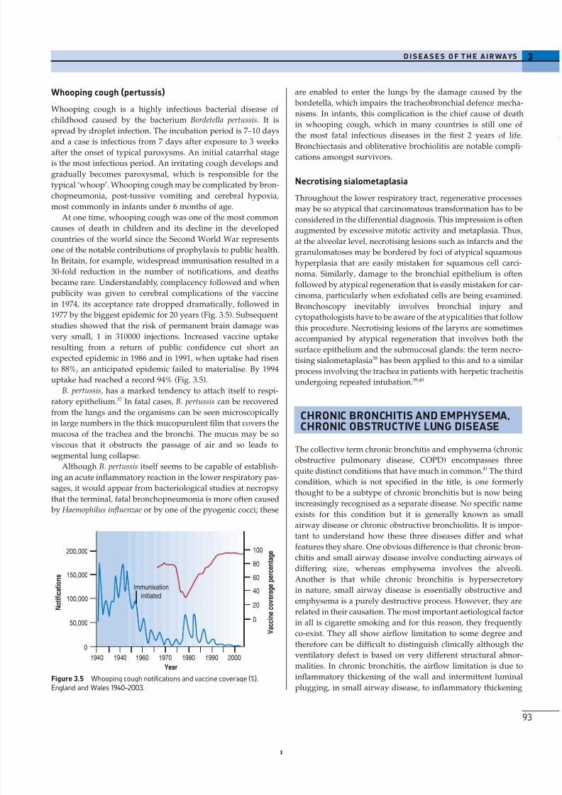
At one time, whooping cough was one of the most common
causes of death in children and its decline in the developed
countries of the world since the Second World War represents
one of the notable contributions of prophylaxis to public health.
In Britain, for example, widespread immunisation resulted in a
30-fold reduction in the number of notifications, and deaths
became rare. Understandably, complacency followed and when
publicity was given to cerebral complications of the vaccine
in 1974, its acceptance rate dropped dramatically, followed in
1977 by the biggest epidemic for 20 years (Fig. 3.5). Subsequent
studies showed that the risk of permanent brain damage was
very small, 1 in 310000 injections. Increased vaccine uptake
resulting from a return of public confidence cut short an
expected epidemic in 1986 and in 1991, when uptake had risen
to 88%, an anticipated epidemic failed to materialise. By 1994
uptake had reached a record 94% (Fig. 3.5).
B. pertussis, has a marked tendency to attach itself to respi-
ratory epithelium.37 In fatal cases, B. pertussis can be recovered
from the lungs and the organisms can be seen microscopically
in large numbers in the thick mucopurulent film that covers the
mucosa of the trachea and the bronchi. The mucus may be so
viscous that it obstructs the passage of air and so leads to
segmental lung collapse.
Although B. pertussis itself seems to be capable of establish-
ing an acute inflammatory reaction in the lower respiratory pas-
sages, it would appear from bacteriological studies at necropsy
that the terminal, fatal bronchopneumonia is more often caused
by Haemophilus influenzae or by one of the pyogenic cocci; these
3D I S E A S E S O F T H E A I R WA YS
93
are enabled to enter the lungs by the damage caused by the
bordetella, which impairs the tracheobronchial defence mecha-
nisms. In infants, this complication is the chief cause of death
in whooping cough, which in many countries is still one of
the most fatal infectious diseases in the first 2 years of life.Bronchiectasis and obliterative brochiolitis are notable compli-
cations amongst survivors.
Necrotising sialometaplasia
Throughout the lower respiratory tract, regenerative processes
may be so atypical that carcinomatous transformation has to be
considered in the differential diagnosis. This impression is often
augmented by excessive mitotic activity and metaplasia. Thus,
at the alveolar level, necrotising lesions such as infarcts and the
granulomatoses may be bordered by foci of atypical squamous
hyperplasia that are easily mistaken for squamous cell carci-
noma. Similarly, damage to the bronchial epithelium is oftenfollowed by atypical regeneration that is easily mistaken for car-
cinoma, particularly when exfoliated cells are being examined.
Bronchoscopy inevitably involves bronchial injury and
cytopathologists have to be aware of the atypicalities that follow
this procedure. Necrotising lesions of the larynx are sometimes
accompanied by atypical regeneration that involves both the
surface epithelium and the submucosal glands: the term necro-
tising sialometaplasia38 has been applied to this and to a similar
process involving the trachea in patients with herpetic tracheitis
undergoing repeated intubation.39,40
CHRONIC BRONCHITIS AND EMPHYSEMA,CHRONIC OBSTRUCTIVE LUNG DISEASE
The collective term chronic bronchitis and emphysema (chronic
obstructive pulmonary disease, COPD) encompasses three
quite distinct conditions that have much in common.41 The third
condition, which is not specified in the title, is one formerly
thought to be a subtype of chronic bronchitis but is now being
increasingly recognised as a separate disease. No specific name
exists for this condition but it is generally known as small
airway disease or chronic obstructive bronchiolitis. It is impor-
tant to understand how these three diseases differ and what
features they share. One obvious difference is that chronic bron-
chitis and small airway disease involve conducting airways of differing size, whereas emphysema involves the alveoli.
Another is that while chronic bronchitis is hypersecretory
in nature, small airway disease is essentially obstructive and
emphysema is a purely destructive process. However, they are
related in their causation. The most important aetiological factor
in all is cigarette smoking and for this reason, they frequently
co-exist. They all show airflow limitation to some degree and
therefore can be difficult to distinguish clinically although the
ventilatory defect is based on very different structural abnor-
malities. In chronic bronchitis, the airflow limitation is due to
inflammatory thickening of the wall and intermittent luminal
plugging, in small airway disease, to inflammatory thickening
50,000
0
100,000
150,000
200,000 100
80
60
40
20
0
1940 1940 1960 1970 1980 1990 2000
Year
N o t i f i c a t i o n s
V a c c i n e c o v e r a g e p e r c e n t a g
e
Immunisation
initiated
Figure 3.5 Whooping cough notifications and vaccine coverage (%),England and Wales 1940–2003.

8/13/2019 Af Ale Cailor Respiratorii
http://slidepdf.com/reader/full/af-ale-cailor-respiratorii 8/24
of the wall and peribronchiolar fibrosis and in emphysema to
premature closure of inherently normal or atrophic airways
because of diminished pulmonary elastic recoil.
The majority of patients with generalised chronic airflow
limitation suffer from both obstructive airway disease andemphysema but a minority of patients have one condition or
another. Two clinical syndromes, types A (‘pink puffer’) and B
(‘blue bloater’), have been described and it is widely believed
that the former indicates emphysema and the latter chronic
bronchitis.42,43 The association of the type A syndrome with
emphysema is fairly well established but the association of type
B with chronic bronchitis is not well substantiated morpholog-
ically. Type A patients show rapid shallow breathing and this
maintains near normal blood gases at the cost of subjective
breathlessness. They are usually thin and because their blood
gases are not severely deranged they tend not to develop poly-
cythaemia or cor pulmonale. Type B patients on the other hand
are hypoxic and therefore suffer from polycythaemia andrepeated bouts of congestive cardiac failure. They are usually
obese and oedematous and have a productive cough but they
are seldom severely breathless. It is important to realise that
most patients with chronic airflow limitation do not fit neatly
into one or other of these types. Nor do these two types reflect
pure bronchitis or pure emphysema.44 The fundamental differ-
ence between type A and type B patients may be in the brain
rather than the lungs: type B patients seem to have a respira-
tory centre that is relatively unresponsive to the usual stimuli,
an abnormality that may be genetically determined.
Chronic bronchitisDefinition
Chronic bronchitis is defined in clinical terms as a persistent or
recurrent excess of secretion in the bronchial tree on most days
for at least 3 months in the year, over at least 2 years.45 The secre-
tions of the normal human respiratory tract are believed to total
less than 100ml in 24h, all of which is swallowed without
conscious need to clear the throat or cough so that the normal
person produces no sputum. The diagnosis of chronic bron-
chitis may be made only when other conditions that cause
expectoration, such as tuberculosis and bronchiectasis, have
been excluded.
Chronic bronchitis was formerly subdivided into simplemucoid bronchitis, mucopurulent bronchitis and obstructive
bronchitis.46 It was widely thought that these subdivisions rep-
resented successive phases of the disease but a strong counter-
argument to this was advanced by British epidemiologists.47
These workers showed that while simple mucoid bronchitis
progresses to the mucopurulent variety, this does not progress
in turn to the obstructive form. In line with this, neither
bronchial gland size nor sputum production are significantly
related to airflow limitation.48 The view that the development
of obstructive bronchitis is independent of the repeated respi-
ratory infections that characterise mucopurulent bronchitis has
been challenged,49 but the obstructive form of the disease is now
3 D I S E A S ES O F T H E A I R WA YS
94
nevertheless widely recognised as a separate condition: small
airway disease, which is dealt with below.
Aetiology
Chronic bronchitis affects mainly the middle-aged and elderlyand is more common in men; cigarette smoking is by far the
most important cause.50–52 The influence of cigarette smoke often
begins in infancy when the child is exposed passively to
parental cigarette smoke. This is generally augmented by active
cigarette smoking when the child emulates parents or school-
mates and acquires the habit, often becoming addicted for life.
However, as with lung cancer, many indulge in smoking with
impunity, indicating that susceptibility to disease varies con-
siderably,41 probably reflecting genetic differences in the control
of such factors as the balance of helper and cytotoxic T-
lymphocytes.53 Marijuana smoke is likely to be recognised as a
further aetiological agent as it has similar morphological effects
on the airways as tobacco smoke.54
Other factors contributing to chronic bronchitis include
general air pollution, which accounts for the higher prevalence
of the disease in urban communities, occupational dust expo-
sure,55 fog and a damp and cold climate. The morbidity from
the disease rises every winter and remains high throughout the
colder, damper months. The occurrence of fog, especially the
form known as smog in which the water vapour becomes
heavily contaminated with smoke and sulphurous gases, causes
a prompt increase in both morbidity and mortality among older
people. The heavy 4-day smog in London in 1952 is believed to
have precipitated 4000 deaths.
Infections by respiratory viruses and bacteria are also of
importance in both initiating and promoting chronic bron-
chitis.56 Some patients may recall a liability in their earlier
years for head colds to go to their chest. 57 Relatives are often
similarly affected. An increased frequency of respiratory infec-
tion in childhood has been identified in adults with chronic
bronchitis.58
These various irritants initiate mucus secretion by a combi-
nation of direct action on the mucous cells and nervous reflexes
involving sensory nerve endings in the airway epithelium and
both local peptidergic and spinal cholinergic pathways. Upreg-
ulation of the mucin (MUC) genes is involved and epidermal
growth factor is a key mediator in the mucous cell hyperplasia.
Clinical features
The excessive bronchial secretion inherent in the definition of
chronic bronchitis is manifest as sputum. This is typically
mucoid and white but the disease is marked by episodes of
acute bronchitis when the sputum becomes purulent and
yellow. Later the sputum may become purulent continuously;
it accumulates in the bronchi during sleep and causes severe
obstruction of the airways until it is coughed up in the morning.
While a change from white to yellow sputum usually signifies
infection it should be noted that large numbers of eosinophils
also render the sputum yellow, a potential pitfall in the clinical
distinction of bronchitis and asthma.
8/13/2019 Af Ale Cailor Respiratorii
http://slidepdf.com/reader/full/af-ale-cailor-respiratorii 9/24
Microbiological examination of the sputum in chronic bron-
chitis has shown that the most frequent and important
pathogens are Haemophilus influenzae, Streptococcus pneumoniae,
Branhamella catarrhalis and Chlamydia pneumoniae.59–63 Purulent
sputum usually contains one or more of these organisms inabundance; they tend to disappear after antimicrobial therapy
when the sputum becomes mucoid again.
The productive cough appears at first only in the winter
months. Later, it is present all through the year, characteristi-
cally with acute exacerbations in winter that are usually pre-
cipitated by a viral infection.64,65
Morbidity and mortality
Chronic obstructive lung disease is a major cause of death
worldwide.52 However, death comes many years after the onset
of the disease and it is therefore also a major cause of sickness
and incapacity for work. The social gradient of the disease is
steep, for the death rate in the poorest section of the population
is some five times that in the most prosperous. Death in chronic
bronchitis is often due to bronchopneumonia. There is also a
4- to 5-fold increased risk of lung cancer in patients with
obstructive lung disease, as compared with controls matched
for cigarette smoking.66,67
Morbid anatomy
When the lungs of a patient with chronic bronchitis are dis-
sected at necropsy, the exposed bronchi, especially those in the
lower lobes, are typically filled with a mixture of mucus and
pus. When the purulent material is washed away from bronchi
that have been opened longitudinally, the underlying mucousmembrane is seen to be a dusky red. The calibre of the main
bronchi may remain unchanged but distal bronchi characteris-
tically are slightly dilated; when they are opened with fine
scissors, the dilation is found to reach almost to the pleura
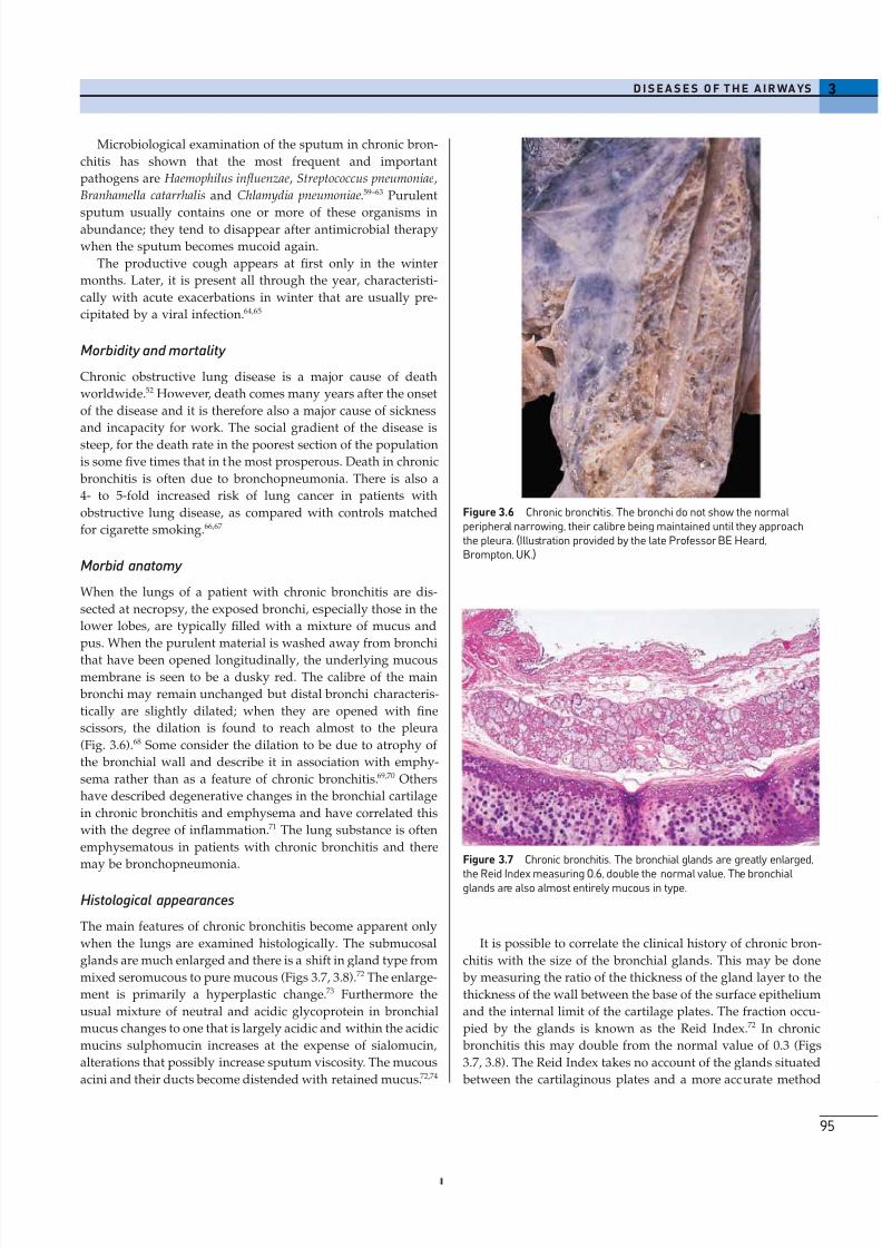
(Fig. 3.6).68 Some consider the dilation to be due to atrophy of
the bronchial wall and describe it in association with emphy-
sema rather than as a feature of chronic bronchitis.69,70 Others
have described degenerative changes in the bronchial cartilage
in chronic bronchitis and emphysema and have correlated this
with the degree of inflammation.71 The lung substance is often
emphysematous in patients with chronic bronchitis and there
may be bronchopneumonia.
Histological appearances
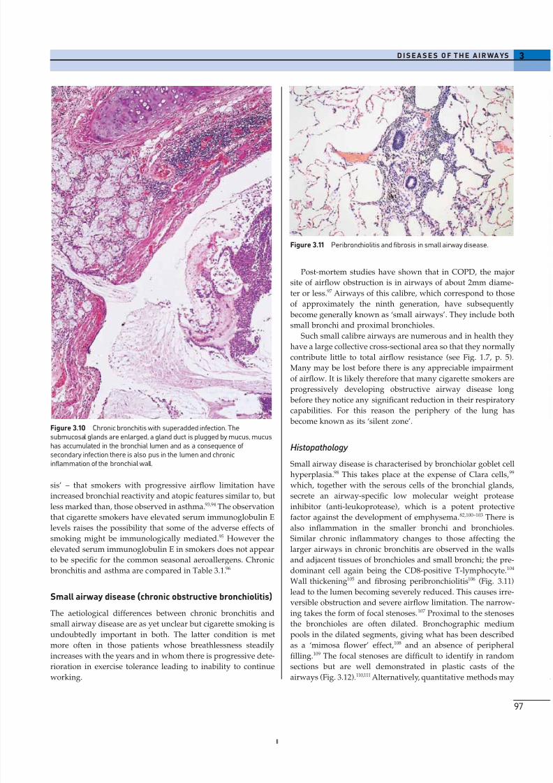
The main features of chronic bronchitis become apparent only
when the lungs are examined histologically. The submucosal
glands are much enlarged and there is a shift in gland type from
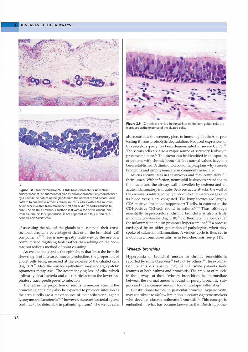
mixed seromucous to pure mucous (Figs 3.7, 3.8).72 The enlarge-
ment is primarily a hyperplastic change.73 Furthermore the
usual mixture of neutral and acidic glycoprotein in bronchial
mucus changes to one that is largely acidic and within the acidic
mucins sulphomucin increases at the expense of sialomucin,
alterations that possibly increase sputum viscosity. The mucous
acini and their ducts become distended with retained mucus.72,74
3D I S E A S E S O F T H E A I R WA YS
95
It is possible to correlate the clinical history of chronic bron-
chitis with the size of the bronchial glands. This may be done
by measuring the ratio of the thickness of the gland layer to the
thickness of the wall between the base of the surface epithelium
and the internal limit of the cartilage plates. The fraction occu-
pied by the glands is known as the Reid Index.72 In chronic
bronchitis this may double from the normal value of 0.3 (Figs
3.7, 3.8). The Reid Index takes no account of the glands situated
between the cartilaginous plates and a more accurate method
Figure 3.6 Chronic bronchitis. The bronchi do not show the normalperipheral narrowing, their calibre being maintained until they approachthe pleura. (Illustration provided by the late Professor BE Heard,Brompton, UK.)
Figure 3.7 Chronic bronchitis. The bronchial glands are greatly enlarged,
the Reid Index measuring 0.6, double the normal value. The bronchialglands are also almost entirely mucous in type.
8/13/2019 Af Ale Cailor Respiratorii
http://slidepdf.com/reader/full/af-ale-cailor-respiratorii 10/24
of assessing the size of the glands is to estimate their cross-
sectional area as a percentage of that of all the bronchial wall
components.75,76 This is now greatly facilitated by the use of a
computerised digitising tablet rather than relying on the accu-
rate but tedious method of point counting.As well as the glands, the epithelium that lines the bronchi
shows signs of increased mucus production, the proportion of
goblet cells being increased at the expense of the ciliated cells
(Fig. 3.9).77 Also, the surface epithelium may undergo patchy
squamous metaplasia. The accompanying loss of cilia, which
ordinarily clear bacteria and dust particles from the lower res-
piratory tract, predisposes to infection.
The fall in the proportion of serous to mucous acini in the
bronchial glands may also be expected to promote infection as
the serous cells are a major source of the antibacterial agents
lysozyme and lactoferrin78,79; however, these antibacterial agents
continue to be detectable in patients’ sputum.80 The serous cells
3 D I S E A S ES O F T H E A I R WA YS
96
also contribute the secretory piece to immunoglobulin A, so pro-
tecting it from proteolytic degradation. Reduced expression of
this secretory piece has been demonstrated in severe COPD.81
The serous cells are also a major source of secretory leukocyte
protease inhibitor.82 This factor can be identified in the sputum
of patients with chronic bronchitis but normal values have not
been established. A diminution could help explain why chronic
bronchitis and emphysema are so commonly associated.
Mucus accumulates in the airways and may completely fill
their lumen. With infection, neutrophil leukocytes are added to
the mucus and the airway wall is swollen by oedema and anacute inflammatory infiltrate. Between acute attacks, the wall of
the airways is infiltrated by lymphocytes and macrophages and
its blood vessels are congested. The lymphocytes are largely
CD8-positive (cytotoxic/suppressor) T cells, in contrast to the
CD4-positive Th2-cells found in asthma.83–85 Thus, although
essentially hypersecretory, chronic bronchitis is also a truly
inflammatory disease (Fig. 3.10).86 Furthermore, it appears that
the inflammation in turn promotes hypersecretion,87,88 a process
envisaged by an older generation of pathologists when they
spoke of catarrhal inflammation. A vicious cycle is thus set in
motion in chronic bronchitis, as in bronchiectasis (see p. 115).
‘Wheezy’ bronchitis
Hyperplasia of bronchial muscle in chronic bronchitis is
reported by some observers89 but not by others.90 The explana-
tion for this discrepancy may be that some patients have
features of both asthma and bronchitis. The amount of muscle
in the airways of these ‘wheezy bronchitics’ is intermediate
between the normal amounts found in purely bronchitic sub-
jects and the increased amount found in atopic asthmatics.91
Constitutional factors, in particular bronchial hyperactivity,
may contribute to airflow limitation in certain cigarette smokers
who develop ‘chronic asthmatic bronchitis’.92 This concept is
embodied in what has become known as the ‘Dutch hypothe-
(a)
(b)Figure 3.8 (a) Normal bronchus. (b) Chronic bronchitis. As well asenlargement of the submucosal glands, chronic bronchitis is characterisedby a shift in the nature of the glands from the normal mixed seromucouspattern to one that is almost entirely mucous, while within the mucousacini there is a shift from mixed neutral and acidic (red/blue) mucus topurely acidic (blue) mucus. A further shift within the acidic mucus, onefrom sialomucin to sulphomucin, is not apparent with this Alcian blue-periodic acid Schiff stain.
Figure 3.9 Chronic bronchitis. In the surface epithelium, goblet cells areincreased at the expense of the ciliated cells.
8/13/2019 Af Ale Cailor Respiratorii
http://slidepdf.com/reader/full/af-ale-cailor-respiratorii 11/24
sis’ – that smokers with progressive airflow limitation have
increased bronchial reactivity and atopic features similar to, but
less marked than, those observed in asthma.93,94 The observation
that cigarette smokers have elevated serum immunoglobulin E
levels raises the possibility that some of the adverse effects of
smoking might be immunologically mediated.95 However the
elevated serum immunoglobulin E in smokers does not appearto be specific for the common seasonal aeroallergens. Chronic
bronchitis and asthma are compared in Table 3.1.96
Small airway disease (chronic obstructive bronchiolitis)
The aetiological differences between chronic bronchitis and
small airway disease are as yet unclear but cigarette smoking is
undoubtedly important in both. The latter condition is met
more often in those patients whose breathlessness steadily
increases with the years and in whom there is progressive dete-
rioration in exercise tolerance leading to inability to continue
working.
3D I S E A S E S O F T H E A I R WA YS
97
Post-mortem studies have shown that in COPD, the majorsite of airflow obstruction is in airways of about 2mm diame-
ter or less.97 Airways of this calibre, which correspond to those
of approximately the ninth generation, have subsequently
become generally known as ‘small airways’. They include both
small bronchi and proximal bronchioles.
Such small calibre airways are numerous and in health they
have a large collective cross-sectional area so that they normally
contribute little to total airflow resistance (see Fig. 1.7, p. 5).
Many may be lost before there is any appreciable impairment
of airflow. It is likely therefore that many cigarette smokers are
progressively developing obstructive airway disease long
before they notice any significant reduction in their respiratory
capabilities. For this reason the periphery of the lung has become known as its ‘silent zone’.
Histopathology
Small airway disease is characterised by bronchiolar goblet cell
hyperplasia.98 This takes place at the expense of Clara cells,99
which, together with the serous cells of the bronchial glands,
secrete an airway-specific low molecular weight protease
inhibitor (anti-leukoprotease), which is a potent protective
factor against the development of emphysema.82,100–103 There is
also inflammation in the smaller bronchi and bronchioles.
Similar chronic inflammatory changes to those affecting the
larger airways in chronic bronchitis are observed in the wallsand adjacent tissues of bronchioles and small bronchi; the pre-
dominant cell again being the CD8-positive T-lymphocyte.104
Wall thickening105 and fibrosing peribronchiolitis106 (Fig. 3.11)
lead to the lumen becoming severely reduced. This causes irre-
versible obstruction and severe airflow limitation. The narrow-
ing takes the form of focal stenoses.107 Proximal to the stenoses
the bronchioles are often dilated. Bronchographic medium
pools in the dilated segments, giving what has been described
as a ‘mimosa flower’ effect,108 and an absence of peripheral
filling.109 The focal stenoses are difficult to identify in random
sections but are well demonstrated in plastic casts of the
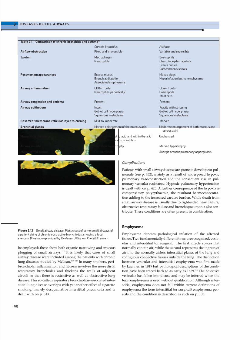
airways (Fig. 3.12).110,111 Alternatively, quantitative methods may
Figure 3.10 Chronic bronchitis with superadded infection. Thesubmucosal glands are enlarged, a gland duct is plugged by mucus, mucushas accumulated in the bronchial lumen and as a consequence of secondary infection there is also pus in the lumen and chronicinflammation of the bronchial wall.
Figure 3.11 Peribronchiolitis and fibrosis in small airway disease.
8/13/2019 Af Ale Cailor Respiratorii
http://slidepdf.com/reader/full/af-ale-cailor-respiratorii 12/24
be employed; these show both organic narrowing and mucous
plugging of small airways.112 It is likely that cases of small
airway disease were included among the patients with chronic
lung diseases studied by McLean.113–115 In many smokers, peri-
bronchiolar inflammation and fibrosis involves the more distal
respiratory bronchioles and thickens the walls of adjacent
alveoli so that there is restrictive as well as obstructive lung
disease. This so-called respiratory bronchiolitis-associated inter-
stitial lung disease overlaps with yet another effect of cigarette
smoking, namely desquamative interstitial pneumonia and is
dealt with on p. 313.
3 D I S E A S ES O F T H E A I R WA YS
98
Complications
Patients with small airway disease are prone to develop cor pul-
monale (see p. 432), mainly as a result of widespread hypoxic
pulmonary vasoconstriction and the consequent rise in pul-monary vascular resistance. Hypoxic pulmonary hypertension
is dealt with on p. 425. A further consequence of the hypoxia is
compensatory polycythaemia, the resultant haemoconcentra-
tion adding to the increased cardiac burden. While death from
small airway disease is usually due to right-sided heart failure,
obstructive respiratory failure and bronchopneumonia also con-
tribute. These conditions are often present in combination.
Emphysema
Emphysema denotes pathological inflation of the affected
tissue. Two fundamentally different forms are recognised, vesic-ular and interstitial (or surgical). The first affects spaces that
normally contain air, while the second represents the ingress of
air into the normally airless interstitial planes of the lung and
contiguous connective tissues outside the lung. The distinction
between vesicular and interstitial emphysema was first made
by Laennec in 1819 but pathological descriptions of the condi-
tion have been traced back to as early as 1679.116 The adjective
vesicular has fallen into disuse and may be inferred when the
term emphysema is used without qualification. Although inter-
stitial emphysema does not fall within current definitions of
emphysema the term interstitial (or surgical) emphysema per-
sists and the condition is described as such on p. 105.
Table 3.1 Comparison of chronic bronchitis and asthma96
Chronic bronchitis Asthma
Airflow obstruction Fixed and irreversible Variable and reversible
Sputum Macrophages EosinophilsNeutrophils Charcot–Leyden crystals
Creola bodiesCurschmann’s spirals
Postmortem appearances Excess mucus Mucus plugsBronchial dilatation Hyperinflation but no emphysemaAssociated emphysema
Airway inflammation CD8+ T cells CD4+ T cellsNeutrophils periodically Eosinophils
Mast cells
Airway congestion and oedema Present Present
Airway epithelium Intact Fragile with strippingGoblet cell hyperplasia Goblet cell hyperplasiaSquamous metaplasia Squamous metaplasia
Basement membrane reticular layer thickening Mild-to-moderate Marked
Bronchial glands Marked enlargement of the mucous acini Moderate enlargement of both mucous andserous acini
Mucin histochemistry Shift from neutral to acid and within the acid Unchangedmucins from sialo- to sulpho-
Airway muscle May show hypertrophy Marked hypertrophy
Major complications Cor pulmonale Allergic bronchopulmonary aspergillosis
Figure 3.12 Small airway disease. Plastic cast of some small airways of a patient dying of chronic obstructive bronchiolitis, showing a focal
stenosis. (Illustration provided by Professor J Bignon, Creteil, France.)

8/13/2019 Af Ale Cailor Respiratorii
http://slidepdf.com/reader/full/af-ale-cailor-respiratorii 13/24
Vesicular emphysema is a common condition. A consecutive
series of 50 male necropsies in London identified emphysema
in more than trace amounts in 37, although few of the patients
had respiratory symptoms.117 Similar findings have been
reported in other English cities.
118
Airways may be normal in emphysema. Alternatively, they
may be atrophic and dilated but prone to collapse prema-
turely,69,70 unless there is also chronic bronchitis with its charac-
teristic thickening of the airway wall by glandular hyperplasia
and inflammatory oedema.
Definition
Emphysema was defined in 1959 as ‘a condition of the lung
characterised by increase beyond the normal in the size of air
spaces distal to the terminal bronchiole either from dilatation
or from destruction of their walls’.45 Subsequently, this was
modified by excluding purely distensive forms of pulmonary
enlargement so that the definition became: ‘an abnormal
increase in the size of air spaces beyond the terminal bronchi-
oles with destruction of air space walls’.119 However, in some
patients, the destruction is secondary to scarring and it has been
suggested that this type of airspace enlargement should also be
excluded from the definition. The American Thoracic Society
accepted this recommendation and adopted the following def-
inition: ‘abnormal, permanent enlargement of the airspaces
distal to the terminal bronchioles, accompanied by destruction
of their walls and without obvious fibrosis’.120,121 The exclusion
of fibrosis is unfortunate for two reasons. First, the term scar
emphysema is a useful one and second, those forms of emphy-
sema that are not secondary to scarring do entail some degree
of fibrosis, albeit slight.122–124
Pathology
Early emphysematous changes can only be detected micro-
scopically. These include an increase in the size and number of
fenestrae (pores of Kohn) in the alveolar walls.125 When the
destruction is moderate in degree, there is loss of alveolar walls,
resulting in fewer alveolar attachments to bronchioles and
consequent premature closure of these airways on expira-
tion.44,126–128 Quantitation of the microscopic changes in the lung
substance can best be achieved by the application of an image
analyser set to calculate factors such as mean linear inter-
cept129,130 or the airspace wall surface area per unit volume. 131,132
More severe changes are characterised by complete loss of most
of the wall of the airspaces, bronchiolar as well as alveolar,
leaving only a network of blood vessels and some interlobular
septa.
These gross changes are better appreciated by the macro-
scopic study of whole lung slices rather than microscopy. If the
lungs are fixed by distension with aqueous formalin at a pres-
sure of 25–30cm of water before slicing, the emphysema can be
appreciated much better than in the collapsed fresh lung. Fixa-
tion overnight is adequate and if time presses a few hours is
beneficial. If the fixed slices are impregnated with barium
sulphate, deficiencies in the lung substance are highlighted and
3D I S E A S E S O F T H E A I R WA YS
99
the amount of destruction and the type of emphysema can be
better appreciated.133 Barium sulphate impregnation is simply
achieved by gently squeezing a slice of lung in a saturated solu-
tion of sodium sulphate and then immersing it in one of bariumnitrate. Paper-mounted whole lung sections can be prepared if
a permanent record is desired.134 Various ways of quantitating
the gross changes have been recommended135 but none is as
accurate or as easy as computerised image analysis.136
Several morphological types of emphysema are distin-
guished according to the part of the acinus that is affected, as
observed in whole lung slices or paper-mounted whole lung
sections. Three of these types, centriacinar, paraseptal and
panacinar are illustrated diagrammatically in Figure 3.13. The
fourth type, scar or irregular emphysema, bears no relation to
the acinar architecture of the lung.
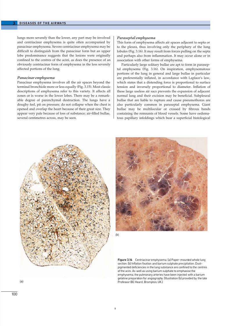
Centriacinar emphysemaThis form of emphysema is characterised by focal lesions con-
fined to the centres of the acini (Fig. 3.14). They are often pig-
mented with dust. The changes are more marked in the upper
lobes, a feature that has been attributed to the greater gravita-
tional forces there, consequent upon our upright posture and
also upon the support afforded to the lower lobes by the
diaphragm. Spaces that exceed 1cm in size are known as bullae
and may be seen in severe cases. The alveolar walls are lost, only
some pulmonary vessels survive to cross the spaces as seem-
ingly bare strands radiating outward from their parent arteries
to supply the alveoli of the periphery of the acini (Fig. 3.14).
Although centriacinar emphysema affects the upper lobes of the
Pleura or
septum
Bronchiolitis
TBTB
TB
TB
AD
AD
AD
AD
AD
AD
AD
AD
AD
AS
AS
AS
ASAS
AS
AS
AS
AS
AS
AS
AS
AS
AS
AS
AS
RB1 RB1
RB1
RB1
RB2
RB2
RB2
RB2
RB2
RB2
RB3
RB3
RB3
RB3
RB3
RB3
RB3
RB3
RB3
RB3
RB3
RB3
Figure 3.13 Morphological types of emphysema in relation to the acinar architecture of the lung. Upper left: The normal acinus. Upper right:
Centriacinar emphysema, in which third order respiratory bronchioles arepredominantly involved. Lower left: Panacinar emphysema in which thereis destructive enlargement of all airspaces distal to the terminal bronchioleand the acinus is affected uniformly. Lower right: Paraseptal emphysema.TB, terminal bronchiole; RB, three orders of respiratory bronchioles; AD,alveolar duct; AS, alveolar sac. (For simplicity, only one order of alveolar duct and one of alveolar sac are drawn and they are not to scale.)
8/13/2019 Af Ale Cailor Respiratorii
http://slidepdf.com/reader/full/af-ale-cailor-respiratorii 14/24
3 D I S E A S ES O F T H E A I R WA YS
100
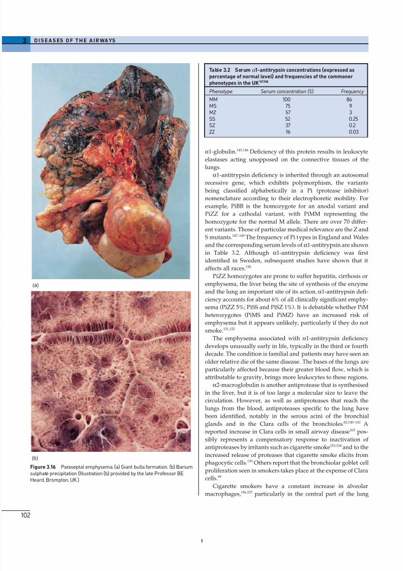
Paraseptal emphysemaThis form of emphysema affects air spaces adjacent to septa or
to the pleura, thus involving only the periphery of the lung
lobules (Fig. 3.16). It may result from forces pulling on the septa
and perhaps also from inflammation. It may occur alone or inassociation with other forms of emphysema.
Particularly large solitary bullae are apt to form in parasep-
tal emphysema (Fig. 3.16). On inspiration, emphysematous
portions of the lung in general and large bullae in particular
are preferentially inflated, in accordance with Laplace’s law,
which states that a distending force is proportional to surface
tension and inversely proportional to diameter. Inflation of
these large useless air sacs prevents the expansion of adjacent
normal lung and their excision may be beneficial. Subpleural
bullae that are liable to rupture and cause pneumothorax are
also particularly common in paraseptal emphysema. Giant
bullae may be multilocular or crossed by fibrous bands
containing the remnants of blood vessels. Some have oedema-tous papillary infoldings which bear a superficial histological
(b)
(a)
Figure 3.14 Centriacinar emphysema. (a) Paper-mounted whole lungsection. (b) Inflation fixation and barium sulphate precipitation. Dust-pigmented deficiencies in the lung substance are confined to the centresof the acini. As well as using barium sulphate to emphasise theemphysema, the pulmonary arteries have been injected with a bariumgelatine preparation for angiography. (Illustration (b) provided by the lateProfessor BE Heard, Brompton, UK.)
lungs more severely than the lower, any part may be involved
and centriacinar emphysema is quite often accompanied by
panacinar emphysema. Severe centriacinar emphysema may be
difficult to distinguish from the panacinar form but an upper
lobe predominance suggests that the lesions were originallyconfined to the centres of the acini, as does the presence of an
obviously centriacinar form of emphysema in the less severely
affected portions of the lung.
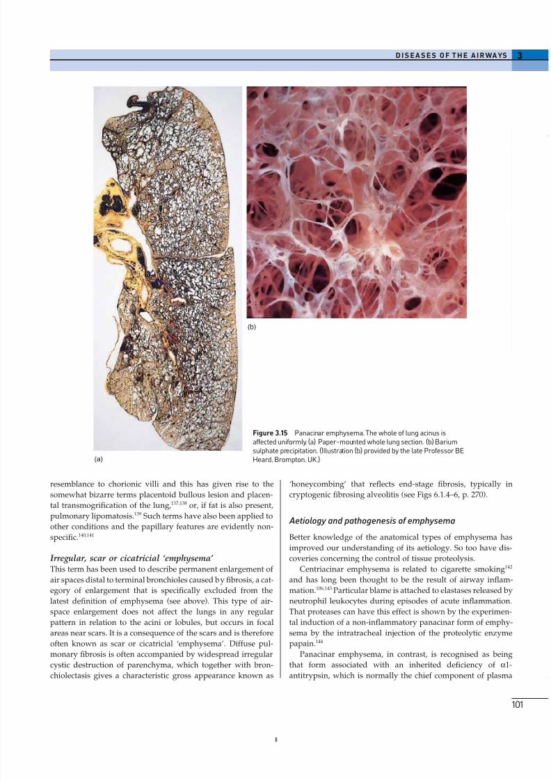
Panacinar emphysemaPanacinar emphysema involves all the air spaces beyond the
terminal bronchiole more or less equally (Fig. 3.15). Most classic
descriptions of emphysema refer to this variety. It affects all
zones or is worse in the lower lobes. There may be a remark-
able degree of parenchymal destruction. The lungs have a
doughy feel, pit on pressure, do not collapse when the chest is
opened and overlap the heart because of their great size. They
appear very pale because of loss of substance; air-filled bullae,several centimetres across, may be seen.
8/13/2019 Af Ale Cailor Respiratorii
http://slidepdf.com/reader/full/af-ale-cailor-respiratorii 15/24
resemblance to chorionic villi and this has given rise to the
somewhat bizarre terms placentoid bullous lesion and placen-
tal transmogrification of the lung,137,138 or, if fat is also present,
pulmonary lipomatosis.139 Such terms have also been applied to
other conditions and the papillary features are evidently non-
specific.140,141
Irregular, scar or cicatricial ‘emphysema’ This term has been used to describe permanent enlargement of
air spaces distal to terminal bronchioles caused by fibrosis, a cat-
egory of enlargement that is specifically excluded from the
latest definition of emphysema (see above). This type of air-
space enlargement does not affect the lungs in any regular
pattern in relation to the acini or lobules, but occurs in focal
areas near scars. It is a consequence of the scars and is therefore
often known as scar or cicatricial ‘emphysema’. Diffuse pul-
monary fibrosis is often accompanied by widespread irregular
cystic destruction of parenchyma, which together with bron-
chiolectasis gives a characteristic gross appearance known as
3D I S E A S E S O F T H E A I R WA YS
101
‘honeycombing’ that reflects end-stage fibrosis, typically in
cryptogenic fibrosing alveolitis (see Figs 6.1.4–6, p. 270).
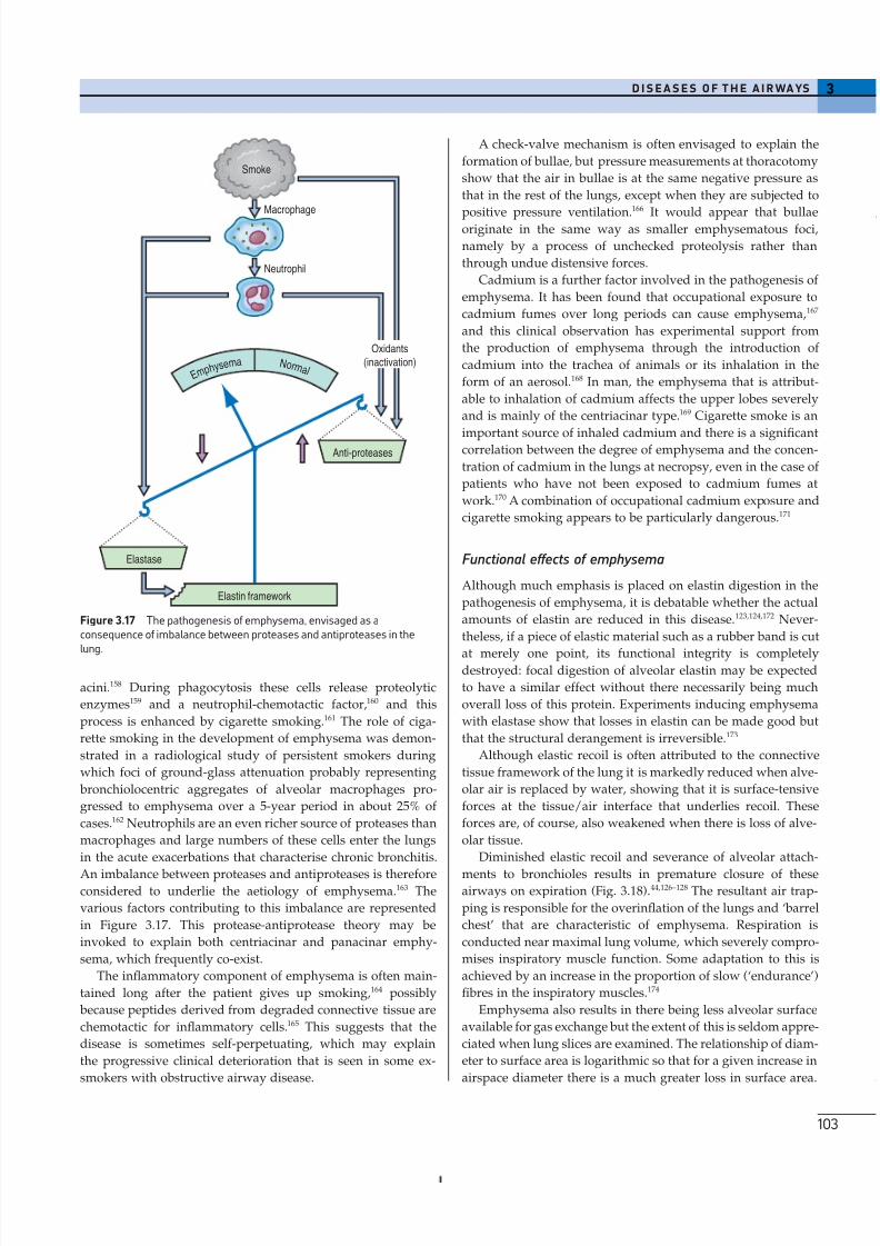
Aetiology and pathogenesis of emphysema
Better knowledge of the anatomical types of emphysema has
improved our understanding of its aetiology. So too have dis-coveries concerning the control of tissue proteolysis.
Centriacinar emphysema is related to cigarette smoking142
and has long been thought to be the result of airway inflam-
mation.106,143 Particular blame is attached to elastases released by
neutrophil leukocytes during episodes of acute inflammation.
That proteases can have this effect is shown by the experimen-
tal induction of a non-inflammatory panacinar form of emphy-
sema by the intratracheal injection of the proteolytic enzyme
papain.144
Panacinar emphysema, in contrast, is recognised as being
that form associated with an inherited deficiency of a1-
antitrypsin, which is normally the chief component of plasma
(a)
(b)
Figure 3.15 Panacinar emphysema. The whole of lung acinus isaffected uniformly. (a) Paper-mounted whole lung section. (b) Bariumsulphate precipitation. (Illustration (b) provided by the late Professor BEHeard, Brompton, UK.)
8/13/2019 Af Ale Cailor Respiratorii
http://slidepdf.com/reader/full/af-ale-cailor-respiratorii 16/24
3 D I S E A S ES O F T H E A I R WA YS
102
a1-globulin.145,146 Deficiency of this protein results in leukocyte
elastases acting unopposed on the connective tissues of the
lungs.
a1-antitrypsin deficiency is inherited through an autosomal
recessive gene, which exhibits polymorphism, the variants
being classified alphabetically in a Pi (protease inhibitor)
nomenclature according to their electrophoretic mobility. Forexample, PiBB is the homozygote for an anodal variant and
PiZZ for a cathodal variant, with PiMM representing the
homozygote for the normal M allele. There are over 70 differ-
ent variants. Those of particular medical relevance are the Z and
S mutants.147–149 The frequency of Pi types in England and Wales
and the corresponding serum levels of a1-antitrypsin are shown
in Table 3.2. Although a1-antitrypsin deficiency was first
identified in Sweden, subsequent studies have shown that it
affects all races.150
PiZZ homozygotes are prone to suffer hepatitis, cirrhosis or
emphysema, the liver being the site of synthesis of the enzyme
and the lung an important site of its action. a1-antitrypsin defi-
ciency accounts for about 6% of all clinically significant emphy-sema (PiZZ 5%; PiSS and PiSZ 1%). It is debatable whether PiM
heterozygotes (PiMS and PiMZ) have an increased risk of
emphysema but it appears unlikely, particularly if they do not
smoke.151,152
The emphysema associated with a1-antitrypsin deficiency
develops unusually early in life, typically in the third or fourth
decade. The condition is familial and patients may have seen an
older relative die of the same disease. The bases of the lungs are
particularly affected because their greater blood flow, which is
attributable to gravity, brings more leukocytes to these regions.
a2-macroglobulin is another antiprotease that is synthesised
in the liver, but it is of too large a molecular size to leave the
circulation. However, as well as antiproteases that reach thelungs from the blood, antiproteases specific to the lung have
been identified, notably in the serous acini of the bronchial
glands and in the Clara cells of the bronchioles.82,100–102 A
reported increase in Clara cells in small airway disease103 pos-
sibly represents a compensatory response to inactivation of
antiproteases by irritants such as cigarette smoke153,154 and to the
increased release of proteases that cigarette smoke elicits from
phagocytic cells.155 Others report that the bronchiolar goblet cell
proliferation seen in smokers takes place at the expense of Clara
cells.99
Cigarette smokers have a constant increase in alveolar
macrophages,156,157 particularly in the central part of the lung
(a)
(b)
Figure 3.16 Paraseptal emphysema. (a) Giant bulla formation. (b) Bariumsulphate precipitation (Illustration (b) provided by the late Professor BEHeard, Brompton, UK.)
Table 3.2 Serum a1-antitrypsin concentrations (expressed aspercentage of normal level) and frequencies of the commonerphenotypes in the UK147,148
Phenotype Serum concentration (%) Frequency
MM 100 86MS 75 9MZ 57 3SS 52 0.25SZ 37 0.2ZZ 16 0.03
8/13/2019 Af Ale Cailor Respiratorii
http://slidepdf.com/reader/full/af-ale-cailor-respiratorii 17/24
acini.158 During phagocytosis these cells release proteolytic
enzymes159 and a neutrophil-chemotactic factor,160 and this
process is enhanced by cigarette smoking.161 The role of ciga-
rette smoking in the development of emphysema was demon-
strated in a radiological study of persistent smokers during
which foci of ground-glass attenuation probably representing
bronchiolocentric aggregates of alveolar macrophages pro-
gressed to emphysema over a 5-year period in about 25% of
cases.162 Neutrophils are an even richer source of proteases than
macrophages and large numbers of these cells enter the lungs
in the acute exacerbations that characterise chronic bronchitis.
An imbalance between proteases and antiproteases is thereforeconsidered to underlie the aetiology of emphysema.163 The
various factors contributing to this imbalance are represented
in Figure 3.17. This protease-antiprotease theory may be
invoked to explain both centriacinar and panacinar emphy-
sema, which frequently co-exist.
The inflammatory component of emphysema is often main-
tained long after the patient gives up smoking,164 possibly
because peptides derived from degraded connective tissue are
chemotactic for inflammatory cells.165 This suggests that the
disease is sometimes self-perpetuating, which may explain
the progressive clinical deterioration that is seen in some ex-
smokers with obstructive airway disease.
3D I S E A S E S O F T H E A I R WA YS
103
A check-valve mechanism is often envisaged to explain the
formation of bullae, but pressure measurements at thoracotomy
show that the air in bullae is at the same negative pressure as
that in the rest of the lungs, except when they are subjected to
positive pressure ventilation.
166
It would appear that bullaeoriginate in the same way as smaller emphysematous foci,
namely by a process of unchecked proteolysis rather than
through undue distensive forces.
Cadmium is a further factor involved in the pathogenesis of
emphysema. It has been found that occupational exposure to
cadmium fumes over long periods can cause emphysema,167
and this clinical observation has experimental support from
the production of emphysema through the introduction of
cadmium into the trachea of animals or its inhalation in the
form of an aerosol.168 In man, the emphysema that is attribut-
able to inhalation of cadmium affects the upper lobes severely
and is mainly of the centriacinar type.169 Cigarette smoke is an
important source of inhaled cadmium and there is a significantcorrelation between the degree of emphysema and the concen-
tration of cadmium in the lungs at necropsy, even in the case of
patients who have not been exposed to cadmium fumes at
work.170 A combination of occupational cadmium exposure and
cigarette smoking appears to be particularly dangerous.171
Functional effects of emphysema
Although much emphasis is placed on elastin digestion in the
pathogenesis of emphysema, it is debatable whether the actual
amounts of elastin are reduced in this disease.123,124,172 Never-
theless, if a piece of elastic material such as a rubber band is cut
at merely one point, its functional integrity is completelydestroyed: focal digestion of alveolar elastin may be expected
to have a similar effect without there necessarily being much
overall loss of this protein. Experiments inducing emphysema
with elastase show that losses in elastin can be made good but
that the structural derangement is irreversible.173
Although elastic recoil is often attributed to the connective
tissue framework of the lung it is markedly reduced when alve-
olar air is replaced by water, showing that it is surface-tensive
forces at the tissue/air interface that underlies recoil. These
forces are, of course, also weakened when there is loss of alve-
olar tissue.
Diminished elastic recoil and severance of alveolar attach-
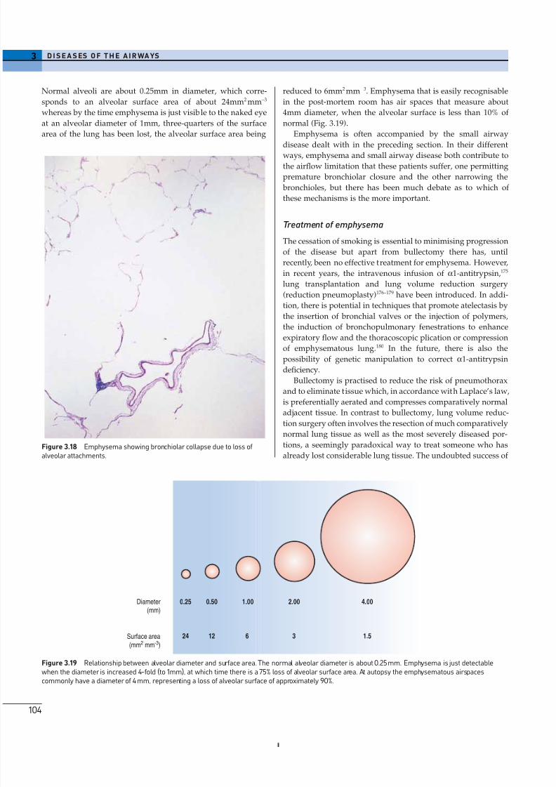
ments to bronchioles results in premature closure of theseairways on expiration (Fig. 3.18).44,126–128 The resultant air trap-
ping is responsible for the overinflation of the lungs and ‘barrel
chest’ that are characteristic of emphysema. Respiration is
conducted near maximal lung volume, which severely compro-
mises inspiratory muscle function. Some adaptation to this is
achieved by an increase in the proportion of slow (‘endurance’)
fibres in the inspiratory muscles.174
Emphysema also results in there being less alveolar surface
available for gas exchange but the extent of this is seldom appre-
ciated when lung slices are examined. The relationship of diam-
eter to surface area is logarithmic so that for a given increase in
airspace diameter there is a much greater loss in surface area.
Elastase
Elastin framework
Anti-proteases
Smoke
Macrophage
Neutrophil
Oxidants
(inactivation)N o r m a l E m
p h y s e ma
Figure 3.17 The pathogenesis of emphysema, envisaged as aconsequence of imbalance between proteases and antiproteases in thelung.
8/13/2019 Af Ale Cailor Respiratorii
http://slidepdf.com/reader/full/af-ale-cailor-respiratorii 18/24
3 D I S E A S ES O F T H E A I R WA YS
104
Figure 3.18 Emphysema showing bronchiolar collapse due to loss of alveolar attachments.
0.25 0.50 1.00 2.00 4.00
24 12 6 3 1.5
Diameter
(mm)
Surface area
(mm2 mm-3)
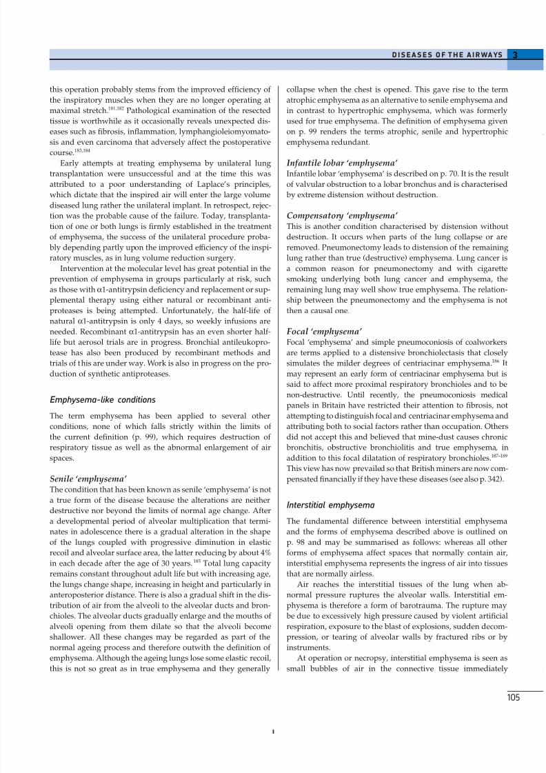
Figure 3.19 Relationship between alveolar diameter and surface area. The normal alveolar diameter is about 0.25mm. Emphysema is just detectablewhen the diameter is increased 4-fold (to 1mm), at which time there is a 75% loss of alveolar surface area. At autopsy the emphysematous airspacescommonly have a diameter of 4 mm, representing a loss of alveolar surface of approximately 90%.
reduced to 6mm2 mm -3. Emphysema that is easily recognisable
in the post-mortem room has air spaces that measure about
4mm diameter, when the alveolar surface is less than 10% of
normal (Fig. 3.19).
Emphysema is often accompanied by the small airwaydisease dealt with in the preceding section. In their different
ways, emphysema and small airway disease both contribute to
the airflow limitation that these patients suffer, one permitting
premature bronchiolar closure and the other narrowing the
bronchioles, but there has been much debate as to which of
these mechanisms is the more important.
Treatment of emphysema
The cessation of smoking is essential to minimising progression
of the disease but apart from bullectomy there has, until
recently, been no effective treatment for emphysema. However,
in recent years, the intravenous infusion of a1-antitrypsin,175
lung transplantation and lung volume reduction surgery
(reduction pneumoplasty)176–179 have been introduced. In addi-
tion, there is potential in techniques that promote atelectasis by
the insertion of bronchial valves or the injection of polymers,
the induction of bronchopulmonary fenestrations to enhance
expiratory flow and the thoracoscopic plication or compression
of emphysematous lung.180 In the future, there is also the
possibility of genetic manipulation to correct a1-antitrypsin
deficiency.
Bullectomy is practised to reduce the risk of pneumothorax
and to eliminate tissue which, in accordance with Laplace’s law,
is preferentially aerated and compresses comparatively normal
adjacent tissue. In contrast to bullectomy, lung volume reduc-tion surgery often involves the resection of much comparatively
normal lung tissue as well as the most severely diseased por-
tions, a seemingly paradoxical way to treat someone who has
already lost considerable lung tissue. The undoubted success of
Normal alveoli are about 0.25mm in diameter, which corre-
sponds to an alveolar surface area of about 24mm2 mm-3
whereas by the time emphysema is just visible to the naked eye
at an alveolar diameter of 1mm, three-quarters of the surface
area of the lung has been lost, the alveolar surface area being
8/13/2019 Af Ale Cailor Respiratorii
http://slidepdf.com/reader/full/af-ale-cailor-respiratorii 19/24
this operation probably stems from the improved efficiency of
the inspiratory muscles when they are no longer operating at
maximal stretch.181,182 Pathological examination of the resected
tissue is worthwhile as it occasionally reveals unexpected dis-
eases such as fibrosis, inflammation, lymphangioleiomyomato-sis and even carcinoma that adversely affect the postoperative
course.183,184
Early attempts at treating emphysema by unilateral lung
transplantation were unsuccessful and at the time this was
attributed to a poor understanding of Laplace’s principles,
which dictate that the inspired air will enter the large volume
diseased lung rather the unilateral implant. In retrospect, rejec-
tion was the probable cause of the failure. Today, transplanta-
tion of one or both lungs is firmly established in the treatment
of emphysema, the success of the unilateral procedure proba-
bly depending partly upon the improved efficiency of the inspi-
ratory muscles, as in lung volume reduction surgery.
Intervention at the molecular level has great potential in theprevention of emphysema in groups particularly at risk, such
as those with a1-antitrypsin deficiency and replacement or sup-
plemental therapy using either natural or recombinant anti-
proteases is being attempted. Unfortunately, the half-life of
natural a1-antitrypsin is only 4 days, so weekly infusions are
needed. Recombinant a1-antitrypsin has an even shorter half-
life but aerosol trials are in progress. Bronchial antileukopro-
tease has also been produced by recombinant methods and
trials of this are under way. Work is also in progress on the pro-
duction of synthetic antiproteases.
Emphysema-like conditions
The term emphysema has been applied to several other
conditions, none of which falls strictly within the limits of
the current definition (p. 99), which requires destruction of
respiratory tissue as well as the abnormal enlargement of air
spaces.
Senile ‘emphysema’ The condition that has been known as senile ‘emphysema’ is not
a true form of the disease because the alterations are neither
destructive nor beyond the limits of normal age change. After
a developmental period of alveolar multiplication that termi-
nates in adolescence there is a gradual alteration in the shape
of the lungs coupled with progressive diminution in elasticrecoil and alveolar surface area, the latter reducing by about 4%
in each decade after the age of 30 years. 185 Total lung capacity
remains constant throughout adult life but with increasing age,
the lungs change shape, increasing in height and particularly in
anteroposterior distance. There is also a gradual shift in the dis-
tribution of air from the alveoli to the alveolar ducts and bron-
chioles. The alveolar ducts gradually enlarge and the mouths of
alveoli opening from them dilate so that the alveoli become
shallower. All these changes may be regarded as part of the
normal ageing process and therefore outwith the definition of
emphysema. Although the ageing lungs lose some elastic recoil,
this is not so great as in true emphysema and they generally
3D I S E A S E S O F T H E A I R WA YS
105
collapse when the chest is opened. This gave rise to the term
atrophic emphysema as an alternative to senile emphysema and
in contrast to hypertrophic emphysema, which was formerly
used for true emphysema. The definition of emphysema given
on p. 99 renders the terms atrophic, senile and hypertrophicemphysema redundant.
Infantile lobar ‘emphysema’ Infantile lobar ‘emphysema’ is described on p. 70. It is the result
of valvular obstruction to a lobar bronchus and is characterised
by extreme distension without destruction.
Compensatory ‘emphysema’ This is another condition characterised by distension without
destruction. It occurs when parts of the lung collapse or are
removed. Pneumonectomy leads to distension of the remaining
lung rather than true (destructive) emphysema. Lung cancer is
a common reason for pneumonectomy and with cigarettesmoking underlying both lung cancer and emphysema, the
remaining lung may well show true emphysema. The relation-
ship between the pneumonectomy and the emphysema is not
then a causal one.
Focal ‘emphysema’ Focal ‘emphysema’ and simple pneumoconiosis of coalworkers
are terms applied to a distensive bronchiolectasis that closely
simulates the milder degrees of centriacinar emphysema.186 It
may represent an early form of centriacinar emphysema but is
said to affect more proximal respiratory bronchioles and to be
non-destructive. Until recently, the pneumoconiosis medical
panels in Britain have restricted their attention to fibrosis, notattempting to distinguish focal and centriacinar emphysema and
attributing both to social factors rather than occupation. Others
did not accept this and believed that mine-dust causes chronic
bronchitis, obstructive bronchiolitis and true emphysema, in
addition to this focal dilatation of respiratory bronchioles.187–189
This view has now prevailed so that British miners are now com-
pensated financially if they have these diseases (see also p. 342).
Interstitial emphysema
The fundamental difference between interstitial emphysema
and the forms of emphysema described above is outlined on
p. 98 and may be summarised as follows: whereas all otherforms of emphysema affect spaces that normally contain air,
interstitial emphysema represents the ingress of air into tissues
that are normally airless.
Air reaches the interstitial tissues of the lung when ab-
normal pressure ruptures the alveolar walls. Interstitial em-
physema is therefore a form of barotrauma. The rupture may
be due to excessively high pressure caused by violent artificial
respiration, exposure to the blast of explosions, sudden decom-
pression, or tearing of alveolar walls by fractured ribs or by
instruments.
At operation or necropsy, interstitial emphysema is seen as
small bubbles of air in the connective tissue immediately
8/13/2019 Af Ale Cailor Respiratorii
http://slidepdf.com/reader/full/af-ale-cailor-respiratorii 20/24
beneath the visceral pleura (see Fig. 2.14, p. 46). Large interstitial
air bubbles are termed blebs, as distinct from bullae, which
represent enlargement of pre-existent air spaces.119 Air in
the interstitial tissues may track along the connective tissue
sheaths about the pulmonary vessels to the hila of the lungs,producing mediastinal emphysema; it may then reach the neck
and present subcutaneously, as surgical emphysema. Systemic
air embolism may complicate interstitial emphysema (see
p. 412).
Microscopically, minute air bubbles appear as seemingly
empty interstitial spaces, particularly in the abundant connec-
tive tissue that surrounds the pulmonary artery and airway
and forms the interlobular septa. The differential diagnosis on
microscopy is from congenital lymphangiectasia and this can be
extremely difficult, not least because the air tracks within lym-
phatics as well as through the surrounding connective tissues.
It is therefore helpful if the nature of the contents of the spaces,
gaseous or fluid, is ascertained at necropsy. If the emphysemahas been present for a few days before death, the diagnosis is
simplified by the development of a foreign body giant cell reac-
tion to the air (see Fig. 2.15, p. 47),190–192 similar to that found
in pneumatosis coli and following the experimental injection of
gases into the subcutaneous tissues.190
PLASTIC BRONCHITIS
Patients with plastic bronchitis are generally well but complain
of fits of coughing that often result in them involuntarily expec-
torating long stringy pieces of sputum, which causes them
much social embarrassment.193–197 The expectorate represents
a bronchial cast of up to eight airway generations (Fig. 3.20).
Microscopically the cast is seen to consist of alternating bands
of fibrin and mucus, with the fibrin containing variable
numbers of lymphocytes.194,195 The appearances suggest that the
cast represents an inspissated fibrinous exudate and the term
fibrinous bronchitis is sometimes applied to the condition.
Several associated conditions have been described194,197,198 and it
is possible that some of these have a causal relationship, notably
heart failure and lymphatic abnormalities. Plastic bronchitis
is often confused with the mucoid impaction of allergic
bronchopulmonary aspergillosis,199 which is characterised by
the expectoration of short stubby gobbets of mucus. The
two conditions are quite different. They are compared in
Table 3.3.194,200,201
3 D I S E A S ES O F T H E A I R WA YS
106
BRONCHIAL ASTHMA
Bronchial asthma is to be distinguished from the aetiologically
distinct condition of cardiac asthma. The latter represents pul-
monary oedema consequent upon a failing left heart. Bronchialasthma, hereafter called simply asthma, is a condition in which
breathing is periodically rendered difficult by widespread nar-
rowing of the bronchi that changes in severity over short
periods of time, either spontaneously or under treatment.45 The
difficulty becomes particularly apparent during expiration
because the airways normally collapse during that phase of res-
piration and because the expiratory muscles are less powerful
than those that act during inspiration.
Figure 3.20 A 15cm long bronchial cast from a patient with plasticbronchitis.
Table 3.3 A comparison of mucoid impaction and plastic bronchitis
Mucoid impaction Plastic bronchitis
Clinical features Aggravation of underlying asthma Expectoration of stringy sputum.Progression to hilar bronchiectasis
Cause Atopy: allergic broncho-pulmonary Unknown. Associations of questionable causal relationshipaspergillosis complicating asthma include heart disease and lymphatic abnormalities
Gross appearance of expectorate A short stubby mucoid plug A long stringy bronchial cast of up to eight airway generationsMicroscopic appearance of plug/cast Alternating layers of mucus and inspissated Alternating layers of mucus and fibrin with variable numbers
eosinophils with numerous Charcot– of lymphocytes. No eosinophils. No fungi.Leyden crystals and scanty aspergillushyphae.
8/13/2019 Af Ale Cailor Respiratorii
http://slidepdf.com/reader/full/af-ale-cailor-respiratorii 21/24
Extrinsic and intrinsic forms of asthma
Asthma is said to be ‘extrinsic’ if allergy to exogenous sub-
stances is recognised and ‘intrinsic’ if no such exogenous factors
can be identified. Extrinsic asthma is the more common. It
usually begins in childhood and is generally paroxysmal, theattack starting suddenly and lasting a few hours or days. Boys
are affected about twice as much as girls. Extrinsic asthma tends
to become less severe as the child grows older and often ceases
during adolescence. However, about 30% of asthmatic children
continue to have symptoms in adult life. Extrinsic asthma is
often familial and some of the genes responsible have been
identified.202–205 This form of asthma is frequently preceded by
flexural infantile eczema and succeeded in adult life by peren-
nial vasomotor rhinitis, although in such families, any one of
these three diseases may affect some members much more than
others. In contrast, intrinsic asthma more often has its onset in
adult life, is chronic, with exacerbations and remissions less
evident and tends to worsen with age. The symptoms are also
apt to become more severe in winter, when the asthmatic
disabilities are likely to be complicated by infection of the res-
piratory tract. In both types of the disease, the dyspnoea is char-
acteristically accompanied by cough, wheezy breathing, some
cyanosis and expectoration. Blood eosinophilia is more promi-
nent in extrinsic asthma but eosinophils are found in the
airways in both. Serum immunoglobulin E is often raised in
extrinsic asthma and normal or low in intrinsic asthma. There
is an increased incidence of nasal polyps in both forms of
asthma, but especially in the intrinsic variety and in certain
asthmatic patients in whom bronchospasm is triggered by
aspirin. Despite these differences, the pathological features
are similar and an immunological basis may be envisaged in
both.206,207
Epidemiology
Over the past few decades the prevalence of asthma has
increased considerably in most developed countries where it
now stands at about 5%. The reason for this is unclear. General
atmospheric pollution has been widely incriminated but there
is little to support this: the increase has been experienced in
countries such as New Zealand which have little atmospheric
pollution and in Philadelphia at a time when atmospheric
pollution declined.208 Atmospheric pollution is more likely toaggravate asthma than cause it. Changes in the home that
encourage the growth of the principal allergen, the house dust
mite (higher temperature and humidity and extensive carpet-
ing), are more likely environmental factors.209,210
Another possibility is the decline in childhood infections in
the developed countries. Infection induces a Th1 response
rather than the Th2 reactions responsible for atopy (see ‘aetiol-
ogy’, below).211 Indirect support for this possibility comes from
studies of family size and birth order. Atopy is less common in
children belonging to large families and, within such families,
in the younger children – the ones that are most likely to be
exposed in infancy to infections brought home by their sib-
3D I S E A S E S O F T H E A I R WA YS
107
lings.212,213 More direct evidence comes from studies showing
less atopy in those who had previously had measles,214 were
seropositive for hepatitis A215,216 or gave a strong tuberculin reac-
tion.217 This raises the attractive possibility of preventive immu-
nisation using harmless bacteria.
218
Mortality figures have fluctuated over the past few decades,
apparently being influenced adversely on occasion by the intro-
duction of new drugs, particularly b-agonists (Fig. 3.21).219,220
The mechanism underlying these fluctuations is not fully
understood but overdosage, cardiac dysrhythmia, refractori-
ness to the bronchodilatory effect of the drug and a false sense
of security afforded by carrying an inhaler are all thought to
have contributed.219 The prognosis is worse in late-onset intrin-
sic asthma. Most asthma deaths occur in the elderly, although
asthma accounts for a greater proportion of deaths in the young
(0.9% of those under 25 vs 0.3% overall). Changes in the mor-
tality rate from 1979 to 1999 are shown in Table 3.4.
The sputum in asthma
The sputum is viscous221 and yellow in asthma. The colour
should not be taken as evidence that the sputum is infected. It
is due to myeloperoxidase, which is found in both eosinophils
and neutrophils. In asthma, the sputum is rich in eosinophils,
but not neutrophils unless there is also infection. As well as
eosinophils, microscopy shows the presence of certain formed
1920 1930 1940 1950 1960 1970 1980 1990
0
1
2
3
4
A s t h m a d e a t h s
/ 1 0 0 0 0 0
Year
New Zealand
England and Wales
Netherlands
AdrenalineIsoprenaline
Isoprenaline
high doseFenoteral
Figure 3.21 Time trends in asthma mortality in relation to theintroduction of various inhaled drugs. (Reproduced from Blauw GJ,Westendorp RGJ. Asthma deaths in New Zealand: whodunnit? The Lancet1995; 345:2–3, with permission from Elsevier.)
Table 3.4 Percentage change in mortality rates for asthma from1979 to 1999 in England and Wales
Age band 1999 mortality rate Percentage change per million 1979–99
0–4 1.8 -405–14 2.4 -3715–44 6.2 -4345–64 23.6 -3865+ 110.2 +3
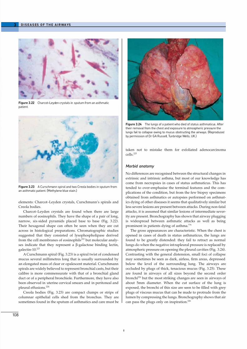
8/13/2019 Af Ale Cailor Respiratorii
http://slidepdf.com/reader/full/af-ale-cailor-respiratorii 22/24
elements: Charcot–Leyden crystals, Curschmann’s spirals and
Creola bodies.
Charcot–Leyden crystals are found when there are large
numbers of eosinophils. They have the shape of a pair of long,
narrow, six-sided pyramids placed base to base (Fig. 3.22).
Their hexagonal shape can often be seen when they are cut
across in histological preparations. Chromatographic studies
suggested that they consisted of lysophospholipase derived
from the cell membranes of eosinophils222 but molecular analy-
ses indicate that they represent a b-galactose binding lectin,galectin-10.223
A Curschmann spiral (Fig. 3.23) is a spiral twist of condensed
mucus several millimetres long that is usually surrounded by
an elongated mass of clear or opalescent material. Curschmann
spirals are widely believed to represent bronchial casts, but their
calibre is more commensurate with that of a bronchial gland
duct or of a peripheral bronchiole. Furthermore, they have also
been observed in uterine cervical smears and in peritoneal and
pleural effusions.224
Creola bodies (Fig. 3.23) are compact clumps or strips of
columnar epithelial cells shed from the bronchus. They are
sometimes found in the sputum of asthmatics and care must be
3 D I S E A S ES O F T H E A I R WA YS
108
taken not to mistake them for exfoliated adenocarcinoma
cells.225
Morbid anatomy
No differences are recognised between the structural changes in
extrinsic and intrinsic asthma, but most of our knowledge has
come from necropsies in cases of status asthmaticus. This hastended to over-emphasise the terminal features and the com-
plications of the condition, but from the few biopsy specimens
obtained from asthmatics or autopsies performed on asthmat-
ics dying of other diseases it seems that qualitatively similar but
less severe lesions are present between attacks. During non-fatal
attacks, it is assumed that similar lesions of intermediate sever-
ity are present. Bronchography has shown that airway plugging
is widespread between asthmatic attacks as well as being
prominent in patients dying of asthma.226
The gross appearances are characteristic. When the chest is
opened in cases of death in status asthmaticus, the lungs are
found to be greatly distended: they fail to retract as normal
lungs do when the negative intrapleural pressure is replaced byatmospheric pressure on opening the pleural cavities (Fig. 3.24).
Contrasting with the general distension, small foci of collapse
may sometimes be seen as dark, airless, firm areas, depressed
below the level of the surrounding lung. The airways are
occluded by plugs of thick, tenacious mucus (Fig. 3.25). These
are found in airways of all sizes beyond the second order
bronchi226 but the most striking changes are seen in airways of
about 5mm diameter. When the cut surface of the lung is
exposed, the bronchi of this size are seen to be filled with grey
plugs of viscous mucus that can be made to protrude from the
lumen by compressing the lungs. Bronchography shows that air
can pass the plugs only on inspiration.226
Figure 3.22 Charcot–Leyden crystals in sputum from an asthmaticpatient.
Figure 3.23 A Curschmann spiral and two Creola bodies in sputum froman asthmatic patient. (Methylene blue stain.)
Figure 3.24 The lungs of a patient who died of status asthmaticus. After their removal from the chest and exposure to atmospheric pressure thelungs fail to collapse owing to mucus obstructing the airways. (Reproducedby permission of Dr GA Russell, Tunbridge Wells, UK.)
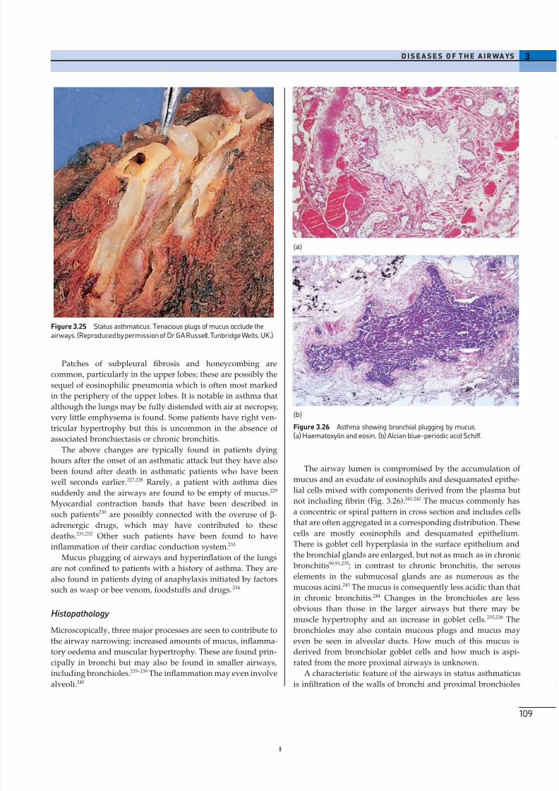
8/13/2019 Af Ale Cailor Respiratorii
http://slidepdf.com/reader/full/af-ale-cailor-respiratorii 23/24
Patches of subpleural fibrosis and honeycombing are
common, particularly in the upper lobes; these are possibly the
sequel of eosinophilic pneumonia which is often most marked
in the periphery of the upper lobes. It is notable in asthma that
although the lungs may be fully distended with air at necropsy,
very little emphysema is found. Some patients have right ven-tricular hypertrophy but this is uncommon in the absence of
associated bronchiectasis or chronic bronchitis.
The above changes are typically found in patients dying
hours after the onset of an asthmatic attack but they have also
been found after death in asthmatic patients who have been
well seconds earlier.227,228 Rarely, a patient with asthma dies
suddenly and the airways are found to be empty of mucus.229
Myocardial contraction bands that have been described in
such patients230 are possibly connected with the overuse of b-
adrenergic drugs, which may have contributed to these
deaths.231,232 Other such patients have been found to have
inflammation of their cardiac conduction system.233
Mucus plugging of airways and hyperinflation of the lungs
are not confined to patients with a history of asthma. They are
also found in patients dying of anaphylaxis initiated by factors
such as wasp or bee venom, foodstuffs and drugs. 234
Histopathology
Microscopically, three major processes are seen to contribute to
the airway narrowing: increased amounts of mucus, inflamma-
tory oedema and muscular hypertrophy. These are found prin-
cipally in bronchi but may also be found in smaller airways,
including bronchioles.235–239 The inflammation may even involve
alveoli.240
3D I S E A S E S O F T H E A I R WA YS
109
The airway lumen is compromised by the accumulation of
mucus and an exudate of eosinophils and desquamated epithe-
lial cells mixed with components derived from the plasma but
not including fibrin (Fig. 3.26).241,242 The mucus commonly has
a concentric or spiral pattern in cross section and includes cells
that are often aggregated in a corresponding distribution. These
cells are mostly eosinophils and desquamated epithelium.
There is goblet cell hyperplasia in the surface epithelium and
the bronchial glands are enlarged, but not as much as in chronic bronchitis90,91,235; in contrast to chronic bronchitis, the serous
elements in the submucosal glands are as numerous as the
mucous acini.243 The mucus is consequently less acidic than that
in chronic bronchitis.244 Changes in the bronchioles are less
obvious than those in the larger airways but there may be
muscle hypertrophy and an increase in goblet cells.235,238 The
bronchioles may also contain mucous plugs and mucus may
even be seen in alveolar ducts. How much of this mucus is
derived from bronchiolar goblet cells and how much is aspi-
rated from the more proximal airways is unknown.
A characteristic feature of the airways in status asthmaticus
is infiltration of the walls of bronchi and proximal bronchioles
Figure 3.25 Status asthmaticus. Tenacious plugs of mucus occlude theairways. (Reproduced by permission of Dr GA Russell, Tunbridge Wells, UK.)
(a)
(b)Figure 3.26 Asthma showing bronchial plugging by mucus.(a) Haematoxylin and eosin, (b) Alcian blue-periodic acid Schiff.
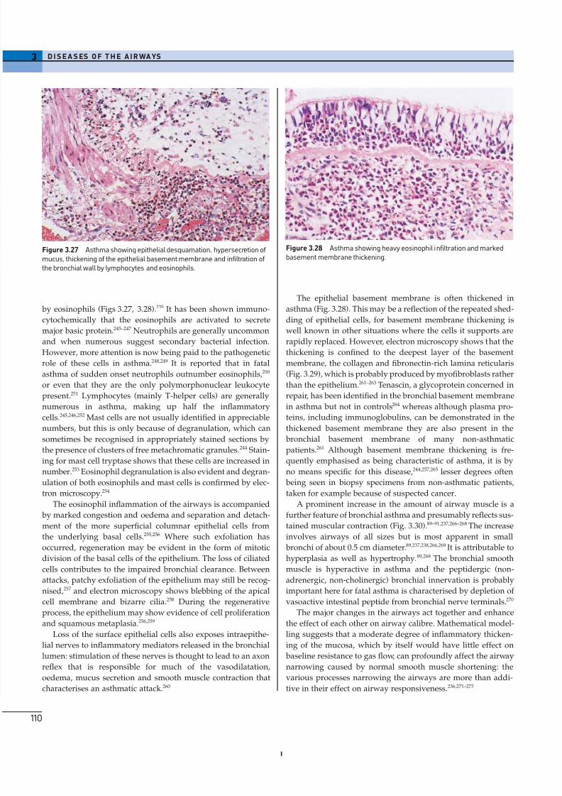
8/13/2019 Af Ale Cailor Respiratorii
http://slidepdf.com/reader/full/af-ale-cailor-respiratorii 24/24
by eosinophils (Figs 3.27, 3.28).235 It has been shown immuno-
cytochemically that the eosinophils are activated to secrete
major basic protein.245–247 Neutrophils are generally uncommon
and when numerous suggest secondary bacterial infection.
However, more attention is now being paid to the pathogenetic
role of these cells in asthma.248,249 It is reported that in fatal
asthma of sudden onset neutrophils outnumber eosinophils,250
or even that they are the only polymorphonuclear leukocyte
present.251 Lymphocytes (mainly T-helper cells) are generally
numerous in asthma, making up half the inflammatory
cells.245,246,252
Mast cells are not usually identified in appreciablenumbers, but this is only because of degranulation, which can
sometimes be recognised in appropriately stained sections by
the presence of clusters of free metachromatic granules.244 Stain-
ing for mast cell tryptase shows that these cells are increased in
number.253 Eosinophil degranulation is also evident and degran-
ulation of both eosinophils and mast cells is confirmed by elec-
tron microscopy.254
The eosinophil inflammation of the airways is accompanied
by marked congestion and oedema and separation and detach-
ment of the more superficial columnar epithelial cells from
the underlying basal cells.255,256 Where such exfoliation has
occurred, regeneration may be evident in the form of mitotic
division of the basal cells of the epithelium. The loss of ciliatedcells contributes to the impaired bronchial clearance. Between
attacks, patchy exfoliation of the epithelium may still be recog-
nised,257 and electron microscopy shows blebbing of the apical
cell membrane and bizarre cilia.258 During the regenerative
process, the epithelium may show evidence of cell proliferation
and squamous metaplasia.256,259
Loss of the surface epithelial cells also exposes intraepithe-
lial nerves to inflammatory mediators released in the bronchial
lumen: stimulation of these nerves is thought to lead to an axon
reflex that is responsible for much of the vasodilatation,
oedema, mucus secretion and smooth muscle contraction that
characterises an asthmatic attack.260
3 D I S E A S ES O F T H E A I R WA YS
110
The epithelial basement membrane is often thickened in
asthma (Fig. 3.28). This may be a reflection of the repeated shed-
ding of epithelial cells, for basement membrane thickening is
well known in other situations where the cells it supports are
rapidly replaced. However, electron microscopy shows that the
thickening is confined to the deepest layer of the basement
membrane, the collagen and fibronectin-rich lamina reticularis
(Fig. 3.29), which is probably produced by myofibroblasts rather
than the epithelium.261–263 Tenascin, a glycoprotein concerned in
repair, has been identified in the bronchial basement membrane
in asthma but not in controls264 whereas although plasma pro-
teins, including immunoglobulins, can be demonstrated in thethickened basement membrane they are also present in the
bronchial basement membrane of many non-asthmatic
patients.261 Although basement membrane thickening is fre-
quently emphasised as being characteristic of asthma, it is by
no means specific for this disease,244,257,265 lesser degrees often
being seen in biopsy specimens from non-asthmatic patients,
taken for example because of suspected cancer.
A prominent increase in the amount of airway muscle is a
further feature of bronchial asthma and presumably reflects sus-
tained muscular contraction (Fig. 3.30).89–91,237,266–268 The increase
involves airways of all sizes but is most apparent in small
bronchi of about 0.5 cm diameter.89,237,238,266,269 It is attributable to
hyperplasia as well as hypertrophy.89,268 The bronchial smoothmuscle is hyperactive in asthma and the peptidergic (non-
adrenergic, non-cholinergic) bronchial innervation is probably
important here for fatal asthma is characterised by depletion of
vasoactive intestinal peptide from bronchial nerve terminals.270
The major changes in the airways act together and enhance
the effect of each other on airway calibre. Mathematical model-
ling suggests that a moderate degree of inflammatory thicken-
ing of the mucosa, which by itself would have little effect on
baseline resistance to gas flow, can profoundly affect the airway
narrowing caused by normal smooth muscle shortening: the
various processes narrowing the airways are more than addi-
tive in their effect on airway responsiveness.236,271–273
Figure 3.27 Asthma showing epithelial desquamation, hypersecretion of mucus, thickening of the epithelial basement membrane and infiltration of the bronchial wall by lymphocytes and eosinophils.
Figure 3.28 Asthma showing heavy eosinophil infiltration and markedbasement membrane thickening.