Embed Size (px)
Citation preview
J Bras Pneumol. 2012;38(4):526-529
Unilateral pulmonary agenesis*Agenesia pulmonar unilateral
Maura Cavada Malcon, Claudio Mattar Malcon, Marina Neves Cavada, Paulo Eduardo Macedo Caruso, Lara Flório Real
AbstractPulmonary agenesis is a rare congenital anomaly. We report the case of an 8-year-old boy with left lung agenesis, without any other congenital malformations. When the patient presented symptoms, including cough, wheezing, and dyspnea, with no clinical improvement after a period of 30 days, imaging studies were conducted and the diagnosis was made.
Keywords: Congenital abnormalities; Respiratory tract diseases; Bronchoscopy.
ResumoA agenesia pulmonar é uma anomalia congênita rara. Relatamos um caso de um menino de 8 anos de idade com agenesia pulmonar à esquerda sem associação com outras malformações. O diagnóstico foi realizado por achados de imagem quando o paciente apresentou sintomas como tosse, sibilância e dispneia sem melhora do quadro clínico após evolução de 30 dias.
Descritores: Anormalidades congênitas; Doenças respiratórias; Broncoscopia.
* Study carried out at the private practice of the first author, Pelotas, Brazil.Correspondence to: Maura Malcon. Rua Félix da Cunha, 916, apto. 1001, CEP 96010-000, Pelotas, RS, Brasil.Tel. 55 53 3222-9875 or 55 53 3222-7338. E-mail: [email protected] support: None.Submitted: 15 April 2010. Accepted, after review: 26 October 2010.
Introduction
Pulmonary agenesis is a rare congenital anomaly, consisting of complete absence of the lung parenchyma, bronchi, and pulmonary vessels.(1-4) The cause is unknown. In 50% of cases, especially in cases of right lung agenesis, cardiovascular, musculoskeletal, gastrointestinal, and renal malformations are also present.(3,5-7) The prognosis is better in cases of left lung agenesis and when there are no cardiac malformations.(3) Herein, we report the clinical case of a boy with left lung agenesis, without any other congenital malformations.
Case report
An 8-year-old White male patient from the city of Jaguarão, Brazil, sought medical attention. He had been born at term, and there had been no complications during pregnancy. He had a family history of asthma. Since early 2006, when he was 7 years old, the patient had been having attacks of wheezing, having been treated at home by his family. When he sought medical attention in February of 2007, the patient
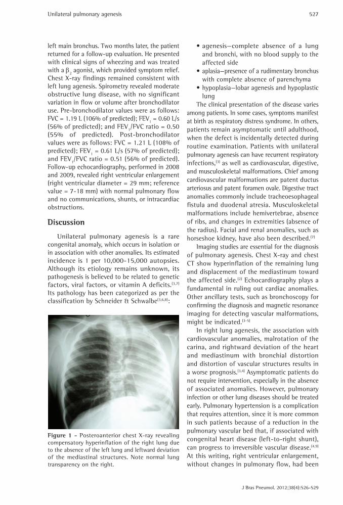
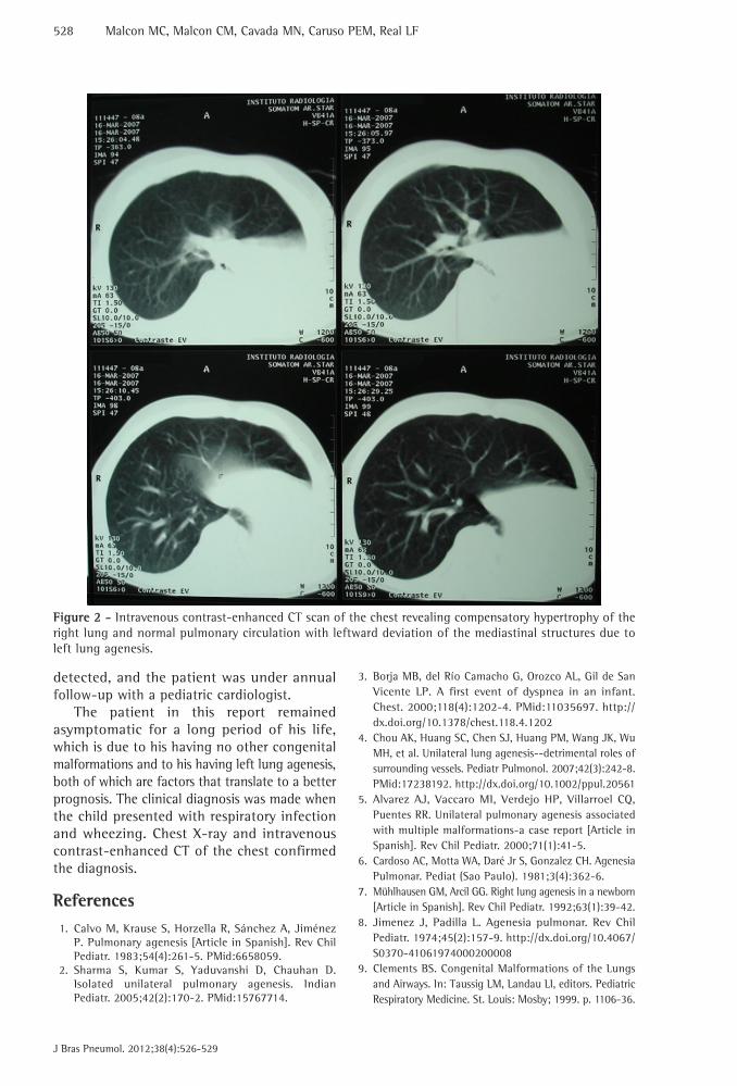
presented with a dry cough that progressed to wheezing and dyspnea for one month. He was hospitalized in his hometown. A chest X-ray performed at admission showed opacity of the left hemithorax, which was treated as bacterial pneumonia (Figure 1). Physical examination by a pulmonologist revealed that the patient was active, acyanotic, and breathing normally; pulmonary auscultation revealed retraction of the left hemithorax, dullness to percussion, and absent breath sounds on the left with wheezing on the right; cardiac auscultation revealed normal, rhythmic heart sounds without heart murmurs. The following tests were requested: chest HRCT; bronchoscopy; Doppler echocardiography; and abdominal ultrasound. Chest HRCT showed left lung agenesis, compensatory hypertrophy of the right lung, and normal right pulmonary circulation with leftward deviation of the mediastinal structures. There were no changes in upper abdominal structures (Figure 2). Abdominal ultrasound and echocardiography showed normal findings. Bronchoscopy revealed a blind-ending
Case Report
Unilateral pulmonary agenesis
J Bras Pneumol. 2012;38(4):526-529
527
•agenesis—complete absence of a lung and bronchi, with no blood supply to the affected side
•aplasia—presence of a rudimentary bronchus with complete absence of parenchyma
•hypoplasia—lobar agenesis and hypoplastic lung
The clinical presentation of the disease varies among patients. In some cases, symptoms manifest at birth as respiratory distress syndrome. In others, patients remain asymptomatic until adulthood, when the defect is incidentally detected during routine examination. Patients with unilateral pulmonary agenesis can have recurrent respiratory infections,(3) as well as cardiovascular, digestive, and musculoskeletal malformations. Chief among cardiovascular malformations are patent ductus arteriosus and patent foramen ovale. Digestive tract anomalies commonly include tracheoesophageal fistula and duodenal atresia. Musculoskeletal malformations include hemivertebrae, absence of ribs, and changes in extremities (absence of the radius). Facial and renal anomalies, such as horseshoe kidney, have also been described.(7)
Imaging studies are essential for the diagnosis of pulmonary agenesis. Chest X-ray and chest CT show hyperinflation of the remaining lung and displacement of the mediastinum toward the affected side.(2) Echocardiography plays a fundamental in ruling out cardiac anomalies. Other ancillary tests, such as bronchoscopy for confirming the diagnosis and magnetic resonance imaging for detecting vascular malformations, might be indicated.(3-5)
In right lung agenesis, the association with cardiovascular anomalies, malrotation of the carina, and rightward deviation of the heart and mediastinum with bronchial distortion and distortion of vascular structures results in a worse prognosis.(3,4) Asymptomatic patients do not require intervention, especially in the absence of associated anomalies. However, pulmonary infection or other lung diseases should be treated early. Pulmonary hypertension is a complication that requires attention, since it is more common in such patients because of a reduction in the pulmonary vascular bed that, if associated with congenital heart disease (left-to-right shunt), can progress to irreversible vascular disease.(4,9) At this writing, right ventricular enlargement, without changes in pulmonary flow, had been
left main bronchus. Two months later, the patient returned for a follow-up evaluation. He presented with clinical signs of wheezing and was treated with a β2 agonist, which provided symptom relief. Chest X-ray findings remained consistent with left lung agenesis. Spirometry revealed moderate obstructive lung disease, with no significant variation in flow or volume after bronchodilator use. Pre-bronchodilator values were as follows: FVC = 1.19 L (106% of predicted); FEV1 = 0.60 L/s (56% of predicted); and FEV1/FVC ratio = 0.50 (55% of predicted). Post-bronchodilator values were as follows: FVC = 1.21 L (108% of predicted); FEV1 = 0.61 L/s (57% of predicted); and FEV1/FVC ratio = 0.51 (56% of predicted). Follow-up echocardiography, performed in 2008 and 2009, revealed right ventricular enlargement (right ventricular diameter = 29 mm; reference value = 7-18 mm) with normal pulmonary flow and no communications, shunts, or intracardiac obstructions.
Discussion
Unilateral pulmonary agenesis is a rare congenital anomaly, which occurs in isolation or in association with other anomalies. Its estimated incidence is 1 per 10,000-15,000 autopsies. Although its etiology remains unknown, its pathogenesis is believed to be related to genetic factors, viral factors, or vitamin A deficits.(3,7) Its pathology has been categorized as per the classification by Schneider & Schwalbe(3,6,8):
Figure 1 - Posteroanterior chest X-ray revealing compensatory hyperinflation of the right lung due to the absence of the left lung and leftward deviation of the mediastinal structures. Note normal lung transparency on the right.
528 Malcon MC, Malcon CM, Cavada MN, Caruso PEM, Real LF
J Bras Pneumol. 2012;38(4):526-529
3. Borja MB, del Río Camacho G, Orozco AL, Gil de San Vicente LP. A first event of dyspnea in an infant. Chest. 2000;118(4):1202-4. PMid:11035697. http://dx.doi.org/10.1378/chest.118.4.1202
4. Chou AK, Huang SC, Chen SJ, Huang PM, Wang JK, Wu MH, et al. Unilateral lung agenesis--detrimental roles of surrounding vessels. Pediatr Pulmonol. 2007;42(3):242-8. PMid:17238192. http://dx.doi.org/10.1002/ppul.20561
5. Alvarez AJ, Vaccaro MI, Verdejo HP, Villarroel CQ, Puentes RR. Unilateral pulmonary agenesis associated with multiple malformations-a case report [Article in Spanish]. Rev Chil Pediatr. 2000;71(1):41-5.
6. Cardoso AC, Motta WA, Daré Jr S, Gonzalez CH. Agenesia Pulmonar. Pediat (Sao Paulo). 1981;3(4):362-6.
7. Mühlhausen GM, Arcil GG. Right lung agenesis in a newborn [Article in Spanish]. Rev Chil Pediatr. 1992;63(1):39-42.
8. Jimenez J, Padilla L. Agenesia pulmonar. Rev Chil Pediatr. 1974;45(2):157-9. http://dx.doi.org/10.4067/S0370-41061974000200008
9. Clements BS. Congenital Malformations of the Lungs and Airways. In: Taussig LM, Landau LI, editors. Pediatric Respiratory Medicine. St. Louis: Mosby; 1999. p. 1106-36.
detected, and the patient was under annual follow-up with a pediatric cardiologist.
The patient in this report remained asymptomatic for a long period of his life, which is due to his having no other congenital malformations and to his having left lung agenesis, both of which are factors that translate to a better prognosis. The clinical diagnosis was made when the child presented with respiratory infection and wheezing. Chest X-ray and intravenous contrast-enhanced CT of the chest confirmed the diagnosis.
References
1. Calvo M, Krause S, Horzella R, Sánchez A, Jiménez P. Pulmonary agenesis [Article in Spanish]. Rev Chil Pediatr. 1983;54(4):261-5. PMid:6658059.
2. Sharma S, Kumar S, Yaduvanshi D, Chauhan D. Isolated unilateral pulmonary agenesis. Indian Pediatr. 2005;42(2):170-2. PMid:15767714.
Figure 2 - Intravenous contrast-enhanced CT scan of the chest revealing compensatory hypertrophy of the right lung and normal pulmonary circulation with leftward deviation of the mediastinal structures due to left lung agenesis.

Unilateral pulmonary agenesis
J Bras Pneumol. 2012;38(4):526-529
529
About the authors
Maura Cavada MalconPulmonologist. Pelotas City Hall, Pelotas, Brazil.
Claudio Mattar MalconPulmonologist. Pelotas, Brazil.
Marina Neves CavadaMedical Student. Federal University of Pelotas, Pelotas, Brazil.
Paulo Eduardo Macedo CarusoMedical Student. Federal University of Pelotas, Pelotas, Brazil.
Lara Flório RealResident in Gynecology and Obstetrics. Hospital Nossa Senhora da Conceição de Porto Alegre, Porto Alegre, Brazil.
![6 Restrepo.ppt [Modo de compatibilidad]uvsalud.univalle.edu.co/pdf/simpsios/decimo/Chatain/6restrepo.pdf · Flujograma para evaluación de Niños con Agenesia Renal Unilateral ( ARU)](https://img.pdfslide.net/doc/110x75/5c03bc7909d3f2c12d8d1c48/6-modo-de-compatibilidaduvsaludunivalleeducopdfsimpsiosdecimochatain6restrepopdf.jpg)