Embed Size (px)
Citation preview
Amino Acids: Structure, Analysis,
and Sequence (in peptides)
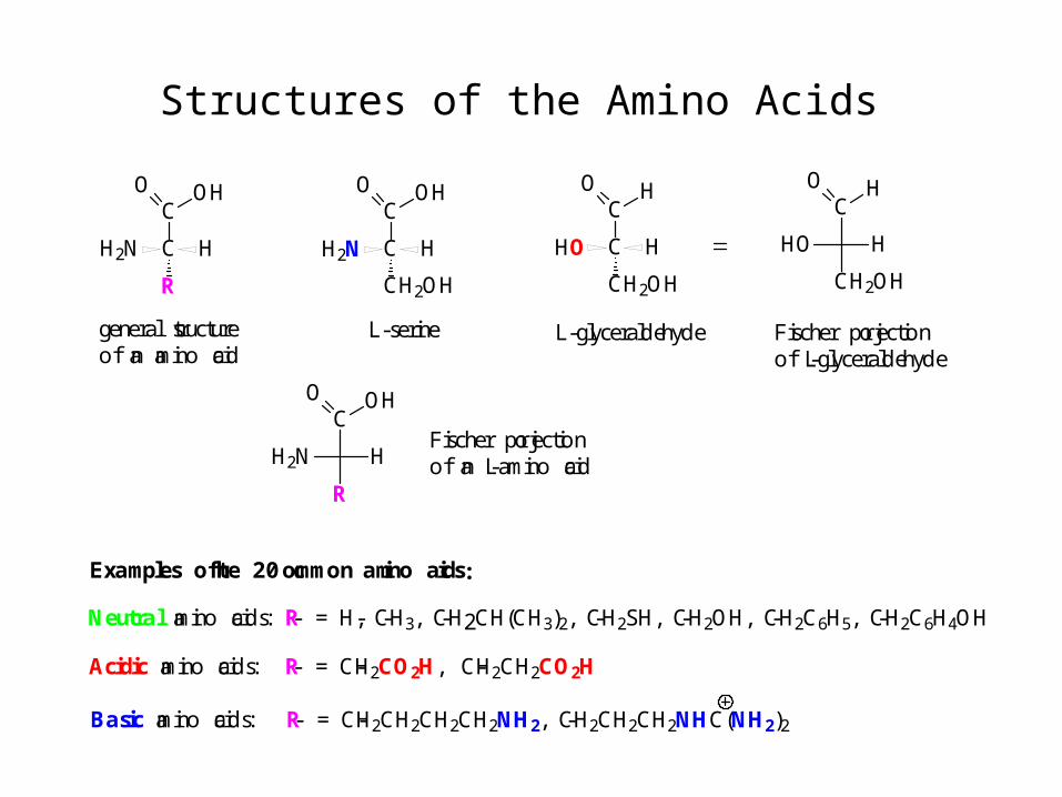
Structures of the Amino Acids
C
C
R
HH2N
O OHC
C
CH2OH
HHO
O H
L-glyceraldehyde
C
C
CH2OH
HH2N
O OH
L-serine
Neutral amino acids: -R = -H, -CH3, -CH2CH(CH3)2, -CH2SH, -CH2OH, -CH2C6H5, -CH2C6H4OH
Acidic amino acids: -R = -CH2CO2H, -CH2CH2CO2H
Basic amino acids: -R = -CH2CH2CH2CH2NH2, -CH2CH2CH2NHC(NH2)2
Examples of the 20 common amino acids:
C
H2N H
R
O OH
C
HO H
CH2OH
O H
general structure of an amino acid
Fischer projectionof L-glyceraldehyde
Fischer projectionof an L-amino acid
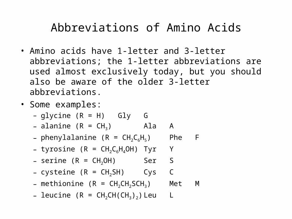
Abbreviations of Amino Acids
• Amino acids have 1-letter and 3-letter abbreviations; the 1-letter abbreviations are used almost exclusively today, but you should also be aware of the older 3-letter abbreviations.
• Some examples:– glycine (R = H) Gly G
– alanine (R = CH3) Ala A
– phenylalanine (R = CH2C6H5) Phe F
– tyrosine (R = CH2C6H4OH) Tyr Y
– serine (R = CH2OH) Ser S
– cysteine (R = CH2SH) Cys C
– methionine (R = CH2CH2SCH3) Met M
– leucine (R = CH2CH(CH3)2) Leu L
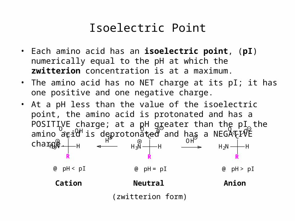
Isoelectric Point
• Each amino acid has an isoelectric point, (pI) numerically equal to the pH at which the zwitterion concentration is at a maximum.
• The amino acid has no NET charge at its pI; it has one positive and one negative charge.
• At a pH less than the value of the isoelectric point, the amino acid is protonated and has a POSITIVE charge; at a pH greater than the pI the amino acid is deprotonated and has a NEGATIVE charge.
Cation Neutral Anion
(zwitterion form)
C
H3N H
R
O OC
H3N H
R
O OHC
H2N H
R
O O
H OH
@ pH = pI@ pH < pI @ pH > pI

Separation and Analysis using pI values
• Differences in isoelectric points (and therefore charges) are used to separate mixtures of amino acids by two common methods:
– Ion exchange chromatography
– Polyacrylamide gel electrophoresis (PAGE)
H3 C CO
OH
CH2CO2
N
A (pI=6.0)
+ H3N
CH3
H O
COC
D (pI=2.8)
+ H3
CH2CH2CH2CH2NH3
H O
COCN
K (pI= 9.7)
Mixture of:buffered at pH 6.0
These methods will be illustrated with a simple mixture of three amino acids having very different isoelectric points:
aspartic acid alanine lysine
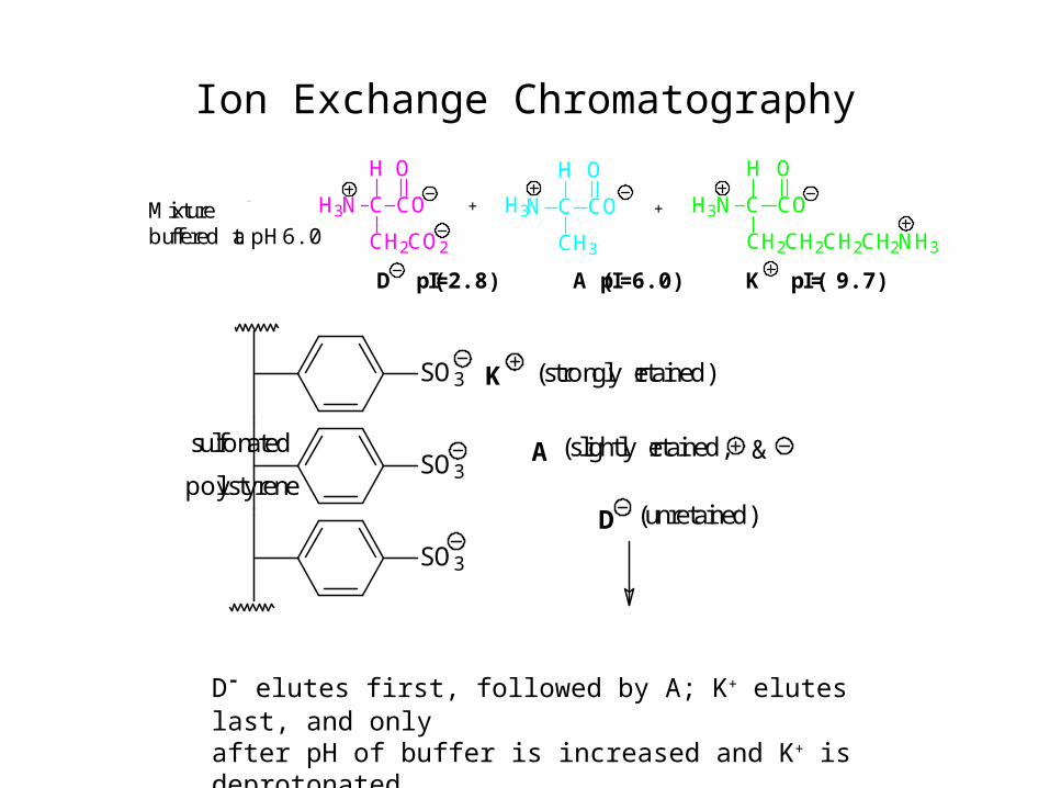
Ion Exchange Chromatography
D- elutes first, followed by A; K+ elutes last, and onlyafter pH of buffer is increased and K+ is deprotonated.
H3 C CO
OH
CH2CO2
N
A (pI=6.0)
+ H3N
CH3
H O
COC
D (pI=2.8)
+ H3
CH2CH2CH2CH2NH3
H O
COCN
K (pI= 9.7)
Mixture of:buffered at pH 6.0
SO3
SO3
SO3
A
D
K (strongly retained)
(unretained)
(slightly retained, &sulfonated
polystyrene
Ion Exchange Chromatography
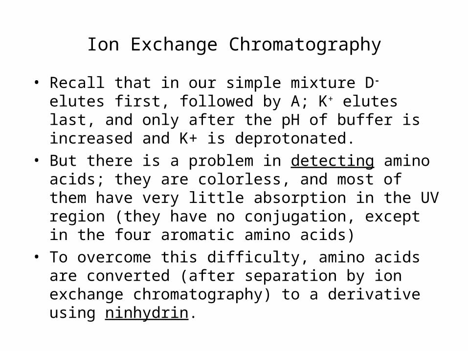
• Recall that in our simple mixture D- elutes first, followed by A; K+ elutes last, and only after the pH of buffer is increased and K+ is deprotonated.
• But there is a problem in detecting amino acids; they are colorless, and most of them have very little absorption in the UV region (they have no conjugation, except in the four aromatic amino acids)
• To overcome this difficulty, amino acids are converted (after separation by ion exchange chromatography) to a derivative using ninhydrin.
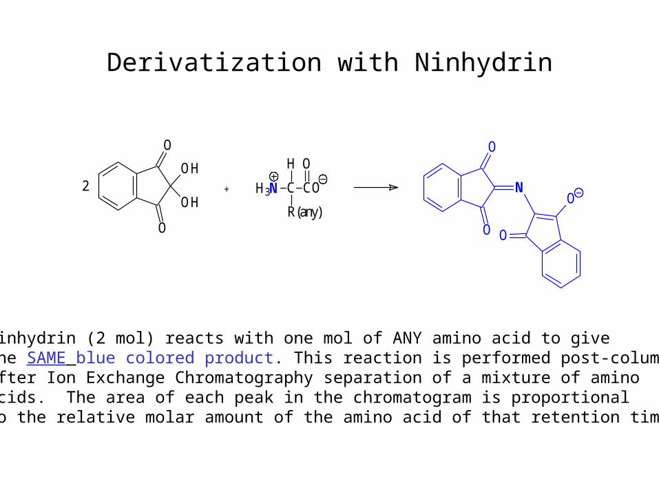
Derivatization with Ninhydrin
H3N C CO
H O
R(any)
O
O
OH
OH2 +
O
ON
O
O
Ninhydrin (2 mol) reacts with one mol of ANY amino acid to givethe SAME blue colored product. This reaction is performed post-column, after Ion Exchange Chromatography separation of a mixture of amino acids. The area of each peak in the chromatogram is proportional to the relative molar amount of the amino acid of that retention time.
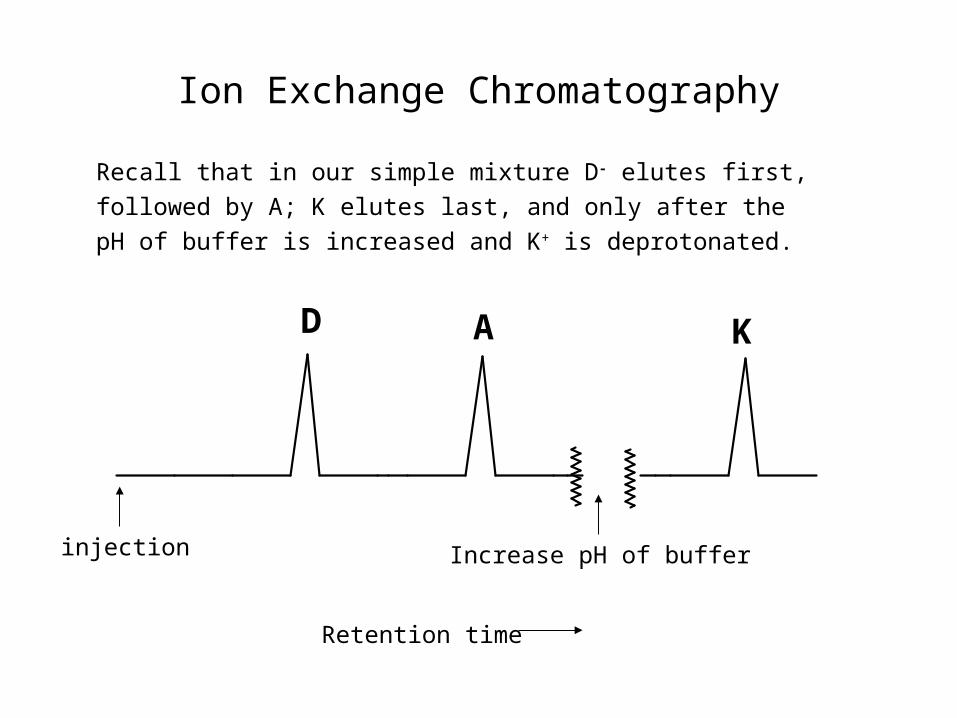
Ion Exchange Chromatography
A KD
injection Increase pH of buffer
Retention time
Recall that in our simple mixture D- elutes first,
followed by A; K elutes last, and only after the
pH of buffer is increased and K+ is deprotonated.
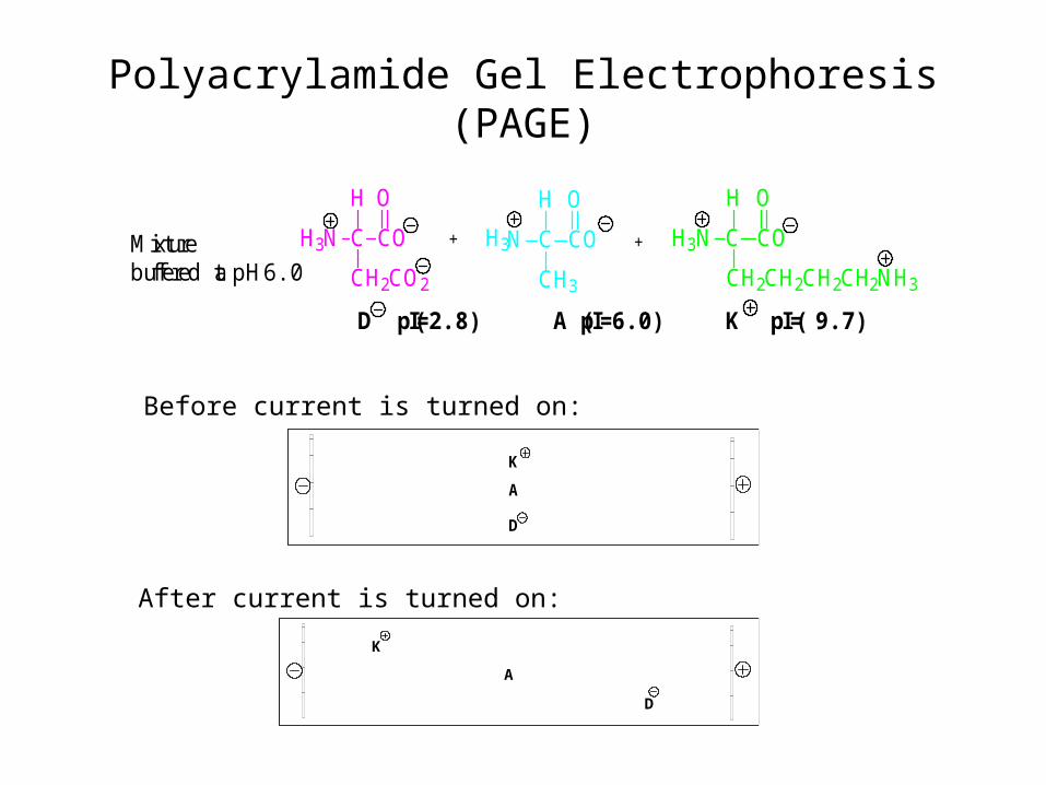
Polyacrylamide Gel Electrophoresis (PAGE)
H3 C CO
OH
CH2CO2
N
A (pI=6.0)
+ H3N
CH3
H O
COC
D (pI=2.8)
+ H3
CH2CH2CH2CH2NH3
H O
COCN
K (pI= 9.7)
Mixture of:buffered at pH 6.0
A
K
D
Before current is turned on:
After current is turned on:
A
K
D
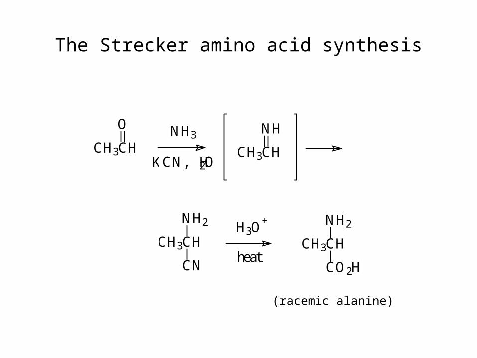
The Strecker amino acid synthesis
CH3CH
O
CH3CH
NH
CH3CH
NH2
CN
CH3CH
NH2
CO2H
NH3
KCN, H2O
H3O+
heat
(racemic alanine)
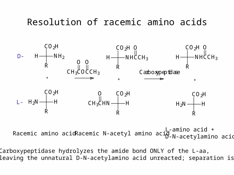
Resolution of racemic amino acids
Racemic amino acid
Carboxypeptidase hydrolyzes the amide bond ONLY of the L-aa, leaving the unnatural D-N-acetylamino acid unreacted; separation is simple
Racemic N-acetyl amino acidL-amino acid +D-N-acetylamino acid
D-
L-
H
R
NH2
CO2H
H2N
R
H
CO2H
+
H
R
NHCCH3
CO2H O
CH3CHN
R
H
CO2HO
+
Carboxypeptidase
H2N
R
H
CO2H
+
CH3COCCH3
O OH
R
NHCCH3
CO2H O
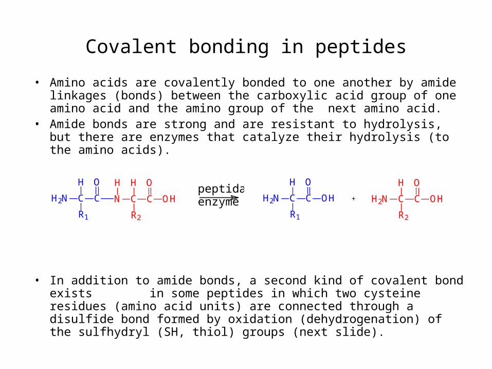
Covalent bonding in peptides
• Amino acids are covalently bonded to one another by amide linkages (bonds) between the carboxylic acid group of one amino acid and the amino group of the next amino acid.
• Amide bonds are strong and are resistant to hydrolysis, but there are enzymes that catalyze their hydrolysis (to the amino acids).
• In addition to amide bonds, a second kind of covalent bond exists in some peptides in which two cysteine residues (amino acid units) are connected through a disulfide bond formed by oxidation (dehydrogenation) of the sulfhydryl (SH, thiol) groups (next slide).
H2N C C
H
R1
O
OH H2N C C
H
R2
O
OH+enzyme
peptidaseH2N C C
H
R1
O
N C C
H
R2
O
OH
H
H2N C C
H
R1
O
OH H2N C C
H
R2
O
OH+enzyme
peptidaseH2N C C
H
R1
O
N C C
H
R2
O
OH
Hpeptidaseenzyme
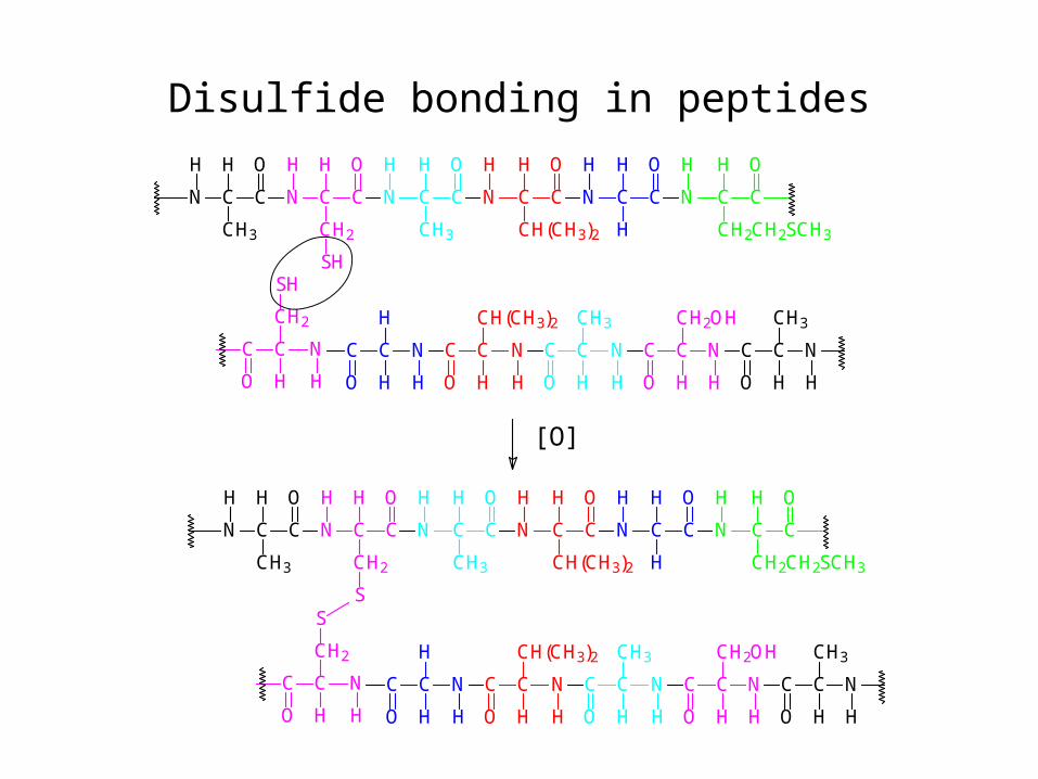
Disulfide bonding in peptides
C C
OH
CH2
H
NN C C
H O
CH3 CH2CH2SCH3
H O
CCN
H
C C
OH
H
N
H
C C
OH
CH(CH3)2
N
HH
N
CH3
H O
CC
SH
H
CC
O H
CH2OH
H
N CC
HO
CH3
CC
O H
H
N
H
CC
O H
CH(CH3)2
N
H H
N
CH3
HO
C C N
SH
H
NCC
O H
CH2
H
C C
OH
CH2
H
NN C C
H O
CH3 CH2CH2SCH3
H O
CCN
H
C C
OH
H
N
H
C C
OH
CH(CH3)2
N
HH
N
CH3
H O
CC
S
H
CC
O H
CH2OH
H
N CC
HO
CH3
CC
O H
H
N
H
CC
O H
CH(CH3)2
N
H H
N
CH3
HO
C C N
S
H
NCC
O H
CH2
H
[O]
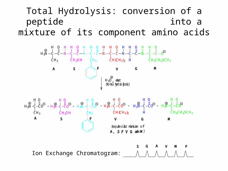
Total Hydrolysis: conversion of a peptide into a mixture of its component amino acids
A F V MG
H3O, heat(total hydrolysis)
S
C C
OH
CH2OH
H
NH3N C C
H O
CH3 CH2CH2SCH3
H O
CC ON
H
C C
OH
H
N
H
C C
OH
CH(CH3)2
N
HH
N
CH2
H O
CC
C CO
OH
CH2OH
NH3N C CO
H O
CH3
N
CH2
H O
COC C CO
OH
CH(CH3)2
N C CO
OH
H
N
CH2CH2SCH3
H O
COCNH3H3H3 H3 H3+ ++ + +
)
A
S
F V G M
A, S, F, V, G, and M(equimolar mixture of
S G A V M F
Ion Exchange Chromatogram:

2. Amino Acid Sequence: Primary Structure Determination of Peptides
• Total hydrolysis followed by and ion exchange chromatography and then ninhydrin derivatization tells us the identity and relative amount of each amino acid present in the peptide
• It gives NO INFORMATION about the sequence, or order of attachment of the amino acids, however.
• For this, we need to perform selective hydrolysis of the peptide.
• We’ll learn three methods:– Sanger’s reagent followed by total hydrolysis– Carboxypeptidase– Leucine aminopeptidase
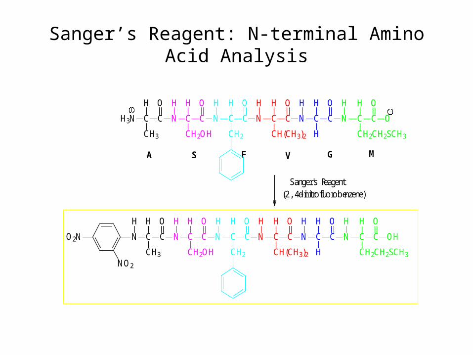
Sanger’s Reagent: N-terminal Amino Acid Analysis
S G MVFA
Sanger's Reagent(2,4-dinitrofluorobenzene)
C C
OH
CH2OH
H
NH3N C C
H O
CH3 CH2CH2SCH3
H O
CC ON
H
C C
OH
H
N
H
C C
OH
CH(CH3)2
N
HH
N
CH2
H O
CC
C C
OH
CH2OH
H
N
H
N C C
H O
CH3
O2N
NO2
CH2CH2SCH3
H O
CC OHN
H
C C
OH
H
N
H
C C
OH
CH(CH3)2
N
HH
N
CH2
H O
CC
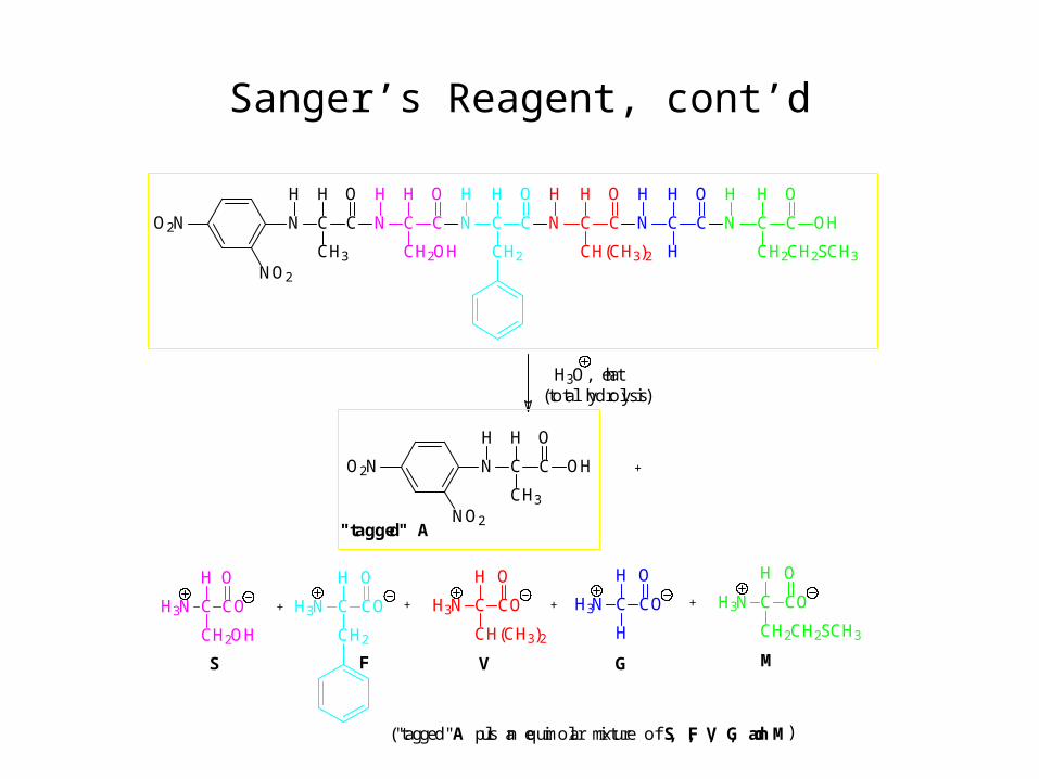
Sanger’s Reagent, cont’d
S
H3O, heat(total hydrolysis)
C C
OH
CH2OH
H
N
H
N C C
H O
CH3
O2N
NO2
CH2CH2SCH3
H O
CC OHN
H
C C
OH
H
N
H
C C
OH
CH(CH3)2
N
HH
N
CH2
H O
CC
C CO
OH
CH2OH
N N
CH2
H O
COC C CO
OH
CH(CH3)2
N C CO
OH
H
N
CH2CH2SCH3
H O
COCNH3H3H3 H3 H3
+
++ + +
"tagged" A
F V G M
) S, F, V, G, and M("tagged" A plus an equimolar mixture of
H
N C C
H O
CH3
O2N
NO2
OH
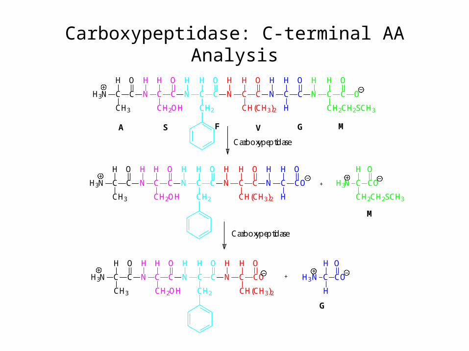
Carboxypeptidase: C-terminal AA Analysis
A F V MG
C C
OH
CH2OH
H
NH3N C C
H O
CH3 CH2CH2SCH3
H O
CC ON
H
C C
OH
H
N
H
C C
OH
CH(CH3)2
N
HH
N
CH2
H O
CC
C CO
OH
H
N
CH2CH2SCH3
H O
COCNH3
H3
+
S
G
M
Carboxypeptidase
C C
OH
CH2OH
H
NH3N C C
H O
CH3
C CO
OH
H
N
H
C C
OH
CH(CH3)2
N
HH
N
CH2
H O
CC
Carboxypeptidase
C C
OH
CH2OH
H
NH3N C C
H O
CH3
C CO
OH
CH(CH3)2
N
HH
N
CH2
H O
CC +
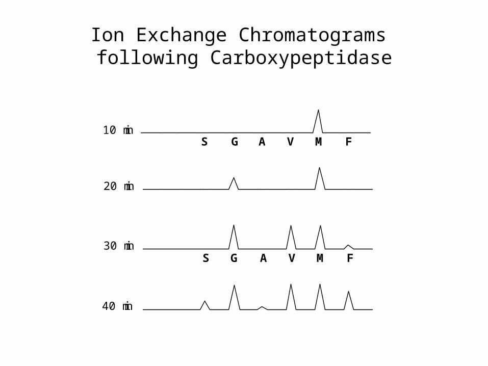
Ion Exchange Chromatograms following Carboxypeptidase
10 min
20 min
30 min
40 min
S G A V M F
S A V M FG
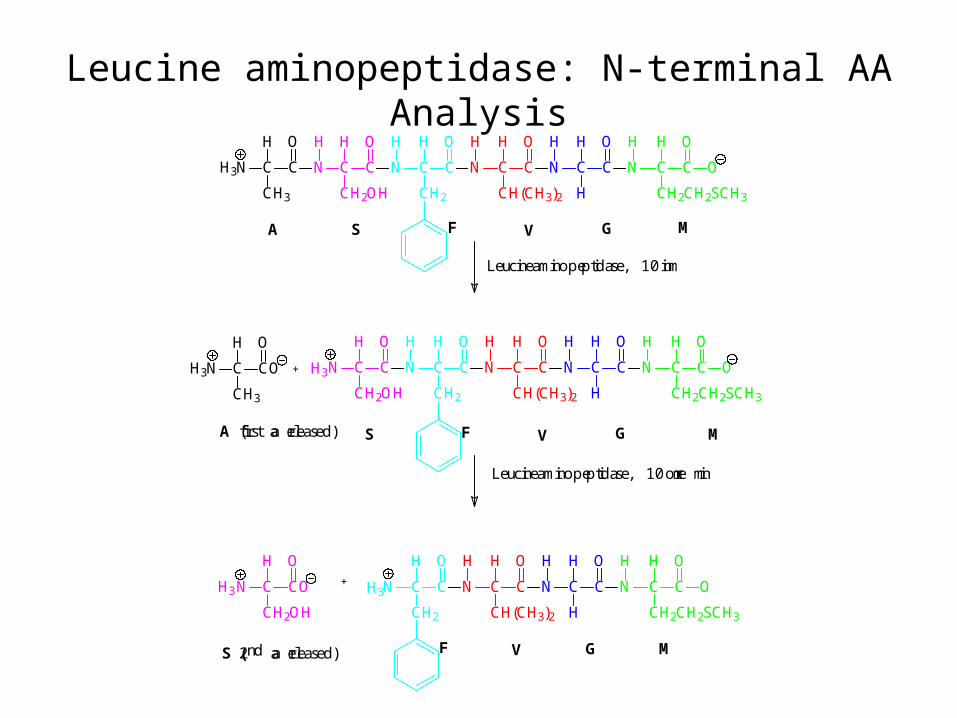
Leucine aminopeptidase: N-terminal AA Analysis
A F V MG
C C
OH
CH2OH
H
NH3N C C
H O
CH3 CH2CH2SCH3
H O
CC ON
H
C C
OH
H
N
H
C C
OH
CH(CH3)2
N
HH
N
CH2
H O
CC
S
A (first aa released) F V GS
Leucineaminopeptidase, 10 min
Leucineaminopeptidase, 10 more min
F VS (2nd aa released) G
+
M
C C
OH
CH2OH
N
CH2CH2SCH3
H O
CC ON
H
C C
OH
H
N
H
C C
OH
CH(CH3)2
N
HH
N
CH2
H O
CCH3N C CO
H O
CH3
H3
C CO
OH
CH2OH
N
M
CH2CH2SCH3
H O
CC ON
H
C C
OH
H
N
H
C C
OH
CH(CH3)2
N
H
N
CH2
H O
CCH3 H3+
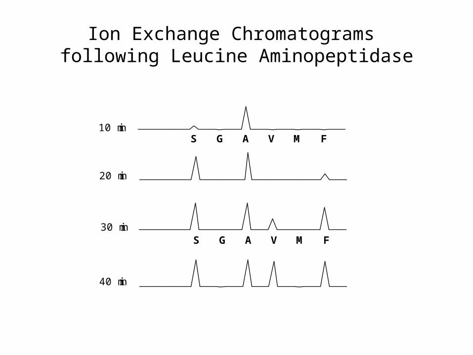
Ion Exchange Chromatograms following Leucine Aminopeptidase
S G A V M F
S G A V M F
10 min
20 min
30 min
40 min
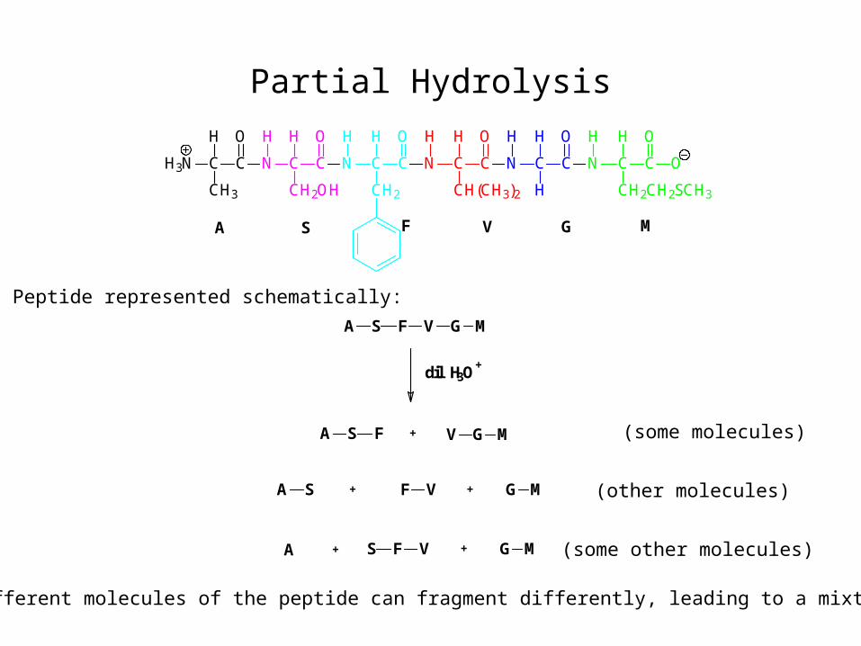
Partial Hydrolysis
A F V MG
C C
OH
CH2OH
H
NH3N C C
H O
CH3 CH2CH2SCH3
H O
CC ON
H
C C
OH
H
N
H
C C
OH
CH(CH3)2
N
HH
N
CH2
H O
CC
S
A S F V G M
A S F
A S
dil H3O+
V G M
F V G M
+
+
+
S F V G M+A
+
Peptide represented schematically:
(different molecules of the peptide can fragment differently, leading to a mixture)
(some molecules)
(other molecules)
(some other molecules)
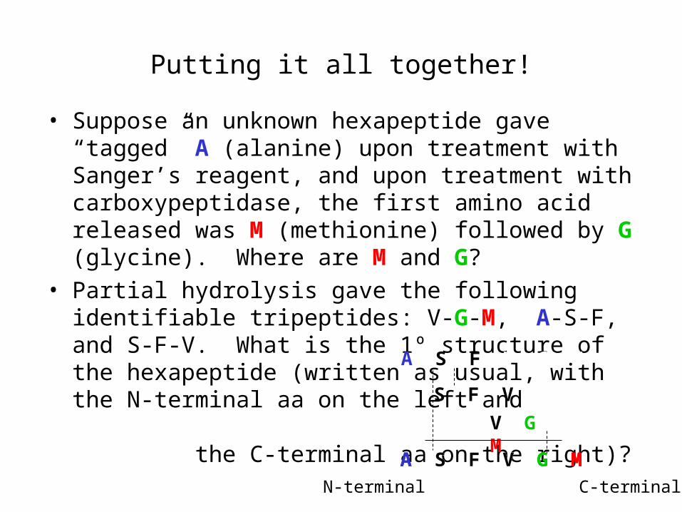
Putting it all together!
• Suppose an unknown hexapeptide gave “tagged” A (alanine) upon treatment with Sanger’s reagent, and upon treatment with carboxypeptidase, the first amino acid released was M (methionine) followed by G (glycine). Where are M and G?
• Partial hydrolysis gave the following identifiable tripeptides: V-G-M, A-S-F, and S-F-V. What is the 1º structure of the hexapeptide (written as usual, with the N-terminal aa on the left and the C-terminal aa on the right)?
A S F V G MA S F V G M
V G M
A S F V G M
A S F V G M
A S F V G M
N-terminal C-terminal