Embed Size (px)
Citation preview
An IL-17–dominant immune profile is shared acrossthe major orphan forms of ichthyosis
Amy S. Paller, MD, MS,a Yael Renert-Yuval, MD,b Maria Suprun, MPH,c Hitokazu Esaki, MD,b,d Margeaux Oliva, BA,b
Thy Nhat Huynh, MD,a Benjamin Ungar, BA,d Norma Kunjravia, MD,d Rivka Friedland, MD,a Xiangyu Peng, MSc,b
Xiuzhong Zheng, MSc,d Yeriel D. Estrada, BSc,b James G. Krueger, MD, PhD,c Keith A. Choate,e
Mayte Su�arez-Fari~nas, PhD,b,c,d,f and EmmaGuttman-Yassky,MD, PhDb,d Chicago, Ill, New York, NY, and NewHaven, Conn
Background: The ichthyoses are rare genetic disordersassociated with generalized scaling, erythema, and epidermalbarrier impairment. Pathogenesis-based therapy is largelylacking because the underlying molecular basis is poorlyunderstood.Objective: We sought to characterize molecularly cutaneousinflammation and its correlation with clinical and barriercharacteristics.Methods: We analyzed biopsy specimens from 21 genotypedpatients with ichthyosis (congenital ichthyosiformerythroderma, n 5 6; lamellar ichthyosis, n 5 7; epidermolyticichthyosis, n 5 5; and Netherton syndrome, n 5 3) usingimmunohistochemistry and RT-PCR and compared them withspecimens from healthy control subjects, patients with atopicdermatitis (AD), and patients with psoriasis. Clinical measuresincluded the Ichthyosis Area Severity Index (IASI), whichintegrates erythema (IASI-E) and scaling (IASI-S);transepidermal water loss; and pruritus.
From athe Departments of Dermatology and Pediatrics, Northwestern University Fein-
berg School of Medicine, Chicago; the Departments of bDermatology and cPopulation
Health Science and Policy and fthe Icahn Institute for Genomics and Multiscale
Biology, Icahn School of Medicine at Mount Sinai, New York; dthe Laboratory for
Investigative Dermatology, Rockefeller University, New York; and ethe Department
of Dermatology, Yale University School of Medicine, New Haven.
Supported by the Foglia Family Foundation Endowment and the National Psoriasis
Foundation (RF fellowship).
This research was supported by the Foglia Family Foundation Endowment and the
National Psoriasis Foundation (RF fellowship). We acknowledge Core resources
provided by the Northwestern University Skin Disease Research Center (NIAMS
P30AR057216).
Disclosure of potential conflict of interest: A. S. Paller has received consultancy fees from
Anacor, Galderma, Stiefel/GlaxoSmithKline, Novartis, Regeneron, and Vitae
Pharmaceuticals and has received grants from Anacor, Astellas, and LEO Pharma.
J. G. Krueger has received personal fees and/or fees to his institution from Novartis,
Pfizer, Amgen, Lilly, Merck, Kadmon, Dermira, Boehringer, Innovaderm, Kyowa,
BMS, Janssen, Serono, Biogen Idec, Delenex, AbbVie, Sanofi, Baxter, Paraxel,
Xenoport, and Kineta. K. A. Choate has received consultancy fees from Alderya
Therapeutics and payment for lectures from Abbvie and Janssen. M. Su�arez-Fari~nas
has received grants from Pfizer, Quorum Consulting, and Genisphere. E. Guttman-
Yassky has received board memberships from Sanofi Aventis, Regeneron, Stiefel/
GlaxoSmithKline, MedImmune, Celgene, Anacor, Leo Pharma, AnaptysBio, Celsus,
Dermira, Galderma, Novartis, Pfizer, and Vitae; consultancy fees from Regeneron,
Sanofi Aventis, Medimmune, Celgene, Stiefel/GlaxoSmithKline, Celsus, BMS,
Amgen, Drais, AbbVie, Anacor, AnaptysBio, Dermira, Galderma, Leo Pharma,
Novartis, Pfizer, Vitae, Mitsubishi Tanabe, and Eli Lilly; and grants/grants pending
from Regeneron, Celgene, BMS, Janssen, Dermira, Leo Pharma, Merck, and Novartis.
The rest of the authors declare that they have no relevant conflicts of interest.
Received for publication May 2, 2016; revised June 18, 2016; accepted for publication
July 19, 2016.
Corresponding author: Amy S. Paller, MD, Departments of Dermatology and Pediatrics,
Northwestern University Feinberg School of Medicine, Chicago, IL 60611. E-mail:
0091-6749/$36.00
� 2016 American Academy of Allergy, Asthma & Immunology
http://dx.doi.org/10.1016/j.jaci.2016.07.019
Results: Ichthyosis samples showed increased epidermalhyperplasia (increased thickness and keratin 16 expression) andT-cell and dendritic cell infiltrates. Increases of generalinflammatory (IL-2), innate (IL-1b), and some TH1/interferon(IFN-g) markers in patients with ichthyosis were comparablewith those in patients with psoriasis or AD. TNF-a levels inpatients with ichthyosis were increased only in those withNetherton syndrome but were much lower than in patients withpsoriasis and those with AD. Expression of TH2 cytokines (IL-13and IL-31) was similar to that seen in control subjects. Thestriking induction of IL-17–related genes or markerssynergistically induced by IL-17 and TNF-a (IL-17A/C, IL-19,CXCL1, PI3, CCL20, and IL36G; P < .05) in patients withichthyosis was similar to that seen in patients with psoriasis.IASI and IASI-E scores strongly correlated with IL-17A(r 5 0.74, P < .001) and IL-17/TNF–synergistic/additive geneexpression. These markers also significantly correlated withtransepidermal water loss, suggesting a link between the barrierdefect and inflammation in patients with ichthyosis.Conclusion: Our data associate a shared TH17/IL-23 immunefingerprint with the major orphan forms of ichthyosis and raisethe possibility of IL-17–targeting strategies. (J Allergy ClinImmunol 2016;nnn:nnn-nnn.)
Key words: Epidermis, ichthyosis, inflammation, autosomalrecessive congenital ichthyosis, congenital ichthyosiform erythro-derma, lamellar ichthyosis, Netherton syndrome, epidermolyticichthyosis, skin, IL-17, TNF-a
Ichthyoses are genetically and clinically heterogeneousdisorders with generalized skin scaling, thickening, anderythema. Other than ichthyosis vulgaris and recessive X-linkedichthyosis subtypes,1-7 the ichthyoses each occur in less than1:100,000 persons. Affected subjects have an extremelycompromised quality of life because of disfigurement and theaccompanying itching, pain, and functional limitation.8,9 Theepidermal barrier is abnormal, with defects in lipids and differenti-ation resulting in increased transepidermalwater loss (TEWL).10-12
Treatment for ichthyosis is largely supportive and unsatisfac-tory. For more severely affected subjects, oral retinoids, vitaminA analogues, are often administered to improve the hyperkera-tosis.13-15 However, retinoids can worsen skin inflammation andpruritus and have deleterious effects (hypertriglyceridemia,teratogenicity, and hyperostosis),16 limiting their use. Topicalanti-inflammatory medications (ie, steroids and calcineurininhibitors) are often ineffective and easily absorbed systemically,restricting chronic use.17,18 Thus a huge unmet need exists forsafe and more effective treatments that will ideally also targetthe erythema/inflammation.
1
J ALLERGY CLIN IMMUNOL
nnn 2016
2 PALLER ET AL
Abbreviations used
AD: A
topic dermatitisAMP: A
ntimicrobial peptideARCI: A
utosomal recessive congenital ichthyosisCIE: C
ongenital ichthyosiform erythrodermaCISI: C
ongenital Ichthyoses Severity IndexDC: D
endritic cellDC-LAMP: D
endritic cell lysosomal-associated membrane proteinDEFB4: b
-Defensin-B4EI: E
pidermolytic ichthyosisFLG: F
ilaggrinhARP: H
uman acidic ribosomal proteinIASI: Ic
hthyosis Area Severity IndexIASI-E: Ic
hthyosis Area Severity Index–ErythemaIASI-S: Ic
hthyosis Area Severity Index–ScalingIHC: Im
munohistochemistryK16: K
eratin 16LCN2: L
ipocalin 2LI: L
amellar ichthyosisLOR: L
oricrinNS: N
etherton syndromePAR2: P
rotease-activated receptor 2PPL: P
eriplakinTEWL: T
ransepidermal water lossTSLP: T
hymic stromal lymphopoietinDespite elucidation of the genetic basis for the various forms ofichthyosis, their underlying molecular mechanisms are poorlyunderstood, with our knowledge predominantly based on cultureand animal models.19-29 These model systems chiefly focus onabnormal barrier function and lipid homeostasis, with little atten-tion paid to immune disturbances.6,30,31 Human studies, largelylimited to Netherton syndrome (NS) and the lamellar ichthyosis(LI) phenotype of autosomal recessive congenital ichthyosis(ARCI), have examined just a few cytokines.32-38 Blood analysesfound inconsistent TH2 skewing39 and increases in levels ofproinflammatory cytokines (TNF-a, IL-1b, IL-2, andIL-18).40-42 Skin studies showed increased expression of TNF-aand IL-1b in patients with ARCI-LI35 and of protease-activatedreceptor 2 (PAR2),32 thymic stromal lymphopoietin (TSLP),TNF-a, IL-8,43 and the TH2 cytokine IL-33 in patients withNS,38 which are often coupled with increased expression ofterminal differentiation products (ie, filaggrin [FLG], loricrin[LOR], and involucrin), and lipid impairement.32,35,37,38 Studiesof response to systemic treatments, including retinoids (n 5 11),anti-TNF (n5 1), and oral corticosteroids combined with omali-zumab (n 5 1), in patients with ARCI-LI and those with NS,respectively,33-35 have only assessed a few cytokines. Therapy-induced decreases in IL-1b, IL-8, TSLP, IL-5, and IL-17A levelswere found in patients with NS, whereas IL-1a and TNF-a levelswere decreased (nonsignificantly) in patients with ARCI-LI.
To elucidate the basis for the cutaneous inflammation seen inpatients with ichthyosis and its correlation with clinicalcharacteristics, we analyzed skin from 21 patients with themost prevalent orphan forms of severe ichthyosis: ARCI-LI,ARCI–congenital ichthyosiform erythroderma (CIE), epidermo-lytic ichthyosis (EI), and NS. All subtypes showed cutaneousskewing of TH17 expression, which correlated with diseaseseverity. This TH17 profile most closely resembled that ofpsoriasis, in which IL-17 antagonism is highly effective inreversing the inflammation and epidermal pathology.44-47 These
data can lead to a new treatment paradigm targeting the TH17/IL-23 pathway in patients with ichthyosis.
METHODS
Patients’ characteristicsTwenty-one patients (aged 10-57 years)with ichthyosis and knownmutations
were enrolled (Tables I and II and see Table E1 and the Methods section in this
article’s Online Repository at www.jacionline.org). Written institutional review
board–approved consent was provided by subjects (>_12 years) and parents
(<18 years). Demographic information, medical history, physical examination,
clinical severity scores, pruritus (5-D itch scale and Itch Numeric Rating Scale),
photography, and TEWLmeasurement were captured. Few scoring instruments
have been used for ichthyosis severity, and the only one tested for reliability is
the Congenital Ichthyoses Severity Index (CISI). In addition to scoring
erythema/redness and hyperkeratosis/scaling, CISI measures alopecia (not a
feature in most patients) and does not score potential differences in body
regions.48Given its limitations,wemodified theCISI scale, eliminating alopecia
and prorating intensity based on body region and extent to create a composite
score similar to the Psoriasis Area and Severity Index.49 This Ichthyosis Area
and Severity Index (IASI) measures the severity of the erythema (Ichthyosis
Area Severity Index–Erythema [IASI-E]) and scaling (Ichthyosis Area
Severity Index–Scaling [IASI-S]), adding them together to a total IASI score
(Tables I and II and see Table E2 and theMethods section in this article’s Online
Repository at www.jacionline.org).
Four-millimeter biopsy specimens were collected and assessed in parallel
with tissue from healthy subjects, patients with atopic dermatitis (AD), and
patients with psoriasis previously published by our group.50-54 Genotyping for
FLGmutations in the AD cohort was previously performed on 4 patients, and
results were negative.51 Four samples of healthy adolescents were included
(see Table E3 in this article’s Online Repository at www.jacionline.org) for
comparison with the younger ichthyosis cohort. Patients’ characteristics are
presented in Tables I and II and Table E1 and E3.
Quantitative RT-PCRRT-PCR was performed, as previously described.55,56 Expression values
were normalized to human acidic ribosomal protein (hARP).
ImmunohistochemistryImmunohistochemistry (IHC) was performed on frozen sections, as previ-
ously described.57 Antibodies are shown in Table E4 and cell counts are shown
in Table E5 in this article’s Online Repository at www.jacionline.org.
Statistical analysesExcept for RT-PCR expression values, no other missing value imputations
were performed. All available observations were included in analyses, which
were performed by using the statistical language R (www.R-project.org).
Differences in expression values (in log2 scale), cell counts, and clinical
variables were assessed by using linear models, which were age adjusted to
account for significant differences in age distributions.
Unsupervised hierarchical clustering of variables or samples/patients was
performed by using the Pearson correlation coefficient as a distance metric
with the McQuitty agglomeration algorithm. The results are represented as a
heat map with a dendrogram and a tree or phylogram (using R package ape).
The uncertainty of the hierarchical clustering analysis was assessed by
using multiscale bootstrap resampling (extended statistics are shown in the
Methods section in this article’s Online Repository).
RESULTS
Demographics and clinical characteristics of
patients with ichthyosisTwenty-one patients aged 10 years or greater with ARCI-CIE
(n 5 6), ARCI-LI (n 5 7), EI (n 5 5), or NS (n 5 3) and with
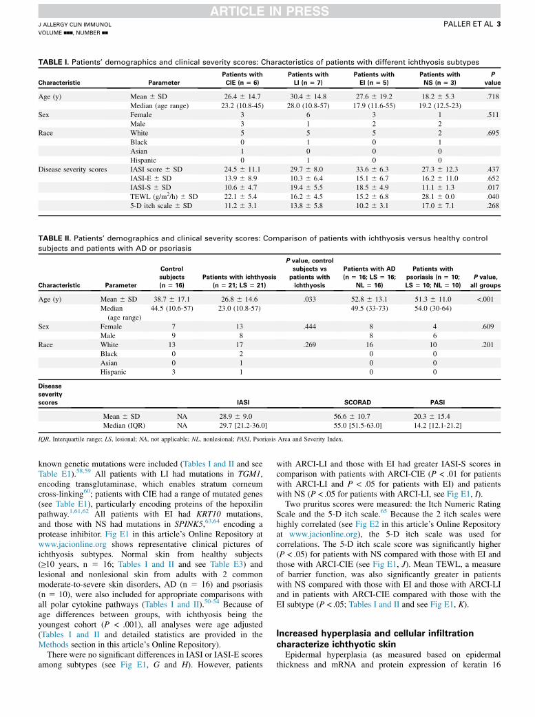
TABLE I. Patients’ demographics and clinical severity scores: Characteristics of patients with different ichthyosis subtypes
Characteristic Parameter
Patients with
CIE (n 5 6)
Patients with
LI (n 5 7)
Patients with
EI (n 5 5)
Patients with
NS (n 5 3)
P
value
Age (y) Mean 6 SD 26.4 6 14.7 30.4 6 14.8 27.6 6 19.2 18.2 6 5.3 .718
Median (age range) 23.2 (10.8-45) 28.0 (10.8-57) 17.9 (11.6-55) 19.2 (12.5-23)
Sex Female 3 6 3 1 .511
Male 3 1 2 2
Race White 5 5 5 2 .695
Black 0 1 0 1
Asian 1 0 0 0
Hispanic 0 1 0 0
Disease severity scores IASI score 6 SD 24.5 6 11.1 29.7 6 8.0 33.6 6 6.3 27.3 6 12.3 .437
IASI-E 6 SD 13.9 6 8.9 10.3 6 6.4 15.1 6 6.7 16.2 6 11.0 .652
IASI-S 6 SD 10.6 6 4.7 19.4 6 5.5 18.5 6 4.9 11.1 6 1.3 .017
TEWL (g/m2/h) 6 SD 22.1 6 5.4 16.2 6 4.5 15.2 6 6.8 28.1 6 0.0 .040
5-D itch scale 6 SD 11.2 6 3.1 13.8 6 5.8 10.2 6 3.1 17.0 6 7.1 .268
TABLE II. Patients’ demographics and clinical severity scores: Comparison of patients with ichthyosis versus healthy control
subjects and patients with AD or psoriasis
Characteristic Parameter
Control
subjects
(n 5 16)
Patients with ichthyosis
(n 5 21; LS 5 21)
P value, control
subjects vs
patients with
ichthyosis
Patients with AD
(n 5 16; LS 5 16;
NL 5 16)
Patients with
psoriasis (n 5 10;
LS 5 10; NL 5 10)
P value,
all groups
Age (y) Mean 6 SD 38.7 6 17.1 26.8 6 14.6 .033 52.8 6 13.1 51.3 6 11.0 <.001
Median
(age range)
44.5 (10.6-57) 23.0 (10.8-57) 49.5 (33-73) 54.0 (30-64)
Sex Female 7 13 .444 8 4 .609
Male 9 8 8 6
Race White 13 17 .269 16 10 .201
Black 0 2 0 0
Asian 0 1 0 0
Hispanic 3 1 0 0
Disease
severity
scores IASI SCORAD PASI
Mean 6 SD NA 28.9 6 9.0 56.6 6 10.7 20.3 6 15.4
Median (IQR) NA 29.7 [21.2-36.0] 55.0 [51.5-63.0] 14.2 [12.1-21.2]
IQR, Interquartile range; LS, lesional; NA, not applicable; NL, nonlesional; PASI, Psoriasis Area and Severity Index.
J ALLERGY CLIN IMMUNOL
VOLUME nnn, NUMBER nn
PALLER ET AL 3
known genetic mutations were included (Tables I and II and seeTable E1).58,59 All patients with LI had mutations in TGM1,encoding transglutaminase, which enables stratum corneumcross-linking60; patients with CIE had a range of mutated genes(see Table E1), particularly encoding proteins of the hepoxilinpathway.1,61,62 All patients with EI had KRT10 mutations,and those with NS had mutations in SPINK5,63,64 encoding aprotease inhibitor. Fig E1 in this article’s Online Repository atwww.jacionline.org shows representative clinical pictures ofichthyosis subtypes. Normal skin from healthy subjects(>_10 years, n 5 16; Tables I and II and see Table E3) andlesional and nonlesional skin from adults with 2 commonmoderate-to-severe skin disorders, AD (n 5 16) and psoriasis(n 5 10), were also included for appropriate comparisons withall polar cytokine pathways (Tables I and II).50-54 Because ofage differences between groups, with ichthyosis being theyoungest cohort (P < .001), all analyses were age adjusted(Tables I and II and detailed statistics are provided in theMethods section in this article’s Online Repository).
There were no significant differences in IASI or IASI-E scoresamong subtypes (see Fig E1, G and H). However, patients
with ARCI-LI and those with EI had greater IASI-S scores incomparison with patients with ARCI-CIE (P < .01 for patientswith ARCI-LI and P < .05 for patients with EI) and patientswith NS (P < .05 for patients with ARCI-LI, see Fig E1, I).
Two pruritus scores were measured: the Itch Numeric RatingScale and the 5-D itch scale.65 Because the 2 itch scales werehighly correlated (see Fig E2 in this article’s Online Repositoryat www.jacionline.org), the 5-D itch scale was used forcorrelations. The 5-D itch scale score was significantly higher(P < .05) for patients with NS compared with those with EI andthose with ARCI-CIE (see Fig E1, J). Mean TEWL, a measureof barrier function, was also significantly greater in patientswith NS compared with those with EI and those with ARCI-LIand in patients with ARCI-CIE compared with those with theEI subtype (P < .05; Tables I and II and see Fig E1, K).
Increased hyperplasia and cellular infiltration
characterize ichthyotic skinEpidermal hyperplasia (as measured based on epidermal
thickness and mRNA and protein expression of keratin 16
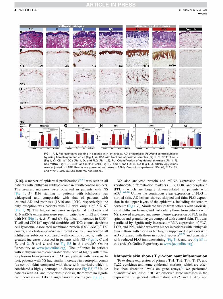
FIG 1. A-E, Representative staining in patients with ichthyoses, AD, or psoriasis (PSO) and control subjects
by using hematoxylin and eosin (Fig 1, A), K16 with fractions of positive samples (Fig 1, B), CD31 T cells
(Fig 1, C), CD11c1 DCs (Fig 1, D), and FLG (Fig 1, E). F-J, Quantification of epidermal thickness (Fig 1, F),K16 mRNA (Fig 1, G), CD31 and CD11c1 cells (Fig 1, H and I), and FLG mRNA (Fig 1, J). mRNA log2 values
were adjusted to hARP. Results are presented as means 6 SEMs. Control comparisons: *P < .05, **P < .01,
and ***P < .001. LS, Lesional; NL, nonlesional.
J ALLERGY CLIN IMMUNOL
nnn 2016
4 PALLER ET AL
[K16], a marker of epidermal proliferation)66,67 was seen in allpatients with ichthyosis subtypes compared with control subjects.The greatest increases were observed in patients with NS(Fig 1, A). K16 staining in patients with ichthyosis waswidespread and comparable with that of patients withlesional AD and psoriasis (16/16 and 10/10, respectively); theonly exception was patients with LI, with only 3 of 7 K161
(Fig 1, B). The highest increases in epidermal thickness andK16 mRNA expression were seen in patients with EI and thosewith NS (Fig 1, A, B, F, and G). Significant increases in CD31
T-cell and CD11c1 myeloid dendritic cell (DC) counts, dendriticcell lysosomal-associated membrane protein (DC-LAMP)1 DCcounts, and elastase-positive neutrophil counts characterized allichthyosis subtypes compared with control subjects, with thegreatest increases observed in patients with NS (Figs 1, C andD, and 2, H and I, and see Fig E3 in this article’s OnlineRepository at www.jacionline.org). The infiltrates in patientswith ichthyosis were comparable with those of highly inflamma-tory lesions from patients with AD and patients with psoriasis. Infact, patients with NS had similar increases in neutrophil counts(vs control skin) compared with those with psoriasis, which isconsidered a highly neutrophilic disease (see Fig E3).54 Unlikepatients with AD and those with psoriasis, there were no signifi-cant increases in CD1a1 Langerhans cell counts (see Fig E3).
We also analyzed protein and mRNA expression of thekeratinocyte differentiation markers (FLG, LOR, and periplakin[PPL]), which are largely downregulated in patients withAD.11,68-70 Unlike the continuous clear expression of FLG innormal skin, AD lesions showed skipped and faint FLG expres-sion in the upper layers of the epidermis, including the stratumcorneum (Fig 1,E). Similar to tissues from patients with psoriasis,most ichthyosis tissues, and particularly those from patients withNS, showed increased and more intense expression of FLG in thespinous and granular layers compared with control skin. This wasparalleled by significantly increased mRNA expression of FLG,LOR, and PPL, which was even higher in patients with ichthyosisthan in thosewith psoriasis but largely suppressed in patients withAD compared with those in control subjects69,71 and consistentwith reduced FLG immunostaining (Fig 1, J, and see Fig E4 inthis article’s Online Repository at www.jacionline.org).
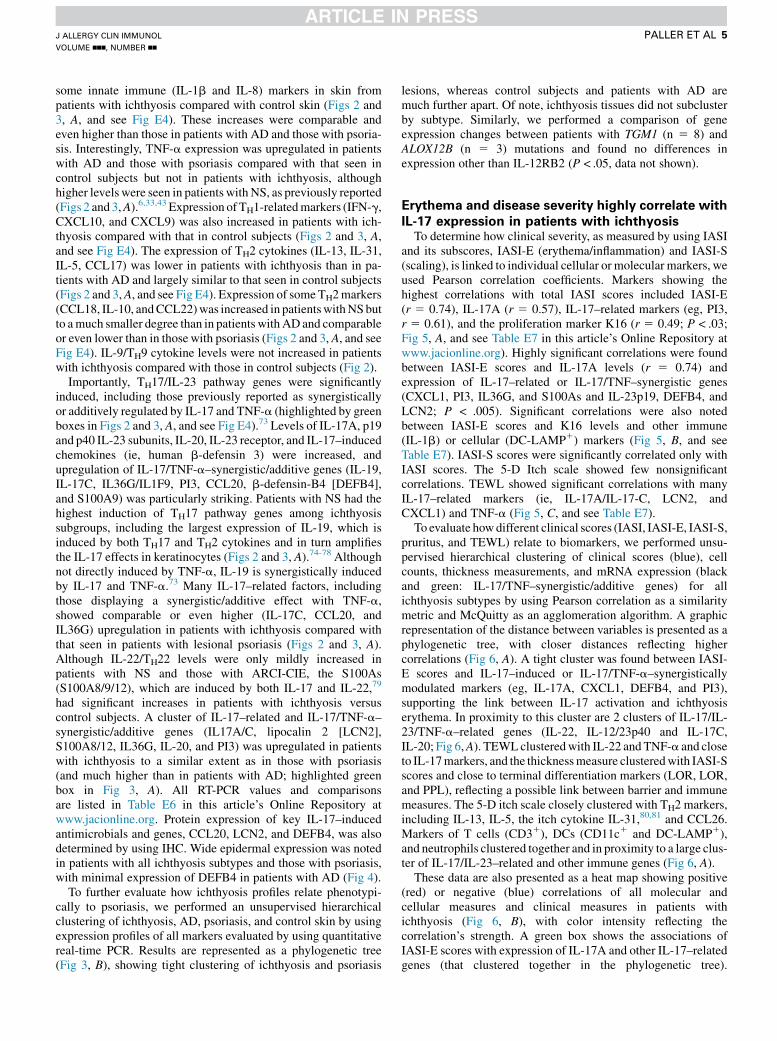
Ichthyotic skin shows TH17-dominant inflammationTo evaluate expression of primary TH1, TH2, TH9, TH17, and
TH22 cytokines and some epidermal markers, which are oftenless than detection levels on gene arrays,72 we performedquantitative real-time PCR. We observed large increases in theexpression of general inflammatory (IL-2 and IL-15) and
J ALLERGY CLIN IMMUNOL
VOLUME nnn, NUMBER nn
PALLER ET AL 5
some innate immune (IL-1b and IL-8) markers in skin frompatients with ichthyosis compared with control skin (Figs 2 and3, A, and see Fig E4). These increases were comparable andeven higher than those in patients with AD and those with psoria-sis. Interestingly, TNF-a expression was upregulated in patientswith AD and those with psoriasis compared with that seen incontrol subjects but not in patients with ichthyosis, althoughhigher levels were seen in patients with NS, as previously reported(Figs 2 and 3,A).6,33,43 Expression of TH1-relatedmarkers (IFN-g,CXCL10, and CXCL9) was also increased in patients with ich-thyosis compared with that in control subjects (Figs 2 and 3, A,and see Fig E4). The expression of TH2 cytokines (IL-13, IL-31,IL-5, CCL17) was lower in patients with ichthyosis than in pa-tients with AD and largely similar to that seen in control subjects(Figs 2 and 3,A, and see Fig E4). Expression of someTH2markers(CCL18, IL-10, andCCL22)was increased in patientswithNSbutto amuch smaller degree than in patients withADand comparableor even lower than in those with psoriasis (Figs 2 and 3, A, and seeFig E4). IL-9/TH9 cytokine levels were not increased in patientswith ichthyosis compared with those in control subjects (Fig 2).
Importantly, TH17/IL-23 pathway genes were significantlyinduced, including those previously reported as synergisticallyor additively regulated by IL-17 and TNF-a (highlighted by greenboxes in Figs 2 and 3, A, and see Fig E4).73 Levels of IL-17A, p19and p40 IL-23 subunits, IL-20, IL-23 receptor, and IL-17–inducedchemokines (ie, human b-defensin 3) were increased, andupregulation of IL-17/TNF-a–synergistic/additive genes (IL-19,IL-17C, IL36G/IL1F9, PI3, CCL20, b-defensin-B4 [DEFB4],and S100A9) was particularly striking. Patients with NS had thehighest induction of TH17 pathway genes among ichthyosissubgroups, including the largest expression of IL-19, which isinduced by both TH17 and TH2 cytokines and in turn amplifiesthe IL-17 effects in keratinocytes (Figs 2 and 3, A).74-78 Althoughnot directly induced by TNF-a, IL-19 is synergistically inducedby IL-17 and TNF-a.73 Many IL-17–related factors, includingthose displaying a synergistic/additive effect with TNF-a,showed comparable or even higher (IL-17C, CCL20, andIL36G) upregulation in patients with ichthyosis compared withthat seen in patients with lesional psoriasis (Figs 2 and 3, A).Although IL-22/TH22 levels were only mildly increased inpatients with NS and those with ARCI-CIE, the S100As(S100A8/9/12), which are induced by both IL-17 and IL-22,79
had significant increases in patients with ichthyosis versuscontrol subjects. A cluster of IL-17–related and IL-17/TNF-a–synergistic/additive genes (IL17A/C, lipocalin 2 [LCN2],S100A8/12, IL36G, IL-20, and PI3) was upregulated in patientswith ichthyosis to a similar extent as in those with psoriasis(and much higher than in patients with AD; highlighted greenbox in Fig 3, A). All RT-PCR values and comparisonsare listed in Table E6 in this article’s Online Repository atwww.jacionline.org. Protein expression of key IL-17–inducedantimicrobials and genes, CCL20, LCN2, and DEFB4, was alsodetermined by using IHC. Wide epidermal expression was notedin patients with all ichthyosis subtypes and those with psoriasis,with minimal expression of DEFB4 in patients with AD (Fig 4).
To further evaluate how ichthyosis profiles relate phenotypi-cally to psoriasis, we performed an unsupervised hierarchicalclustering of ichthyosis, AD, psoriasis, and control skin by usingexpression profiles of all markers evaluated by using quantitativereal-time PCR. Results are represented as a phylogenetic tree(Fig 3, B), showing tight clustering of ichthyosis and psoriasis
lesions, whereas control subjects and patients with AD aremuch further apart. Of note, ichthyosis tissues did not subclusterby subtype. Similarly, we performed a comparison of geneexpression changes between patients with TGM1 (n 5 8) andALOX12B (n 5 3) mutations and found no differences inexpression other than IL-12RB2 (P < .05, data not shown).
Erythema and disease severity highly correlate with
IL-17 expression in patients with ichthyosisTo determine how clinical severity, as measured by using IASI
and its subscores, IASI-E (erythema/inflammation) and IASI-S(scaling), is linked to individual cellular or molecular markers, weused Pearson correlation coefficients. Markers showing thehighest correlations with total IASI scores included IASI-E(r 5 0.74), IL-17A (r 5 0.57), IL-17–related markers (eg, PI3,r 5 0.61), and the proliferation marker K16 (r 5 0.49; P < .03;Fig 5, A, and see Table E7 in this article’s Online Repository atwww.jacionline.org). Highly significant correlations were foundbetween IASI-E scores and IL-17A levels (r 5 0.74) andexpression of IL-17–related or IL-17/TNF–synergistic genes(CXCL1, PI3, IL36G, and S100As and IL-23p19, DEFB4, andLCN2; P < .005). Significant correlations were also notedbetween IASI-E scores and K16 levels and other immune(IL-1b) or cellular (DC-LAMP1) markers (Fig 5, B, and seeTable E7). IASI-S scores were significantly correlated only withIASI scores. The 5-D Itch scale showed few nonsignificantcorrelations. TEWL showed significant correlations with manyIL-17–related markers (ie, IL-17A/IL-17-C, LCN2, andCXCL1) and TNF-a (Fig 5, C, and see Table E7).
To evaluate howdifferent clinical scores (IASI, IASI-E, IASI-S,pruritus, and TEWL) relate to biomarkers, we performed unsu-pervised hierarchical clustering of clinical scores (blue), cellcounts, thickness measurements, and mRNA expression (blackand green: IL-17/TNF–synergistic/additive genes) for allichthyosis subtypes by using Pearson correlation as a similaritymetric and McQuitty as an agglomeration algorithm. A graphicrepresentation of the distance between variables is presented as aphylogenetic tree, with closer distances reflecting highercorrelations (Fig 6, A). A tight cluster was found between IASI-E scores and IL-17–induced or IL-17/TNF-a–synergisticallymodulated markers (eg, IL-17A, CXCL1, DEFB4, and PI3),supporting the link between IL-17 activation and ichthyosiserythema. In proximity to this cluster are 2 clusters of IL-17/IL-23/TNF-a–related genes (IL-22, IL-12/23p40 and IL-17C,IL-20; Fig 6,A). TEWLclusteredwith IL-22 andTNF-a and closeto IL-17markers, and the thicknessmeasure clusteredwith IASI-Sscores and close to terminal differentiation markers (LOR, LOR,and PPL), reflecting a possible link between barrier and immunemeasures. The 5-D itch scale closely clustered with TH2 markers,including IL-13, IL-5, the itch cytokine IL-31,80,81 and CCL26.Markers of T cells (CD31), DCs (CD11c1 and DC-LAMP1),and neutrophils clustered together and in proximity to a large clus-ter of IL-17/IL-23–related and other immune genes (Fig 6, A).
These data are also presented as a heat map showing positive(red) or negative (blue) correlations of all molecular andcellular measures and clinical measures in patients withichthyosis (Fig 6, B), with color intensity reflecting thecorrelation’s strength. A green box shows the associations ofIASI-E scores with expression of IL-17A and other IL-17–relatedgenes (that clustered together in the phylogenetic tree).
FIG 2. A-W, Comparison of immune markers in patients with ichthyosis subtypes, AD, or psoriasis (PSO)and control subjects by using RT-PCR. IL-17/TNF-a–synergistic/additive genes are highlighted in green.
mRNA log2 values were adjusted to hARP expression levels. Asterisks without bars denote comparison
with control subjects. Asterisks above bars denote P values, with comparators defined by the bar. Values
are presented as least-square means (log2 expression/hARP) 6 SEMs. 1P < .1, *P < .05, **P < .01, and
***P < .001.
J ALLERGY CLIN IMMUNOL
nnn 2016
6 PALLER ET AL
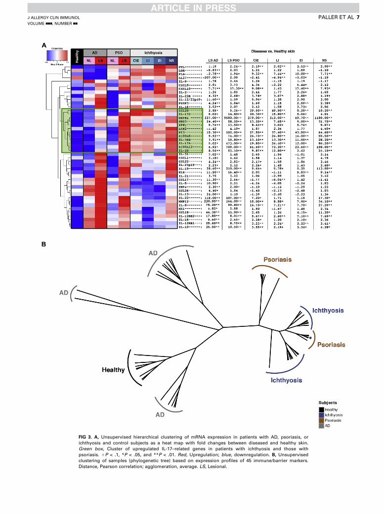
FIG 3. A, Unsupervised hierarchical clustering of mRNA expression in patients with AD, psoriasis, or
ichthyosis and control subjects as a heat map with fold changes between diseased and healthy skin.
Green box, Cluster of upregulated IL-17–related genes in patients with ichthyosis and those with
psoriasis. 1P < .1, *P < .05, and **P < .01. Red, Upregulation; blue, downregulation. B, Unsupervised
clustering of samples (phylogenetic tree) based on expression profiles of 45 immune/barrier markers.
Distance, Pearson correlation; agglomeration, average. LS, Lesional.
J ALLERGY CLIN IMMUNOL
VOLUME nnn, NUMBER nn
PALLER ET AL 7
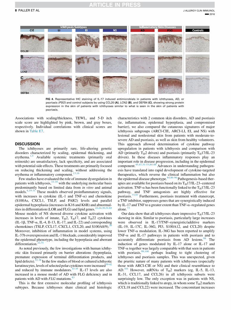
FIG 4. Representative IHC staining of IL-17 induced antimicrobials in patients with ichthyoses, AD, or
psoriasis (PSO) and control subjects by using CCL20 (A), LCN2 (B), and DEFB4 (C), showing strong protein
expression in the skin of patients with ichthyoses similar to what is seen in the skin of patients with
psoriasis.
J ALLERGY CLIN IMMUNOL
nnn 2016
8 PALLER ET AL
Associations with scaling/thickness, TEWL, and 5-D itchscale score are highlighted by pink, brown, and gray boxes,respectively. Individual correlations with clinical scores areshown in Table E7.
DISCUSSIONThe ichthyoses are primarily rare, life-altering genetic
disorders characterized by scaling, epidermal thickening, anderythema.1,5 Available systemic treatments (primarily oralretinoids) are unsatisfactory, lack specificity, and are associatedwith potential side effects. These treatments are primarily focusedon reducing thickening and scaling, without addressing theerythema or inflammatory component.13,16
Few studies have evaluated the role of immune dysregulation inpatients with ichthyosis,33-35 with the underlying molecular basispredominantly based on limited data from in vitro and animalmodels.6,19-31 These models observed proinflammatory signals,with increases in cytokine (IL-1 and TNF-a) and chemokine(S100As, CXCL1, TSLP, and PAR2) levels and parallelepidermal hyperplasia (increases in K16 and K6B) and abnormal-ities in differentiation (LOR and FLG) and lipid genes.22,24,30,31,82
Mouse models of NS showed diverse cytokine activation withincreases in levels of innate, TH2, TH17, and TH22 cytokines(IL-1b, TNF-a, IL-4, Il-13, IL-17, and IL-22) and correspondingchemokines (TSLP, CCL17, CXCL1, CCL20, and S100A8/9).20
Moreover, inhibition of inflammation in model systems, usingIL-37b overexpression and IL-1 blockade, considerably improvedthe epidermal phenotype, including the hyperplasia and aberrantdifferentiation.22,31
As noted previously, the few investigations with human ichthy-otic skin focused primarily on barrier alterations (hyperplasia,premature expression of terminal differentiation products, andlipid defects).32-38 In the few studies of blood or cultured ichthyotickeratinocytes, levels of selected polar cytokineswere increased39-42
and reduced by immune modulators.33-35 IL-17 levels are alsoincreased in a mouse model of AD with FLG deficiency and inpatients with AD with FLG mutations.83
This is the first extensive molecular profiling of ichthyosissubtypes. Because ichthyoses share clinical and histologic
characteristics with 2 common skin disorders, AD and psoriasis(ie, inflammation, epidermal hyperplasia, and compromisedbarrier), we also compared the cutaneous signatures of majorichthyosis subgroups (ARCI-CIE, ARCI-LI, EI, and NS) withlesional and nonlesional skin from patients with moderate-to-severe AD and psoriasis, as well as skin from healthy volunteers.This approach allowed determination of cytokine pathwayupregulation in patients with ichthyosis and comparison withAD (primarily TH2 driven) and psoriasis (primarily TH17/IL-23driven). In these diseases inflammatory responses play animportant role in disease progression, including in the epidermalcomponent.45,47,51-53,84-88 Advances in understanding pathogen-esis have translated into rapid development of cytokine-targetedtherapeutics, which reverse the clinical inflammation but alsothe epidermal disease phenotype.84,86-96 Pathogenesis-based ther-apies are available for psoriasis based on its TH17/IL-23–centeredactivation. TNF-a has been functionally linked to the TH17/IL-23pathway, and TNF antagonists are highly effective forpsoriasis.73,97 Furthermore, psoriasis treatment with etanercept,a TNF inhibitor, suppresses genes that are synergistically inducedby IL-17 and TNF to a greater extent than TNF-a–regulated genesalone.73
Our data show that all ichthyoses share impressive TH17/IL-23skewing in skin. Similar to psoriasis, particularly large increaseswere observed in IL-17/TNF–synergistic/additive markers(IL-19, IL-17C, IL-36G, PI3, S100A12, and CCL20) despitelower TNF-a modulation. IL-36G has been reported to amplifyTNF-a and IL-17 pathways in patients with psoriasis and toaccurately differentiate psoriasis from AD lesions.98 Theinduction of genes modulated by IL-17 alone or IL-17 andTNF-a together was largely comparable with that seen in patientswith psoriasis,99,100 perhaps leading to tight clustering ofichthyoses and psoriasis samples. This was unexpected, giventhe pruritic nature of many patients with ichthyoses (especiallythose with ARCI-CIE or NS) and their clinical resemblance toAD.101 However, mRNAs of TH2 markers (eg, IL-5, IL-13,IL-31, CCL17, and CCL26) in all ichthyosis subsets weresurprisingly low. The only exception was in patients with NS,which is traditionally linked to atopy, in whom some TH2markers(CCL18 and CCL22) were increased. The concomitant increases
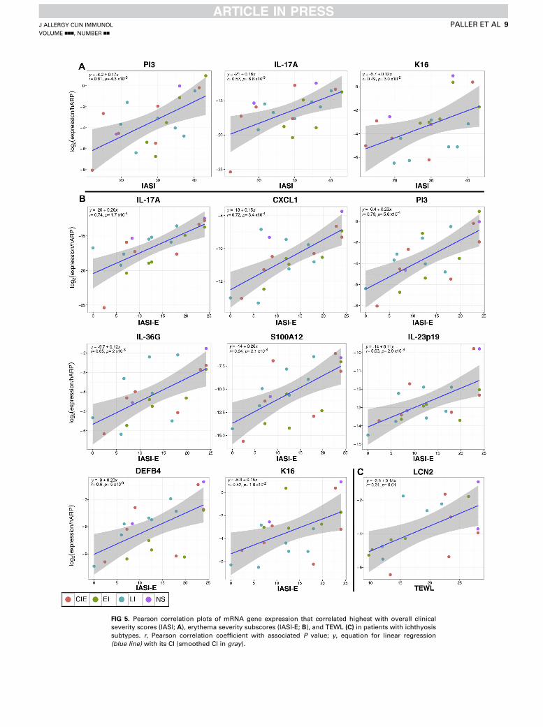
FIG 5. Pearson correlation plots of mRNA gene expression that correlated highest with overall clinical
severity scores (IASI; A), erythema severity subscores (IASI-E; B), and TEWL (C) in patients with ichthyosis
subtypes. r, Pearson correlation coefficient with associated P value; y, equation for linear regression
(blue line) with its CI (smoothed CI in gray).
J ALLERGY CLIN IMMUNOL
VOLUME nnn, NUMBER nn
PALLER ET AL 9
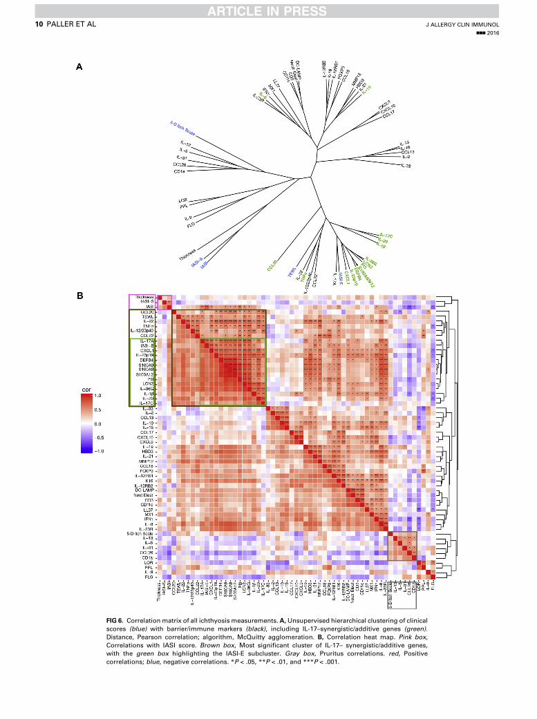
FIG 6. Correlationmatrix of all ichthyosis measurements. A,Unsupervised hierarchical clustering of clinical
scores (blue) with barrier/immune markers (black), including IL-17–synergistic/additive genes (green).Distance, Pearson correlation; algorithm, McQuitty agglomeration. B, Correlation heat map. Pink box,Correlations with IASI score. Brown box, Most significant cluster of IL-17– synergistic/additive genes,
with the green box highlighting the IASI-E subcluster. Gray box, Pruritus correlations. red, Positive
correlations; blue, negative correlations. *P < .05, **P < .01, and ***P < .001.
J ALLERGY CLIN IMMUNOL
nnn 2016
10 PALLER ET AL
J ALLERGY CLIN IMMUNOL
VOLUME nnn, NUMBER nn
PALLER ET AL 11
in TH17- and TH2-related markers in patients with NS might alsocontribute to the large increases in IL-19 levels in this subtype,given that TH17 but also TH2 cytokines can induce IL-19.74-78
IL-19 induces epidermal hyperplasia and S100As,74-78 whichwere highest in patients with NS. Interestingly, although mouseNS models demonstrate increased TH2/TH17 responses,6,20,21
TH2 inhibition through PAR2/TSLP suppression did not improvecutaneous inflammation.30 Expression of TH1 markers varied inpatients with different ichthyoses but was mostly lower than inpatients with AD and those with psoriasis. Increases in innatemarkers (IL-1b and IL-8) were also seen in patients withichthyoses and largely comparable with those in patients withAD and those with psoriasis.
Importantly, IASI scores, particularly the erythema subscoreIASI-E (reflecting clinical severity), were highly correlated withIL-17A and IL-17/TNF–regulated genes (CXCL1 and IL-36G).Significant correlations were also found between IASI andIASI-E scores and epidermal hyperplasia, as measured byK16.66,102 Although in patients with AD and those with psoriasisepidermal hyperplasia is linked to IL-22, IL-22 activation was farlower in patients with ichthyosis than in those with AD orpsoriasis. Other hyperplasia-inducing IL-20 family cytokines(ie, IL-19) might contribute to increased the epidermal thicknessin patients with ichthyosis.103-106
The ichthyoses are recognized as resulting in significantepidermal abnormalities,67,71,107,108 including epidermalhyperplasia, and lipid and differentiation abnormalities leadingto barrier impairment, which is reflected by increased TEWL.1,3
Hyperplasia and differentiation abnormalities, particularly inpatients with ARCI and NS subtypes, are similar to those seenin patients with psoriasis, with hyperplastic epidermis and largelyincreased expression of differentiation proteins (LOR, FLG, andPPL) in the upper epidermis.32,43,109 We found higher expressionof these markers in patients with ichthyosis and psoriasisbut much reduced expression in patients with AD. Thesignificant correlations between TEWL with IL-17A and IL-17/TNF–regulated genes (IL-17C, CXCL1, LCN2, and IL-36G)might link the immune activation and functional barrierabnormalities in patients with ichthyosis.
Finally, similar to those with psoriasis, patients with ichthyosisare able tomount significant IL-17–induced antimicrobial peptide(AMP) responses, as shown by high mRNA expression of LL37,DEFB4, human b-defensin 3, and CCL20. Our IHC studies ofDEFB4, LCN2, and CCL20 show increased protein expression ofIL-17–related proteins/AMPs. AMPs were recently shown toupregulate tight junctions and keratinocyte differentiation,110,111
providing an explanation beyond antimicrobial function for in-creases in these products in patients with ichthyosis and thosewith psoriasis versus patients with AD. A critical considerationin interpreting these results is whether the increase in TH17/IL-23 pathway expression is merely a compensatory mechanism inan effort to reduce the risk of infection versus a driver of inflam-mation. Based on case reports and our experience, Staphylo-coccus aureus, dermatophyte, and candidal infections occur notinfrequently in patients with ichthyosis, although S aureus infec-tions are less common than in those with moderate-to-severeAD.99,112-116 Even with an anti–IL-17 drug (secukinumab) forpsoriasis, only about 1%of patients havemild-to-moderatemuco-cutaneous infections, predominantly candidal.117,118 The reactiverole of TH17/IL-23 skewing to the barrier defects versus a primarypathogenic role in patients with ichthyosis can only be clarified
through future studies with targeted antagonists, which includecareful monitoring for mucocutaneous infections.
Limitations of our data include the small sample size, reflectingthe rarity of ichthyoses, and the use of a new, nonvalidatedseverity score (chosen because no acceptable validated score wasavailable). Nevertheless, a large and significant effect wasobserved for IL-17–modulated or IL-17/TNF synergisticallyregulated markers and their association with disease severity.
Although 25% of control subjects were children 10 years orolder, the age difference between patients with ichthyosis andhealthy subjects was statistically significant. Our AD andpsoriasis samples were obtained from subjects 18 years or older,although 8 of 21 patients with ichthyosis were less than 18 yearsold. Although the adolescent skin phenotype in patients with ADand those with psoriasis is commonly considered close to that inadults, there are no studies comparing the two. Thus, for propercomparisons, all analyses were age adjusted, and a sensitivityanalysis including only patients with 18 years old and oldersuggested similar findings (see Table E8 in this article’s OnlineRepository at www.jacionline.org). Furthermore, given thatIL-17 expression increases with age in healthy skin,119,120 ourresults might actually underestimate the increased TH17activation in patients with ichthyosis. Future studies shouldaddress the effect of age on the observed differences.
Our data link the TH17/IL-23 pathway and IL-17/TNFsynergistic interactions with ichthyosis severity andinflammation, providing evidence that ichthyosis more closelyresembles psoriasis in its immune profile. The linkage betweenimmune alterations and functional barrier abnormalities inpatients with ichthyoses potentially suggests a similar model topsoriasis and AD, in which increased cytokine productionperpetuates the barrier alterations. These results imply thatpsoriasis therapeutics might be applicable for patients withichthyosis. One patient with NS demonstrated clinicalimprovement and reduction in IL-17 levels after administrationof infliximab (anti–TNF-a used to treat psoriasis).33,121,122
IL-17/IL-23–targeting strategies94,123-125 have been shown to bemore effective than TNF-a inhibitors in patients with psoriasis,dramatically improving Psoriasis Area and Severity Indexscores.92,126 Specific IL-17/IL-23 targeting will elucidate afunctional role of IL-17 in patients with ichthyoses and mightestablish a novel treatment paradigm for ichthyoses.
We thank Drs Adam Berry, Jayla Gray, Isabel Haugh, Lisa Shen, Anjali
Shroff, and Robalee Wanderman for helping with patient enrollment. We are
grateful to the Foundation for Ichthyosis and Related Skin Types for allowing
this research to be performed in part at its Family Conference in 2014.
Clinical implications: The link between increased expression ofTH17 pathway cytokines and clinical disease severity raises thepossibility of a new therapeutic paradigm of targeted IL-17/IL-23 intervention for patients with ichthyosis.
REFERENCES
1. Oji V, Tadini G, Akiyama M, Blanchet Bardon C, Bodemer C, Bourrat E, et al.
Revised nomenclature and classification of inherited ichthyoses: results of the
First Ichthyosis Consensus Conference in Soreze 2009. J Am Acad Dermatol
2010;63:607-41.
2. DiGiovanna JJ, Robinson-Bostom L. Ichthyosis: etiology, diagnosis, and
management. Am J Clin Dermatol 2003;4:81-95.
3. Oji V, Traupe H. Ichthyoses: differential diagnosis and molecular genetics. Eur J
Dermatol 2006;16:349-59.
J ALLERGY CLIN IMMUNOL
nnn 2016
12 PALLER ET AL
4. Craig WY, Roberson M, Palomaki GE, Shackleton CH, Marcos J, Haddow JE.
Prevalence of steroid sulfatase deficiency in California according to race and
ethnicity. Prenat Diagn 2010;30:893-8.
5. Traupe H, Fischer J, Oji V. Nonsyndromic types of ichthyoses—an update.
J Dtsch Dermatol Ges 2014;12:109-21.
6. Briot A, Deraison C, Lacroix M, Bonnart C, Robin A, Besson C, et al.
Kallikrein 5 induces atopic dermatitis-like lesions through PAR2-mediated
thymic stromal lymphopoietin expression in Netherton syndrome. J Exp Med
2009;206:1135-47.
7. Thyssen JP, Godoy-Gijon E, Elias PM. Ichthyosis vulgaris: the filaggrin mutation
disease. Br J Dermatol 2013;168:1155-66.
8. Ganemo A, Lindholm C, Lindberg M, Sjoden PO, Vahlquist A. Quality of life in
adults with congenital ichthyosis. J Adv Nurs 2003;44:412-9.
9. Dreyfus I, Pauwels C, Bourrat E, Bursztejn AC, Maruani A, Chiaverini C, et al.
Burden of inherited ichthyosis: a French national survey. Acta Derm Venereol
2015;95:326-8.
10. Williams ML. Ichthyosis: mechanisms of disease. Pediatr Dermatol 1992;9:
365-8.
11. Elias PM, Williams ML, Holleran WM, Jiang YJ, Schmuth M. Pathogenesis of
permeability barrier abnormalities in the ichthyoses: inherited disorders of lipid
metabolism. J Lipid Res 2008;49:697-714.
12. Eckl KM, Weindl G, Ackermann K, Kuchler S, Casper R, Radowski MR, et al.
Increased cutaneous absorption reflects impaired barrier function of
reconstructed skin models mimicking keratinisation disorders. Exp Dermatol
2014;23:286-8.
13. Hernandez-Martin A, Aranegui B, Martin-Santiago A, Garcia-Doval I.
A systematic review of clinical trials of treatments for the congenital ichthyoses,
excluding ichthyosis vulgaris. J Am Acad Dermatol 2013;69:544-9.e8.
14. Vahlquist A, Ganemo A, Virtanen M. Congenital ichthyosis: an overview of
current and emerging therapies. Acta Derm Venereol 2008;88:4-14.
15. Fleckman P. Management of the ichthyoses. Skin Therapy Lett 2003;8:3-7.
16. Digiovanna JJ, Mauro T, Milstone LM, Schmuth M, Toro JR. Systemic retinoids
in the management of ichthyoses and related skin types. Dermatol Ther 2013;26:
26-38.
17. Halverstam CP, Vachharajani A, Mallory SB. Cushing syndrome from
percutaneous absorption of 1% hydrocortisone ointment in Netherton syndrome.
Pediatr Dermatol 2007;24:42-5.
18. Allen A, Siegfried E, Silverman R, Williams ML, Elias PM, Szabo SK, et al.
Significant absorption of topical tacrolimus in 3 patients with Netherton
syndrome. Arch Dermatol 2001;137:747-50.
19. Descargues P, Deraison C, Bonnart C, Kreft M, Kishibe M, Ishida-Yamamoto A,
et al. Spink5-deficient mice mimic Netherton syndrome through degradation of
desmoglein 1 by epidermal protease hyperactivity. Nat Genet 2005;37:56-65.
20. Furio L, de Veer S, Jaillet M, Briot A, Robin A, Deraison C, et al. Transgenic
kallikrein 5 mice reproduce major cutaneous and systemic hallmarks of Netherton
syndrome. J Exp Med 2014;211:499-513.
21. Furio L, Pampalakis G, Michael IP, Nagy A, Sotiropoulou G, Hovnanian A.
KLK5 inactivation reverses cutaneous hallmarks of Netherton syndrome. PLoS
Genet 2015;11:e1005389.
22. O’Shaughnessy RF, Choudhary I, Harper JI. Interleukin-1 alpha blockade
prevents hyperkeratosis in an in vitro model of lamellar ichthyosis. Hum Mol
Genet 2010;19:2594-605.
23. Jensen JM, Schutze S, Neumann C, Proksch E. Impaired cutaneous permeability
barrier function, skin hydration, and sphingomyelinase activity in keratin 10
deficient mice. J Invest Dermatol 2000;115:708-13.
24. Roth W, Kumar V, Beer HD, Richter M, Wohlenberg C, Reuter U, et al. Keratin 1
maintains skin integrity and participates in an inflammatory network in skin
through interleukin-18. J Cell Sci 2012;125:5269-79.
25. Rosenberger S, Dick A, Latzko S, Hausser I, Stark HJ, Rauh M, et al. A mouse
organotypic tissue culture model for autosomal recessive congenital ichthyosis.
Br J Dermatol 2014;171:1347-57.
26. Krieg P, Rosenberger S, de Juanes S, Latzko S, Hou J, Dick A, et al. Aloxe3
knockout mice reveal a function of epidermal lipoxygenase-3 as hepoxilin syn-
thase and its pivotal role in barrier formation. J Invest Dermatol 2013;133:172-80.
27. Zuo Y, Zhuang DZ, Han R, Isaac G, Tobin JJ, McKee M, et al. ABCA12
maintains the epidermal lipid permeability barrier by facilitating formation of
ceramide linoleic esters. J Biol Chem 2008;283:36624-35.
28. Smyth I, Hacking DF, Hilton AA, Mukhamedova N, Meikle PJ, Ellis S, et al.
A mouse model of harlequin ichthyosis delineates a key role for Abca12 in lipid
homeostasis. PLoS Genet 2008;4:e1000192.
29. Aufenvenne K, Rice RH, Hausser I, Oji V, Hennies HC, Rio MD, et al. Long-term
faithful recapitulation of transglutaminase 1-deficient lamellar ichthyosis in a
skin-humanized mouse model, and insights from proteomic studies. J Invest
Dermatol 2012;132:1918-21.
30. Briot A, Lacroix M, Robin A, Steinhoff M, Deraison C, Hovnanian A. Par2
inactivation inhibits early production of TSLP, but not cutaneous inflammation,
in Netherton syndrome adult mouse model. J Invest Dermatol 2010;130:2736-42.
31. Cottle DL, Ursino GM, Ip SC, Jones LK, Ditommaso T, Hacking DF, et al. Fetal
inhibition of inflammation improves disease phenotypes in harlequin ichthyosis.
Hum Mol Genet 2015;24:436-49.
32. Descargues P, Deraison C, Prost C, Fraitag S, Mazereeuw-Hautier J, D’Alessio
M, et al. Corneodesmosomal cadherins are preferential targets of stratum
corneum trypsin- and chymotrypsin-like hyperactivity in Netherton syndrome.
J Invest Dermatol 2006;126:1622-32.
33. Fontao L, Laffitte E, Briot A, Kaya G, Roux-Lombard P, Fraitag S, et al.
Infliximab infusions for Netherton syndrome: sustained clinical improvement
correlates with a reduction of thymic stromal lymphopoietin levels in the skin.
J Invest Dermatol 2011;131:1947-50.
34. Yalcin AD. A case of Netherton syndrome: successful treatment with omalizumab
and pulse prednisolone and its effects on cytokines and immunoglobulin levels.
Immunopharmacol Immunotoxicol 2016;38:162-6.
35. Pavez Lorie E, Ganemo A, Borgers M, Wouters L, Blockhuys S, van de
Plassche L, et al. Expression of retinoid-regulated genes in lamellar ichthyo-
sis vs. healthy control epidermis: changes after oral treatment with liarozole.
Acta Derm Venereol 2009;89:12-20.
36. Hannula-Jouppi K, Laasanen SL, Ilander M, Furio L, Tuomiranta M, Marttila R,
et al. Intrafamily and interfamilial phenotype variation and immature immunity in
patients with Netherton syndrome and Finnish SPINK5 founder mutation. JAMA
Dermatol 2016;152:435-42.
37. de Oliveira GV, Hawkins HK, Guedes AC, Pinto LF, Oliveras G, Kitten GT, et al.
Comel-Netherton syndrome in brothers and expression of cytokeratins. J Am
Acad Dermatol 2005;52:725-6.
38. Konishi T, Tsuda T, Sakaguchi Y, Imai Y, Ito T, Hirota S, et al. Upregulation of
interleukin-33 in the epidermis of two Japanese patients with Netherton
syndrome. J Dermatol 2014;41:258-61.
39. Van Gysel D, Koning H, Baert MR, Savelkoul HF, Neijens HJ, Oranje AP.
Clinico-immunological heterogeneity in Comel-Netherton syndrome. Derma-
tology 2001;202:99-107.
40. Renner ED, Hartl D, Rylaarsdam S, Young ML, Monaco-Shawver L, Kleiner G,
et al. Comel-Netherton syndrome defined as primary immunodeficiency. J Allergy
Clin Immunol 2009;124:536-43.
41. Hosomi N, Fukai K, Nakanishi T, Funaki S, Ishii M. Caspase-1 activity of stratum
corneum and serum interleukin-18 level are increased in patients with Netherton
syndrome. Br J Dermatol 2008;159:744-6.
42. Akagi A, Kitoh A, Moniaga CS, Fujimoto A, Fujikawa H, Shimomura Y, et al.
Case of Netherton syndrome with an elevated serum thymus and
activation-regulated chemokine level. J Dermatol 2013;40:752-3.
43. Lacroix M, Lacaze-Buzy L, Furio L, Tron E, Valari M, Van der Wier G, et al.
Clinical expression and new SPINK5 splicing defects in Netherton syndrome:
unmasking a frequent founder synonymous mutation and unconventional intronic
mutations. J Invest Dermatol 2012;132:575-82.
44. Lowes MA, Russell CB, Martin DA, Towne JE, Krueger JG. The IL-23/T17
pathogenic axis in psoriasis is amplified by keratinocyte responses. Trends
Immunol 2013;34:174-81.
45. Noda S, Krueger JG, Guttman-Yassky E. The translational revolution and use of
biologics in patients with inflammatory skin diseases. J Allergy Clin Immunol
2015;135:324-36.
46. Martin DA, Towne JE, Kricorian G, Klekotka P, Gudjonsson JE, Krueger JG,
et al. The emerging role of IL-17 in the pathogenesis of psoriasis: preclinical
and clinical findings. J Invest Dermatol 2013;133:17-26.
47. Wang CQ, Suarez-Farinas M, Nograles KE, Mimoso CA, Shrom D, Dow ER,
et al. IL-17 induces inflammation-associated gene products in blood monocytes,
and treatment with ixekizumab reduces their expression in psoriasis patient blood.
J Invest Dermatol 2014;134:2990-3.
48. Kamalpour L, Rice ZP, Pavlis M, Veledar E, Chen SC. Reliable methods to
evaluate the clinical severity of ichthyosis. Pediatr Dermatol 2010;27:148-53.
49. Fredriksson T, Pettersson U. Severe psoriasis—oral therapy with a new retinoid.
Dermatologica 1978;157:238-44.
50. Noda S, Suarez-Farinas M, Ungar B, Kim SJ, de Guzman Strong C, Xu H, et al.
The Asian atopic dermatitis phenotype combines features of atopic dermatitis and
psoriasis with increased TH17 polarization. J Allergy Clin Immunol 2015;136:
1254-64.
51. Khattri S, Shemer A, Rozenblit M, Dhingra N, Czarnowicki T, Finney R, et al.
Cyclosporine in patients with atopic dermatitis modulates activated inflammatory
pathways and reverses epidermal pathology. J Allergy Clin Immunol 2014;133:
1626-34.
52. Johnson-Huang LM, Suarez-Farinas M, Sullivan-Whalen M, Gilleaudeau P,
Krueger JG, Lowes MA. Effective narrow-band UVB radiation therapy
J ALLERGY CLIN IMMUNOL
VOLUME nnn, NUMBER nn
PALLER ET AL 13
suppresses the IL-23/IL-17 axis in normalized psoriasis plaques. J Invest
Dermatol 2010;130:2654-63.
53. Tintle S, Shemer A, Suarez-Farinas M, Fujita H, Gilleaudeau P, Sullivan-Whalen
M, et al. Reversal of atopic dermatitis with narrow-band UVB phototherapy
and biomarkers for therapeutic response. J Allergy Clin Immunol 2011;128:
583-93, e1-4.
54. Dhingra N, Suarez-Farinas M, Fuentes-Duculan J, Gittler JK, Shemer A, Raz A,
et al. Attenuated neutrophil axis in atopic dermatitis compared to psoriasis
reflects TH17 pathway differences between these diseases. J Allergy Clin
Immunol 2013;132:498-501.e3.
55. Esaki H, Ewald DA, Ungar B, Rozenblit M, Zheng X, Xu H, et al. Identification
of novel immune and barrier genes in atopic dermatitis by means of laser capture
microdissection. J Allergy Clin Immunol 2015;135:153-63.
56. Suarez-Farinas M, Ungar B, Correa da Rosa J, Ewald DA, Rozenblit M, Gonzalez
J, et al. RNA sequencing atopic dermatitis transcriptome profiling provides
insights into novel disease mechanisms with potential therapeutic implications.
J Allergy Clin Immunol 2015;135:1218-27.
57. Czarnowicki T, Gonzalez J, Bonifacio KM, Shemer A, Xiangyu P, Kunjravia N,
et al. Diverse activation and differentiation of multiple B-cell subsets in patients
with atopic dermatitis but not in patients with psoriasis. J Allergy Clin Immunol
2016;137:118-29.e5.
58. Taieb A, Labreze C. Collodion baby: what’s new. J Eur Acad Dermatol Venereol
2002;16:436-7.
59. Van Gysel D, Lijnen RL, Moekti SS, de Laat PC, Oranje AP. Collodion
baby: a follow-up study of 17 cases. J Eur Acad Dermatol Venereol 2002;
16:472-5.
60. Russell LJ, DiGiovanna JJ, Rogers GR, Steinert PM, Hashem N, Compton JG,
et al. Mutations in the gene for transglutaminase 1 in autosomal recessive lamellar
ichthyosis. Nat Genet 1995;9:279-83.
61. Eckl KM, de Juanes S, Kurtenbach J, Natebus M, Lugassy J, Oji V, et al.
Molecular analysis of 250 patients with autosomal recessive congenital
ichthyosis: evidence for mutation hotspots in ALOXE3 and allelic heterogeneity
in ALOX12B. J Invest Dermatol 2009;129:1421-8.
62. Fischer J. Autosomal recessive congenital ichthyosis. J Invest Dermatol 2009;
129:1319-21.
63. Chavanas S, Bodemer C, Rochat A, Hamel-Teillac D, Ali M, Irvine AD, et al.
Mutations in SPINK5, encoding a serine protease inhibitor, cause Netherton
syndrome. Nat Genet 2000;25:141-2.
64. Hachem JP, Wagberg F, Schmuth M, Crumrine D, Lissens W, Jayakumar A, et al.
Serine protease activity and residual LEKTI expression determine phenotype in
Netherton syndrome. J Invest Dermatol 2006;126:1609-21.
65. Elman S, Hynan LS, Gabriel V, Mayo MJ. The 5-D itch scale: a new measure of
pruritus. Br J Dermatol 2010;162:587-93.
66. Mansouri Y, Guttman-Yassky E. Immune pathways in atopic dermatitis, and
definition of biomarkers through broad and targeted therapeutics. J Clin Med
2015;4:858-73.
67. Suarez-Farinas M, Tintle SJ, Shemer A, Chiricozzi A, Nograles K, Cardinale I,
et al. Nonlesional atopic dermatitis skin is characterized by broad terminal
differentiation defects and variable immune abnormalities. J Allergy Clin
Immunol 2011;127:954-64, e1-4.
68. Rice RH, Bradshaw KM, Durbin-Johnson BP, Rocke DM, Eigenheer RA,
Phinney BS, et al. Distinguishing ichthyoses by protein profiling. PLoS
One 2013;8:e75355.
69. Leung DY, Guttman-Yassky E. Deciphering the complexities of atopic dermatitis:
shifting paradigms in treatment approaches. J Allergy Clin Immunol 2014;134:
769-79.
70. Deraison C, Bonnart C, Lopez F, Besson C, Robinson R, Jayakumar A, et al.
LEKTI fragments specifically inhibit KLK5, KLK7, and KLK14 and control
desquamation through a pH-dependent interaction. Mol Biol Cell 2007;18:
3607-19.
71. Guttman-Yassky E, Suarez-Farinas M, Chiricozzi A, Nograles KE, Shemer A,
Fuentes-Duculan J, et al. Broad defects in epidermal cornification in atopic
dermatitis identified through genomic analysis. J Allergy Clin Immunol 2009;
124:1235-44.e58.
72. Suarez-Farinas M, Lowes MA, Zaba LC, Krueger JG. Evaluation of the psoriasis
transcriptome across different studies by gene set enrichment analysis (GSEA).
PLoS One 2010;5:e10247.
73. Chiricozzi A, Guttman-Yassky E, Suarez-Farinas M, Nograles KE, Tian S,
Cardinale I, et al. Integrative responses to IL-17 and TNF-alpha in human
keratinocytes account for key inflammatory pathogenic circuits in psoriasis.
J Invest Dermatol 2011;131:677-87.
74. Witte E, Kokolakis G, Witte K, Philipp S, Doecke WD, Babel N, et al. IL-19 is a
component of the pathogenetic IL-23/IL-17 cascade in psoriasis. J Invest
Dermatol 2014;134:2757-67.
75. Liao SC, Cheng YC, Wang YC, Wang CW, Yang SM, Yu CK, et al. IL-19 induced
Th2 cytokines and was up-regulated in asthma patients. J Immunol 2004;173:
6712-8.
76. Hsing CH, Hsu CC, Chen WY, Chang LY, Hwang JC, Chang MS. Expression of
IL-19 correlates with Th2 cytokines in uraemic patients. Nephrol Dial Transplant
2007;22:2230-8.
77. Gallagher G, Eskdale J, Jordan W, Peat J, Campbell J, Boniotto M, et al. Human
interleukin-19 and its receptor: a potential role in the induction of Th2 responses.
Int Immunopharmacol 2004;4:615-26.
78. Huang F, Wachi S, Thai P, Loukoianov A, Tan KH, Forteza RM, et al.
Potentiation of IL-19 expression in airway epithelia by IL-17A and IL-4/IL-13:
important implications in asthma. J Allergy Clin Immunol 2008;121:1415-21,
e1-3.
79. Nograles KE, Zaba LC, Guttman-Yassky E, Fuentes-Duculan J, Suarez-Farinas
M, Cardinale I, et al. Th17 cytokines interleukin (IL)-17 and IL-22 modulate
distinct inflammatory and keratinocyte-response pathways. Br J Dermatol 2008;
159:1092-102.
80. Sonkoly E, Muller A, Lauerma AI, Pivarcsi A, Soto H, Kemeny L, et al. IL-31: a
new link between T cells and pruritus in atopic skin inflammation. J Allergy Clin
Immunol 2006;117:411-7.
81. Cedeno-Laurent F, Singer EM, Wysocka M, Benoit BM, Vittorio CC,
Kim EJ, et al. Improved pruritus correlates with lower levels of
IL-31 in CTCL patients under different therapeutic modalities. Clin
Immunol 2015;158:1-7.
82. Hewett DR, Simons AL, Mangan NE, Jolin HE, Green SM, Fallon PG, et al.
Lethal, neonatal ichthyosis with increased proteolytic processing of filaggrin in
a mouse model of Netherton syndrome. Hum Mol Genet 2005;14:335-46.
83. Bonefeld CM, Petersen TH, Bandier J, Agerbeck C, Linneberg A, Ross-Hansen
K, et al. Epidermal filaggrin deficiency mediates increased systemic Th17
immune response. Br J Dermatol 2016 [Epub ahead of print].
84. Papp KA, Langley RG, Lebwohl M, Krueger GG, Szapary P, Yeilding N,
et al. Efficacy and safety of ustekinumab, a human interleukin-12/23
monoclonal antibody, in patients with psoriasis: 52-week results from a
randomised, double-blind, placebo-controlled trial (PHOENIX 2). Lancet
2008;371:1675-84.
85. Chiricozzi A, Saraceno R, Chimenti MS, Guttman-Yassky E, Krueger JG. Role of
IL-23 in the pathogenesis of psoriasis: a novel potential therapeutic target? Expert
Opin Ther Targets 2014;18:513-25.
86. Hamilton JD, Suarez-Farinas M, Dhingra N, Cardinale I, Li X, Kostic A, et al.
Dupilumab improves the molecular signature in skin of patients with
moderate-to-severe atopic dermatitis. J Allergy Clin Immunol 2014;134:
1293-300.
87. Hamilton JD, Ungar B, Guttman-Yassky E. Drug evaluation review: dupilumab in
atopic dermatitis. Immunotherapy 2015;7:1043-58.
88. Beck LA, Thaci D, Hamilton JD, Graham NM, Bieber T, Rocklin R, et al.
Dupilumab treatment in adults with moderate-to-severe atopic dermatitis.
N Engl J Med 2014;371:130-9.
89. Thaci D, Simpson EL, Beck LA, Bieber T, Blauvelt A, Papp K, et al. Efficacy and
safety of dupilumab in adults with moderate-to-severe atopic dermatitis
inadequately controlled by topical treatments: a randomised, placebo-
controlled, dose-ranging phase 2b trial. Lancet 2016;387:40-52.
90. Lopez-Ferrer A, Vilarrasa E, Puig L. Secukinumab (AIN457) for the treatment of
psoriasis. Expert Rev Clin Immunol 2015;11:1177-88.
91. Griffiths CE, Reich K, Lebwohl M, van de Kerkhof P, Paul C, Menter A, et al.
Comparison of ixekizumab with etanercept or placebo in moderate-to-severe
psoriasis (UNCOVER-2 and UNCOVER-3): results from two phase 3 randomised
trials. Lancet 2015;386:541-51.
92. Langley RG, Elewski BE, Lebwohl M, Reich K, Griffiths CE, Papp K, et al.
Secukinumab in plaque psoriasis—results of two phase 3 trials. N Engl J Med
2014;371:326-38.
93. Papp KA, Tyring S, Lahfa M, Prinz J, Griffiths CE, Nakanishi AM, et al. A global
phase III randomized controlled trial of etanercept in psoriasis: safety, efficacy,
and effect of dose reduction. Br J Dermatol 2005;152:1304-12.
94. Krueger JG, Ferris LK, Menter A, Wagner F, White A, Visvanathan S, et al.
Anti-IL-23A mAb BI 655066 for treatment of moderate-to-severe psoriasis:
Safety, efficacy, pharmacokinetics, and biomarker results of a single-rising-
dose, randomized, double-blind, placebo-controlled trial. J Allergy Clin Immunol
2015;136:116-24.e7.
95. Lebwohl M. Psoriasis. Lancet 2003;361:1197-204.
96. Gordon KB, Langley RG, Gottlieb AB, Papp KA, Krueger GG, Strober BE, et al.
A phase III, randomized, controlled trial of the fully human IL-12/23 mAb
briakinumab in moderate-to-severe psoriasis. J Invest Dermatol 2012;132:304-14.
97. Zaba LC, Suarez-Farinas M, Fuentes-Duculan J, Nograles KE, Guttman-Yassky E,
Cardinale I, et al. Effective treatment of psoriasis with etanercept is linked to
J ALLERGY CLIN IMMUNOL
nnn 2016
14 PALLER ET AL
suppression of IL-17 signaling, not immediate response TNF genes. J Allergy Clin
Immunol 2009;124:1022-110, e1-395.
98. D’Erme AM, Wilsmann-Theis D, Wagenpfeil J, Holzel M, Ferring-Schmitt S,
Sternberg S, et al. IL-36gamma (IL-1F9) is a biomarker for psoriasis skin lesions.
J Invest Dermatol 2015;135:1025-32.
99. Guttman-Yassky E, Lowes MA, Fuentes-Duculan J, Zaba LC, Cardinale I,
Nograles KE, et al. Low expression of the IL-23/Th17 pathway in atopic
dermatitis compared to psoriasis. J Immunol 2008;181:7420-7.
100. Chiricozzi A, Nograles KE, Johnson-Huang LM, Fuentes-Duculan J, Cardinale I,
Bonifacio KM, et al. IL-17 induces an expanded range of downstream genes in
reconstituted human epidermis model. PLoS One 2014;9:e90284.
101. Samuelov L, Sprecher E. Peeling off the genetics of atopic dermatitis-like
congenital disorders. J Allergy Clin Immunol 2014;134:808-15.
102. de Mare S, van Erp PE, Ramaekers FC, van de Kerkhof PC. Flow cytometric
quantification of human epidermal cells expressing keratin 16 in vivo after stan-
dardized trauma. Arch Dermatol Res 1990;282:126-30.
103. He M, Liang P. IL-24 transgenic mice: in vivo evidence of overlapping
functions for IL-20, IL-22, and IL-24 in the epidermis. J Immunol 2010;184:
1793-8.
104. Wang F, Lee E, Lowes MA, Haider AS, Fuentes-Duculan J, Abello MV, et al.
Prominent production of IL-20 by CD681/CD11c1 myeloid-derived cells in
psoriasis: gene regulation and cellular effects. J Invest Dermatol 2006;126:
1590-9.
105. Boniface K, Bernard FX, Garcia M, Gurney AL, Lecron JC, Morel F.
IL-22 inhibits epidermal differentiation and induces proinflammatory
gene expression and migration of human keratinocytes. J Immunol 2005;174:
3695-702.
106. Sa SM, Valdez PA, Wu J, Jung K, Zhong F, Hall L, et al. The effects of IL-20
subfamily cytokines on reconstituted human epidermis suggest potential roles
in cutaneous innate defense and pathogenic adaptive immunity in psoriasis.
J Immunol 2007;178:2229-40.
107. Ye L, Lv C, Man G, Song S, Elias PM, Man MQ. Abnormal epidermal barrier
recovery in uninvolved skin supports the notion of an epidermal pathogenesis
of psoriasis. J Invest Dermatol 2014;134:2843-6.
108. Takahashi H, Tsuji H, Minami-Hori M, Miyauchi Y, Iizuka H. Defective barrier
function accompanied by structural changes of psoriatic stratum corneum.
J Dermatol 2014;41:144-8.
109. Kawashima J, Akiyama M, Takizawa Y, Takahashi S, Matsuo I, Shimizu H.
Structural, enzymatic and molecular studies in a series of nonbullous congenital
ichthyosiform erythroderma patients. Clin Exp Dermatol 2005;30:429-31.
110. Akiyama T, Niyonsaba F, Kiatsurayanon C, Nguyen TT, Ushio H, Fujimura T,
et al. The human cathelicidin LL-37 host defense peptide upregulates tight
junction-related proteins and increases human epidermal keratinocyte barrier
function. J Innate Immun 2014;6:739-53.
111. Kiatsurayanon C, Niyonsaba F, Smithrithee R, Akiyama T, Ushio H, Hara M,
et al. Host defense (Antimicrobial) peptide, human beta-defensin-3, improves
the function of the epithelial tight-junction barrier in human keratinocytes.
J Invest Dermatol 2014;134:2163-73.
112. Ong PY, Ohtake T, Brandt C, Strickland I, Boguniewicz M, Ganz T, et al.
Endogenous antimicrobial peptides and skin infections in atopic dermatitis.
N Engl J Med 2002;347:1151-60.
113. Boguniewicz M, Leung DY. Recent insights into atopic dermatitis and
implications for management of infectious complications. J Allergy Clin Immunol
2010;125:4-15.
114. Damsky WE, Leventhal JS, Khalil D, Vesely MD, Craiglow BG, Milstone LM,
et al. Recurrent coxsackievirus infection in a patient with lamellar ichthyosis.
Pediatr Dermatol 2016;33:e140-2.
115. Vyas NS, Kannan S, N Jahnke M, Hu RH, Choate KA, Shwayder TA. Congenital
ichthyosiform erythroderma superimposed with chronic dermatophytosis: a report
of three siblings. Pediatr Dermatol 2016;33:e6-9.
116. Freitas CF, Mulinari-Brenner F, Fontana HR, Gentili AC, Hammerschmidt M.
Ichthyosis associated with widespread tinea corporis: report of three cases. An
Bras Dermatol 2013;88:627-30.
117. van de Kerkhof PCM, Griffiths CEM, Reich K, Leonardi CL, Blauvelt A, Tsai TF,
et al. Secukinumab long-trem safety experience: a pooled analysis of 10 phase II
and III clinical studies in patients with moderate to severe plaque psoriasis. J Am
Acad Dermatol 2016;75:83-98.e4.
118. Cosentyx (secukinumab) prescribing information. Available at: https://www.pharma.
us.novartis.com/sites/www.pharma.us.novartis.com/files/cosentyx.pdf. Accessed
June 17, 2016.
119. Dunbar GC, Kuchibhatla RV, Lee G. TC-1734 (AZD3480) AAMI Clinical
Study Group (USA). A randomized double-blind study comparing 25 and 50
mg TC-1734 (AZD3480) with placebo, in older subjects with age-associated
memory impairment. J Psychopharmacol 2011;25:1020-9.
120. Lee JS, Lee WW, Kim SH, Kang Y, Lee N, Shin MS, et al. Age-associated
alteration in naive and memory Th17 cell response in humans. Clin Immunol
2011;140:84-91.
121. Chaudhari U, Romano P, Mulcahy LD, Dooley LT, Baker DG, Gottlieb AB.
Efficacy and safety of infliximab monotherapy for plaque-type psoriasis:
a randomised trial. Lancet 2001;357:1842-7.
122. Schopf RE, Aust H, Knop J. Treatment of psoriasis with the chimeric monoclonal
antibody against tumor necrosis factor alpha, infliximab. J Am Acad Dermatol
2002;46:886-91.
123. Sofen H, Smith S, Matheson RT, Leonardi CL, Calderon C, Brodmerkel C, et al.
Guselkumab (an IL-23-specific mAb) demonstrates clinical and molecular
response in patients with moderate-to-severe psoriasis. J Allergy Clin Immunol
2014;133:1032-40.
124. Russell CB, Rand H, Bigler J, Kerkof K, Timour M, Bautista E, et al.
Gene expression profiles normalized in psoriatic skin by treatment with
brodalumab, a human anti-IL-17 receptor monoclonal antibody. J Immunol
2014;192:3828-36.
125. Chiricozzi A, Krueger JG. IL-17 targeted therapies for psoriasis. Expert Opin
Investig Drugs 2013;22:993-1005.
126. Griffiths CE, Strober BE, van de Kerkhof P, Ho V, Fidelus-Gort R, Yeilding N,
et al. Comparison of ustekinumab and etanercept for moderate-to-severe
psoriasis. N Engl J Med 2010;362:118-28.


![harlequin ichthyosis and functional recovery by · of ichthyosis, harlequin ichthyosis (HI) (Mendelian Inheritance of Man [MIM] 242500) is the most serious subtype (Figure 1); it](https://img.pdfslide.net/doc/110x75/5d4f332b88c993257d8be9c0/harlequin-ichthyosis-and-functional-recovery-by-of-ichthyosis-harlequin-ichthyosis.jpg)