Embed Size (px)
Citation preview
60 Recognition of Propofol Infusion Syndrome under Anesthesia Bithal et al.
DiscussionCurrently, PRIS is assumed to be related to disruption of fatty acid oxidation caused by impaired entry of long-chain acylcarnitine ester into the mitochondria and failure of the mitochondrial respiratory chain.6 Isolated hyperkalemia and acidosis, components of PRIS have been reported from even smaller or average doses of propofol infused over a shorter period of time.7 Exact mechanism of isolated hyperkale-mia from propofol administration is unclear. In the body, the main store of potassium are the muscles and liver. PRIS usually leads to rhabdomyolysis, with secondary hyperka-lemia. However, hyperkalemia has been reported without rhabdomyolysis.8 It is hypothesized that mitochondrial fatty acid oxidation disorder could disturb potassium homeostasis because of its relationship with mitochondrial metabolism.9 Meanwhile propofol could produce lactic acidosis through the impairment of free fatty acid utilization and mitochon-drial activity in the muscles and heart.10 The patient was hemodynamically stable without evidence of hypovolemia or any pre-existing infection, thereby ruling out other possible causes of lactic acidosis. Hyperkalemia and acidosis during propofol infusion are harbingers of impending catastrophe, and failure to recognize these early warning signs and discontinuing its infusion would lead to florid PRIS with dan-gerous consequences.
In the light of the clinical circumstances, blood chemistry and recovery of deranged parameters following discontinua-tion of propofol infusion, it can be assumed that the patient may have developed PRIS. In this context, the role of serial ABGs and careful ECG monitoring cannot be overemphasized.
Conflict of InterestNone declared.
References
1 Parke TJ, Stevens JE, Rice ASC, et al. Metabolic acidosis and fatal myocardial failure after propofol infusion in children: five case reports. BMJ 1992;305(6854):613–616
2 Cremer OL, Moons KGM, Bouman EAC, Kruijswijk JE, de Smet AM, Kalkman CJ. Long-term propofol infusion and cardiac fail-ure in adult head-injured patients. Lancet 2001;357(9250): 117–118
3 Bray RJ. Propofol infusion syndrome in children. Paediatr Anaesth 1998;8(6):491–499
4 Haase R, Sauer H, Eichler G. Lactic acidosis following short-term propofol infusion may be an early warning of propofol infu-sion syndrome. J Neurosurg Anesthesiol 2005;17(2):122–123
5 Lee JH, Ko YS, Shin HJ, Yi JH, Han SW, Kim HJ. Is there any relationship between hyperkalemia and propofol? Electrolyte Blood Press 2011;9(1):27–31
6 Wolf A, Weir P, Segar P, Stone J, Shield J. Impaired fatty acid oxi-dation in propofol infusion syndrome. Lancet 2001;357(9256): 606–607
7 Mali AR, Patil VP, Pramesh CS, Mistry RC. Hyperkalemia during surgery: is it an early warning of propofol infusion syndrome? J Anesth 2009;23(3):421–423
8 Kam PC, Cardone D. Propofol infusion syndrome. Anaesthesia 2007;62(7):690–701
9 Wasant P, Matsumoto I, Naylor E, Liammongkolkul S. Mitochon-drial fatty acid oxidation disorders in Thai infants: a report of 3 cases. J Med Assoc Thai 2002;85(Suppl 2):S710–S719
10 Bonhomme V, Demoitie J, Schaub I, Hans P. Acid-base status and hemodynamic stability during propofol and sevoflurane-based anesthesia in patients undergoing uncomplicated intracranial surgery. J Neurosurg Anesthesiol 2009;21(2):112–119
THIEME
Case Report
An Infant with Apert Syndrome and Tetralogy of Fallot for Craniosynostosis Correction: Anesthetic ChallengesKaren R. Lionel1 Satish K. Sundararajan1 Ranjith K. Moorthy2 Ramamani Mariappan1
1Department of Anaesthesia, Christian Medical College, Vellore, Tamil Nadu, India
2Department of Neurological Sciences, Christian Medical College, Vellore, Tamil Nadu, India
Address for correspondence Ramamani Mariappan, MD, DNB, Department of Anaesthesia, Christian Medical College, Vellore 632004, Tamil Nadu, India (e-mail: [email protected]).
J Neuroanaesthesiol Crit Care:2021;8:60–62
Published onlineJuly 1, 2020
DOI https://doi.org/ 10.1055/s-0039-1692735 ISSN 2348-0548.
©2020. Indian Society of Neuroanaesthesiology and Critical Care.This is an open access article published by Thieme under the terms of the Creative Commons Attribution-NonDerivative-NonCommercial-License, permitting copying and reproduction so long as the original work is given appropriate credit. Contents may not be used for commercial purposes, or adapted, remixed, transformed or built upon. (https://creativecommons.org/licenses/by-nc-nd/4.0/).Thieme Medical and Scientific Publishers Pvt. Ltd. A-12, 2nd Floor, Sector 2, Noida-201301 UP, India
Published online: 2019-07-01
61
Journal of Neuroanaesthesiology and Critical Care Vol. 8 No. 1/2021 © 2020. Indian Society of Neuroanaesthesiology and Critical Care.
Anesthetic Challenges in AS and Tetralogy of Fallot Lionel et al.
Apert syndrome (AS) is an autosomal dominant disorder characterized by craniosyn-ostosis, craniofacial anomalies, and symmetrical syndactyly of hands and feet. Ten percent children with AS can have associated congenital cardiac anomalies. Association of complex cyanotic heart disease with craniosynostosis is very rare. So far, only one case has been reported in the literature. The craniosynostosis corrective surgery is associated with the risk of massive bleeding or venous air embolism, which can cause paradoxical air embolism and precipitate cyanotic spell, which makes the anesthesia more challenging. In this report, we present the anesthetic challenges of an 8-month-old infant with AS and tetralogy of Fallot for craniosynostosis correction.
Abstract
Keywords ►Apert’s syndrome ► tetralogy of Fallot ► craniosynostosis ►paradoxical air embolism ► tet spell
Apert syndrome (AS), popularly known as acrocephalosyn- dactylia, a rare autosomal dominant disorder characterized by craniosynostosis, craniofacial anomalies, and severe symmetri- cal syndactyly of the hands and feet.1 Ten percent of children with AS can have associated congenital cardiac anomalies.2 AS with complex cyanotic heart diseases for craniostenostosis correction surgery is not reported in the literature. So far, only one case of hypoplastic left heart syndrome for cranios-tenostosis correction has been reported in the literature.3 The craniosynostosis corrective surgery is associated with the risk of massive bleeding or venous air embolism (VAE), which can precipitate cyanotic spell and can cause paradoxical air embolism in children with tetralogy of Fallot (TOF) which makes the anaesthetic management a challenging one. We present the anesthetic challenges of an infant with AS and TOF for craniosynostosis correction.
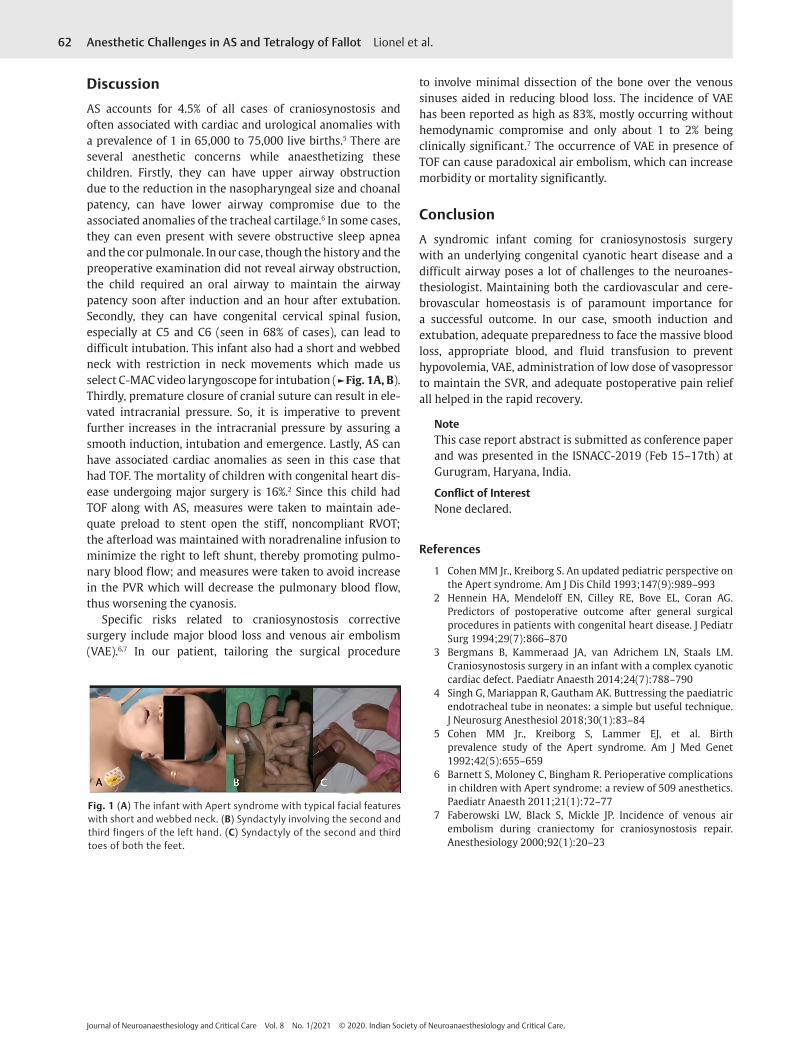
Case ReportAn 8-month-old infant (weight: 8 kg) presented with the typical features of AS, such as flattening of bilateral frontal prominenc-es with fused cranial bones, ocular hypertelorism, down-slant-ing palpebral fissures, and proptotic eyes, depressed nasal bridge, wide nose with a bulbous tip, fused middle and third fingers on the left hand, and fused second and third toes in both feet (►Fig. 1A-C). The child had central cyanosis, with oxygen sat-uration of 82%. Airway examination revealed short webbed neck with restriction of neck movements. Cardiac examination revealed features of TOF. Echocardiography showed a subaor-tic ventricular septal defect (5-mm defect) with bidirectional shunting, 20% aortic overriding with severe infundibular ste-nosis with a peak gradient of 107 mm Hg. Computerized tomog-raphy of skull revealed fused coronal suture with open sagittal, metopic, and lambdoid sutures. She was posted for bifrontal craniotomy with right frontal orbitotomy. The child was on propranolol 5 mg bid for preventing infundibular spasm.
After consultation with a multidisciplinary team, it was decided to proceed with craniostenostosis correc-tion before the surgical correction for TOF. All standard monitors were connected and induction was carried out using 4% sevoflurane with 100% oxygen. Once the child
was asleep, an intravenous access was established and the anesthetic plane was deepened with fentanyl, ketamine, and a small dose of propofol. Phenylephrine (5 µg) bolus was given along with induction drugs to maintain the sys-temic vascular resistance (SVR). After confirming the ade-quate bag and mask ventilation, the child was paralyzed with atracurium, was intubated using a video laryngoscope (C-MAC size 1, video laryngoscope, KARL STORZ, Germany) with 4.5-uncuffed endotracheal tube (EET) which was rein-forced with 5.5-mm ETT to prevent kinking (from the angle of mouth to connector level) as described by Singh et al.4 A 22-G peripheral venous access, a 24-G right radial arte-rial line, and a 5.5-F right subclavian triple lumen central venous catheter were inserted and strict measures were taken to avoid air entrainment. Tranexamic acid (20 mg/kg) bolus followed by an infusion at 1 mg/kg/h was given to reduce the bleeding. Bilateral scalp block was given using 8 mL of 0.2% ropivacaine before the skin incision. Fentan-yl (up to 8 µg/kg) and morphine (0.75 mg) and 20 mg/kg of paracetamol were given for analgesia. Intraoperatively, measures were taken to avoid “Tet Spell” by maintaining the SVR using low dose of noradrenaline (0.02–0.05 µg/kg/min) and by avoiding factors that can increase the pulmonary vascular resistance (PVR), such as hypercarbia, hypoxia, acidosis, hypothermia, and high positive end expiratory pressure. Intraoperatively, the saturation was maintained between 85% and 93% with an inspired oxygen concentra-tion of 60%. The procedure lasted for 4 hours with 170 mL of blood loss (30% of blood volume) which was replaced with 100 mL packed red cell concentrates and 50 mL of fresh frozen plasma and 25 mL of cryoprecipitate. Since we were expecting another 50 to 60 mL of blood loss in the post-operative wound drain and we did not want to dilute the blood with crystalloids, and to avoid postoperative hypo-tension and coagulopathy, we had given the blood products. The child was hemodynamically stable, warm, and awake at the end of surgery. The trachea was extubated and shifted to high dependency unit with fentanyl (0.4–0.5 µg/kg/h) and ketamine (0.02–0.03 mg/kg/h) infusion for postoperative analgesia. Child had good recovery and got discharged on the 7th postoperative day.
62
Journal of Neuroanaesthesiology and Critical Care Vol. 8 No. 1/2021 © 2020. Indian Society of Neuroanaesthesiology and Critical Care.
Anesthetic Challenges in AS and Tetralogy of Fallot Lionel et al.
DiscussionAS accounts for 4.5% of all cases of craniosynostosis and often associated with cardiac and urological anomalies with a prevalence of 1 in 65,000 to 75,000 live births.5 There are several anesthetic concerns while anaesthetizing these children. Firstly, they can have upper airway obstruction due to the reduction in the nasopharyngeal size and choanal patency, can have lower airway compromise due to the associated anomalies of the tracheal cartilage.6 In some cases, they can even present with severe obstructive sleep apnea and the cor pulmonale. In our case, though the history and the preoperative examination did not reveal airway obstruction, the child required an oral airway to maintain the airway patency soon after induction and an hour after extubation. Secondly, they can have congenital cervical spinal fusion, especially at C5 and C6 (seen in 68% of cases), can lead to difficult intubation. This infant also had a short and webbed neck with restriction in neck movements which made us select C-MAC video laryngoscope for intubation (►Fig. 1A, B). Thirdly, premature closure of cranial suture can result in ele-vated intracranial pressure. So, it is imperative to prevent further increases in the intracranial pressure by assuring a smooth induction, intubation and emergence. Lastly, AS can have associated cardiac anomalies as seen in this case that had TOF. The mortality of children with congenital heart dis-ease undergoing major surgery is 16%.2 Since this child had TOF along with AS, measures were taken to maintain ade-quate preload to stent open the stiff, noncompliant RVOT; the afterload was maintained with noradrenaline infusion to minimize the right to left shunt, thereby promoting pulmo-nary blood flow; and measures were taken to avoid increase in the PVR which will decrease the pulmonary blood flow, thus worsening the cyanosis.
Specific risks related to craniosynostosis corrective surgery include major blood loss and venous air embolism (VAE).6,7 In our patient, tailoring the surgical procedure
to involve minimal dissection of the bone over the venous sinuses aided in reducing blood loss. The incidence of VAE has been reported as high as 83%, mostly occurring without hemodynamic compromise and only about 1 to 2% being clinically significant.7 The occurrence of VAE in presence of TOF can cause paradoxical air embolism, which can increase morbidity or mortality significantly.
ConclusionA syndromic infant coming for craniosynostosis surgery with an underlying congenital cyanotic heart disease and a difficult airway poses a lot of challenges to the neuroanes-thesiologist. Maintaining both the cardiovascular and cere-brovascular homeostasis is of paramount importance for a successful outcome. In our case, smooth induction and extubation, adequate preparedness to face the massive blood loss, appropriate blood, and fluid transfusion to prevent hypovolemia, VAE, administration of low dose of vasopressor to maintain the SVR, and adequate postoperative pain relief all helped in the rapid recovery.
NoteThis case report abstract is submitted as conference paper and was presented in the ISNACC-2019 (Feb 15–17th) at Gurugram, Haryana, India.
Conflict of InterestNone declared.
References
1 Cohen MM Jr., Kreiborg S. An updated pediatric perspective on the Apert syndrome. Am J Dis Child 1993;147(9):989–993
2 Hennein HA, Mendeloff EN, Cilley RE, Bove EL, Coran AG. Predictors of postoperative outcome after general surgical procedures in patients with congenital heart disease. J Pediatr Surg 1994;29(7):866–870
3 Bergmans B, Kammeraad JA, van Adrichem LN, Staals LM. Craniosynostosis surgery in an infant with a complex cyanotic cardiac defect. Paediatr Anaesth 2014;24(7):788–790
4 Singh G, Mariappan R, Gautham AK. Buttressing the paediatric endotracheal tube in neonates: a simple but useful technique. J Neurosurg Anesthesiol 2018;30(1):83–84
5 Cohen MM Jr., Kreiborg S, Lammer EJ, et al. Birth prevalence study of the Apert syndrome. Am J Med Genet 1992;42(5):655–659
6 Barnett S, Moloney C, Bingham R. Perioperative complications in children with Apert syndrome: a review of 509 anesthetics. Paediatr Anaesth 2011;21(1):72–77
7 Faberowski LW, Black S, Mickle JP. Incidence of venous air embolism during craniectomy for craniosynostosis repair. Anesthesiology 2000;92(1):20–23
Fig. 1 (A) The infant with Apert syndrome with typical facial features with short and webbed neck. (B) Syndactyly involving the second and third fingers of the left hand. (C) Syndactyly of the second and third toes of both the feet.