Embed Size (px)
Citation preview
1
Kanika Sharma and Randhir Singh, “An optimized and robust iris recognition algorithm for biometric authentication systems,” International
Journal of Scientific and Technical Advancements, Volume 4, Issue 4, pp. 1-8, 2018.
International Journal of Scientific and Technical Advancements ISSN: 2454-1532
An Optimized and Robust Iris Recognition Algorithm
for Biometric Authentication Systems
Kanika Sharma1,#, Randhir Singh1
1Department of Electronics and Communication Engineering, Sri Sai College of Engineering and Technology,
Badhani, Pathankot, India #Email address: [email protected]
Abstract—Biometric authentication systems are based on the concept of matching some unique features of human body such as fingerprints,
facial features, hand geometry, speech etc. One such feature viz. the Iris, a thing circular diaphragm lying between the cornea and retina of
human eye, has been considered in this paper. A new optimized and robust iris recognition algorithm has been proposed in this paper for
application in various biometric authentication applications. The acquired image first undergoes segmentation using the circular Hough
transform for localizing the iris and pupil regions, and the linear Hough transform for localizing occluding eyelids. Next, the segmented iris
region will be normalized by Daugman's rubber sheet model to eliminate dimensional inconsistencies between iris regions. Finally, features
of the iris will be encoded by convolving the normalized iris region with 1-D Log-Gabor filters and phase quantizing the output in order to
produce a bit-wise biometric template. The Hamming distance is proposed to be chosen as a matching metric, which can give a measure of
how many bits disagree between two templates. Based on the simulation results, it is observed that the proposed algorithm works accurately
for different test images and thus can be used in different biomedical applications.
Keywords— Iris Recognition, Biometric Authentication, Image Processing, Segmentation, Feature Extraction
I. INTRODUCTION
biometric authentication system provides
automatic recognition of an individual based on
some sort of unique feature or characteristic
possessed by the individual. Biometric systems work by first
capturing a sample of the feature, such as recording a digital
sound signal for voice recognition, or taking a digital colour
image for face recognition. The sample is then transformed
using some sort of mathematical function into a biometric
template. The biometric template will provide a normalised,
efficient and highly discriminating representation of the
feature, which can then be objectively compared with other
templates in order to determine identity. Biometric systems
have been developed based on fingerprints [1,2], facial
features [3,4], voice [5,6], hand geometry [7,8], the retina
[9,10], and the one presented in this paper, the iris.
The iris is a thin circular diaphragm, which lies between
the cornea and the lens of the human eye. The function of the
iris is to control the amount of light entering through the pupil,
and this is done by the sphincter and the dilator muscles,
which adjust the size of the pupil. The average diameter of the
iris is 12 mm, and the pupil size can vary from 10% to 80% of
the iris diameter. The iris is an externally visible, yet protected
organ whose unique epigenetic pattern remains stable
throughout adult life. These characteristics make it very
attractive for use as a biometric for identifying individuals.
Image processing techniques can be employed to extract the
unique iris pattern from a digitised image of the eye, and
encode it into a biometric template, which can be stored in a
database. This biometric template contains an objective
mathematical representation of the unique information stored
in the iris, and allows comparisons to be made between
templates. When a subject wishes to be identified by an iris
recognition system, their eye is first photographed, and then a
template created for their iris region. This template is then
compared with the other templates stored in a database until
either a matching template is found and the subject is
identified, or no match is found and the subject remains
unidentified.
II. LITERATURE REVIEW
In [11] authors have proposed modified Log-Gabor filters
for iris recognition. The proposed filter in general is similar to
Daugman algorithm proposed in [12] but the Log-Gabor filters
are used to extract the iris phase information instead of
complex Gabor filters used in Daugman’s [12]. The advantage
of Log-Gabor filters over complex Gabor filters is the former
are strictly bandpass filters and the latter are not. The property
of strictly bandpass makes the Log-Gabor filters more suitable
to extract the iris phase features regardless of the background
brightness. Special attention has not been paid to a modified
system in which a more accurate segmentation process is
applied to an already existing efficient algorithm thereby
increasing the overall reliability and accuracy of iris
recognition. In [13] an improvement of the already existing
wavelet packet decomposition for iris recognition with a
Correct Classification Rate (CCR) is proposed. It involves
changing the segmentation technique used for this
implementation from the integro-differential operator
approach (John Daugman’s model [12]) to the Hough
transform (Wilde’s model). This research extensively
compared the two segmentation techniques to show which is
better in the implementation of the wavelet packet
decomposition. Vatsa et al. [14] proposed algorithms for iris
segmentation, quality enhancement, match score fusion, and
indexing to improve both the accuracy and the speed of iris
recognition. A curve evolution approach is proposed to
A
2
Kanika Sharma and Randhir Singh, “An optimized and robust iris recognition algorithm for biometric authentication systems,” International
Journal of Scientific and Technical Advancements, Volume 4, Issue 4, pp. 1-8, 2018.
International Journal of Scientific and Technical Advancements ISSN: 2454-1532
effectively segment a non-ideal iris image using the modified
Mumford-Shah functional. Different enhancement algorithms
are concurrently applied on the segmented iris image to
produce multiple enhanced versions of the iris image.
Similarly in [15], two powerful sets of features are introduced
to be used for iris recognition: scattering transform-based
features and textural features. PCA is also applied on the
extracted features to reduce the dimensionality of the feature
vector while preserving most of the information of its initial
value. A new iris recognition algorithm is proposed in [16],
which adopts Independent Component Analysis (ICA) to
extract iris texture feature and a competitive learning
mechanism to recognize iris patterns. Experimental results
show that the algorithm is efficient and adaptive to the
environment, e.g. it works well even for blurred iris images,
variable illumination, and interference of eyelids and
eyelashes.
III. PROPOSED METHODOLOGY
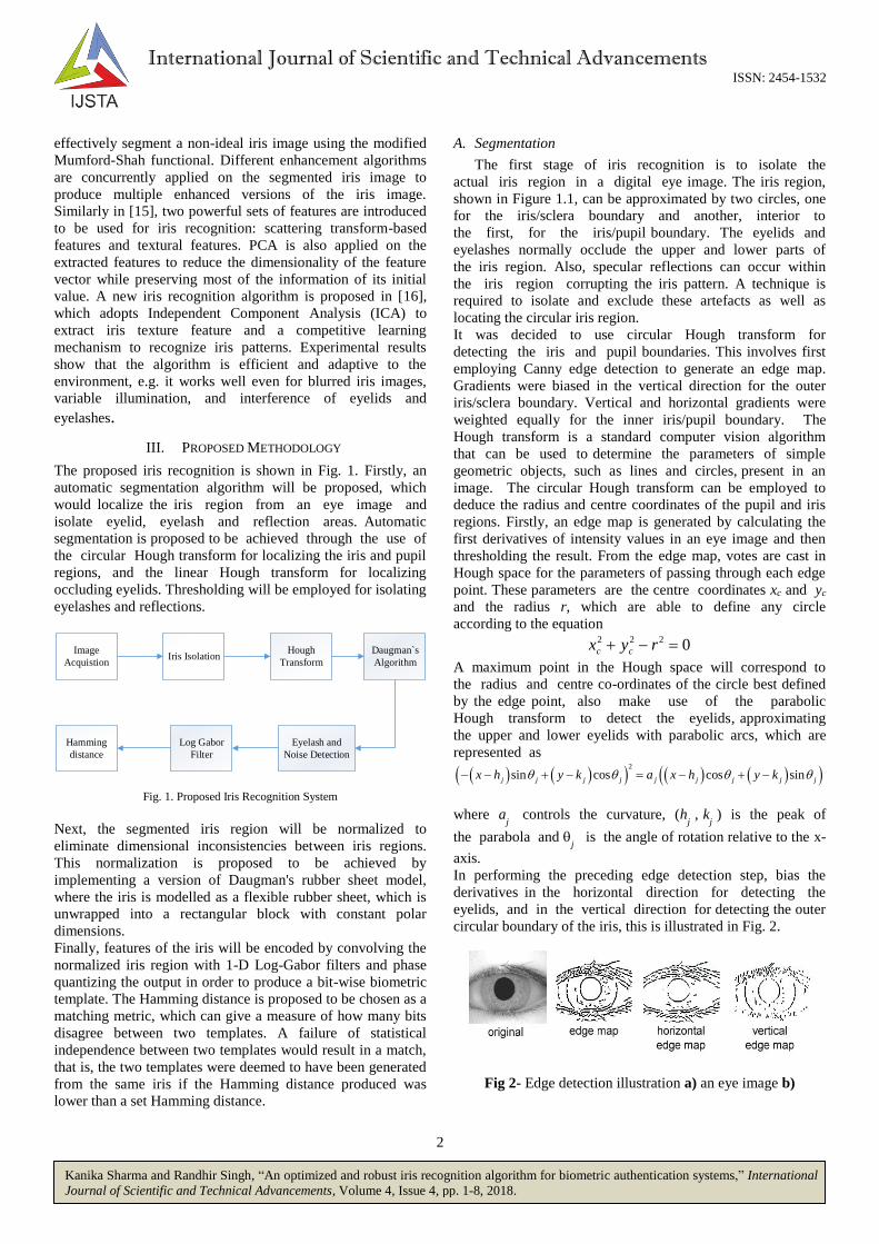
The proposed iris recognition is shown in Fig. 1. Firstly, an
automatic segmentation algorithm will be proposed, which
would localize the iris region from an eye image and
isolate eyelid, eyelash and reflection areas. Automatic
segmentation is proposed to be achieved through the use of
the circular Hough transform for localizing the iris and pupil
regions, and the linear Hough transform for localizing
occluding eyelids. Thresholding will be employed for isolating
eyelashes and reflections.
Image
AcquistionIris Isolation
Hough
Transform
Daugman`s
Algorithm
Eyelash and
Noise Detection
Log Gabor
Filter
Hamming
distance
Fig. 1. Proposed Iris Recognition System
Next, the segmented iris region will be normalized to
eliminate dimensional inconsistencies between iris regions.
This normalization is proposed to be achieved by
implementing a version of Daugman's rubber sheet model,
where the iris is modelled as a flexible rubber sheet, which is
unwrapped into a rectangular block with constant polar
dimensions.
Finally, features of the iris will be encoded by convolving the
normalized iris region with 1-D Log-Gabor filters and phase
quantizing the output in order to produce a bit-wise biometric
template. The Hamming distance is proposed to be chosen as a
matching metric, which can give a measure of how many bits
disagree between two templates. A failure of statistical
independence between two templates would result in a match,
that is, the two templates were deemed to have been generated
from the same iris if the Hamming distance produced was
lower than a set Hamming distance.
A. Segmentation
The first stage of iris recognition is to isolate the
actual iris region in a digital eye image. The iris region,
shown in Figure 1.1, can be approximated by two circles, one
for the iris/sclera boundary and another, interior to
the first, for the iris/pupil boundary. The eyelids and
eyelashes normally occlude the upper and lower parts of
the iris region. Also, specular reflections can occur within
the iris region corrupting the iris pattern. A technique is
required to isolate and exclude these artefacts as well as
locating the circular iris region.
It was decided to use circular Hough transform for
detecting the iris and pupil boundaries. This involves first
employing Canny edge detection to generate an edge map.
Gradients were biased in the vertical direction for the outer
iris/sclera boundary. Vertical and horizontal gradients were
weighted equally for the inner iris/pupil boundary. The
Hough transform is a standard computer vision algorithm
that can be used to determine the parameters of simple
geometric objects, such as lines and circles, present in an
image. The circular Hough transform can be employed to
deduce the radius and centre coordinates of the pupil and iris
regions. Firstly, an edge map is generated by calculating the
first derivatives of intensity values in an eye image and then
thresholding the result. From the edge map, votes are cast in
Hough space for the parameters of passing through each edge
point. These parameters are the centre coordinates xc and yc
and the radius r, which are able to define any circle
according to the equation 2 2 2 0c cx y r
A maximum point in the Hough space will correspond to
the radius and centre co-ordinates of the circle best defined
by the edge point, also make use of the parabolic
Hough transform to detect the eyelids, approximating
the upper and lower eyelids with parabolic arcs, which are
represented as
2
sin cos cos sinj j j j j j j j jx h y k a x h y k
where aj controls the curvature, (h
j , k
j ) is the peak of
the parabola and j is the angle of rotation relative to the x-
axis.
In performing the preceding edge detection step, bias the
derivatives in the horizontal direction for detecting the
eyelids, and in the vertical direction for detecting the outer
circular boundary of the iris, this is illustrated in Fig. 2.
Fig 2- Edge detection illustration a) an eye image b)
3
Kanika Sharma and Randhir Singh, “An optimized and robust iris recognition algorithm for biometric authentication systems,” International
Journal of Scientific and Technical Advancements, Volume 4, Issue 4, pp. 1-8, 2018.
International Journal of Scientific and Technical Advancements ISSN: 2454-1532
corresponding edge map c) edge map with only horizontal
gradients d) edge map with only vertical gradients.
The motivation for this is that the eyelids are usually
horizontally aligned, and also the eyelid edge map will
corrupt the circular iris boundary edge map if using all
gradient data. Taking only the vertical gradients for
locating the iris boundary will reduce influence of the
eyelids when performing circular Hough transform, and not all
of the edge pixels defining the circle are required for
successful localisation. Not only does this make circle
localisation more accurate, it also makes it more efficient,
since there are less edge points to cast votes in the Hough
space.
The range of radius values to search for was set manually,
depending on the database used. In order to make the circle
detection process more efficient and accurate, the Hough
transform for the iris/sclera boundary was performed first,
then the Hough transform for the iris/pupil boundary was
performed within the iris region, instead of the whole eye
region, since the pupil is always within the iris region. After
this process was complete, six parameters are stored, the
radius, and x and y centre coordinates for both circles.
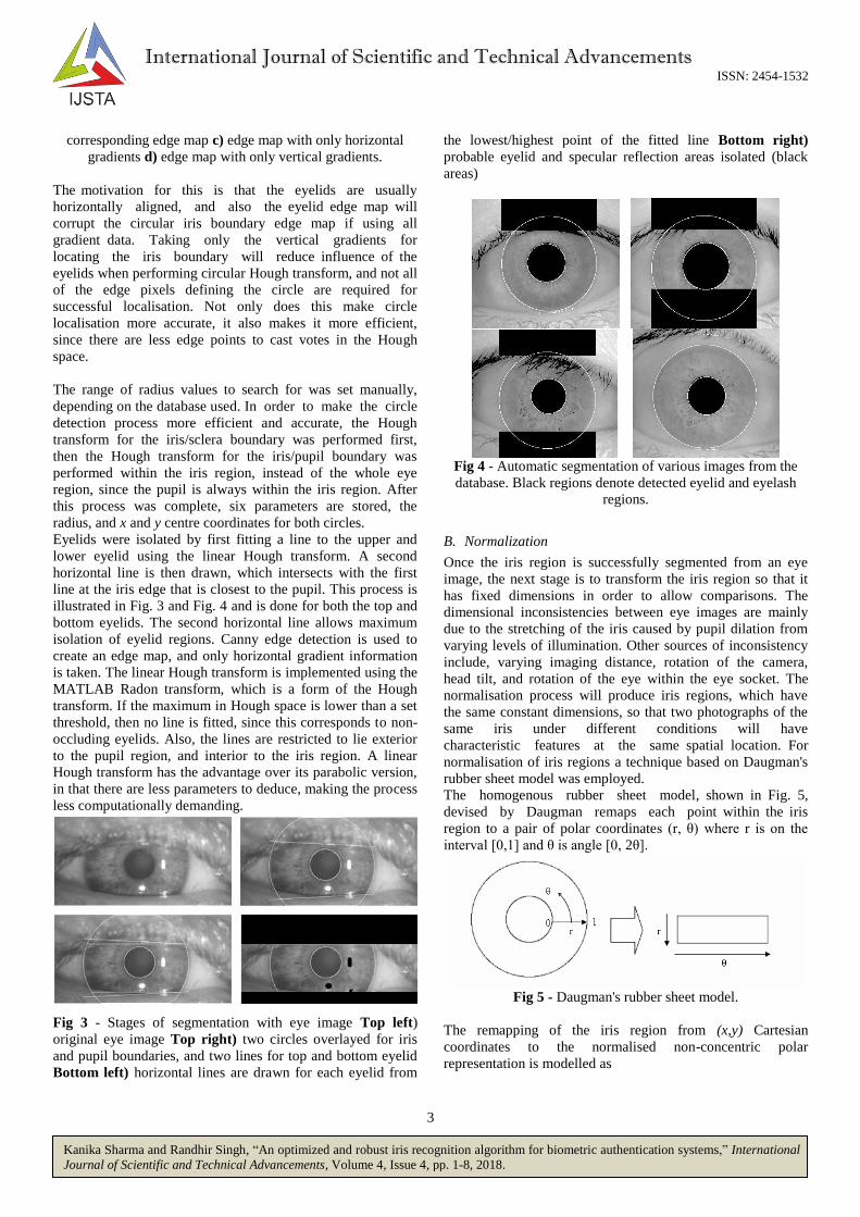
Eyelids were isolated by first fitting a line to the upper and
lower eyelid using the linear Hough transform. A second
horizontal line is then drawn, which intersects with the first
line at the iris edge that is closest to the pupil. This process is
illustrated in Fig. 3 and Fig. 4 and is done for both the top and
bottom eyelids. The second horizontal line allows maximum
isolation of eyelid regions. Canny edge detection is used to
create an edge map, and only horizontal gradient information
is taken. The linear Hough transform is implemented using the
MATLAB Radon transform, which is a form of the Hough
transform. If the maximum in Hough space is lower than a set
threshold, then no line is fitted, since this corresponds to non-
occluding eyelids. Also, the lines are restricted to lie exterior
to the pupil region, and interior to the iris region. A linear
Hough transform has the advantage over its parabolic version,
in that there are less parameters to deduce, making the process
less computationally demanding.
Fig 3 - Stages of segmentation with eye image Top left)
original eye image Top right) two circles overlayed for iris
and pupil boundaries, and two lines for top and bottom eyelid
Bottom left) horizontal lines are drawn for each eyelid from
the lowest/highest point of the fitted line Bottom right)
probable eyelid and specular reflection areas isolated (black
areas)
Fig 4 - Automatic segmentation of various images from the
database. Black regions denote detected eyelid and eyelash
regions.
B. Normalization
Once the iris region is successfully segmented from an eye
image, the next stage is to transform the iris region so that it
has fixed dimensions in order to allow comparisons. The
dimensional inconsistencies between eye images are mainly
due to the stretching of the iris caused by pupil dilation from
varying levels of illumination. Other sources of inconsistency
include, varying imaging distance, rotation of the camera,
head tilt, and rotation of the eye within the eye socket. The
normalisation process will produce iris regions, which have
the same constant dimensions, so that two photographs of the
same iris under different conditions will have
characteristic features at the same spatial location. For
normalisation of iris regions a technique based on Daugman's
rubber sheet model was employed.
The homogenous rubber sheet model, shown in Fig. 5,
devised by Daugman remaps each point within the iris
region to a pair of polar coordinates (r, θ) where r is on the
interval [0,1] and θ is angle [0, 2θ].
Fig 5 - Daugman's rubber sheet model.
The remapping of the iris region from (x,y) Cartesian
coordinates to the normalised non-concentric polar
representation is modelled as

4
Kanika Sharma and Randhir Singh, “An optimized and robust iris recognition algorithm for biometric authentication systems,” International
Journal of Scientific and Technical Advancements, Volume 4, Issue 4, pp. 1-8, 2018.
International Journal of Scientific and Technical Advancements ISSN: 2454-1532
, , , ,I x r y r I r
With
, 1
, 1
p l
p l
x r r x rx
y r r y ry
where I(x,y) is the iris region image, (x,y) are the original
Cartesian coordinates, (r,) are the corresponding normalised
polar coordinates, and xp , yp and x
l , y
l are the coordinates
of the pupil and iris boundaries along the direction. The
rubber sheet model takes into account pupil dilation and size
inconsistencies in order to produce a normalised
representation with constant dimensions. In this way the iris
region is modelled as a flexible rubber sheet anchored at the
iris boundary with the pupil centre as the reference point.
Even though the homogenous rubber sheet model accounts for
pupil dilation, imaging distance and non-concentric pupil
displacement, it does not compensate for rotational
inconsistencies. In the Daugman system, rotation is accounted
for during matching by shifting the iris templates in the
direction until two iris templates are aligned.
The centre of the pupil was considered as the reference
point, and radial vectors pass through the iris region, as
shown in Fig. 6. A number of data points are selected along
each radial line and this is defined as the radial resolution. The
number of radial lines going around the iris region is
defined as the angular . Since the pupil can be non-
concentric to the iris, a remapping formula is needed to rescale
points depending on the angle around the circle. This is given
by
, 2 2
Ir r
With 2 2
x yo o
cos arctany
x
o
o
where displacement of the centre of the pupil relative to the
centre of the iris is given by ox, o
y and r' is the distance
between the edge of the pupil and edge of the iris at an angle,
around the region, and rI is the radius of the iris. The
remapping formula first gives the radius of the iris region
'doughnut' as a function of the angle .
A constant number of points are chosen along each radial
line, so that a constant number of radial data points are
taken, irrespective of how narrow or wide the radius is at a
particular angle. The normalised pattern was created by
backtracking to find the Cartesian coordinates of data points
from the radial and angular position in the normalised
pattern. From the 'doughnut' iris region, normalisation
produces a 2D array with horizontal dimensions of
angular resolution and vertical dimensions of radial
resolution. Another 2D array was created for marking
reflections, eyelashes, and eyelids detected in the
segmentation stage. In order to prevent non-iris region data
from corrupting the normalised representation, data points
which occur along the pupil border or the iris border are
discarded. As in Daugman's rubber sheet model, removing
rotational inconsistencies is performed at the matching stage.
Fig. 6 - Outline of the normalisation process with radial
resolution of 10 pixels, and angular resolution of 40 pixels.
Pupil displacement relative to the iris centre is exaggerated for
illustration purposes.
Normalisation of two eye images of the same iris is shown in
Fig. 7. The pupil is smaller in the bottom image, however the
normalisation process is able to rescale the iris region so that it
has constant dimension. In this example, the rectangular
representation is constructed from 10,000 data points in each
iris region. Note that rotational inconsistencies have not been
accounted for by the normalisation process, and the two
normalised patterns are slightly misaligned in the
horizontal (angular) direction. Rotational inconsistencies will
be accounted for in the matching stage.
Fig. 7 - Illustration of the normalisation process for two
images of the same iris taken under varying conditions.
5
Kanika Sharma and Randhir Singh, “An optimized and robust iris recognition algorithm for biometric authentication systems,” International
Journal of Scientific and Technical Advancements, Volume 4, Issue 4, pp. 1-8, 2018.
International Journal of Scientific and Technical Advancements ISSN: 2454-1532
C. Feature Encoding
In order to provide accurate recognition of
individuals, the most discriminating information present in
an iris pattern must be extracted. Only the significant features
of the iris must be encoded so that comparisons between
templates can be made. Most iris recognition systems make
use of a band pass decomposition of the iris image to create a
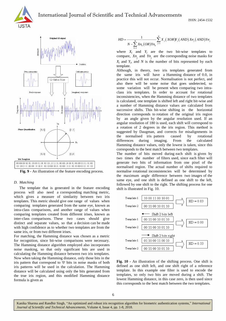
biometric template. Feature encoding was implemented by
convolving the normalised iris pattern with 1D Log-Gabor
wavelets.
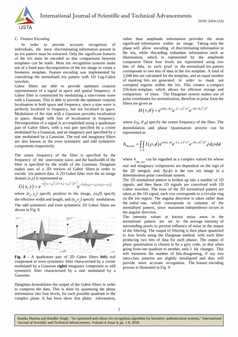
Gabor filters are able to provide optimum conjoint
representation of a signal in space and spatial frequency. A
Gabor filter is constructed by modulating a sine/cosine wave
with a Gaussian. This is able to provide the optimum conjoint
localisation in both space and frequency, since a sine wave is
perfectly localised in frequency, but not localised in space.
Modulation of the sine with a Gaussian provides localisation
in space, though with loss of localisation in frequency.
Decomposition of a signal is accomplished using a quadrature
pair of Gabor filters, with a real part specified by a cosine
modulated by a Gaussian, and an imaginary part specified by a
sine modulated by a Gaussian. The real and imaginary filters
are also known as the even symmetric and odd symmetric
components respectively.
The centre frequency of the filter is specified by the
frequency of the sine/cosine wave, and the bandwidth of the
filter is specified by the width of the Gaussian. Daugman
makes uses of a 2D version of Gabor filters in order to
encode iris pattern data. A 2D Gabor filter over the an image
domain (x,y) is represented as
2 22 20 0 0 0 0 02
,x x y y i u x x v y y
G x y e e
where (xo, y
o) specify position in the image, (,) specify
the effective width and length, and (uo,v
o) specify modulation.
The odd symmetric and even symmetric 2D Gabor filters are
shown in Fig. 8.
Fig. 8 - A quadrature pair of 2D Gabor filters left) real
component or even symmetric filter characterised by a cosine
modulated by a Gaussian right) imaginary component or odd
symmetric filter characterised by a sine modulated by a
Gaussian.
Daugman demodulates the output of the Gabor filters in order
to compress the data. This is done by quantising the phase
information into four levels, for each possible quadrant in the
complex plane. It has been show that phase information,
rather than amplitude information provides the most
significant information within an image. Taking only the
phase will allow encoding of discriminating information in
the iris, while discarding redundant information such as
illumination, which is represented by the amplitude
component. These four levels are represented using two
bits of data, so each pixel in the normalised iris pattern
corresponds to two bits of data in the iris template. A total of
2,048 bits are calculated for the template, and an equal number
of masking bits are generated in order to mask out
corrupted regions within the iris. This creates a compact
256-byte template, which allows for efficient storage and
comparison of irises. The Daugman system makes use of
polar coordinates for normalisation, therefore in polar form the
filters are given as
2 22 2
0 0 0 0/ /,
i r r iH r e e e
where (r0, 0) specify the centre frequency of the filter. The
demodulation and phase Quantisation process can be
represented as
2 22 2
0 0 0/ /
Re,Im,
i rh I e e e d d
where h{Re, Im}
can be regarded as a complex valued bit whose
real and imaginary components are dependent on the sign of
the 2D integral, and, I(,) is the raw iris image in a
dimensionless polar coordinate system.
The 2D normalised pattern is broken up into a number of 1D
signals, and then these 1D signals are convolved with 1D
Gabor wavelets. The rows of the 2D normalised pattern are
taken as the 1D signal, each row corresponds to a circular ring
on the iris region. The angular direction is taken rather than
the radial one, which corresponds to columns of the
normalised pattern, since maximum independence occurs in
the angular direction.
The intensity values at known noise areas in the
normalised pattern are set to the average intensity of
surrounding pixels to prevent influence of noise in the output
of the filtering. The output of filtering is then phase quantised
to four levels using the Daugman method, with each filter
producing two bits of data for each phasor. The output of
phase quantisation is chosen to be a grey code, so that when
going from one quadrant to another, only 1 bit changes. This
will minimise the number of bits disagreeing, if say two
intra-class patterns are slightly misaligned and thus will
provide more accurate recognition. The feature encoding
process is illustrated in Fig. 9
6
Kanika Sharma and Randhir Singh, “An optimized and robust iris recognition algorithm for biometric authentication systems,” International
Journal of Scientific and Technical Advancements, Volume 4, Issue 4, pp. 1-8, 2018.
International Journal of Scientific and Technical Advancements ISSN: 2454-1532
Fig. 9 - An illustration of the feature encoding process.
D. Matching
The template that is generated in the feature encoding
process will also need a corresponding matching metric,
which gives a measure of similarity between two iris
templates. This metric should give one range of values when
comparing templates generated from the same eye, known as
intra-class comparisons, and another range of values when
comparing templates created from different irises, known as
inter-class comparisons. These two cases should give
distinct and separate values, so that a decision can be made
with high confidence as to whether two templates are from the
same iris, or from two different irises.
For matching, the Hamming distance was chosen as a metric
for recognition, since bit-wise comparisons were necessary.
The Hamming distance algorithm employed also incorporates
noise masking, so that only significant bits are used in
calculating the Hamming distance between two iris templates.
Now when taking the Hamming distance, only those bits in the
iris pattern that correspond to '0' bits in noise masks of both
iris patterns will be used in the calculation. The Hamming
distance will be calculated using only the bits generated from
the true iris region, and this modified Hamming distance
formula is given as
, ,
1
1
1 N
j j j jN
jk k
k
HD X XOR Y AND Xn AND Yn
N Xn OR Yn
where Xj and Yj are the two bit-wise templates to
compare, Xnj and Yn
j are the corresponding noise masks for
Xj and Yj, and N is the number of bits represented by each
template.
Although, in theory, two iris templates generated from
the same iris will have a Hamming distance of 0.0, in
practice this will not occur. Normalisation is not perfect, and
also there will be some noise that goes undetected, so
some variation will be present when comparing two intra-
class iris templates. In order to account for rotational
inconsistencies, when the Hamming distance of two templates
is calculated, one template is shifted left and right bit-wise and
a number of Hamming distance values are calculated from
successive shifts. This bit-wise shifting in the horizontal
direction corresponds to rotation of the original iris region
by an angle given by the angular resolution used. If an
angular resolution of 180 is used, each shift will correspond to
a rotation of 2 degrees in the iris region. This method is
suggested by Daugman, and corrects for misalignments in
the normalised iris pattern caused by rotational
differences during imaging. From the calculated
Hamming distance values, only the lowest is taken, since this
corresponds to the best match between two templates.
The number of bits moved during each shift is given by
two times the number of filters used, since each filter will
generate two bits of information from one pixel of the
normalised region. The actual number of shifts required to
normalise rotational inconsistencies will be determined by
the maximum angle difference between two images of the
same eye, and one shift is defined as one shift to the left,
followed by one shift to the right. The shifting process for one
shift is illustrated in Fig. 10.
Fig. 10 - An illustration of the shifting process. One shift is
defined as one shift left, and one shift right of a reference
template. In this example one filter is used to encode the
templates, so only two bits are moved during a shift. The
lowest Hamming distance, in this case zero, is then used since
this corresponds to the best match between the two templates.

7
Kanika Sharma and Randhir Singh, “An optimized and robust iris recognition algorithm for biometric authentication systems,” International
Journal of Scientific and Technical Advancements, Volume 4, Issue 4, pp. 1-8, 2018.
International Journal of Scientific and Technical Advancements ISSN: 2454-1532
IV. SIMULATION RESUTLS
Fig. 11 – GUI of the proposed Iris Recognition System.
Fig. 12 – Illustration of selection of an eye image for the
matching process by the proposed Iris Recognition System.
Fig. 13 – Illustration of segmentation of eye image by the
proposed Iris Recognition System.
Fig. 14 – Illustration of matching process by the proposed Iris
Recognition System.
Fig. 15 – Illustration of matching confirmation by the
proposed Iris Recognition System.
Fig. 16 – Illustration of matching rejection by the proposed
Iris Recognition System.

8
Kanika Sharma and Randhir Singh, “An optimized and robust iris recognition algorithm for biometric authentication systems,” International
Journal of Scientific and Technical Advancements, Volume 4, Issue 4, pp. 1-8, 2018.
International Journal of Scientific and Technical Advancements ISSN: 2454-1532
V. CONCLUSION
A new optimized and robust iris recognition algorithm has
been proposed in this paper for application in various
biometric authentication applications. The acquired image first
undergoes segmentation using the circular Hough transform
for localizing the iris and pupil regions, and the linear Hough
transform for localizing occluding eyelids. Next, the
segmented iris region will be normalized by Daugman's rubber
sheet model to eliminate dimensional inconsistencies between
iris regions. Finally, features of the iris will be encoded by
convolving the normalized iris region with 1-D Log-Gabor
filters and phase quantizing the output in order to produce a
bit-wise biometric template. The Hamming distance is
proposed to be chosen as a matching metric, which can give a
measure of how many bits disagree between two templates.
Based on the simulation results, it is observed that the
proposed algorithm works accurately for different test images
and thus can be used in different biomedical applications.
REFERENCES
[1] James Wayman, Anil Jain, Davide Maltoni, and Dario Maio. "An
introduction to biometric authentication systems." In Biometric Systems, pp. 1-20. Springer, London, 2005.
[2] Obi Ogbanufe, and Dan J. Kim. "Comparing fingerprint-based
biometrics authentication versus traditional authentication methods for e-payment." Decision Support Systems, vol. 106, 2018, pp. 1-14.
[3] Marios Savvides, BVK Vijaya Kumar, and Pradeep K. Khosla.
"Cancelable biometric filters for face recognition." In Proc. of the 17th IEEE International Conference on Pattern Recognition, 2004, pp. 922-
925 [4] Wang Feng, Jiyan Zhou, Chen Dan, Zhou Peiyan, and Zhang Li.
"Research on mobile commerce payment management based on the face
biometric authentication." International Journal of Mobile Communications, vol. 15, no. 3, 2017, pp. 278-305.
[5] Girija Chetty and Michael Wagner. "Multi-level liveness verification for
face-voice biometric authentication." In proc. of Biometrics Symposium:
Special Session on Research at the IEEE Biometric Consortium
Conference, 2006, pp. 1-6. [6] Felipe Gomes Barbosa, and Washington Luís Santos Silva. "Support
vector machines, Mel-Frequency Cepstral Coefficients and the Discrete
Cosine Transform applied on voice based biometric authentication." In proc. of IEEE SAI Intelligent Systems Conference (IntelliSys), 2015,
pp. 1032-1039.
[7] Puneet Gupta, Saurabh Srivastava, and Phalguni Gupta. "An accurate infrared hand geometry and vein pattern based authentication
system." Knowledge-Based Systems, vol. 103, 2016, pp. 143-155.
[8] Aditya Nigam and Phalguni Gupta. "Designing an accurate hand biometric based authentication system fusing finger knuckleprint and
palmprint." Neurocomputing, vol. 151, 2015, pp. 1120-1132.
[9] M. Islamuddin Ahmed, M. Ashraful Amin, Bruce Poon, and Hong Yan. "Retina based biometric authentication using phase
congruency." International Journal of Machine Learning and
Cybernetics, vol. 5, no. 6, 2014, pp. 933-945. [10] Jarina B. Mazumdar and S. R. Nirmala. "Retina Based Biometric
Authentication System: A Review" International Journal of Advanced
Research in Computer Science, vol. 9, no. 1, 2018, pp. 711-718.
[11] P. Yao, J. Le, X.Ye, Z. Zhuang, B. Li, “Iris Recognition Algorithm
using Modified Log-Gabor Filters,” Proc. of IEEE International
Conference on Pattern Recognition, Hong Kong China, 2006, pp. 1-4. [12] J. Daugman, “How Iris Recognition Works,” IEEE Transactions on
Circuits and Systems for Video Technology, vol. 14, no. 1, 2004, pp. 21-
30. [13] K. Okokpujie, E. Noma-Osaghae, S. Hohn, A. Ajulibe, “An Improved
Iris Segmentation Technique using Circular Hough Transform,” in IT
Convergence and Security, Springer, 2017. [14] M. Vatsa, R. SIgnh, Afzel Noore, “Improving Iris Recognition
Performance using Segmentation, Quality Enhancement, Match Score
Fusion, and Indexing,” IEEE Transactions on Systems, Man, and Cybernetics, Part B (Cybernetics), vol. 38, no. 4, 2008, pp. 1021-1035.
[15] S. Minaee, A. Abdolrashidi, Y. Wang, “Iris recognition using scattering transform and textural features,” in proc. of IEEE International
Conference on Signal Processing and Signal Processing Education, Salt
Lake City, US, 2015. [16] Y. Huang, S. Luo, E. Chen, “An efficient iris recognition system,” in
proc. of IEEE International conference on Machine Learning and
Cybernetics, Beijing, China, 2002.


![Biometric Standards documents/Standards... · Biometric Profiles Biometric [Application] Profile – a conforming subset or combination of base standards used to effect specific biometric](https://img.pdfslide.net/doc/110x75/5f711372ce578d4ee02aea91/biometric-standards-documentsstandards-biometric-profiles-biometric-application.jpg)