Embed Size (px)
Citation preview

Analiza uloge međustanične komunikacije u simbioziRhizobium-Leguminosae
Busch, Franka
Master's thesis / Diplomski rad
2016
Degree Grantor / Ustanova koja je dodijelila akademski / stručni stupanj: University of Zagreb, Faculty of Science / Sveučilište u Zagrebu, Prirodoslovno-matematički fakultet
Permanent link / Trajna poveznica: https://urn.nsk.hr/urn:nbn:hr:217:417404
Rights / Prava: In copyright
Download date / Datum preuzimanja: 2021-11-29
Repository / Repozitorij:
Repository of Faculty of Science - University of Zagreb

University of Zagreb
Faculty of Science
Department of Biology
Franka Busch
ANALYSIS OF THE QUORUM SENSING ROLE IN THE Rhizobium-
Leguminosae SYMBIOSIS
Graduation Thesis
Zagreb, 2016

This Thesis was made at the Centre for Plant Biotechnology and Genomics (CBGP),
Technical University of Madrid, in the Microbiology laboratory under the guidance of
Professor José Manuel Palacios Alberti and Dr. Jasna Hrenović, Assoc. Professor at the
University of Zagreb, Faculty of Science, Microbiology Department. The Thesis was
submitted for evaluation at the Department of Biology, Faculty of Science, University
of Zagreb, in order to obtain Master Degree in Environmental Sciences and Ecology.

Acknowledgements
This Thesis was made at the Centre for Plant Biotechnology and Genomics (CBGP),
Technical University of Madrid, in the Microbiology laboratory under the guidance of
Professor José Manuel Palacios Alberti and Dr. Jasna Hrenović, Assoc. Professor at the
University of Zagreb, Faculty of Science, Microbiology Department.
Firstly I would like to thank my mentor Professor Palacios for the opportunity for
making my Graduation Thesis at the CBGP in Madrid and for guidance during the
whole realization of my Thesis.
I would also like to thank my mentor Associate Professor Hrenović who was always
able to help as quickly as possible during the making of this Thesis.
Moreover, special thank you to my colleagues from the laboratory at CBGP in Madrid;
Ana, David, Alba, Alejandro and Hakim, who helped me from the beginning in every
segment of my work.
Finally, I would like to thank Professor Emeritus Tomás Ruiz Argüeso for his great
knowledge, experience and for trying to make my stay in Laboratory more pleasant.
TEMELJNA DOKUMENTACIJSKA KARTICA
Sveučilište u Zagrebu
Prirodoslovno-matematički fakultet
Biološki odsjek
Diplomski rad
ANALIZA ULOGE MEĐUSTANIČNE KOMUNIKACIJE U SIMBIOZI Rhizobium-
Leguminosae
Franka Busch
Rooseveltov trg 6,10 000 Zagreb, Hrvatska
U istraživanju je provedeno ispitivanje uloge meĎustanične komunikacije u simbiozi
Rhizobium-Leguminosae u svrhu boljeg razumijevanja meĎustanične komunikacije u
procesu fiksacije dušika. Metodama molekularnog inženjerstva konstruirani su mutanti
cinR i traI gena koji su dio genoma bakterije soja Rhizobium leguminosarum bv. viciae
UPM791. Geni cinR i traI su izabrani iz razloga što imaju kontrolu nad acil-homoserin
lakton ovisnom sistemu meĎustanične komunikacije. Mutanti gena cinR i traI su
uspješno dobiveni i podvrgnuti daljnjim istraživanjem utjecaja mutanata na interakciju s
biljkama. Preliminarni rezultati dobiveni TLC analizom ukazuju da cinR mutant
smanjeno proizvodi C14-HSL molekule kao i kratkolančane AHL molekule. Analize traI
mutanta ukazuju na smanjenu proizvodnju kratkolančanih AHL molekula. Preliminarne
rezultate potrebno je potvrditi nastavkom istraživanja radi procjenjivanja značaja
mutacija.
(54 stranica,18 slika, 8 tablica, 49 literaturnih navoda, jezik izvornika: engleski)
Rad je pohranjen u Središnjoj biološkoj knjižnici
Ključne riječi: bakterije, signalne molekule, homoserin lakton, acil-homoserin lakton,
nodulacija
Voditelj: izv. prof. dr. sc. Jasna Hrenović
Ocjenitelji: izv. prof. dr. sc. Jasna Hrenović
doc. dr. sc. Petar Kružić
doc. dr. sc. Ana Galov
Zamjena: izv. prof. dr. sc. Renata Matoničkin Kepčija
Rad prihvaćen: 18.02.2016.
BASIC DOCUMENTATION CARD
University of Zagreb
Faculty of Science
Department of Biology
Graduation Thesis
ANALYSIS OF THE QUORUM SENSING ROLE IN THE Rhizobium-Leguminosae
SYMBIOSIS
Franka Busch
Rooseveltov trg 6, 10 000 Zagreb, Croatia
In this study the role of quorum sensing communication in Rhizobium-Leguminosae
symbiosis was examined in the process of nitrogen fixation. In order to understand
better the quorum sensing communication with the methods of molecular engineering
mutants in cinR and traI genes, which are the part of the bacterial strains genome of
Rhizobium leguminosarum bv. viciae UPM791, were constructed. Genes cinR and traI
are chosen because they control the AHL-dependent quorum sensing system. Mutants
of genes cinI and traI are successfully obtained and further characterized at the
laboratory for phenotypes in interactions with plants. Preliminary results obtained by
TLC analysis showed that the cinR mutant is producing less C14-HSL signals, also
showing a reduction on the level of short-chain AHLs. Analysis of traI gene showed no
significant alteration on C14-HSL production but the level of short-chain AHLs is
reduced. Preliminary results have to be confirmed in further analysis to assess better
examination of the significance of mutations.
(54 pages, 18 figures, 8 tables, 49 references, origin in: English)
Key words: bacteria, signal molecules, homoserin lactone, aycl-homoserin lactone,
nodulation
Supervisor: Dr. Jasna Hrenović, Assoc. Prof.
Reviewers: Dr. Jasna Hrenović, Assoc. Prof.
Dr. Petar Kružić, Asst. Prof.
Dr. Ana Galov, Asst. Prof.
Replacement: Dr. Renata Matoničkin Kepčija, Assoc. Prof.
Thesis accepted: 18.02.2016.
Abbreviations and acronyms
3OC6-HSL 3-oxo-hexanoyl-homoserine lacone
3-OH-C14:1-HSL N-(3-hydroxy-7-cis-tetradodecyl)-L-homoserine lactone
ABD autoinducer binding domain
ACP acyl carrier proteins
AHL(s) acyl-homoserine lactone(s)
AI-2 autoinducer-2
ApR
ampillicin resistance
BLAST Basic Local Alignment Search Tool
BNF biological nitrogen fixation
bp base pairs
bv. biovar
C carbon
C14-HSL N-tetradecanoyl-L-homoserine lactone
C6-HSL N-hexanoyl-L-homoserine lactone
C7-HSL N-heptanoyl-L-homoserine lactone
C8-HSL N-octanoyl-L-homoserine lactone
CaCl2 calcium chloride
CSPD chemiluminescent substrate
DNA deoxyribonucleic acid
EDTA ethylenediaminetetraacetic acid
Et-Br ethidium bromide
h hours
HCl hydrochloric
HSL homoserine lactone
HTH helix turn helix
K2HPO4 potassium phosphate
kb kilobase
Km kanamycin
KmR
kanamycin resistance
LB Luria-Betrani broth
M molar
Mb megabase

MgSO4 magnesium sulphate
min minutes
mM miliMolar
N nitrogen
N2 molecular nitrogen (nitrogen gas)
NaCl Sodium chloride
NaOH sodium hydroxide
NCBI National Centre for Biotechnology Information
Nf nitrofurantoin
NH4+
ammonium cations
NO3-
nitrates
Nod/nod nodultion
NT3 neutrotrophin-3
PCR polymerase chain reaction
QS quorum sensing
Rlv Rhizobium leguminosarum bv. viciae
RNase ribonuclease
rpm rotation per minute
s seconds
SAM S-adenosyl-methionin
SDS sodium dodecyl sulphate
Sp spectinomycin
SpR
spectinomycin resistance
Str streptomycin
StrR
streptomycin resistance
TBE Tris base-boric acid-EDTA
Tc tetracycline
TcR
tetracycline resistance
TLC thin-layer chromatography
Tm annealing temperature
TY tryptone-yeast extract
UV ultra violet light/ radiation
WT wild type

YMB yeast mannitol broth
Δ mutants
Table of Contents
1. INTRODUCTION ........................................................................................................ 1
1.1. Relevance of nitrogen fixation ............................................................................... 1
1.2. Establishment of the Rhizobium-legume symbiosis ............................................... 2
1.2.1. The plant-bacteria recognition ......................................................................... 2
1.2.2. The process of infection .................................................................................. 3
1.2.3. The nodule development ................................................................................. 3
1.3. Rhizobial genome ................................................................................................... 4
1.4. Quorum sensing system ......................................................................................... 5
1.4.1. Molecular basis of quorum sensing ................................................................. 6
1.4.2. Gram-negative bacteria: the LuxRI signaling system ..................................... 7
1.4.3. Quorum sensing genes in rhizobia .................................................................. 7
1.4.4. Quorum sensing systems in Rhizobium leguminosarum bv viciae ................. 8
2. OBJECTIVES ............................................................................................................. 13
3. MATERIALS AND METHODS ................................................................................ 14
3.1. BIOLOGICAL MATERIAL ................................................................................ 14
3.1.1. Bacteria and plasmids .................................................................................... 14
3.2. MEDIA AND GROWTH CONDITIONS ........................................................... 15
3.3. DNA manipulation techniques ............................................................................. 15
3.3.1. DNA extraction ............................................................................................. 15
3.3.4. PCR amplification and electrophoresis ......................................................... 17
3.3.5. DNA sequencing, analyzing and primer design ............................................ 18
3.3.6. Restriction enzyme digestion ........................................................................ 19
3.3.7. Cloning .......................................................................................................... 19
3.3.8. Ligation .......................................................................................................... 19
3.3.9. Plasmid transfer by transformation ............................................................... 19
3.3.10. Plasmid transfer by mating .......................................................................... 20
3.3.11. Southern blot ............................................................................................... 20
4. RESULTS ................................................................................................................... 22
4.1. CHARACTERIZATION OF cinR AND traI GENES FROM R. leguminosarum
UPM791 ...................................................................................................................... 22
4.1.1. Study of the sequence of the cinR and traI genes from R. leguminosarum
strains and design of the primers to amplify the target genes ................................. 22

4.1.2. Amplification of the target genes .................................................................. 23
4.1.3. Analysis of the sequence of the amplified genes and comparison with other
genes in the database ............................................................................................... 25
4.2. GENERATION OF MUTANT DERIVATIVES DEFECTIVE IN cinR AND traI
GENES ........................................................................................................................ 33
4.2.1. Subcloning of the fragment by using a suicide plasmid containing an internal
fragment ................................................................................................................... 33
4.2.2. Mating into R. leguminosarum strains ........................................................... 36
4.2.3. Analysis of the transconjugants through PCR and Southern blot analysis ... 38
5. DISCUSSION ............................................................................................................. 42
6. CONCLUSIONS ........................................................................................................ 48
7. LITERATURE ............................................................................................................ 49
8. CURICULUM VITAE ............................................................................................... 54

1
1. INTRODUCTION
1.1. Relevance of nitrogen fixation
Nitrogen is a very important element in agriculture, and along with water is one of the
most relevant limiting factors in plant growth. It is also a limiting factor for growth of
many ecosystems. Even though the atmosphere contains enormous amounts of nitrogen,
its molecular form (N2) cannot be used by most of living organisms. When the
concentration of nitrogen in soil is really low, plants cannot reach their maximum
growth potential. Today, when the maximum of crop productivity is the priority, the
agricultural systems are using additional chemically produced nitrogen fertilizers to
improve poor N-content soils.
This human activity has an enormous impact on the global nitrogen cycle. Agriculture
alone contributes with around 2.4*1012
mol N per year because of cultivation-induced
nitrogen fixation (Canfield et al., 2010). Plants can use nitrogen for their growth only in
two forms, ammonium cations (NH4+) or nitrates (NO3
-). The process of converting
molecular nitrogen to other forms is called nitrogen fixation. Nitrogen fixation is a very
important process in which molecular nitrogen (N2) from the atmosphere, that is
relatively inert, is converted into ammonium cations (NH4+). In order to avoid the
excessive use of chemically produced nitrogen fertilizers, because of their negative
impact to the environment, organisms capable for nitrogen fixation are again in the
centre of interest.
There are several ways to convert nitrogen to other forms and one of them is biological
transformation. In the process of biological nitrogen fixation (BNF) atmospheric
nitrogen is converted to ammonia due to the activity of nitrogenase enzymatic complex.
The process is carried out by microorganisms, all belonging to a biological group
known as prokaryotes (Bacteria and Archaea). The BNF can be done in two different
ways; symbiotic and non-symbiotic nitrogen fixation. Symbiotic nitrogen fixation,
nowadays, is very important from an agricultural point of view. The most relevant
biological nitrogen fixation system is the symbiotic association between legumes and
bacteria belonging to the Rhizobium group (Herridge et al., 2008).
2
1.2. Establishment of the Rhizobium-legume symbiosis
Rhizobium is a genus of soil bacteria, whose members are able to establish nitrogen-
fixing symbiosis interaction with legume, plant members of Fabaceae family (Blažinkov
et al., 2007). Following the interaction between the bacteria and the host plant the result
is formation of nitrogen-fixing structures called nodules. Usually they are developed on
the root of the host plant. In the nodule structure, nitrogen-fixing forms of rhizobia
called bacteroids, in exchange of photosynthetic products of the plant, support plant
growth by the reduction of the atmospheric nitrogen gas into ammonia, later
metabolized by the host plant (Kereszt et al., 2011).
1.2.1. The plant-bacteria recognition
The symbiosis Rhizobium-Leguminosae is a very specific process that depends not only
on the specificity of bacteria but also on the specificity of the host plant, and the process
depends on many other different factors.
The development of nitrogen-fixing nodules is a complex process that generally starts
with chemotaxis towards the plant roots, because of the specific exchange of signal
compounds between the plant roots and the rhizobia bacteria. The host plant produces
phytochemical signals in the form of flavonoids that passively diffuse through the root
system in the rhizosphere (Downie, 2010). Only specific bacteria are attracted by these
flavonoids, which are recognized by the bacterial transcriptional factor nodD that is
bound to the promoters of the rhizobial nod genes, which are activated in that moment.
Proteins encoded by the rhizobial nod (nodulation) genes (nod, nol, and noe genes) are
involved in Nod factors synthesis and secretion (Geurts and Bisseling, 2002). Nod
factors, chemically lipochitin oligosaccharides, are key bacterial determinants of
symbiotic specificity and they play the induction of the initial stages of nodulation.
They are determining which legumes the bacteria will be able to nodulate. This is the
reason why this symbiosis is very specific (Oldroyd and Downie, 2008). The reason
why they have the ability of making the symbiosis with limited number of legume
species is because most bacteria produce only several different Nod factors (Topol and
Kanižai Šarić, 2013).

3
1.2.2. The process of infection
Once the plant and bacteria are recognized, the infection process starts. The infection
process is regulated by a very complicated chemical communication between the plant
and bacteria. Bacteria produce Nod factors that are crucial in the process of infection
because they initiate and regulate the phases of the process. nod genes are also activated
in plants. When rhizobia colonize the root surface of the plant host, morphological
changes in the root epidermis are induced (Geurts and Bisseling, 2002). As a result of
signal exchange, root hairs curl and bacteria become entrapped in curls. The plant cell
wall is hydrolyzed in the curled region, the plasma membrane invaginates and the new
plant cell wall material is deposited (Mylona et al., 1995). Trapped rhizobia are able to
enter the root hair through a tubular structure called infection threads formed by the
plant. The infection threads grow into the developing nodule tissue transporting the
bacteria to the nodule primordium. The bacterial release into the plant cells is initiated
by the formation of an infection droplet, usually formed at the tip of infection threads.
The plant cell membrane then outgrows and bacteria are taken up into the plant cell
lumen by endocytosis (Prell and Polle, 2006). Bacteria intensively divide and transform
in the organelle-like structures, called symbiosomes that are released into the cytoplasm.
Symbiosomes are composed of nitrogen-fixing forms of rhizobia called bacteroids, the
peribacteroid space and the enveloping peribacteroid membrane of plant origin
(Kereszt, 2011).
1.2.3. The nodule development
Nodules induced by rhizobia are of two general kinds, determinate and indeterminate,
that differ primarily in the activity of the nodule meristem. In determinate nodules, such
as those observed in Lotus japonicus, Gylcine max and Phaseolus vulgaris, or usually in
other temperate legumes, the meristem functions until the formation of the nodule
primordium. In these nodules, individual symbiosomes fuse and bacteroids further
divide within the symbiosome, which results in symbiosomes that contain several
bacteroids. These bacteroids are similar in size and morphology to free-living cells. In
contrast, in indeterminate nodules, such as those from Medicago sativa, Pisum sativum
or Vicia sativa, an active meristem persists and individual symbiosomes further divide,

4
together with the bacteroid, resulting in single bacteroids within a symbiosome. These
bacteroids are strongly elongated and most of them branch as well. The new generations
of cells are produced in a development gradient and these nodules are composed of
different zones: the apical meristem (zone I), the invasion zone (zone II), the inter zone
II-III, the nitrogen-fixing zone (zone III), and the senescence zone (zone IV) in older
nodules (Kereszt et al. 2011). The majority of nodulating legumes have indeterminate
nodules (Sprent, 2008).
The nodulation process ends with the formation of the nodules on the surface of the
legume’s root. In mature nodules, the bacteroids synthesize nitrogenase and other
essential proteins, required for nitrogen fixation process. Bacteroids have the possibility
to fix more nitrogen than the plant can use. In that case the nitrogen’s excess is left in
the rhizosphere, increasing the nutrient content of the soil. The knowledge of that
characteristic is very important in ecological sustainable agricultural practice that allows
a decreased use of applied fertilizers.
In the beginning of the formation the nodules are very fine and white to gray color from
the inside. The color is showing the fact that the nitrogen fixation still has not started.
Once the nodules are bigger, and the inside color is changing from light pink to red,
showing that the nitrogen fixation has started. Once the fixation has finished, the
nodule’s color is gradually changing to the green. After the change to green color the
nodules can be thrown away (Topol and Kanižai Šarić, 2013).
1.3. Rhizobial genome
Rhizobia are used to living in many different conditions of soil environment and are
able to take all the advantages the environment is offering (Downie, 2010). Knowing
the genetic structure of this characteristics can help us understand the whole symbiotic
system better such as bacterial adaptation to the rhizosphere, the complex
communication with legumes and communications among bacteria itself (quorum
sensing), synthesis of signal molecules or how bacteria enter in the host system
(Downie, 2010).
5
The database with the genome sequences of rhizobia strains is increasing but there is
still not enough information to get the complete genome structures.
Only three complete sequences of Rhizobim leguminosarum strains have been
published: one strain from R. leguminosarum bv. viciae (3841) and two from R.
leguminosarum bv. trifolii (WSM1325 and WSM2304). The genome size of R.
leguminosarum bv. viciae 3841 is 7.75 Mb. It is distributed in a circular chromosome of
5 Mb and six circular plasmids (Young et al., 2006).
1.4. Quorum sensing system
The symbiotic relationship between the nitrogen-fixing rhizobia and legume hosts is a
result of complex signaling communication between the symbiont and the plant host
(Gonzalez and Marketon, 2003). Not only does communication between bacteria and
the host plant exist, there is also communication among bacteria. This is known as
quorum sensing (QS), a mechanism of chemical cell to cell communication that permits
coordination of gene expression as a function of the local population density (Schuster,
2013). It is a widespread process in bacteria that coordinates a wide range of activities
in different bacterial species (Whitehead et al., 2001). As the bacterial culture grows,
signal molecules known as autoinducers, are synthesized intracellularly, and released
into the extracellular environment, where they accumulate. Once the concentration of
the autoinducers and the density of population are achieved, a coordinated change in the
bacterial behavior is started (Williams et al., 2007). Processes that are regulated by QS
are productive only when a group of bacteria acts together (Henke and Bassler, 2004).
Often the gene encoding the enzyme that synthesizes the signaling molecule, is
activated by quorum sensing (Sanchez Contreras et al., 2007).

6
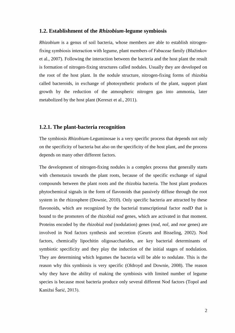
Figure 1. Potential impact of QS signals in nodule development on legume roots In the beginning, the interaction between the root and rhizobial bacteria is a result of the
chemical communication through different signals. It is presumed that QS has a role in the early
steps of the process. Communication consists of flavonoids secreted by the plant and Nod
factors produced by the bacteria. The result of this communication is formation of an infection
thread, which progresses into the inner cortex, where cells proliferate and lead to nodule
formation (taken from Sánchez Cañizares, 2013).
1.4.1. Molecular basis of quorum sensing
There are many different types of bacterial quorum sensing systems (Schuster, 2013).
We can distinguish QS in Gram-negative and Gram-positive bacteria. In Gram-negative
bacteria, the most used signal molecules are N-acyl-homoserine lactones (AHLs) and
autoinducer-2 (AI-2). In Gram-positive bacteria small peptide molecules are used as a
signaling system (Waters and Bassler, 2005).

7
1.4.2. Gram-negative bacteria: the LuxRI signaling system
The first QS system was discovered in the marine bacterium Vibrio fischeri (Nealson et
al., 1970), and it was used to describe the LuxRI system. The luciferase operon in V.
fischeri is regulated by two proteins, LuxI, a cytoplasmic enzyme responsible for the
synthesis of the signaling molecules called autoinducers, and LuxR that binds to its
cognate autoinducer and promotes transcription of the luciferase operon (Wisniewski-
Dyé and Downie, 2002). Quorum sensing is used to control a set of genes by LuxR and
3-oxo-hexanoyl-homoserine lactone (3OC6-HSL). Activated LuxR binds to a 20 bp
DNA element, known as lux box, leading to increased transcription of the luciferase
operon (Whitehead et al., 2001). Most of LuxR-type proteins function as activators. The
luxI gene is activated by 3OC6-HSL-bound LuxR, resulting in a positive autoinduction
mechanism (Schuster, 2013). The products of the luxI and luxR genes are regulators of
bioluminescence.
The chemical signals synthesized by the bacteria are based on a modified amino acid
(homoserine lactone) carrying a variable acyl chain substituent, called acyl-homoserine
lactones (AHLs). By detecting and reacting to these chemicals, individual cells can
sense how many cells surround them, and whether there are enough bacteria to initiate
the change towards acting in a multicellular way
(http://www.jic.ac.uk/science/molmicro/Rhizo.html/).
1.4.3. Quorum sensing genes in rhizobia
Two known protein families are shown to catalyse the synthesis of AHLs. The most
common group is the LuxI family and the other one is the LuxM family, but in rhizobia
are also indentified the regulators belonging to the LuxR protein family (Sanchez
Contreras et al., 2007), and are often encoded to their nearby corresponding LuxI-type
AHL synthase. Many more LuxR homologues are revealed, and many of these
regulatory genes are orphans, ˝extra˝ LuxR-type proteins because LuxI-type protein has
more than one specific LuxR-type protein that binds its AHL (Fuqua, 2006). The LuxR
orphans are proteins that are not directly controlling the synthesis of autoinducers but
are able to interact with them in the way of expanding the regulatory network of the
bacterium (Patankar and Gonzalez, 2009). Quorum sensing signals are found in many

8
species of legume-nodulating rhizobia. It is evident that some aspects of rhizobial
physiology related to the interaction between rhizobia and legumes are influenced by
quorum sensing. Rhizobium leguminosarum bv. viciae, which nodulates peas, vetch and
lentils, synthesizes many different autoinducers. All of them have been identified as N-
acyl-homoserine lactones (AHLs) (Sanchez Contreras et al., 2007).
1.4.4. Quorum sensing systems in Rhizobium leguminosarum bv viciae
Rhizobium leguminosarum bv. viciae (Rlv) establishes symbiosis with different
legumes (Pisum, Lens, Vicia and Lathyrus). The Rhizobium has a genome that contains
a large chromosome and 2-6 plasmids. Rlv UPM791 genome’s size is estimated as 7.79
Mb and it is organized in one chromosome and five extrachromosomal replicons
(Downie and Young, 2001). The chromosome has an estimated size of 4.75 Mb,
representing 61% of UPM791 genome. The estimated size of the plasmid DNA is 3.04
Mb, corresponding to 39% of the genome (Sánchez Cañizares, 2013).
In the search for transcriptional regulators of the LuxR-type in the genome of Rlv
UPM791, proteins with two characteristic domains were found. The first one is an
autoinducer-binding domain (ABD) which binds AHLs in the N-terminal region, and
the other one is the LuxR-family DNA-binding, in the form of helix-turn-helix (HTH)
domain in the C-terminal region (Whitehead et al., 2001). Both systems are similar to
the one described in Rhizobium leguminosarum A34 and 3841 strains. The CinRI is
located in the chromosome and RhiRI is encoded in the symbiotic plasmid (Sánchez
Cañizares, 2013). The LuxI-type transcriptional proteins are consisted of amino-
terminal and C-terminal half and are responsible for the production of AHLs from S-
adenosyl-methionine (SAM) and acyl groups supplied by acyl-carrier proteins (ACP).
Amino acids in the C-terminal half of LuxI are not necessary for acyl ACP selection,
but disruption of amino-terminal half can cause a significant reduction of synthase
activity. Conserved amino acids were thought to be necessary for acyl ACP selection
but when mutations were made a loss of activity was not a result (Whitehead et al.
2001).
QS systems have been studied in three different R. leguminosarum bv. viciae strains:
A34, which was the focus of pioneering studies; 3841, whose genome has been
9
sequenced, and UPM791. The UPM 791 was the reference strain in our laboratory from
the research made by Sánchez Cañizares (2013). The following four QS systems have
been described in these bacterial species (Wisniewski-Dyé and Downie, 2002; Gonzalez
and Marketon, 2003).
1.5.1. The RhiRI system
RhiRI system was the first one described in R. leguminosarum. The system is encoded
in the symbiotic plasmid. The first identified LuxR-type regulator was RhiR that
positively controls the rhiABC operon responsible for encoding RhiA protein. This
expression is repressed by flavonoids that normally induce nod gene expression (Cubo
et al., 1992). RhiA protein has the highest level of expression in the rhizosphere of
legume roots, but not in bacteroids. These rhizosphere induced genes function is
unknown. They are found between the nodulation genes and the genes encoding the
nitrogenase complex. It is assumed that RhiA is specific to Rhizobium leguminosarum
bv. viciae strains. RhiI, the LuxI homologue, produces the short-chain AHLs: C6-HSL,
C7-HSL and C8-HSL (Rodelas et al., 1999). rhiI is regulated by RhiR in a way that is
dependent of the cell density and it is positively autoregulated (Wisniewski-Dyé and
Downie, 2002). In different R. leguminosarum strains with affected rhiI genes
nodulation efficiency has been reduced (Sanchez Contreras et al., 2009). Effect of
RhiRI system on symbiotic performance in Rlv UPM791ΔrhiI showed, in the root
system analysis, that a different types of nodules grew. The effect also depends on a
host, which Rlv associate with. Mutants in rhiR/rhiI were able to form the nodules but
the nodules were not able to fix nitrogen (Sánchez Cañizarez, 2013).
1.5.2. The CinRIS system
CinRIS system is encoded in the chromosome in A34 and 3841 strains. The system has
been described as the main control for the other AHL-dependent QS systems in R.
leguminosarum strains (Lithgow et al., 2000). CinI, the LuxI homologue, is responsible
for the synthesis of N-(3-hydroxy-7-cis-tetradecenoyl)-L-homoserine lactone (3-OH-
C14:1-HSL). The molecule 3-OH-C14:1-HSL, previously known as a small bacteriocin
10
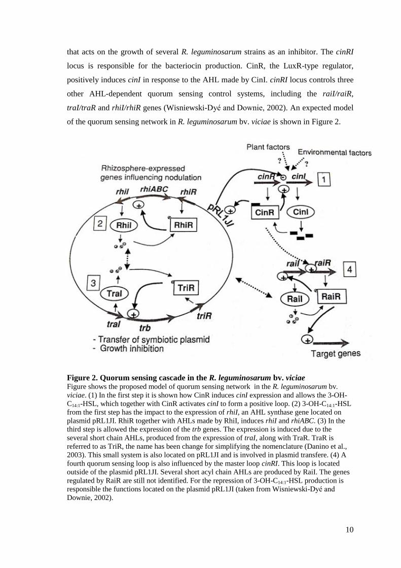
that acts on the growth of several R. leguminosarum strains as an inhibitor. The cinRI
locus is responsible for the bacteriocin production. CinR, the LuxR-type regulator,
positively induces cinI in response to the AHL made by CinI. cinRI locus controls three
other AHL-dependent quorum sensing control systems, including the raiI/raiR,
traI/traR and rhiI/rhiR genes (Wisniewski-Dyé and Downie, 2002). An expected model
of the quorum sensing network in R. leguminosarum bv. viciae is shown in Figure 2.
Figure 2. Quorum sensing cascade in the R. leguminosarum bv. viciae Figure shows the proposed model of quorum sensing network in the R. leguminosarum bv.
viciae. (1) In the first step it is shown how CinR induces cinI expression and allows the 3-OH-
C14:1-HSL, which together with CinR activates cinI to form a positive loop. (2) 3-OH-C14:1-HSL
from the first step has the impact to the expression of rhiI, an AHL synthase gene located on
plasmid pRL1JI. RhiR together with AHLs made by RhiI, induces rhiI and rhiABC. (3) In the
third step is allowed the expression of the trb genes. The expression is induced due to the
several short chain AHLs, produced from the expression of traI, along with TraR. TraR is
referred to as TriR, the name has been change for simplifying the nomenclature (Danino et al.,
2003). This small system is also located on pRL1JI and is involved in plasmid transfere. (4) A
fourth quorum sensing loop is also influenced by the master loop cinRI. This loop is located
outside of the plasmid pRL1JI. Several short acyl chain AHLs are produced by RaiI. The genes
regulated by RaiR are still not identified. For the repression of 3-OH-C14:1-HSL production is
responsible the functions located on the plasmid pRL1JI (taken from Wisniewski-Dyé and
Downie, 2002).

11
Small bacteriocin with the other N-acyl-homoserine lactones produced by these three
AHL-based control systems, regulate growth inhibition of sensitive strains, transfer of
the symbiotic plasmid pRL1JI and expression of the rhizosphere-expressed (rhi) genes
which influence nodulation (Wisniewski-Dyé and Downie, 2002). Mutations done in
cinI and cinR genes suppress the production of 3-OH-C14:1-HSL decreasing the
concentration of all the short-chain AHLs and by that reducing the expression of rhiI
and rhiA genes. Expression of RhiA is also controlled by QS in R. leguminosarum bv.
viciae UPM791 (Cantero et al., 2006). This is the main reason, the cinRI system is on
the top of the regulatory cascade that influences these three AHL-regulated QS systems
(Wisniewski-Dyé and Downie, 2002; Lithgow et al., 2000). In the strain Rlv 3841 an
additional small gene, cinS, has been involved in the expression of QS systems by
acting with an expR-like gene similar to the one from S. meliloti. A R. leguminosarum
cinI mutant is not impaired in nodulation in A34 (Lithgow et al., 2000), neither cinR,
cinI or cinS mutants in 3841 (Edwards et al., 2009), showing that 3-OH-C14:1-HSL is
not required for symbiosis (Wisniewski-Dyé and Downie, 2002). The genetic structure
of the system in Rlv UPM791 is homologous of the one in R. leguminosarum 8401. Rlv
UPM791 mutants with the ΔcinRIS deletion lost the ability to produce 3-OH-C14:1-HSL,
but the rhiRI system still produced small signals. On the other hand, mutation in cinS
resulted in the increased amount of 3-OH-C14:1-HSL production and all the detected
signals were described also in the wild type. The plants inoculated with the UPM791
mutants affected with cinRIS were small and yellow, with white and inefficient
nodules, and the one inoculated with the wild type was green and healthy (Sánchez
Cañizares, 2013).
1.5.3. The TraRI system
TraRI is present in Agrobacterium tumefaciens, Rhizobium NGR234 and R.
leguminosarum A34 that devastates TraI AHL synthase, which is responsible for the
synthesis of the signal 3-oxo-C8-HSL (Wilkinson et al., 2002; Danino et al., 2003). The
system induces the trb operon that is located in the symbiotic plasmid pRL1JI and
influences its conjugal transfer in R. leguminosarum A34. Downstream on the trb
operon bifunctional signaling regulator, bisR, is located. It shares the identity with a
LuxR-type regulator CinR (Wilkinson et al., 2002). cinRI system controls the traR

12
expression by binding BisR to 3-OH-C14:1-HSL. In the strains carrying the plasmid
pRL1JL, BisR represses cinI expression, and reduces the induction of traR (Danino et
al., 2003). When bacteria carrying pRL1JI is close to strains producing 3-OH-C14:1-
HSL, BisR detects this signal inducing traR and plasmid transfer. Both Rlv strains,
UPM791 and 3841 contain TraI and TraR, but the regulator BisR has not been found
(Sanchez Contreras et al., 2007).
1.5.4. The RaiRI system
RaiRI system is described in the strain A34 and it is not present in other R.
leguminosarum strains that have been analyzed (Sanchez Contreras et al., 2007). RaiRI
is located in one of the non-symbiotic plasmids. raiI is responsible for the synthesis of
the main product of the system, 3-OH-C8-HSL (Gonzalez and Marketon, 2003). In
addition, raiI is positively regulated by raiR and its signal, 3-OH-C8-HSL (Wisniewski-
Dyé et al., 2002).
13
2. OBJECTIVES
Quorum sensing (QS) is a widely spread process in bacteria described as bacterial
communication. It is a mechanism of chemical cell to cell communication that allows
coordination of gene expression as a function depending on local population density.
The process relies upon the interaction of signal molecules known as autoinducers that
can regulate the gene expression, with cell population density. The main role of QS is
regulation of many relevant activities in different bacterial species. Due to those QS
nowadays is under a high interest of manipulation in agriculture, but also in other fields
of science. In previous work was showed that R. leguminosarum bv. viciae (Rlv)
UPM791 is producing AHLs and the systems influenced by AHLs (cinRI, traRI and
rhiRI).
The general objective of this work is to acquire more knowledge of quorum sensing
system from the endosymbiotic bacterium R. leguminosarum UPM791.
This general objective was carried out through the following specific objectives:
Objective 1. Characterization of cinR and traI genes from Rlv UPM791
Objective 2. Construction of insertion mutants of cinR and traI
Objective 3. Characterization of mutants
Objective 4: Phenotype characterization in free living cells.

14
3. MATERIALS AND METHODS
3.1. BIOLOGICAL MATERIAL
3.1.1. Bacteria and plasmids
Strains and plasmids used in this work are listed in Table 1.
Table 1. Bacteria and plasmids used in this work
Abbreviations: Ap, ampicillin; Km, kanamycin; Str, streptomycin; Tc, tetracycline;
Sp, spectinomycin
Strain or plasmid Genotype or relevant
characteristic(s)
Source or
reference
Escherichia coli
DH5α recA1 endA1 gyrA96 thi hsdR17
supE44 relA1 Δ(lacZYA-argF)U169
(Ф80lacZΔM15) deoRphoA
(Hanahan,1983)
Rhizobium leguminosarum bv.
viciae
UPM791 128C53 StrR; Nod
+ Fix
+ Hup
+ (Leyva et al., 1987)
UPM1253 UPM791ΔcinRIS, SpR
(Sánchez
Cañizares, 2013)
UPM1255 UPM791ΔrhiI, TcR
(Sánchez
Cañizares, 2013)
Plasmids
pCR2.1-TOPO PCR product cloning vector; ApR,
KmR
Invitrogen
pRK2073 Helper strain for plasmid
mobilization; Mob+Tra
+Sp
R
(Figurski and
Helinski, 1979)
pK18mobsac Integrative vector pUC18 derative;
lacZ mob site sacB, KmR
(Schäfer et al.
1994)
15
3.2. MEDIA AND GROWTH CONDITIONS
Rhizobium leguminosarum bv. viciae (Rlv) cultures were grown at 28˚C, and
Escherichia coli strains at 37˚C.
For Rlv the following media were used: YMB and TY, and for Escherichia coli LB
medium.
YMB (yeast mannitol broth, Vincent, 1970): yeast extract 0.4 g/l, mannitol 1 g/l, NaCl
0.1 g/l, K2HPO4 0.5 g/l, Mg SO4 0.2 g/l. Once prepared the pH has to be adjusted to 6.8
with HCl 1N.
TY (tryptone-yeast extract, Beringer, 1974): yeast extract 3 g/l, tryptone 6 g/l, CaCl2 0.5
g/l.
LB (Luria-Bertani broth, Sambrook and Russell, 2001): tryptone 10 g/l, yeast extract 5
g/l, NaCl 5 g/l.
Liquid cultures were grown in a rotary shaker at 200 rpm. Media for plating contained
1.5% agar unless otherwise specified. The media were sterilized by autoclaving at
120˚C for 20 min. For plasmid maintenance, selection of transformants and
transconjugants, media were supplemented with the appropriate antibiotics at the
following concentrations: kanamycin, 50 µg/ml for E. coli strains and Rhizobium
strains; tetracycline, 2 µg/ml for Rhizobium strains; spectinomycin, 50 µg/ml for
Rhizobium strains. We also used nitrofurantoin (5 µg/ml) to inhibit the growth of E. coli
donor and helper strains on the plates for selection of transconjugants.
Strains were preserved by freezing at -80˚C in media supplemented with 20% glycerol.
3.3. DNA manipulation techniques
3.3.1. DNA extraction
Plasmid DNA preparations were obtained from E. coli cells carried out by two different
methods.
16
3.3.2. Alkaline Lysis Mini-Preparation
Cells of 1.5 ml of liquid medium were collected by centrifugation during 5 min at 10
000 rpm. Once the cell pellet was obtained, 200 µl of solution I (10 mM EDTA, 25 M
Tris at pH 8) were added. Later, the tubes were vortexed and left for 5 min at room
temperature, and 100 µl of freshly prepared solution II (1 ml NaOH 2N, 1 ml SDS 10%,
8 ml H2O) were added, mixed well and left for 5 min on ice. Then, 150 ml of solution
III (K-Acetate 3M at pH 4.8) were added. After adding the last solution, into the sample
3 µl of RNase (1 mg/ml) was added to ensure degradation of RNA. Around 2 min was
necessary for the enzyme to start working. The next step was mixing the sample and ice
incubation for 10 min. The sample needed to be centrifuged at 13 000 rpm for 10 min
and the supernatant was moved to a new tube with the addition of 1 ml of cold 100%
ethanol. After homogenization, the tubes were centrifuged at 13 000 rpm for 10 min, the
supernatant was thrown away. The DNA pellet was washed with 0.5 ml of 70% ethanol.
The sample needed to be put on centrifugation for 5 min at 13 000 rpm, the supernatant
was eliminated and the pellet was left to dry the DNA extract. Once the DNA pellet was
dry, it was dissolved with 20 µl of water.
3.3.3. Extraction with a commercial kit
When needed for sequencing or cloning, plasmid purification was carried with the kit
NucleoSpin Plasmid DNA (Macherey-Magel, Germany), used according to
manufacturer instructions. The kit contains everything that is necessary, from protocol
to reactants, needed to extract the plasmid DNA. This method has a higher efficiency
and the DNA extraction is cleaner. The disadvantage of the method is the quantity of
the DNA that is in this case lower. This method was used in case that the plasmid DNA
was needed for cloning.
Genomic DNA for R. leguminosarum was extracted from cultures grown in TY medium
using DNeasy Blood & Tissue Kit columns (QIAGEN Ltd.) following manufacturer
instructions. For standard PCR procedures, a faster DNA extraction was made by
recovery of culture from a plate with a toothpick, followed by the addition of 20 µl of a
lysis solution (0.05M NaOH, 0.25% SDS). The samples were incubated at 90˚C for 15

17
min. After incubation, 100 µl of water was added and the samples were centrifuged 5
min at 13 000 rpm. The DNA was recovered from the supernatant.
3.3.4. Extraction of DNA from agarose gels
DNA extraction from agarose gels was done by using NucleoSpin® Gel and PCR
Clean-up (Macherey-Nagel, Cultek). For gel extraction, first the band from
electrophoresis gel had to be weighed. To every 100 mg of the gel 200 µL of NTI
solution was added to a tube with DNA-binding membrane. The sample was put 30 s in
the centrifuge at 11 000 rpm. To wash the membrane 700 µL NT3 solution was added
and put 30 s in the centrifuge at 11 000 rpm. Second wash is recommended. To dry the
membrane the sample was centrifuged 1 min at 11 000 rpm. After centrifugation the
supernatant was discarded and 30 µL NE desorption buffer were added. Tubes were
incubated at room temperature for 1 min, and then centrifuged at 11 000 rpm for 1 min.
3.3.4. PCR amplification and electrophoresis
Purified DNA (5 to 20 ng) was used as a template for PCR amplifications. Standard
PCR amplifications were carried out with Taq Polymerase (Roche). All PCR reactions
were carried out in total volume (25 or 50 µL) following the manufacturer’s
instructions. For amplifying the target genes, these PCR reaction settings were used:
93˚C for 5 min; 30 cycles of 93˚C for 45 s, 60˚C for 45 s, 72˚C for 120 s and a final
extension of 72˚C for 5 min.
The size of DNA fragments was checked by electrophoresis, using horizontal 1%
agarose gel submerged in TBE buffer (Sambrook and Russell, 2001). DNA samples
were prepared with ¼ volume of loading buffer (glycerol 60%, EDTA 20 mM, orange
G, ribonuclease A 20 U/ml). Gels were stained with ethidium bromide (1 µg/ml), that
was added directly to the gel in the moment of the preparation. Fragment sizes were
estimated by comparing the rates of migration to those from the reference pattern
obtained from either 1kb DNA ladder (Nippon Genetics, Cultek), or Lambda Phage
DNA digested with Hind III (Roche). Interpretation of 1kb DNA ladder is in Figure 3.
18
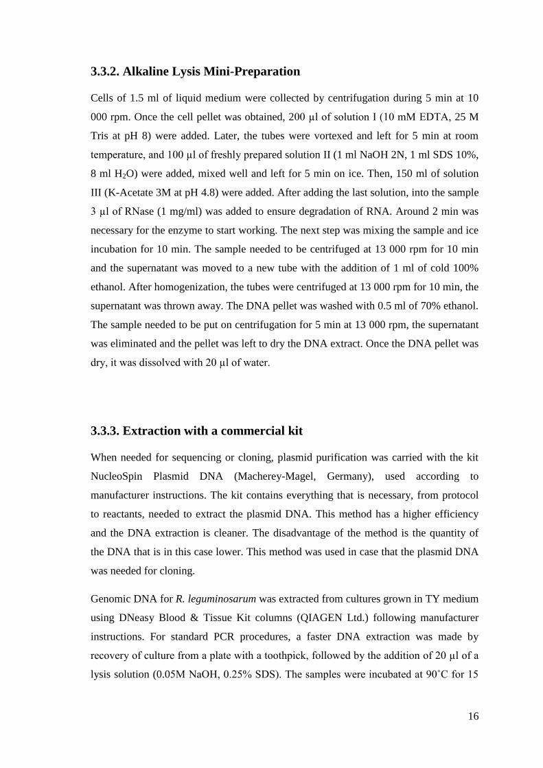
Figure 3. Chart of the 1kb DNA ladder Molecular-weight size marker in the form of a 1kb DNA ladder. It is use as a set of exact sizes
of DNA sequences for an identification of the approximate size of a molecule run on a gel
during electrophoresis. The DNA includes fragments raning from 250-10 000 base pairs. The
approximate mass of DNA in each band is provided (0.5 µg a load) approximating the mass of
DNA to compare the intensity of samples of similar size (taken from
https://www.nippongenetics.eu/).
3.3.5. DNA sequencing, analyzing and primer design
Primer design was carried out using NCBI webpage utility (National Centre for
Biotechnology Information). DNA sequencing was used to confirm the correct sequence
of plasmids and PCR fragments. Sequencing reactions were carried out at Stabvida
(Lisboa, Portugal). The samples were sent in 1.5 ml Eppendorf tubes. Every tube
contained 15 µl of DNA (50-100 ng/µl) together with the primer required for the
sequencing. Sequences were analyzed using BLAST (Basic Local Alignment Search
Tool, Altschul et al. 1990) or LALIGN (ExPASy, Bioinformatics Resource Portal).

19
3.3.6. Restriction enzyme digestion
Restriction enzyme digestions were carried out by standard protocols (Sambrook and
Russell, 2001). In a typical digestion reaction, 2 µl of the DNA, 2 µl of buffer H or B
(depending which enzyme is used), 11 µl of water and 1µl of enzyme XbaI, BamHI or
EcoRI up to final volume of 20 µl. After adding the enzyme the samples were incubated
for 60 to 120 min at 37˚C. The result of the digestion was checked by agarose gel
electrophoresis.
3.3.7. Cloning
Cloning for PCR products was done by using pCRTM
2.1-TOPO® TA Cloning®Kit
(Invitrogen, Life Technologies), according to the instructions provided in the kit.
3.3.8. Ligation
Ligations were done with T4 DNA ligase (Roche) following the manufacturer’s
protocol and normally the samples were left over night at 16˚C. During this procedure
the vector (pK18mobsac) and the insert (cinR/traI-containing fragments) were mixed in
a V:I=1:3 ratio.
3.3.9. Plasmid transfer by transformation
To make the transformation, the plasmid DNA (50-100 ng) was added to 100 µl of E.
coli competent cells, and the mix was incubated on ice for 30 min. Later the samples
were moved to thermo block (1 min at 42˚C) to achieve a heat-shock. Then 1 ml of LB
liquid medium was added, and left on incubation 60 min at 37˚C. After the incubation,
100 µl of the transformation mix were spread on LB plates supplemented with the
corresponding antibiotics to the resistance of plasmid. When the frequency of
transformation was predicted to be low, the remaining 900 µl were centrifuged,
resuspended in 100 µl of LB liquid medium and spread on the same type of plate. The
plates were incubated at 37˚C for 24 h.

20
3.3.10. Plasmid transfer by mating
Plasmids were transferred to R. leguminosarum strains by triparental mating using E.
coli-DH5α as a donor strain, wild type of R. leguminosarum bv. viciae or one of two
mutant strains UPM1255 and UPM1253 as recipients, and an additional E. coli strain
with the helper plasmid pRK2073 was included to supply the conjugation machinery.
In one Eppendorf tube (1.5 ml) were mixed 0.5 ml of liquid culture of donor, 0.5 ml of
recipient and 0.5 ml of helper. The tubes with the mix were put 5 min on centrifugation
at 13 000 rpm, the pellet was resuspended in 50 µl of TY liquid medium after that the
content of the tubes was put on small filters (diameter 0.22 µm) placed on TY plates,
and incubated overnight at 28˚C. After incubation, filters were moved to a tube with 2
ml of NaCl (0.85%)/Tween 20 (0.1%) and vortexed. From the content of the tubes 100
µl were plated into YMB plates supplemented with kanamycin (50 µg/ml) and
nitrofurantoin (5 µg/ml) to avoid E. coli growth. The rest of the content of the tubes was
centrifuged; the pellet was resuspended in 100 µl of NaCl (0.85%)/Tween 20 (0.1%)
and also spread on a plate with the same, above mentioned, antibiotics. The plates were
incubated at 28˚C for five days.
3.3.11. Southern blot
For carrying out this method first the electrophoresis of a specific DNA sequences in
DNA samples that are going to be detected, is made. Before the electrophoresis, in this
case, the restriction enzyme digestion was made using enzyme EcoRI. The DNA
fragments were resolved by slow electrophoresis (overnight at 15 V) on a 1% agarose
gel. After the electrophoresis of the specific DNA sequences, a picture of the gel was
made using UV light and the lane with the sample was marked with ink. Then the gel
was submerged in the Solution I (HCl 0.25M) 15 min at room temperature with a slow
agitation to break the DNA. In the bath the gel was washed with water for 1 min.
Denaturing Solution II (NaOH 0.5N, NaCl 1.5M) was then added, and the gel is left
submerged in this Solution II for 30 min. After this step the gel was washed with water
for 1 min, twice. The next step was neutralization with Solution III (Tris 0.5 N pH 7,
NaCl 3N) for 30 min. Once 30 min passed, the gel was washed again with water for 1
min, twice. To transfer the DNA from the gel a “sandwich” construction was made. To
21
make a “sandwich” it was necessary to put one crystal glass on the flat surface. On top
of the crystal, papers were put, then the gel and on top of the gel the nylon membrane
was placed. The membrane was again covered with MM sheets and towel papers, and
the papers were covered with another crystal glass. The whole construction was pressed
evenly with a weight to ensure good contact between gel and membrane. The transfer
lasted around 2.30-3 h. Then, the construction was open to the nylon membrane and the
lanes where the samples and the marker are loaded are marked with a pen. The
membrane was exposed to the ultraviolet radiation (UV light) 3 min in the way that the
DNA was turned upside down to permanently attach the transferred DNA to the
membrane. To make sure that the DNA was permanently transferred to the membrane
from the gel, the test was made by exposing only the gel to the UV light. Lack of
detection of DNA on the gel was a proof that the entire DNA was transferred. The
membrane was then incubated for 1 h to 12.5 ml of pre-hybridization buffer and later
hybridization probe was added. The DNA probes were made by PCR amplification with
the addition of labelled nucleotides (Digoxigenin-11-dUTP, Roche). The specific DNA
probes were denaturized at 100˚C for 1 min, or 30 min at 65˚C in the case of a second
use. In each case, the membrane was prehybridized at 42˚C for 1 h with hybridization
buffer (NaCl 0.75M, sodium citrate 75mM pH 7.0, formamide 50%, SDS 7%, sodium
phosphate 50mM pH 7.0 and n-lauroilsarcosine 0.1%). The membrane was hybridized
with the labelled probe overnight at 42˚C. After hybridization, the membrane was
washed twice from the membrane using 2*SCC buffer and 0.1% SDS following this
condition: 5-10 min at 42˚C. The next step was washing the membrane, twice, with
0,1*SSC and 0.1% SDS, 5-10 min at 68˚C. The last washing was done with buffer 1
(Tris 100mM pH 7.5; NaCl 150mM) with agitation. The membrane was put in the
buffer 1 (50 ml) with the 5% milk (2.5 g of commercial powder milk) for 1 h. The step
was repeated (1 h: buffer 1 + 5% milk) but 5 ml of antibodies Anti-Dioxigenin-A
(Roche) were added. The membrane was then washed three times for 10 min with
buffer 1 and after 5 min with alkaline phosphatase buffer (Tris/HCl 100mM pH 9.5,
NaCl 100mM, MgCl2 50mM). In next step 6 ml of alkaline phosphatase buffer with 4
ml of chemiluminescent substrate (CSPD) was put in the bath with the membrane for 1
min. The pattern of hybridization was visualized on x-ray film by development of color
on the membrane during 2 h at room temperature in darkness.
22
4. RESULTS
4.1. CHARACTERIZATION OF cinR AND traI GENES FROM R.
leguminosarum UPM791
4.1.1. Study of the sequence of the cinR and traI genes from R.
leguminosarum strains and design of the primers to amplify the target
genes
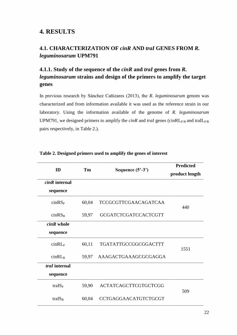
In previous research by Sánchez Cañizares (2013), the R. leguminosarum genom was
characterized and from information available it was used as the reference strain in our
laboratory. Using the information available of the genome of R. leguminosarum
UPM791, we designed primers to amplify the cinR and traI genes (cinRLF/R and traILF/R
pairs respectively, in Table 2.).
Table 2. Designed primers used to amplify the genes of interest
ID Tm Sequence (5’-3’) Predicted
product length
cinR internal
sequence
cinRSF 60,04 TCCGCGTTCGAACAGATCAA 440
cinRSR 59,97 GCGATCTCGATCCACTCGTT
cinR whole
sequence
cinRLF 60,11 TGATATTGCCGGCGGACTTT 1551
cinRLR 59,97 AAAGACTGAAAGCGCGAGGA
traI internal
sequence
traISF 59,90 ACTATCAGCTTCGTGCTCGG
509
traISR 60,04 CCTGAGGAACATGTCTGCGT

23
traI whole
sequence
traILF 59,97 TCGTTGGTCAGAAAGGGACG 1378
traILR 60,32 GTACCGATCACCATCTCCGC
Abbreviations: cinRSF, cinR small sequence forward primer; cinRSR, cinR small sequence
reverse primer; cinRLF, cinR large sequence forward primer; cinRLR, cinR large sequence
reverse primer; traISF, traI small sequence forward primer; traISR, traI small sequence reverse
primer; traILF, traI large sequence forward primer; traILR, traI large sequence reverse primer;
Tm, annealing temperature
4.1.2. Amplification of the target genes
PCR reactions were carried out with both sets of primers using genomic DNA from R.
leguminosarum UPM791 as template. In these reactions we obtained PCR amplification
products of the expected size shown in Figure 4. In all cases, the sizes of PCR products
were consistent with the expected ones assuming a similar sequence in UPM791 and
3841 strains. Taking into account the quality and amount of amplified DNA we selected
the combinations shown in Table 3. to continue the work.
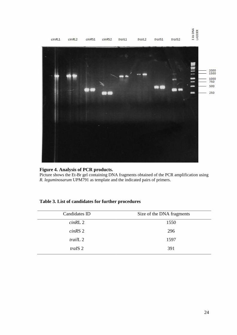
24
Figure 4. Analysis of PCR products. Picture shows the Et-Br gel containing DNA fragments obtained of the PCR amplification using
R. leguminosarum UPM791 as template and the indicated pairs of primers.
Table 3. List of candidates for further procedures
Candidates ID Size of the DNA fragments
cinRL 2 1550
cinRS 2 296
trailL 2 1597
traIS 2 391

25
4.1.3. Analysis of the sequence of the amplified genes and comparison
with other genes in the database
PCR products representing the best candidates according to quality and amount of
amplified DNA, shown in Table 3., were cloned in Topo vector and sequenced using
universal primers flanking the cloned region in the vector. Analysis of the sequence
allowed the identification of cinR and traI genes. DNA sequencing was used to confirm
the correct sequence of PCR fragments and analyzed using BLAST (Basic Local
Alignment Search Tool, Altschul et al. 1990) and LALIGN (ExPASy, Bioinformatics
Resource Portal). The outcome of these sequences is shown in Figure 5.
cinR and flanking region
TGATATTGCCGGCGGACTTTCTCGGCATGGTCGGTAAAGCCGATATCGAGAT
GGGTCTTGAAGACCAGATGCACGCGCTTTTCAGTCATTCCATTTCACACATC
GGTGATGACAGTCGAAACAGTCTGCAGCCAGTGGCGGTACAGGTGAGGAGT
GTAACCGTTTCCACAAGCACCCTTGCTCCTGCCGCAAACGCTCCGTCAACCG
TTTTGGCACAAATCCATAAATTAATGCGGATTTCTTAACCATATATCCAGAA
CGCAGCGTCCGATCCTTTCGCCGCCAAATCGAATCACGACATGAATGAAGA
CGTGTTCATTGGCAATTTTGCAGAGCAGAAAGGCGGTGGGATTTCCCTGCCG
ACCGGCGTGGGTGGCCCGCCTAAATTGCGGAGAAAGGGCTGAGCCGTTCGG
GAAGGCCGATCGTCAGAAAGCGTTGGTTTCCTGGCAGAGCGCCCGGTGAAC
CTCGGTCCAGAACAACGCCAGCGGCCTGTCGCCATTGCGTTCGGCAACGAG
GGCGCGGATCAGCGCTTCGGAGCCTGCCATGTCCTGCCAGCTGGATTTCAG
GCCCTTGGCGGCCTGTTGGCATTCGGCAATCTCGAATGGTCTCTTGCTGTCC
ATATGAGGTCTCCTCATCACAGGCGCTAGCAGATGGGACCCGGATTGTCATT
CCCCGCATATGTGGGGATGCCTTGTATTTGTGACGGCCCTCGCGGTAGGATT
GAGGGAATTGGGGCAGTGGAAATGATTGAGAATACCTATAGCGAAAAGTTC
GAGTCCGCGTTCGAACAGATCAAGGCGGCGGCCAACGTGGATGCCGCCATC
CGTATTCTCCAGGCGGAATATAACCTCGATTTCGTCACCTACCATCTCGCCC
AGACGATCGCGAGCAAGATCGATTCGCCCTTCGTGCGCACCACCTATCCGG
ATGCCTGGGTTTCCCGCTACCTCCTCAACAGCTATGTGAAGGTCGATCCGAT
CGTCAAGCAGGGCTTCGAACGCCAGCTGCCCTTCGACTGGAGCGAGGTCGA

26
ACCGACGCCGGAGGCCTATGCCATGCTGGTCGACGCCCAGAAACACGGCAT
CGGTGGCAATGGCTACTCCATCCCCGTCGCCGACAAGGCGCAGCGCCGCGC
CCTGCTGTCGCTGAATGCCCGTATACCGGCCGACGAATGGACCGAGCTCGT
GCGCCGCTGCCGCAACGAGTGGATCGAGATCGCCCATCTGATCCACCGCAA
GGCCGTCTATGAGCTGCATGGCGAAAACGATCCGGTGCCGGCATTGTCGCC
GCGCGAGATCGAGTGTCTGCACTGGACCGCCCTCGGCAAGGATTACAAGGA
TATTTCGGTCATCCTGGGCATATCAGAGCATACCACACGCGATTACCTGAAG
ACCGCCCGCTTCAAGCTCGGCTGCGCCACGATCTCGGCCGCCGCGTCGCGG
GCTGTTCAATTGCGCATCATCAATCCCTAGCCCTTTGTGCGTCCAAACGGAC
GCACGGCGCTCTAAAGCGGGTCGCGATCTTTCAGATTCGCTCCTCGCGCTTT
CAGTCT
traI and flanking region
TCGTTGGTCAGAAAGGGACGCAGGGTCTTTTCCGATTTTGGCGGATACATAC
GAGTTCTGAGCTGCTGCAATTGCCCGGACAGAAGCCCGGCATGGCGCATGA
TTTTGCTGCCGGCATCTTCCTTCGAGCCAACACTTGCTTCTACTCGGTTTGCT
ATCGCCATCTGTCCCTCTTGGCGGCTTTTCGCCGGTTTACCGGCCATTGCCCG
CTAAAAATAGAACACGATTCTCCGAACTGGTCCAGAGGTTTAGGGTTAACA
AAAGGTTAACGACCGGGGACAATCTGCATAAGGCTGTTCGGATTTTTGCTTT
TCCGCTCCGGGAGTTAGCCTCAAATGCCCGGCTCCCTGATCCCGGAATGTTC
TCCTGTGAATCCTACAAATCCAAACAGTGTAGCAGCAAGTGGATTAGGCGG
CTCTGTAGCGCCACACCCCTTATGAGCCGCGACTCACCTCGCTACGTGCATG
TTGGATCCTACAACTGCCCTCCGATTCCAAACTGTGCTGATTTCCCGCCGAC
CAACAACGACGGAGAAAGATCATGCGGGCTCTCGCGCTCTCAACACCCCGG
ACGATCCAAGAGGCGCATCTCCTACACATCCACTATCAGCTTCGTGCTCGGG
TCTTTTCCGATCGCCTGGGTTGGGAAGTCGATGTAACGGCGGGGTGCGAGTC
CGATCGTTTCGACGCGCTTCGGCCGACCTATATTCTCGCCATCGCAGAGACC
GGCGAATTGGCGGGGTGCGCAAGGCTTCTTCCTGCGCTCGGACCGACAATG
GTGGCCGACGTTTTCCCGTCGCTGCTCCCCGACGGCCAACTCAAGGGGCATG
CCGCGATGATCGAGAGTTCTCGCTTCTGTGTCGACACGGCTCTCGCGGAGGG
GAGGGGAGCCGGCTCGGTCCATGAAGCGACGCTGACCATGTTCGCTGGCAT
CATCGAATGGTGCATGGCAAATGGGTACACTGAGATTGTTACGGTGACCGA
TCTTCGGTTTGAGCGCATCCTCGCCCGCGTGGGGTGGCAGCTGCATCGTTTA

27
GGCGAACCCAAGAAGATCGGCGTGACGACGGCCGTAGCGGGCACGCTGGC
CGCCGACGCAGACATGTTCCTCAGGCTTCGCCCCTCCAAGTACCGTTCTGAA
CTCGCCCCCTGTCAGCCAGGCAGCGTAAGGAGAAATCCGTGAACCAGCTTC
GCTCTCACCCTCGGCTCGTCCGCAAACTTCAGGAGGCGCTCGGCGACCAGCT
TTGTGTCGCCCTGGACGACGCGAACGTCGTCGAGATCATGCTTAATCCGGAC
GGAAAGTTGTTCATCGAACGGCTCGGTCACGGCGTTACGCCCGCCGGCCAG
ATGTCATCCGCTGCAGCGGAGATGGTGATCGGTAC
Figure 5. Outcome of the sequences Red sequence indicates the coding sequence of cinR/traI gene. Primers are marked in blue and
green color.
28
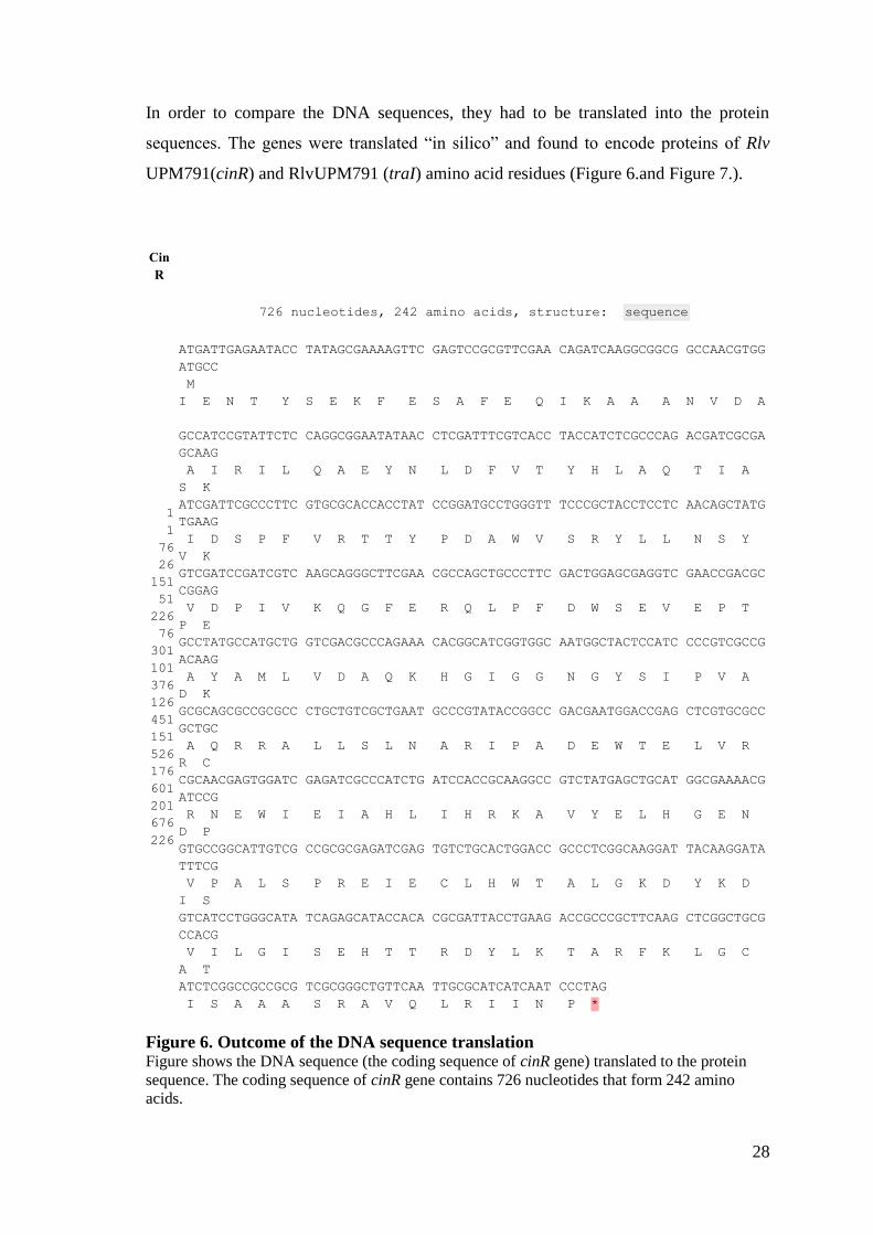
In order to compare the DNA sequences, they had to be translated into the protein
sequences. The genes were translated “in silico” and found to encode proteins of Rlv
UPM791(cinR) and RlvUPM791 (traI) amino acid residues (Figure 6.and Figure 7.).
Cin
R
726 nucleotides, 242 amino acids, structure: sequence
1
1
76
26
151
51
226
76
301
101
376
126
451
151
526
176
601
201
676
226
ATGATTGAGAATACC TATAGCGAAAAGTTC GAGTCCGCGTTCGAA CAGATCAAGGCGGCG GCCAACGTGG
ATGCC
M
I E N T Y S E K F E S A F E Q I K A A A N V D A
GCCATCCGTATTCTC CAGGCGGAATATAAC CTCGATTTCGTCACC TACCATCTCGCCCAG ACGATCGCGA
GCAAG
A I R I L Q A E Y N L D F V T Y H L A Q T I A
S K
ATCGATTCGCCCTTC GTGCGCACCACCTAT CCGGATGCCTGGGTT TCCCGCTACCTCCTC AACAGCTATG
TGAAG
I D S P F V R T T Y P D A W V S R Y L L N S Y
V K
GTCGATCCGATCGTC AAGCAGGGCTTCGAA CGCCAGCTGCCCTTC GACTGGAGCGAGGTC GAACCGACGC
CGGAG
V D P I V K Q G F E R Q L P F D W S E V E P T
P E
GCCTATGCCATGCTG GTCGACGCCCAGAAA CACGGCATCGGTGGC AATGGCTACTCCATC CCCGTCGCCG
ACAAG
A Y A M L V D A Q K H G I G G N G Y S I P V A
D K
GCGCAGCGCCGCGCC CTGCTGTCGCTGAAT GCCCGTATACCGGCC GACGAATGGACCGAG CTCGTGCGCC
GCTGC
A Q R R A L L S L N A R I P A D E W T E L V R
R C
CGCAACGAGTGGATC GAGATCGCCCATCTG ATCCACCGCAAGGCC GTCTATGAGCTGCAT GGCGAAAACG
ATCCG
R N E W I E I A H L I H R K A V Y E L H G E N
D P
GTGCCGGCATTGTCG CCGCGCGAGATCGAG TGTCTGCACTGGACC GCCCTCGGCAAGGAT TACAAGGATA
TTTCG
V P A L S P R E I E C L H W T A L G K D Y K D
I S
GTCATCCTGGGCATA TCAGAGCATACCACA CGCGATTACCTGAAG ACCGCCCGCTTCAAG CTCGGCTGCG
CCACG
V I L G I S E H T T R D Y L K T A R F K L G C
A T
ATCTCGGCCGCCGCG TCGCGGGCTGTTCAA TTGCGCATCATCAAT CCCTAG
I S A A A S R A V Q L R I I N P *
Figure 6. Outcome of the DNA sequence translation Figure shows the DNA sequence (the coding sequence of cinR gene) translated to the protein
sequence. The coding sequence of cinR gene contains 726 nucleotides that form 242 amino
acids.

29
Tra
I
639 nucleotides, 213 amino acids, structure: sequence
1
1
76
26
151
51
226
76
301
101
376
126
451
151
526
176
601
201
ATGCGGGCTCTCGCG CTCTCAACACCCCGG ACGATCCAAGAGGCG CATCTCCTACACATC CACTATCAGCT
TCGT
M R A L A L S T P R T I Q E A H L L H I H Y Q L
R
GCTCGGGTCTTTTCC GATCGCCTGGGTTGG GAAGTCGATGTAACG GCGGGGTGCGAGTCC GATCGTTTCGA
CGCG
A R V F S D R L G W E V D V T A G C E S D R F D
A
CTTCGGCCGACCTAT ATTCTCGCCATCGCA GAGACCGGCGAATTG GCGGGGTGCGCAAGG CTTCTTCCTGC
GCTC
L R P T Y I L A I A E T G E L A G C A R L L P A
L
GGACCGACAATGGTG GCCGACGTTTTCCCG TCGCTGCTCCCCGAC GGCCAACTCAAGGGG CATGCCGCGAT
GATC
G P T M V A D V F P S L L P D G Q L K G H A A M
I
GAGAGTTCTCGCTTC TGTGTCGACACGGCT CTCGCGGAGGGGAGG GGAGCCGGCTCGGTC CATGAAGCGAC
GCTG
E S S R F C V D T A L A E G R G A G S V H E A T
L
ACCATGTTCGCTGGC ATCATCGAATGGTGC ATGGCAAATGGGTAC ACTGAGATTGTTACG GTGACCGATCT
TCGG
T M F A G I I E W C M
A N G Y T E I V T V T D L R
TTTGAGCGCATCCTC GCCCGCGTGGGGTGG CAGCTGCATCGTTTA GGCGAACCCAAGAAG ATCGGCGTGAC
GACG
F E R I L A R V G W Q L H R L G E P K K I G V T
T
GCCGTAGCGGGCACG CTGGCCGCCGACGCA GACATGTTCCTCAGG CTTCGCCCCTCCAAG TACCGTTCTGA
ACTC
A V A G T L A A D A D M F L R L R P S K Y R S E
L
GCCCCCTGTCAGCCA GGCAGCGTAAGGAGA AATCCGTGA
A P C Q P G S V R R N P *
Figure 7. Outcome of the DNA sequence translation Figure shows the DNA sequence (the coding sequence of traI gene) translated to the protein
sequence. The coding sequence of traI contains 639 nucleotides that form 213 amino acids.

30
DNA sequences, translated into the protein sequences, were compared with the protein
sequences in other similar strains using BLAST at EXPASY server.
Comparison of the deduced proteins with similar proteins from other rhizobia is
presented in Figure 8. and Figure 9.
Figure 8. Multiple alignment of cinR-like proteins from different Rhizobiaceae Sequence correspond to Rhizobium leguminosarum bv. viciae UPM791, Rhizobium etli CFN42
(RHIEC), Rhizobium leguminosarum bv viciae 3841 (RHILE3), and Agrobacterium
tumefaciens 5A (ATUM). Multiple alignment was constructed using ClustalW.
31
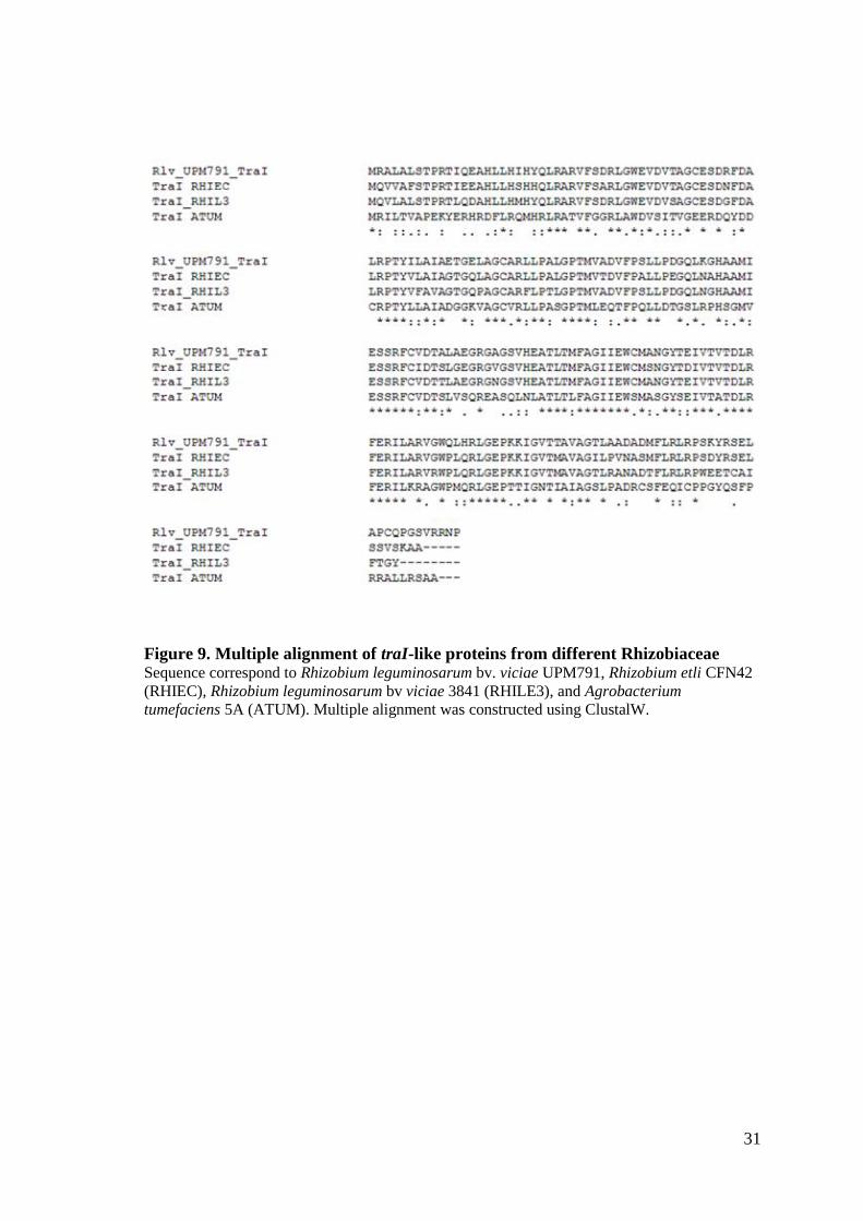
Figure 9. Multiple alignment of traI-like proteins from different Rhizobiaceae Sequence correspond to Rhizobium leguminosarum bv. viciae UPM791, Rhizobium etli CFN42
(RHIEC), Rhizobium leguminosarum bv viciae 3841 (RHILE3), and Agrobacterium
tumefaciens 5A (ATUM). Multiple alignment was constructed using ClustalW.

32
In order to have an idea of the conservation of this system in other Rhizobiaceae the
protein comparison was made. According to the identity percentages, the system found
in Rlv UPM791 genome seems to be widely spread and highly conserved in rhizobia. As
it is shown in Table 4., AHL system for cinR and traI genes is highly conserved in
comparison with Rlv 3841 and R. etli CFN42 strains.
In case of R. leguminosarum strains, cinR gene is located on the chromosome and CinR
regulates the expression of cinI in response to 3-OH-C14:1-HSL made by CinI (Lithgow
et al. 2000). The CinR, the LuxR-type protein, in Rlv UPM791 is most similar to the
AHL synthase from Rlv 3841, with which it shares 98% identity. TraR induces traI in
response to TraI-made 3-oxo-C8-HSL. The TraI is a protein similar to the LuxI family
(Danino et al., 2003). The comparison of the TraI protein from Rlv UPM791 showed
that is 87% identical to autoinducer transcriptional regulator protein from the Rlv 3841.
Table 4. Conservation of cinR- and traI-like proteins
Strain Rlv 3841 R. etli
CFN42 A. tumefaciens
AHL synthase similar to
CinR
98 97 54
Autoinducer transcriptional
regulator similar to TraI
87 85 55
Numbers correspond to the percentages of amino acid identity as derived from BLAST
comparison
33
4.2. GENERATION OF MUTANT DERIVATIVES DEFECTIVE IN
cinR AND traI GENES
4.2.1. Subcloning of the fragment by using a suicide plasmid containing
an internal fragment
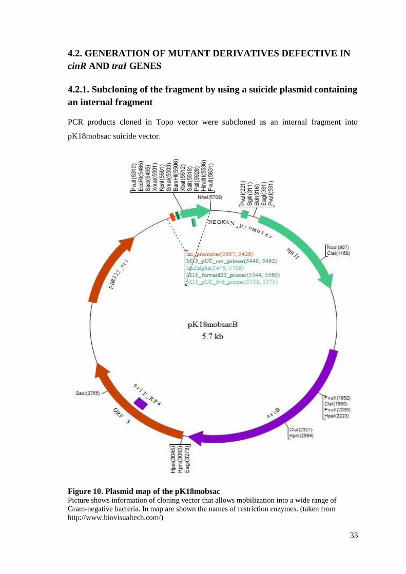
PCR products cloned in Topo vector were subcloned as an internal fragment into
pK18mobsac suicide vector.
Figure 10. Plasmid map of the pK18mobsac Picture shows information of cloning vector that allows mobilization into a wide range of
Gram-negative bacteria. In map are shown the names of restriction enzymes. (taken from
http://www.biovisualtech.com/)
34
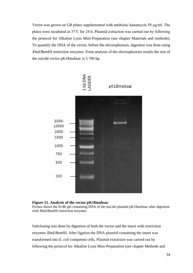
Vector was grown on LB plates supplemented with antibiotic kanamycin 50 µg/ml. The
plates were incubated at 37˚C for 24 h. Plasmid extraction was carried out by following
the protocol for Alkaline Lysis Mini-Preparation (see chapter Materials and methods).
To quantify the DNA of the vector, before the electrophoresis, digestion was done using
XbaI/BamHI restriction enzymes. From analysis of the electrophoresis results the size of
the suicide vector pK18mobsac is 5 700 bp.
Figure 11. Analysis of the vector pK18mobsac Picture shows the Et-Br gel containing DNA of the suicide plasmid pK18mobsac after digestion
with XbaI/BamHI restriction enzymes.
Subcloning was done by digestion of both the vector and the insert with restriction
enzymes XbaI/BamHI. After ligation the DNA plasmid containing the insert was
transformed into E. coli competent cells. Plasmid extraction was carried out by
following the protocol for Alkaline Lysis Mini-Preparation (see chapter Methods and

35
Materials). To quantify the DNA of subcloning, before the electrophoresis, digestion
was done using EcoRI restriction enzyme. Results were positive and are shown in
Figure 12.
Figure 12. Results of the subcloning Picture shows the Et-Br gel containing DNA of the clones. Before the electrophoresis, digestion
with EcoRI was made. In first case (traI:k18), in the vector pK18mobsac was inserted the small
sequence of traI gene and in the second case (cinR:k18), in the vector pK18mobsac was inserted
the small sequence of cinR gene.
36
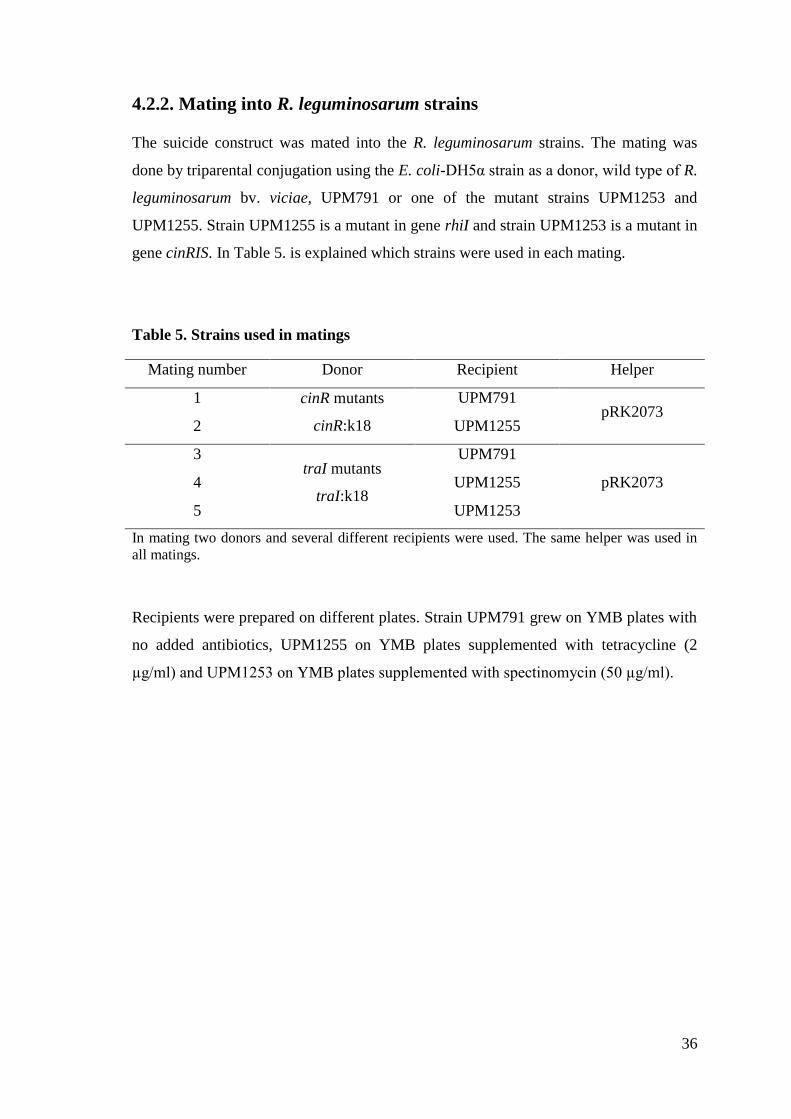
4.2.2. Mating into R. leguminosarum strains
The suicide construct was mated into the R. leguminosarum strains. The mating was
done by triparental conjugation using the E. coli-DH5α strain as a donor, wild type of R.
leguminosarum bv. viciae, UPM791 or one of the mutant strains UPM1253 and
UPM1255. Strain UPM1255 is a mutant in gene rhiI and strain UPM1253 is a mutant in
gene cinRIS. In Table 5. is explained which strains were used in each mating.
Table 5. Strains used in matings
Mating number Donor Recipient Helper
1 cinR mutants
cinR:k18
UPM791 pRK2073
2 UPM1255
3 traI mutants
traI:k18
UPM791
4 UPM1255 pRK2073
5 UPM1253
In mating two donors and several different recipients were used. The same helper was used in
all matings.
Recipients were prepared on different plates. Strain UPM791 grew on YMB plates with
no added antibiotics, UPM1255 on YMB plates supplemented with tetracycline (2
µg/ml) and UPM1253 on YMB plates supplemented with spectinomycin (50 µg/ml).

37
Mating was done following the protocol described in chapter Methods and materials.
Results of mating was done by analyzing YMB plates supplemented with kanamycin
(50 µg/ml) and nitrofurantoin (5 µg/ml) that were incubated for three days at 28˚C. The
plates, where more concentrated sample was plated, had more colonies, in the case of all
five matings, comparing to the ones in which less concentrated sample was plated. Only
in matings 1 and 3 positive results were obtained (Table 6.) and for that reason plates
were exposed two days more to the incubation at 28˚C. After two days extra, more
colonies appeared and positive results were obtained for mating number 4 (results
shown in Table 7.).
Table 6. Results of matings after three days
Mating number Concentration 1 Concentration 2
1 6 colonies 4 colonies
2 / /
3 26 colonies 11 colonies
4 / /
5 / /
In table are shown the results of mating. For every mating is described how many colonies have
grown. In the case of concentration 1 more concentrated sample was plated, and in the
concentration 2 less concentrated sample was plated.
Table 7. Results of mating after five days
Mating number Concentration 1 Concentration 2
1 11 colonies 5 colonies
2 / /
3 34 colonies 21 colonies
4 4 colonies /
5 / /
In table are shown the results of mating after 5 days. For every mating is described how many
colonies have grown after five days. The number of new grown colonies was added to the
number of colonies after three days. In the case of concentration 1, more concentrated sample
was plated, and in the concentration 2, less concentrated sample was plated.
For mating 2 and 5, results were not obtained. In matings 1 and 3 more colonies grew.
The reason being that is because the wild type, Rlv UPM791, grows faster than other
strains.

38
4.2.3. Analysis of the transconjugants through PCR and Southern blot
analysis
Colonies arising were just potential mutants. The candidates had to be analyzed by PCR
or Southern blot. Only those showing adequate DNA profiles could be defined as
mutants. Insertions were confirmed by PCR using four sets of primers (Table 8.) for
cinR and traI mutants located outside of the flanking region of the marker gene and with
genomic DNA of the wild type. These PCR reaction settings were used: 93˚C for 5 min,
10 cycles of 93˚C for 45 s, 60˚C for 45 s, 72˚C for 120 s; 20 cycles of 93˚C for 45 s,
60˚C for 45 s, 72˚C for 120 s with an elongation of 5 s/ cycle; and a final extension of
72˚C for 5 min.
Table 8. Primer sets used in PCR to confirm the insertion
Strains Nr. of primer sets Primers
ΔcinR, WT
Set 1 cinRLF
cinRLR
Set 2 cinRLF
M13PUC-R
Set 3 cinRLR
M13PUC-R
Set 4 traILF
traILR
ΔtraI, WT
Set 1.1 trailLF
traILR
Set 2.1 trailLF
M13PUC-R
Set 3.1 traILR
M13PUC-R
Set 4.1 cinRLF
cinRLR
Abbreviations: ΔcinR, cinR mutants; ΔtraI, traI mutants; WT,wild type (genomic DNA)
39
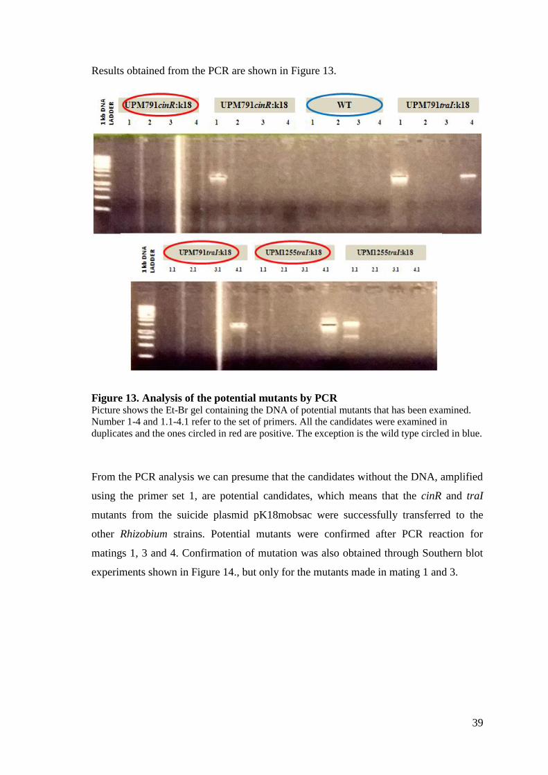
Results obtained from the PCR are shown in Figure 13.
Figure 13. Analysis of the potential mutants by PCR Picture shows the Et-Br gel containing the DNA of potential mutants that has been examined.
Number 1-4 and 1.1-4.1 refer to the set of primers. All the candidates were examined in
duplicates and the ones circled in red are positive. The exception is the wild type circled in blue.
From the PCR analysis we can presume that the candidates without the DNA, amplified
using the primer set 1, are potential candidates, which means that the cinR and traI
mutants from the suicide plasmid pK18mobsac were successfully transferred to the
other Rhizobium strains. Potential mutants were confirmed after PCR reaction for
matings 1, 3 and 4. Confirmation of mutation was also obtained through Southern blot
experiments shown in Figure 14., but only for the mutants made in mating 1 and 3.

40
Figure 14. Construction of mutants affected in traI and cinR genes Pictures correspond to autoradiograms of Southern blot membrane containing total DNA from
wild type (UPM791) and candidate mutant derivates obtained by insertion of pK18mobsac.
Southern blots were hybridized with specific DNA probes for each gene. Red stars indicate the
position of hybridizing bands in the wild type and mutant strains.
DNA probe for each gene was denaturized in the process of Southern blot and during
the hybridization the probe recognized the complementary segment of the DNA. When
the complementary segment is found then a double stranded structure is formed again
(renaturation). The renaturation is detectable by the bands on the autoradiograms. In the
case of of cinR, the signals are very faint, although it is detectable. For traI is visible
how the band changes size.
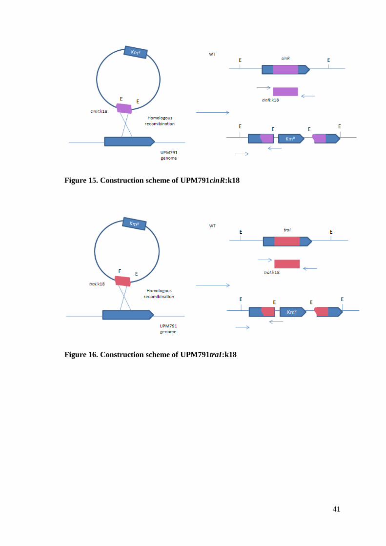
From positive results of confirmations, transconjugant strains were designed:
UPM791cinR, and UPM791traI (Figure 15. and Figure 16.). These mutants are
currently being characterized at the laboratory for phenotypes in free-living cells and in
bacteroids.
41
Figure 15. Construction scheme of UPM791cinR:k18
Figure 16. Construction scheme of UPM791traI:k18

42
5. DISCUSSION
The quorum sensing population-density regulation of gene expression involves a
complex cascade in endosymbiotic bacteria (Sanchez Contreras et al., 2007). In this
work, we have isolated DNA regions containing genes encoding a LuxR-type regulator
(CinR) and a LuxI-type synthase (TraI) from Rhizobium leguminosarum UPM791, and
we have used these DNA fragments to generate insertion mutants in each of these
genes.
A previous search for LuxR- type regulators identified 5 genes that might have a
potential role in quorum sensing in R. leguminosarum UPM791, 4 of which were
previously described by Cantero (2005). Two of these regulators were associated to
LuxRI-type functional systems in this strain. The first one is cinRI system, placed in the
chromosome that produces 3OH-C14:1-HSL. Another system is the rhiRI system, which
produces C6-HSL, C7-HSL and C8-HSL and is located in symbiotic plasmid. A third
system (traRI) was found in the symbiotic plasmid. TraI might contribute to the
production of AHLs (Wilkinson et al., 2002), but its cognate regulator TraR is not in
functional in this strain.
In previous research by Sánchez Cañizares (2013), a R. leguminosarum UPM791
cinRIS-deletion mutant was characterized. The analysis of this mutant revealed that the
deletion of the entire cinRIS operon caused a strong symbiotic defect, since only white,
ineffective nodules were induced in pea plants. This phenotype was unexpected.
Although the CinRI system, located in chromosome in R. leguminosarum strains, has
been described as the master control for the other AHL-dependent QS systems in other
R. leguminosarum strains (Lithgow et al., 2000), the research carried out by Lithgow et
al. (2000) in strain 8401 revealed that mutants affected in cinI or cinR had normal
growth rate in different growth media, and the nodulation phenotype was essentially
normal. In the strain A34, cinRI locus controls three other AHL-dependent quorum-
sensing control systems, including the raiIR, traIR and rhiIR genes (Wisniewski-Dyé
and Downie, 2002). The presence of cinI and cinR genes was identified in Rlv UPM791
in the research by Cantero (2005). R. leguminosarum Cin system has an additional small
regulatory protein, CinS that is coexpressed with CinI (Lithgow et al., 2000). Mutants in
cinRIS lost the ability to produce 3-OH-C14:1-HLS. In the same mutant the level of the
short-chain signals (C6 through C8-HSL) produced by the RhiRI system was reduced
43
but not suppressed. In the research by Lithgow et al. (2000) mutations in cinI and cinR
suppressed the production of 3-OH-C14:1-HLS, whereas lower levels of short-chain
AHLs were detected, likely resulting from reduced expression of rhiI gene. In the
review by Sanchez Contreras (2007) the results from several researches were compared
(included the research by Lithgow et al. 2000 mentioned above, and also those from
Rodelas et al., 1999; Wisniewski Dyé et al., 2002; Danino et al., 2003) and the
conclusion was that mutation in cinI and cinR decrease the expression of all other AHL
synthase genes, without significant alterations in symbiotic performance.
Based on the differences found in the cinRIS and traRI systems, we hypothesized that
the quorum sensing-dependent regulation in R. leguminosarum strain UPM791 might
present significant differences with that from other strains, and concluded that a more
detailed analysis was required. To start this work we decided to isolate and mutagenized
cinR and traI genes from this strain.
As shown in the results section, genomic DNA from R. leguminosarum UPM791 was
used as a template for PCR amplification with specific primers for cinR and traI. These
experiments allowed the cloning and sequencing of the two genes. The data obtained
from DNA sequences of cinI and traI genes were used for a comparative genomic
analysis carried with the reference sequences that are available in the web: Rhizobium
etli CFN42, R. leguminosarum bv. viciae 3841 and Agrobacterium tumefaciens 5A.
This sequence analysis of the amplified DNA region revealed that R. leguminosarum
UPM791 cinR gene is highly conserved as regarding the homologous genes from other
strains previously characterized, such as Rlv 3841 or A34 (Lithgow et al, 2000). Rlv
UPM791 CinR is almost identical to CinR from R. leguminosarum bv. viciae 3841
(98% identity). Protein comparison also showed that CinR in Rlv UPM791 shares 54%
identity to the LuxR protein in Agrobacterium tumefaciens. Multiple alignment of CinR
sequences revealed that the C-terminal part of the protein displays a high number of
conserved residues. This region in LuxR is known to be responsible for DNA binding
and activation of LuxI expression (Choi and Greenberg, 1991).
The number of bacteria in which QS is identified is increasing. In research by Case et
al. (2008) it was estimated that 66% of bacteria containing QS circuits had more LuxR
than LuxI homologues. In the same research the frequency and function of AHL-driven
quorum-sensing circuits were compared among the Proteobacteria, and the results

44
allowed the construction of a phylogenic tree (Figure 17.) including Rhizobium and
Agrobacterium proteins. Agrobacterium genomes share a highly conserved CinR
protein as compared to the one in R. leguminosarum bv. viciae. This CinR of R.
leguminosarum 8401 shown in phylogenetic tree is virtually identical to the one that
was sequenced in this Thesis.
Figure 17. Phylogenetic tree of LuxR types proteins from Agrobacterium and
Rhizobium In phylogenetic tree are compared Agrobacterium and Rhizobium strains, showing how related
are those strains. The value written above the nodes represents the percentage of similarity
obtained from 100 replicates of the original data (taken from Case et al., 2008).
45
In the case of TraI, the Rlv UPM791 protein showed an 87% identity to the TraI
homologue in R. leguminosarum bv. viciae 3841, and 55% identity when compared to
the homologues protein in Agrobacterium tumefaciens. traRI systems are present in
Agrobacterium tumefaciens, Rhizobium NG234 and R. leguminosarum A34.
In Rhizobium leguminosarum luxI homologues rhiI and traI, and luxR homologues
(bisR, rhiR), encoded in the symbiotic plasmid pRL1JI, have been originated from a
separate source. For BisR an endogenous origin from the chromosomal CinR is
supposed (Gray et al., 2001). Previous studies by Wilkinson et al. (2002) in R.
leguminosarum have shown that traI can be induced by cinI-dependent 3-OH-C14:1-
HSL. The TraRI system induces the trb operon located in the symbiotic plasmid
pRL1JL, and it is involved in the regulation of the conjugative transfer of this plasmid.
It has been discovered that a gene for a bifunctional signaling regulator (bisR) is located
downstream on trb operon. Mutations on traRI genes in R. leguminosarum A34 stops
plasmid transfer. In Sánchez Cañizares Thesis (2013) during the analysis of mutation in
RhiRI system on the genome was discovered that UPM791 also contains a traI gen
located on symbiotic plasmid. The traI gen is responsible for the LuxI-like synthase of
short-chain AHLs. Although traR, the LuxR-like regulator of this system, is inactivated,
traI synthase is supposed to be responsible for the synthesis of low-molecular-weight
AHL signaling molecules that were found in detection of rhiI-deficient derivative of Rlv
UPM791. To prove the Sánchez Cañizares statement mutants were made in traI gene.
The single-recombination-based mutagenesis carried out in this work with
pK18mobsac-derived plasmids carrying the internal region of cinR gene produced two
incomplete copies of the corresponding gene. One of these copies lacked 253 bp of the
3′ end, where DNA binding domain of the protein is encoded. So the resulting cinR
gene is likely possible to occur in an inactive protein form. This kind of single-
recombination mutagenesis has been extensively used for generating mutants in
Rhizobium (Brom et al., 2004).Since a Rlv UPM791-derivative strain unable to produce
C14-HSL signals is symbiotically proficient (Cantero et al., 2006), the phenotype
induced by the deletion of the entire operon might be due to a potential role of the cinR
regulator when not bound to autoninducer. The cinR mutant generated here will allow to
study this possibility. A single-recombination based mutagenesis of traI was carried out
in this work with pK18mobsac, as for the cinR gene; derived plasmids carrying the
internal region of traI gene produced again two incomplete copies of the corresponding

46
gene. So the traI gene is resulting in disruption of amino-terminal half and causing the
reduction in synthase activity (Whitehead et al., 2001).
The phenotypic characterization of mutants in cinR and traI made by this Thesis have
yet to be fully defined since the lack of time at the UPM lab did not allow to perform
the functional analysis. These analyses are currently being carried out at the laboratory
using free-living cells and bacteroids. Preliminary results (R. García, personal
communication) obtained by TLC analysis indicate that the cinR mutant obtained is
indeed impaired in the production of C14–HSL signals, also showing a reduction on the
level of short-chain AHLs (see Fig. 18). No significant alteration on C14–HSL was
observed in the traI mutant. In this mutant, the level of short-chain AHLs is also
reduced, although further analysis is required to properly quantify the level of reduction.
If that preliminary data are confirmed, then we can conclude that traI is being expressed
and that this synthase is responsible for a fraction of autoinducers synthesized in free-
living cultures. Further studies will indicate the relevance of the mutation on the
interaction with the plant. Next step will be the inoculation of plants and analyzing how
the mutations affect nodule formation. As mentioned above, many possibilities could
arise as result of mutation on quorum sensing systems, including changes in the ability
of forming nodules and thus on the efficiency of nitrogen fixation.
Figure 18. Analysis of AHL production by cinR and traI mutants
Ethyl-acetate extracts were loaded into C18 Thin-layer Chromatography plates and separated
using 60% methanol-water as solvent. AHLs were developed using a bioassay based on A.
tumefaciens strain expressing an AHL-inducible lacZ fusion.
UPM
791
791cinR::k18
791DcinRIS
791traI:k18
C14-HSL
C6/C8-HSL

47
Potential applications of results
Nowadays, as the environmental contamination is very high, the scientists are looking
how to prevent and reduce a negative impact on the environment. In order to reach
maximum crop productivity, agricultural systems are using addition of chemical
nitrogen fertilizers. The reason for that are the soils insufficiently supplemented with
nitrogen. Fertilizers are added to improve poor N-content soil in order to provide plants
enough nutrients for maximum growth potential.
Due to the negative effects on the environment, the development of alternatives that are
ecofriendly allowing the reduction of nitrogen fertilizers is a major concern. Organisms
capable of carrying out nitrogen fixation have an important role in accumulation of
nitrogen in biomass and soil organic matter. In global ecological context, the biological
nitrogen fixation can contribute to reduce the problems linked to nitrogen fertilizers.
From an agricultural point of view, the most relevant biological nitrogen fixation system
is the symbiotic association between legumes and bacteria belonging to Rhizobium
group (Herridge et al., 2008). For that reason effort in research and understanding the
Rhizobium-Leguminosae symbiosis are needed, and one of the components in the
symbiosis is quorum sensing. Increasing the existing knowledge of the quorum sensing
system and due to the recombinant DNA techniques we will be able to understand better
the process of biological and to provide plants with better characteristics.
The results obtained in this Thesis are giving the opportunity to examine the
possibilities how the mutations are affecting the nitrogen fixation possibility. Further
studies are required to clarify the role of quorum sensing in Rhizobium-Leguminosae
symbiosis, one of the very important processes in agriculture.
48
6. CONCLUSIONS
From the comparison of the DNA sequences it was concluded that cinR and traI genes
are highly conserved in R. leguminosarum bv. viciae.
Insertion mutants affected in cinR and traI are now available and will allow the
assessment of the relevance of quorum sensing on free living and symbiotic cells of this
endosymbiotic bacterium.
The resulting truncated genes are possibly inactive proteins, cinR gene is possibly not
able to produce C14-HSL signals and traI gene is potentially going to reduce synthase
activity. The obtained mutants need to be further examined to confirm the assumption.
The phenotypic characterization of mutants in cinR and traI make by this Thesis is
being currently examined at the UPM lab using free living cells and bacteroids.
Preliminary results obtained in UPM lab by TLC analysis show that the cinR mutant is
really damaged in the production of C14-HSL signals. The level of short-chain HSLs is
reduced. No significant change of C14-HSL signal production was observed for traI
gene. In traI mutant the level of short-chain AHLs is also reduced.
Preliminary data have to be confirmed in further analysis to assess the relevance of the
mutation on the interaction with the plant.
49
7. LITERATURE
1. Altschul S.F., Gish W., Miller W., Myers E.W: and Lipman D.J. (1990). Basic
local alignment search tool. Journal of Molecular Biology 215(3): 403-410
2. Beringer J.E. (1974). R factor transfer in Rhizobium leguminosarum. Journal of
general Microbiology 84: 188-198.
3. Blažinkov M., Sikora S., Uher D., Mačešić D. and Redžepović S. (2007).
Genotypic characterization of indigenous Rhizobium leguminosarum bv. viciae
field population in Croatia. Agriculture Conspectus Scientificus 72: 153-157.
4. Brom S., Girard L., Tun Garrido C., Garcia de los Santos A., Bustos P.,
González V. and Romero D. (2004). Transfer of the symbiotic plasmid of
Rhizobium etli CFN42 requires cointegration with p42a, which may be mediated
by site-specific recombination. Journal of Bacteriology 186(22): 7538-7548
5. Canfield D.E., Glazer A.N. and Falkowski P.G. (2010). The evolution and future
of Earth’s nitrogen cycle. Science 330(6001): 192-196.
6. Cantero L. (2005). Análísis genetic y proteómico de la regulación de los
sistemas de auto inducción en Rhizobium leguminosarum bv. viciae UPM791 y
3841. Tesis doctoral, Universidad Politécnica de Madrid.
7. Cantero L., Palacios J.M., Ruiz-Argüeso T., Imperial J. (2006). Proteomic
analysis of quorum sensing in Rhizobium leguminosarum biovar viciae
UPM791. Proteomics 6: S97-S106.
8. Case R.J., Labbate M. and Kjelleberg S. (2008). AHL-driven quorum-sensing
circuits: their frequency and function among the Proteobacteria. The ISME
Journal 2(4): 345-349.
9. Choi S.H. and Greenberg E.P. (1991): The C-terminal region of the Vibrio
fischeri LuxR protein contains an inducer-independent lux gene activating
domain. Proceedings of the National Academy of Science 88(24):11115-11119.
10. Cubo M.T., Economou A., Murphy G., Johnston A.W. and Downie J.A. (1992).
Molecular characterization and regulation of the rhizospehre-expressed genes
rhiABCr that can influence nodulation by Rhizobium leguminosarum biovar
viciae. Jourmal of Bacteriology 174(12): 4026-4035.
50
11. Danino V.E., Wilkinson A., Edwards A. and Downie J.A. (2003). Recipient-
induced transfer of the symbiotic plasmid pRL1JI in Rhizobium leguminosarum
bv. viciae is regulated by a quorum-sensing relay. Molecular Microbiology
50(2): 511-525.
12. Downie J.A. (2010). The roles of extracellular proteins, polysaccharides and
signal in the interactions of rhizobia with legume roots. FEMS Microbiology
Reviews 34 (2): 150-170.
13. Downie J.A. and Young P. (2001). Genome sequencing. The ABC of symbiosis.
Nature 412(6847): 597-598.
14. Edwards A., Frederix M., Wisniewski-Dyé F., Jones J., Zorreguieta A. and
Downie J.A. (2009). The cin and rai quorum-sensing regulatory systems in
Rhizobium leguminosarum are coordinated by ExpR and CinS, a small
regulatory protein coexpressed with CinI. Journal of Bacteriology 191(9): 3059-
3067.
15. Figurski D.H. and Helinski (1979). Replication of an origin-containing derivate
of plasmid RK2 dependent on a plasmid function provided in trans. Proceedings
of the National Academy of Sciences of the USA 76(4): 1648-1652.
16. Fuqua C. (2006). The QscR quorum-sensing regulon of Pseudomonas
aeruginosa: an orphan claims its identity. Journal of Bacteriology 188(9): 3169-
3171.
17. Geurts R. and Bisseling T. (2002). Rhizobium Nod factor perception and
signaling. The Plant cell 14(1): S239-S249.
18. Gonzalez J.E. and Marketon M.M. (2003). Quorum Sensing in Nitrogen-Fixing
Rhizobia. Microbiology and Molecular Biology Reviews 67(4): 574-592.
19. Gray K.M. and Garey J.G. (2001). The evolution of bacteria LuxI and LuxR
quorum sensing regulators. Microbiology 147: 2379-2387.
20. Hanahan D. (1983). Studies on transformation of Escherichia coli with
plasmids. Journal of Molecular Biology 166: 557-580.
21. Henke J.M. and Bassler B.L. (2004). Bacterial social engagements. Trends in
Cell Biology 14(11): 648-656.
51
22. Herridge D.F, Peoples M.B. and Boddey R.M. (2008). Global inputs of
biological nitrogen fixation in agricultural systems. Marschner Review 311: 1-
18.
23. Hirsch P.R. (1979). Plasmid-determined bacteriocin production by Rhizobium
leguminosarum. Journal of General Microbiology 113(2): 219-228.
24. Kereszt A., Mergaert P. and Kondorosi E. (2011). Bacteroid development in
legume nodules: evolution of mutual benefit or of sacrificial victims?. Molecular
Plant-Microbe Interaction 24 (11): 1300-1309.
25. Leyva A., Palacios J.M., Mozo T. and Ruiz-Argüeso T. (1987). Cloning and
characterization of hydrogen uptake genes from Rhizobium leguminosarum.
Journal of Bacteriology 169: 4929-4934.
26. Lithgow J.K., Wilkinson A., Hardman A., Rodelas B., Wisniewski-Dyé F.,
Wiliams P. and Downie J.A. (2000). The regulatory locus cinRI in Rhizobium
leguminosarum controls a network of quorum-sensing loci. Molecular
Microbiology 37(1): 81-97.
27. Mylona P., Pawlowski K., and Bisseling T. (1995). Symbiotic nitrogen fixation.
The Plant Cell 7: 869-885, American Society of Plant Physiologists.
28. Nealson K.H., Platt T. and Hastings J.W. (1970). Cellular control of the
synthesis and activity of the bacterial luminescent system. Journal of
Bacteriology 104: 313-322.
29. Oldroyd G.E. and Downie J.A. (2008). Coordinating nodule morphogenesis with
rhizobial infection in legumes. Annual Review of Plant Biology 59: 519-546.
30. Patankar A.V. and González J.E. (2009). Orphan LuxR regulators of quorum
sensing. FEMS Microbiology Reviews 33(4): 739-756.
31. Prell J., and Poole P. (2006). Metabolic changes of rhizobia in legume nodules.
Trends In Microbiology 14(4): 161-168.
32. Rodelas B., Lithgow J.K., Wisniewski-Dyé F., Hardman A., Wilkinson A.,
Economou A., Williams A., and Downie J.A. (1999). Analysis of quorum-
sensing-dependent control of rhizosphere expressed (rhi) genes in Rhizobium
leguminosarum bv. viciae, Journal of Bacteriology 181(12): 3816-3823.
33. Sambrook J. and Russell D.W. (2001). Molecular Cloning: A Laboratory
Manual. Cold Spring Harbor Laboratory Press.
52
34. Sánchez Cañizares C. (2013). Análisis genómico y functional de los sistemas de
quorum sensing en Rhizobium leguminosarum. Doctoral Thesis, Technical
University of Madrid.
35. Sanchez Contreras M., Bauer W.D., Gao M., Robinson J.B. and Downie J.A.
(2007). Quorum sensing regulation in rhizobia and its role in symbiotic
interactions with legumes. Philosophical Transactions of the Royal Society B
Biological Sciences 362(14839): 1149-1163.
36. Schäfer A.A., Tauch W., Jäger W., Kalinowski J., Theirbach G. and Pühler A.
(1994). Small mobilizable multi-purpose cloning vectors derived from
Escherichia coli plasmid pK18 and pK19: section of defined deletions in the
chromosome of Corynebacterium glutamicum. Gene 145(1): 69-73.
37. Schuster M., Sexton D.J., Diggle S.P. and Greenberg E.P. (2013). Acyl-
homoserin lactone quorum sensing: From evolution to Application. Annual
Review of Microbiology 67: 43-63.
38. Sprent J.I. (2008). 60Ma of legume nodulation. What’s new? What’s changing?
Journal of Experimental Botany 59: 1081-1084.
39. Topol J. and Kanižai Šarić G. (2013). Simbiotska fiksacija dušika u ekološkoj
poljoprivrednoj proizvodnji. Agronomski glasnik 75: 117-134.
40. Vincent J.M. (1970). A manual for the practical study of root-nodule bacteria.
Oxford, Blackwell Scientific Publications, Ltd.
41. Waters C.M. and Bassler B.L. (2005). Quorum sensing: Cell to cell
communication in bacteria. Annual Review of Cell and Development Biology
21: 319-346.
42. Whitehead N.A., Barnard A.M., Slater H., Simpson N.J. and Salmond G.P.
(2001). Quorum sensing in Gram-negative bacteria. FEMS Microbiology
Reviews 25(4): 365-404.
43. Wilkinson A., Danino V., Wisniewski-Dyé F., Lithgow J.K. and Downie J.A.
(2002). N-acyl-homoserine lactone inhibition of Rhizobial growth is mediated
by two quorum-sensing genes that regulate plasmid transfer. Journal of
Bacteriology 184(16): 4510-4519.
44. Wisniewski-Dyé F. and Downie J.A. (2002). Quorum-sensing in Rhizobium.
Kluwer van Leeuwenhoek 81: 397-407.
53
45. Wisniewski-Dyé F., Jones J., Chhabra S.R. and Downie J.A. (2002). raiIR genes
are part of a quorum –sensing network controlled by cinI and cinR in Rhizobium
leguminosarum. Journal of Bacteriology 184: 1597-1606.
46. Young J.P., Crossman L., Johnston A.W.B., Thomas N.R., Ghazoui Z.F., Hull
K.H., Wexler M., Curson A.R.J., Todd J.D., Poole P.S., Mauchline T.H., East
A.K., Quail M.A., Churcher C., Aeeowsmith C., Cherevach I., Cilingworth T.,
Clarke K., Cronin A., Davis P., Fraser A., Hance Z., Hauser H., Jagels K.,
Moule S., Mungall K., Norbertczak H., Rabbinowitsch E., Sanders M.,
Simmonds M., Whitehead S. and Parkhill J. (2006). The genome of Rhizobium
leguminosarum has recognizable core and accessory components. Genome
Biology 7(4): R34.
47. http://www.biovisualtech.com/.
48. http://www.jic.ac.uk/science/molmicro/Rhizo.html/.
49. https://www.nippongenetics.eu/.
54
8. CURICULUM VITAE
I was born in Zagreb on 4th
June 1991. I finished my grammar school and high school
education in Zagreb, and in September 2013 I obtained my Bachelor Degree in Biology
at the University of Zagreb. In 2013 I started my Master Degree in Environmental
Science and Ecology at the Faculty of Science, University of Zagreb, and passed all my
exams prior to my Master thesis. In 2015 I spent an exchange year at the University of
Madrid through the Erasmus+ program, where I did my master thesis research. I worked
as an unpaid trainee in multiple laboratories (Andrija Stampar Teaching Institute of
Public Health, Pliva Hrvatska Ltd., Zagreb Wastewater Ltd.).
Contact details:
Franka Busch, mag. oec. et prot. nat.
Mobile: (+)385915946774
E-mail: [email protected]