Embed Size (px)
Citation preview

Analysis of Albumin-Associated Peptides andProteins from Ovarian Cancer Patients
Mark S. Lowenthal,1 Arpita I. Mehta,3 Kristina Frogale,1 Russell W. Bandle,1
Robyn P. Araujo,1 Brian L. Hood,5 Timothy D. Veenstra,5 Thomas P. Conrads,5
Paul Goldsmith,1 David Fishman,4 Emanuel F. Petricoin III,2* and Lance A. Liotta2*
Background: Albumin binds low–molecular-weight mol-ecules, including proteins and peptides, which thenacquire its longer half-life, thereby protecting the boundspecies from kidney clearance. We developed an exper-imental method to isolate albumin in its native state andto then identify [mass spectrometry (MS) sequencing]the corresponding bound low–molecular-weight mole-cules. We used this method to analyze pooled sera froma human disease study set (high-risk persons withoutcancer, n� 40; stage I ovarian cancer, n � 30; stage IIIovarian cancer, n � 40) to demonstrate the feasibility ofthis approach as a discovery method.Methods: Albumin was isolated by solid-phase affinitycapture under native binding and washing conditions.Captured albumin-associated proteins and peptideswere separated by gel electrophoresis and subjected toiterative MS sequencing by microcapillary reversed-phase tandem MS. Selected albumin-bound proteinfragments were confirmed in human sera by Westernblotting and immunocompetition.Results: In total, 1208 individual protein sequenceswere predicted from all 3 pools. The predicted se-quences were largely fragments derived from proteinswith diverse biological functions. More than one third
of these fragments were identified by multiple peptidesequences, and more than one half of the identifiedspecies were in vivo cleavage products of parent pro-teins. An estimated 700 serum peptides or proteins werepredicted that had not been reported in previous serumdatabases. Several proteolytic fragments of larger mol-ecules that may be cancer-related were confirmed im-munologically in blood by Western blotting and peptideimmunocompetition. BRCA2, a 390-kDa low-abundancenuclear protein linked to cancer susceptibility, was repre-sented in sera as a series of specific fragments bound toalbumin.Conclusion: Carrier-protein harvesting provides a richsource of candidate peptides and proteins with potentialdiverse tissue and cellular origins that may reflectimportant disease-related information.© 2005 American Association for Clinical Chemistry
The circulatory proteome holds great promise as a reser-voir of information useful for disease detection and ther-apeutic monitoring (1 ). Despite this potential, compre-hensive characterization of the circulatory proteome isdifficult because of the wide dynamic range of proteinconcentrations that exist between larger molecules such asalbumin (g/L) and the sought after biomarkers (belowng/L) (2 ). Here we describe a method for amplifying theyield of low-abundance, low–molecular-weight proteinsand peptide fragments in serum as a means of providinga new window into the information content containedwithin the circulation. This method takes advantage of thetendency for low-abundance molecules to associate withhigh-abundance proteins such as albumin and therebyacquire the carrier’s longevity in the serum (3 ). Thisreport describes the application of this method to identifycandidate proteins and peptides in serum pools fromdiseased and control ovarian cancer patients.
Current protocols for investigation of the serum pro-teome often recommend prefractionation by native de-pletion of high-abundance species (e.g., albumin, im-
1 Laboratory of Pathology, Center for Cancer Research, National CancerInstitute, National Institutes of Health, Bethesda, MD.
2 Center for Applied Proteomics and Molecular Medicine, George MasonUniversity, Manassas, VA.
3 Tufts University School of Medicine, Howard Hughes Medical Institute,Boston MA.
4 National Ovarian Cancer Early Detection Program, New York University,New York, NY.
5 National Cancer Institute Biomedical Proteomics Program, SAIC, NCI,Frederick, MD.
*Address correspondence to Dr. Petricoin or Dr. Liotta at: Center forApplied Proteomics and Molecular Medicine, George Mason University,Manassas, VA 20110. E-mail [email protected] or [email protected].
Received April 25, 2005; accepted August 1, 2005.Previously published online at DOI: 10.1373/clinchem.2005.052944
Clinical Chemistry 51:101933–1945 (2005) Oak Ridge Conference
1933

munoglobulins, lipoproteins) (4–8). This approach caninadvertently remove a high percentage of candidatelow-abundance proteins and peptides because most low–molecular-weight molecules in serum form complexeswith high-abundance proteins, which protect the boundmolecules from clearance and allow them to remain incirculation. Albumin is the most abundant protein inplasma and serum, present at 50 g/L, and has a half-life of19 days in humans (9–11). Because the kidney generallyand efficiently filters out molecules �60 kDa, smallerproteins and peptides will be protected from clearance byassociation with albumin (9, 10). Plasma protein bindingcan be an effective means of extending the pharmacoki-netic properties of otherwise short-lived molecules (9, 11–13). Dennis et al. (9 ) demonstrated that a 58-fold increasein peptide longevity could be attained through albuminbinding and association. Albumin binding of biologicallyimportant proteins and peptides is well documented. Forexample, the amino-terminal peptide of HIV-1, gp41, andthe 14-kDa fragment of streptococcal protein G are knownto specifically bind with human serum albumin (14, 15).Vitamin A transport and homeostasis is controlled by aspecific protein–protein interaction between the 21-kDaretinol-binding protein and transthyretin, which wasshown to reduce glomerular filtration of the low–molec-ular-mass retinol-binding protein (16 ).
It has recently been shown that most low–molecular-weight molecules that have been visualized by massspectrometry (MS)6 profiling methods in the past existunder native conditions in a complexed state with high–molecular-weight proteins (17, 18). Direct analysis of thelow–molecular-weight portion of size-fractionated nativeserum revealed few unbound molecular species. Theseprevious studies (3, 17, 18) demonstrated that most of thelow–molecular-weight species were found in the high–molecular-weight fraction because they were all com-plexed to other proteins. These same investigators wenton to denature the protein complexes and then sequencedthe size-fractionated low–molecular-weight repository.The selectivity of binding of low–molecular-weight con-stituents for different classes of carrier proteins wasexplored further (17, 18), revealing that subsets of boundlow–molecular-weight peptides and sequenced proteinfragments associated with albumin were different fromthose bound to immunoglobulin, apolipoprotein, andtransferrin (18 ). These previous studies established thescientific rationale for the present investigation into thecarrier-protein–bound information archive. To evaluatethe possibility that this repository of bound moleculescontains potential disease-related information, we reportthe results of an investigation comparing the albumin-
associated peptides in serum study sets derived frompatients with pathologically documented ovarian cancervs healthy but high-risk patients who had been disease-free for 5 years after serum collection.
Two key properties of carrier proteins enable them tofunction as efficient harvesters of lower-abundance puta-tive biomarkers in the circulation: overwhelming relativeabundance and long half-lives compared with their cargo.Considering the binding reaction between a biomarker (B)and its carrier protein (C):
kf
B � C 34 BC
kr
it is reasonable to suppose that the reaction dynamics aregoverned by mass action kinetics because a carrier proteinwould not usually bind more than one molecule of thebiomarker of interest if the carrier existed in high relativeabundance.
Thus, the rate of formation of the biomarker–carrier-protein complex (BC) is proportional to the product of thebiomarker concentration [B] and the carrier-protein con-centration [C], with a constant of proportionality kf. Thedissociation of the biomarker–carrier-protein complexinto free biomarker and carrier protein, on the other hand,is proportional to the concentration of the bound biomar-ker [BC], with a constant of proportionality kr. The for-ward and reverse reaction rates are given by:
Rf � kf [B][C]
and
Rr � kr [BC]
respectively. The vast excess of carrier protein means thatonly a tiny proportion of its total concentration will beconsumed in the reaction with the biomarker, so that thefree carrier-protein concentration [C] may be considered aconstant. The free carrier-protein concentration may thenbe absorbed into the forward rate constant to yield anamplified effective forward rate constant, kf*, giving riseto the forward reaction rate:
Rf �kf* [B]
where
kf* � kf [C]
The reaction arrives at an equilibrium when Rf � Rr, or:
kf* [B] � kr [BC]
Importantly, because the effective forward rate constantkf* has been amplified by the (very large) carrier-proteinconcentration, it is expected to be many orders of magni-tude larger than the reverse rate constant kr, so that thereaction between the biomarker and its carrier protein isstrongly biased in the forward direction. The equilibrium
6 Nonstandard abbreviations: MS, mass spectrometry; �LC-MS/MS micro-capillary reversed-phase tandem MS; and PVDF, polyvinylidene difluoride.
1934 Lowenthal et al.: Albumin-Associated Peptides and Proteins from Ovarian Cancer

ratio between the bound and free forms of the biomarkeris given by:
�eq �[BC][B]
�kf
*
kr
Thus, the putative biomarker is likely to exist almostentirely in complexed association with a carrier protein,even when its innate binding affinity, characterized by the“natural” forward rate constant, kf, is low (3 ). Indeed,with an albumin concentration in the blood of 6 � 10�4
mol/L (19 ), molecules with low-affinity binding con-stants, kf/kr, of 15 � 104 L/mol [reported for flavonoidinteraction with albumin (20 )] and 9.34 � 106 L/mol[reported for interactions between protoporphyrins andalbumin (21 )] correspond to 98.9% and 99.98% probabil-ity, respectively, that the molecule of interest will becomplexed with albumin.
Moreover, the relatively long half-life of the carrierprotein [�19 days for albumin compared with a fewhours or less for an unbound molecule (9, 22–25)] facili-tates the prolonged presence of smaller peptides andproteins in the bloodstream. Mehta et al. (3 ) showed thatif a protein is secreted into the bloodstream at a constantrate p, and is subsequently eliminated through the kid-neys at a rate proportional to its concentration (with aconstant of proportionality of e), its steady-state concen-tration in the circulation is given by the ratio p/e . In theabsence of carrier-protein binding, the steady-state con-centration of biomarker in the circulation would be p
b/e
b,
where pb and eb are the production and elimination ratesof the biomarker, respectively. When bound to a carrierprotein, however, the biomarker acquires the carrier pro-tein’s longer half-life, so that the steady-state concentra-tion becomes p
b/e
c, where ec is the relatively small elim-
ination rate of the carrier protein. Because the eliminationrate of a protein is inversely proportional to its half-life, amplification of the biomarker’s concentration inthe bloodstream as a result of carrier-protein bindingis e
b/e
c� t
�1/ 2�c/t
�1/ 2�b, where t(1/2)c and t(1/2)b are the
half-lives of the carrier protein and biomarker, respec-tively. Thus, the longer the half-life of the carrier protein,the greater is its ability to amplify the concentration ofbound species in the bloodstream. In this way, the relativeabundance of large carrier proteins and their longerhalf-lives work together to increase the total concentrationof complexed molecules in the bloodstream. This reportdescribes the application of the carrier-protein sequestra-tion principle to the isolation and mass spectrometricidentification of candidate low–molecular-weight pro-teins and peptides that exist in the serum of ovariancancer patient disease study sets.
Materials and Methodsclinical serum samplesSerum samples were collected under full patient consentand Institutional Review Board approval. Serum was
collected before physical evaluation, diagnosis, and treat-ment and stored at �80 °C. The ovarian study set con-sisted of 40 samples from unaffected, high-risk patient, 30samples from patients with stage I ovarian cancer, and 40samples from patients with stage III ovarian cancer. Thegynecologic oncology clinic where the samples from thecases were collected is located in a building that isseparate but contiguously linked to the building in whichthe National Ovarian Cancer Early Detection Program atNorthwestern University is located, where the high-riskcontrol samples were collected. A special attribute of thissample set is that the same personnel were involved in theblood collection, handling, and storage of all biospeci-mens. In addition, all blood specimens were processed inan identical manner under the same methodology.Healthy control sera were collected from unaffectedwomen determined to be at increased risk for ovariancancer. These women were enrolled in the NationalOvarian Cancer Early Detection Program and had noevidence of any cancer for 5 years as determined by twiceyearly 3-dimensional color Doppler ultrasound and ex-tensive clinical evaluation by a board-certified gyneco-logic oncologist.
Increased risk was determined by classic genetic ped-igree analysis and by the presence or absence of factorssuch as BRCA mutation status. The determination ofincreased risk was based on commercially available com-puter-generated risk algorithm programs such as BRCAPro. All patients in the high-risk clinic were seen byboard-certified genetic counselors and geneticists whodefined the risk. Similarly, the serum specimens fromwomen with ovarian cancer were procured in a gyneco-logic oncology clinic from symptomatic women who werelater surgically staged and found to have epithelial ovar-ian carcinoma. Each sample was accompanied by a veri-fied pathology diagnosis. Briefly, specimens were col-lected in red-top Vacutainer Tubes and allowed to clot for1 h on ice, followed by centrifugation at 4 °C for 10 min at1210g. The serum supernatant was divided in aliquotsand stored at�80 °C until needed. Samples were selectedfor our analysis by a random process categorized by thepathology diagnosis as cancer or benign.
capture of native serum albumin and elutionof complexed protein speciesA schematic representation of the basic experimentaltechnique is illustrated in Fig. 1. Native, diluted serum isintroduced into an affinity column so that the carrierprotein (albumin) is captured along with any boundmolecules. The bound subproteome consisting of thecarrier proteins and their peptide “cargo” is eluted, dis-sociated, and separated by 1-dimensional gel electro-phoresis. Each entire gel lane is cut out, finely subdividedinto molecular mass regions, subjected to in-gel trypsindigestion, and prepared for electrospray mass spectro-metric analysis.
Clinical Chemistry 51, No. 10, 2005 1935
purification of albumin and bound peptideTypically, 25 �L of human stage-specific (pooled) cancerserum (�3.1 mg of protein) was diluted to 200 �L withEquilibration Buffer (Millipore) and run through a (Mon-tage) albumin-specific affinity column twice. The boundprotein was washed thoroughly with two 200-�L volumesof proprietary wash buffer (provided by the manufac-turer). These fractions were combined and labeled as a“flow-through” fraction. The bound proteins were elutedfrom the column by equilibrating with acetonitrile–H2O–trifluoroacetic acid (70:30:0.2 by volume) for 30 min,followed by a slow spin-through of the elution mixture,repeated once. The eluate (retentate fraction) was lyoph-ilized to �10 �L in a HetoVac roto (CT 110) and recon-stituted in an H2O–acetonitrile–formic acid (95:5:0.1 byvolume) buffer. Samples were desalted with a ZipTipcleanup and reconstituted in a 1:1 mixture of water and
sodium dodecyl sulfate sample buffer (20 �L total vol-ume).
1-dimensional protein gel separation anddigestionThe flow-through and retentate fractions were kept on icein 20 �L of sample buffer from 25 �L of original serum,and then were heated for 5 min at 95 °C and loaded on1-dimensional precast gels to separate albumin from theproteins/peptides/fragments of interest. The proteinsand fragments were visualized with a Gel Code Blue StainReagent (Pierce) according to the manufacturer’s proto-cols. The entire lane was excised from the gel and finelysliced into very small molecular-weight regions (�35slices/lane). Gel bands were reduced, alkylated, anddigested with porcine modified trypsin according to a
Fig. 1. Schematic representation of the experimental technique.Raw serum is introduced into a Millipore albumin depletion column. Albumin complexed with other proteins and peptides is retained. After gentle washing under nativeconditions, the protein and peptide cargo are dissociated with organic solvent. Eluted proteins are fractionated by 1-dimensional (1D) gel electrophoresis followed byin-gel trypsin digestion and subjected to �LC-MS/MS analysis.
1936 Lowenthal et al.: Albumin-Associated Peptides and Proteins from Ovarian Cancer

standard protocol (26 ), and peptides were concentratedand prepped for mass spectrometric analysis.
microcapillary reversed-phase tandem msSamples were lyophilized to near dryness and reconsti-tuted in 6.3 �L of Buffer A (H2O–acetonitrile–formic acid;95:5:0.5 by volume) for MS analysis. Microcapillary re-versed-phase tandem MS (�LC-MS/MS) analysis wasperformed with a Dionex LC Packings liquid chromatog-raphy system coupled on-line to a ThermoFinnigan LCQClassic ion trap mass spectrometer with a modified nano-spray source. Reversed-phase separations were per-formed on an in-house, slurry-packed capillary column.The C18 silica-bonded column was a 10-cm long (75-�mi.d.) fused-silica column packed with 5-�m beads (poresize, 300 Å; Vydac). A PepMap C18 cartridge (5-mm;Dionex) acted as a desalting column. Sample was injectedin microliter pick-up mode and washed with Buffer A for5 min before elution with a linear gradient with buffer B(acetonitrile–H2O–formic acid; 95:5:0.1 by volume) up to85% over 95 min at a flow rate of 200 nL/min. Full MSscans were followed by 4 MS/MS scans of the mostabundant peptide ions (in a data-dependent mode), andcollision-induced dissociation was performed at a colli-sion energy of 38% with the ion spray voltage set to1.80 kV, capillary voltage set to 22.80 V and temperatureset to 180 °C.
data analysis and repetitive sequencingData analysis was performed by searching MS/MS spec-tra against the European Bioinformatics Institute of thenonredundant proteome set of Swiss-Prot, TrEMBL, andEnsembl entries through the Sequest Bioworks Browser(ThermoFinnigan), with a static modification of �57 Daon cysteine residues and a dynamic modification foroxidation of methionine of �15.9994 Da. Peptides wereconsidered legitimate hits after the correlation scores(refer below) were filtered and the MS/MS data weremanually inspected. The criteria used to filter data in thisreport are at least as stringent as most literature citations(17, 18, 27–30):
Charge Xcorr Cn Rsp
�1 1.9 0.1 �1
�2 2.2 0.1 �1
�3 3.5 0.1 �1
Accepted peptide hits were required to have an Xcorr
ranking � 1 relative to all other peptides in the database.The albumin extraction, gel electrophoresis, protein diges-tion/extraction, and �LC-MS/MS analysis were repeatedin 5 subsequent trials, each time yielding diminishingreturns of new identifications for low-abundance peptidehits. Repetitive sequencing of peptides in multiple trialswas an additional means to validate the reproducibility ofour experimental procedure, both within and betweencancer stages.
validation by serum western blottingThe primary antibody that recognized BRCA2 was syn-thesized in house. Rabbits were immunized with a pep-tide corresponding to an exact antigenic region of BRCA2,and the resulting polyclonal anti-BRCA2 antibody wasaffinity-purified (see below). The specificity of the anti-body was verified against the full-length (390 kDa)BRCA2 protein extracted from HeLa cell nuclear extract.Subsequent preincubation of the primary antibody withan immunizing synthetic peptide overlapping the anti-genic region of interest successfully competed away therepresentative band of native BRCA2 at 390 kDa. Afterverification of the specificities of the antibody and com-petition peptide, this experimental procedure was appliedto pooled ovarian cancer and control serum samples.
Prepared serum samples were heated for 5 min at 95 °Cin sample buffer containing 20 mL/L �-mercaptoethanol,followed by centrifugation at 10 000g for 1 min to removeinsoluble material. Samples were then subjected to 1-dimensional electrophoresis and electroblotting at 30 Vfor 2 h on ice. Membranes were incubated overnight at4 °C in 50 g/L nonfat dry milk, 75 g/L glycine, and 1mL/L Tween 20 in water to block unoccupied proteinbinding sites.
The blocked membranes were rinsed twice with washbuffer [10 mmol/L Tris (pH 7.5), 150 mmol/L NaCl, 1 g/Lbovine serum albumin, 1 mL/L Tween 20] and thenincubated with 1 mg/L primary antibody in wash buffercontaining 50 g/L nonfat dry milk, with rocking, for 2 h atroom temperature. For peptide blocking/competition as-says, 10 �g of primary antibody was incubated with 100�g of the corresponding immunization peptide in 400 �Lof wash buffer for 1 h at room temperature with end-over-end mixing. The peptide-treated antibody solution wasdiluted to 10 mL (1 mg/L final antibody concentration) inwash buffer containing 50 g/L nonfat dry milk beforeincubation with polyvinylidene difluoride (PVDF) mem-brane.
The membranes were washed 5 times (3 min each) in50 mL of wash buffer and subsequently incubated in10 mL of horseradish peroxidase–conjugated goat anti-rabbit IgG (1:50 000 in wash buffer) for 1 h at roomtemperature. After the PVDF membranes were washedthoroughly, signals were developed by enhanced chemi-luminescence.
peptide-specific antibodiesA peptide representing amino acid residues 980–993(DKIPEKNNDYMNKW) of the BRCA2 sequence wassynthesized (Anaspec) and conjugated to keyhole limpethemocyanin for immunization as described previously(31 ). The resulting antisera were affinity-purified overcolumns of peptides conjugated to Affigel 15 (Bio-Rad)and concentrated in stirred cells with YM-30 membranes(Millipore). The concentrates were subjected to gel-filtra-tion chromatography on 2.6 � 60 cm Superdex 200columns (GE Healthcare) in phosphate-buffered saline,
Clinical Chemistry 51, No. 10, 2005 1937

and the monomeric IgG fractions were pooled and con-centrated. The protein concentrations were determined bythe Bradford assay (Bio-Rad).
ResultsThe ovarian cancer study set was divided according todisease category; serum samples within each diseasecategory were pooled into sets. A total of 110 sampleswere classified based on pathology into high-risk (n � 40),stage I (n � 30), and stage III (n � 40) ovarian cancerpools, and 5 separate aliquots per disease stage wereiteratively sequenced by the experimental procedure inFig. 1. A total of 1208 unique proteins were predicted inall 3 pools, 446 of these from multiple peptide sequences.An iterative sequencing approach examines the repetitiveyield and variability between runs and between stageclassifications. The aggregate yield of low-abundanceprotein identifications is expected to increase with re-peated iterations of the experimental method. The corre-lation between the number of sequencing iterations per-formed and the total number of peptide sequences, andcorresponding protein identifications obtained are de-scribed in Table 1. Overall, the number of unique proteinsidentified by multiple peptide sequences increased at adiminishing rate relative to the number of iterationsperformed. The rates of single and multiple peptide hitsaccumulated vs the number of experimental iterations areshown in Fig. 2. The ability to identify new peptides withmultiple hits began to diminish by the third iteration(Table 1 and Fig. 2); however, the total number of newpeptide identifications and single hit identifications con-tinued to increase even after 5 iterations. Previous workby Liu et al. (32 ) revealed that saturation of new peptide
identification occurs at around the 10th iteration. Theresults presented here support the conclusion that greatercoverage of lower-abundance proteins can be achieved byincreasing the number of experiments performed on agiven sample.
Table 1. Representation of the total number of peptide hits and protein identifications acquired after subsequent iterationsof the experimental method for ovarian cancer stage-specific and normal sera.
Ovarian cancerpool
No. of patientsamples in pool
No. ofiterations
No. of uniqueprotein IDsb
No. of peptidessequenced
No. ofmultipeptideprotein IDs
No. ofsingle peptide
protein IDs
High risk 40 5 359 935 147 2124 334 908 144 1903 282 838 140 1422 235 720 133 1021 164 565 106 58
Stage I 30 5 453 1027 149 3044 425 998 148 2773 386 921 142 2442 298 732 132 1661 168 594 111 57
Stage III-IV 40 5 396 896 150 2464 358 848 148 2103 301 761 144 1572 253 694 140 1131 185 560 123 62
a The number of patient samples pooled for each group is indicated. We performed 5 iterations for each category and report the total number of peptides sequencedcorrelating to the total number of unique proteins identified. The protein identifications are classified as single or multiple peptide hits.
b ID, identification. Identified proteins are listed in the online Data Supplement.
Fig. 2. Number of protein identifications and peptide sequences foundrelative to the number of iterations of the experimental procedure.The recurring yield of peptide identifications (IDs) is plotted vs the number ofindependent experimental sample iterations. Each successive iteration repre-sents a separate, new aliquot of the serum pool. Multipeptide identificationsreached a plateau earlier in the iteration cycle. �, total identifications; f,multiple peptide identifications; Œ, single peptide identifications.
1938 Lowenthal et al.: Albumin-Associated Peptides and Proteins from Ovarian Cancer

We have identified 700 different tryptic peptidesderived from proteins not previously reported to exist inthe blood in published databases from sera from womenwith various stages of ovarian cancer; many of theseproteins are of low abundance (see Tables 1a–1c of theData Supplement that accompanies the online version ofthis article at http://www.clinchem.org/content/vol51/issue10/). More than 100 proteins falling into putativefunctional categories (Fig. 3) previously known to berelated to cancer were identified by single or multiplepeptide hits. In this study, unique single peptide hits wereoften discovered more than once from the same diseasecategory (Table 1b of the online Data Supplement). Thatis, the same single peptide hit was generated more thanonce from different aliquots of the same disease category.
Low-abundance proteins or peptides derived fromtissues entering the serum compartment can becomecomplexed with high abundance proteins (3, 9–17, 32). Atleast one half of all the proteins identified, which werebound to albumin and thus sequenced by the presentmethod, must exist as peptide fragments of whole pro-teins. This is supported by two points of evidence: (a) thepeptides were sequenced from a molecular-weight regionof a gel that did not correspond to the predicted mass ofthe intact protein; and (b) passive diffusion of proteinspecies through the vascular walls is hindered aboveapparent molecular masses of 60 kDa (25 ). Thus, largetissue proteins can be represented in the blood circulation
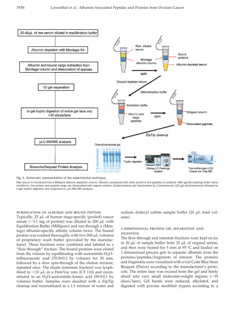
only if they are actively secreted or if they are in vivocleavage products of the parent protein. BRCA2, whichwas sequenced by LC-MS/MS and represented by 4distinct peptides (example BRCA2 spectra from 2 itera-tively found peptides are presented in Fig. 4) and subse-quently validated by competition Western blotting (Fig.5C), is not predicted to be in the blood circulation as anintact protein. In fact, the parent protein with a knownmolecular mass of 390 kDa was not found by serumWestern blotting (Fig. 5D). Shown in Fig. 5C are immu-nocompetition results for 2 cleavage products of BRCA2(�12 and 25 kDa) that were identified by Western blotanalysis of pooled ovarian cancer sera. A rabbit wasimmunized with a synthetic peptide representing aminoacid residues 980–993 (DKIPEKNNDYMNKW) of theBRCA2 protein to produce a polyclonal antibody thatultimately detected the BRCA2 fragments in serum. Com-petition with the cognate peptide completely extin-guished the 12- and 25-kDa bands. The estimated massesof the immuno-fragments (12 and 25 kDa) represent �9%by mass of the total parent protein. Furthermore, 3 otheradditional and distinct peptides were sequenced (residues1942–1959, 2390–2401, and 2447–2459) in a separate re-gion of the parent protein. The close proximity of thepeptides containing amino acids 2390–2401 and 2447–2459 suggests that they likely originate from a singleserum fragment of BRCA2 (Fig. 5, A and B).
Candidate proteins bound to albumin (Table 1; see also
Fig. 3. Functional categories of the predicted albumin-associated proteins identified for each of the stage-specific ovarian cancer and high-riskcontrol serum pools.Serum pools are designated as high risk, stage I (stage 1), and stage III-IV (stage 3). IDs, identifications.
Clinical Chemistry 51, No. 10, 2005 1939

Fig. 4. Representative MS/MS spectra of BRCA2 peptides.(A), result for the BRCA2 peptide identified as MTLGQDLKSDISLNIDK. (B), result for the BRCA2 peptide identified as VSKISPCDVSLETSDICK.
1940 Lowenthal et al.: Albumin-Associated Peptides and Proteins from Ovarian Cancer
Tables 1a–1c of the online Data Supplement) are appar-ently diverse in origin and span a wide variety of pre-dicted physiologic functions (Fig. 3). The breadth ofbiological and functional classes ranges from defense andimmunity to transcription regulation, apoptosis, and celladhesion. More than one half of the predicted proteinsthat have been sequenced have no known function re-ported in the literature. The relative abundances of thedifferent functional classes of those proteins found inovarian cancer samples are shown in Fig. 3. The diversityof categories represented is characteristic of a typicalserum proteome (23, 26). The ovarian cancer serum pro-teins that have been identified are listed in Tables 1a–1c ofthe online Data Supplement, and are shown classified aseither multiple-peptide or single-peptide identifications.A sequence for CA125, a known ovarian cancer marker
(33, 34), was predicted to exist in the stage III ovariancancer pool, but it was not found in either the stage I orthe high-risk serum pools.
DiscussionThe data presented here indicate that high-abundanceserum carrier proteins such as albumin may act to seques-ter low–molecular-weight peptide fragments in the blood.Such sequestered peptides may provide a potentially richsource of candidate disease-associated biomarkers forsubsequent clinical validation and provide a new oppor-tunity to expand the knowledge base for the molecularcomposition of the circulation. Although the present workemphasizes the use of albumin, nonalbumin carrier pro-teins, such as IgG or fibronectin, are associated with adistinct subproteome within human sera (23 ). The
Fig. 5. Confirmation of the presence of BRCA2 peptide in serum.(A), locations of the 4 peptide sequences of BRCA2 found by MS and the serum pool population origins. (B), synthetic peptide corresponding to the amino acid sequenceoverlapping with the sequence found by MS was used as a blocking agent in immunocompetition Western blotting. All sequenced peptides are shown in a spatialorientation within native BRCA2. (C), peptide identification by MS was confirmed by competition Western blot analysis using the peptide shown in B. Western blots ofpooled ovarian cancer are displayed in a dilution curve. Top row, standard Western blot using a polyclonal anti-rabbit primary antibody synthesized in housecorresponding to the antigenic region of BRCA2 found by MS. Middle row, the primary antibody is first incubated with a competition peptide specific for the identicalamino acid region of BRCA2 before Western blotting. Bottom row, negative control/background of the goat anti-rabbit secondary. The BRCA2 peptides highlighted areevident at �12 and 25 kDa. The indication of peptide fragments in Western blots of serum is expected because the molecular mass of the parent BRCA2 protein is390 kDa, an apparent mass much larger than what is known to be able to passively enter the blood stream. (D), Western blot using polyclonal anti-BRCA 2 antibodiesshowing 2 example ovarian cancer sera (OS1 and OS2) compared with total nuclear lysate of HeLa cells (HNL). Absence of the full-length BRCA2 protein in the serais shown.
Clinical Chemistry 51, No. 10, 2005 1941

method of albumin capture, elution of bound peptide andprotein fragments, fractionation, digestion, and MS se-quencing was repeated on 5 independent aliquots of eachdisease category serum pool. Because shown in Table 1,this iterative-sequencing approach provided a means toassess sequence discovery reproducibility within thesame serum pool sample. Iterative sample sequencingindicated that after 5 rounds of iterations, the yield ofnovel multiple-peptide predictions not identified in pre-vious iterations appeared to increase more slowly. Thiswould be expected if the peptide sequences are beingrandomly resampled from the same large population oflimited original candidates.
In theory, a similar list of protein fragments could begenerated by denaturing and size-fractionating of serabecause albumin fractionation and isolation would occurfirst. This is, in fact, exactly what previous investigatorsdid in their analyses (17, 18). However, because manycurrent biomarker efforts begin by depletion of nativealbumin (as well as depletion of other high-abundanceproteins such as immunoglobulins), the findings of thisstudy begin to more fully describe the molecular informa-tion that is being lost by this method of sample prepara-tion. Moreover, direct analysis of the albumin-boundmaterial allows for clearer understanding of the natureand existence of the carrier-protein–bound low–molecu-lar-weight information archive while minimizing issuesof small amounts of free/unbound material contaminat-ing the analysis.
The total list of identified albumin-bound entities areshown in Tables 1a–1c of the online Data Supplement(http://www.clinchem.org/content/vol51/issue10/). Thislist reveals a rich and previously undescribed informationarchive of putative analytes, many of which had not beendescribed as being represented in the circulation. Thesemolecules are mostly fragments of larger proteins; thus, theirvalidation will necessitate the development of new immu-nodetection reagents such as anti-peptide antibodies.
It is well known that highly abundant proteins areidentified by a larger number of peptides than are lower-abundance proteins. The probability of selecting andidentifying a peptide from a low-abundance, and poten-tially more interesting, protein is therefore much less. Theinherent complexity and large dynamic range of proteinconcentrations of global proteome samples represents aconsiderable barrier to obtaining multiple peptide identi-fications for each protein. The large number of peptidespresent in such mixtures greatly exceeds the capacity ofcurrent data-dependent tandem mass spectrometers, evenwhen multidimensional fractionation is used (4, 35, 36).In the present method we have endeavored to reduce thisbarrier by isolating the major high-abundance proteinalbumin and then examining the identity of its boundspecies.
Predicted sequences according to the criteria outlinedin the Materials and Methods section fell into 2 categories:(a) multiple peptide hits, and (b) single peptide hits.
Multiple peptide hits according to disease pool categoryare presented in Table 1a of the online Data Supplementby protein identity. Single peptide hits are listed in Table1b and 1c of the online Data Supplement, along with thecorresponding specific predicted sequences. Low-abun-dance proteins in blood are statistically likely to bediscovered by only a single peptide sequence as a func-tion of the dynamic range of proteins in serum; conse-quently, their probability of detection is low. Further-more, many of the predicted serum species are peptidefragments of larger parent proteins. This can be concludedbecause a high percentage of the predicted proteins inTables 1a–1c of the online Data Supplement have afull-length mass larger than albumin. Peptide fragmentshave reduced numbers of trypsin domains comparedwith the intact parent protein. Thus, a single peptideprediction, if correct, can correspond to a low-abundancepeptide or a protein fragment with a reduced number oftrypsin domains compared with the parent molecule.Because we conducted 5 independent isolation and se-quence determinations for each disease category, singlehits that were discovered more than once could be tabu-lated (Table 1b of the online Data Supplement). Singlepeptide hits discovered more than once may have ahigher probability of being valid serum biomarkers thando single-instance single peptide hits.
The relevance of single peptide hits has been addressedrecently (37 ). These investigators pointed out that theelimination of single peptide hits from experimental re-sults is scientifically unwarranted and detrimental to thefield in general (37 ). In fact, when so-called “one-hitwonders” identified by isotope-coded affinity tag pro-teomics were later assessed for validation by immunoas-say detection, 90% were found to retain the differentialexpression state determined by the initial MS-based assay.Moreover, in the present study, many of the same single-hit tryptic fragments were iteratively and reproduciblyfound in the same disease category. Finally, for small,low-abundance fragments (with a low number of trypsincleavage sites), it is statistically likely that only a single hitwould be obtained in any given experimental cycle.
Although we identified 4 separate fragments forBRCA2, which increased our confidence in the finding,the fact that a majority of our protein identifications werebased on one-hit MS analysis does not diminish theirpotential for potential validation. It is well known thathighly abundant proteins are identified by a larger num-ber of peptides than are lower-abundance proteins. Theprobability of selecting and identifying a peptide from alow abundance, and potentially more interesting, proteinis therefore much less. The inherent complexity and largedynamic range of protein concentrations of global pro-teome samples represents a considerable barrier to obtain-ing multiple-peptide identifications for each protein. Thelarge number of peptides present in such mixtures greatlyexceeds the capacity of current data-dependent tandemmass spectrometers, even when multidimensional frac-
1942 Lowenthal et al.: Albumin-Associated Peptides and Proteins from Ovarian Cancer

tionation is used. Moreover, global proteomic studieshave consistently shown a low false-positive rate (i.e.,�5%) for peptide identification; therefore; for every incor-rect one-hit wonder that is thrown out, �19 correctidentifications are also excluded. Excluding one-hit won-ders from possible validation would decimate quantita-tive proteomics studies that use isotope-coded affinity tagreagents and phosphoproteomic experiments, which typ-ically produce only single phosphopeptide identifications.Many investigations have performed orthogonal valida-tion experiments (e.g., Western analysis) that have con-firmed that these qualitative and quantitative proteomicresults forthcoming from one-hit wonders are indeedvalid identifications (27, 38–42).
Ultimately all predicted peptide sequences, whethersingle or multiple, must remain designated as “candi-date” components of the human serum until they areconfirmed immunologically. Many of the predicted se-quences were derived from molecular-weight fractionssmaller than the predicted size of the protein. We there-fore concluded that the sequenced species existed as afragment of a larger parent molecule. Thus, the gelmigration location of the band could not be used toidentify the protein. Western blotting with peptide com-petition was performed on the selected fragments identi-fied by MS as belonging to BRCA2 to verify the identity ofselected mass spectrometric identifications (Fig. 5C).
BRCA2 is a well-studied tumor suppressor proteinrelated to the p53 pathway that is directly implicated infamilial breast cancer and ovarian cancer (43–46). Al-though the role of BRCA2 in breast and ovarian cancerpredisposition is poorly understood, it is known thatmutations of the BRCA2 gene are responsible for one thirdof hereditary breast cancer cases (45 ). The data in Fig. 5suggest that at least 2 fragments of the BRCA2 parentprotein exist in blood. A single predicted trypsin-cleavedpeptide at amino acids 965–981 was identified in bothstage I and stage III ovarian cancer serum pools (Fig. 4Aand Fig. 5A). This peptide is represented in competitionWestern blots by 2 distinct molecular-weight bands (Fig.5C). It is likely that a fragment containing the peptide atamino acids 965–981 would be cleaved at various residuesin vivo and could therefore be represented at multiplemolecular weights. This peptide was not predicted inpooled high-risk ovarian cancer serum by MS analysis.On the basis of evidence from the Western blots using aBRCA2 polyclonal antibody and the fact that BRCA2 istoo large (390 kDa) to enter the blood circulation in itsnative form, we conclude that BRCA2 can be representedin the serum as one or more fragments and that at least 2amino-terminal peptide fragments of BRCA2 �25 kDacan be validated by peptide competition for antibodiesthat recognize an amino acid sequence adjacent to, andoverlapping with, the predicted peptide sequence. Addi-tionally, 2 separate BRCA2 fragments encompassing noless than the amino acid region from 2390–2459 and1942–1959 (Fig. 4B) are predicted to exist in the serum
from mass spectral evidence of multiple peptide se-quences identified in this region of BRCA2.
The current methodology applies carrier-protein se-questering to a disease category pool of combined sera.The methodology described here appears to be a success-ful means to identify candidate proteins and peptides inthe sera of patients with known disease states. On thebasis of the predicted physiologic functions of the paren-tal protein containing the peptide sequence, these proteinsand peptides can potentially be derived from tumor cells,from the host microenvironment, or from interactionsbetween these two tissue compartments. Each pool con-tained multiple serum specimens procured before pathol-ogy-based diagnosis. The rationale for using a pool ofmultiple serum samples is based on the conservativeassumption that even within the same histopathologicdiagnosis, cancer is a heterogeneous disease. Thus, abiologically relevant molecule may be expressed onlywithin a subfraction of the cancer population. Poolingsamples is statistically the best means to identify a list ofcandidate peptides and proteins that exist within subsetsof the pooled population.
The first disadvantage of using a pool of sera todiscover markers is that molecules present in a subset ofthe combined samples are diluted within the pool. Al-though an individual sample may have a high concentra-tion of the putative protein or peptide, this sample isdiluted within the entire pool. Consequently, this averag-ing effect will lower the concentration of any individualmolecule in the pool before sequence analysis. Lastly, acandidate protein that is identified in one disease pool(e.g., high risk) but not found in a second disease category(e.g., stage I cancer) may still exist in the second categoryat a concentration below a threshold for statistical sam-pling probability. Currently we are performing competi-tion Western blots on a large set of serum samples fromindividual patients diagnosed as either high risk or withstage I, III, or IV ovarian cancer to determine the extent bywhich pooling samples limits the effectiveness of MSsequencing detection. Despite these drawbacks, as shownin Table 1 and in Tables 1a–1c of the online Data Supple-ment, carrier-protein sequestration appears to yield a listof predicted sequences derived from proteins predicted inthe literature to have diverse physiologic functions.
The predicted sequences presented in Tables 1a–1c ofthe online Data Supplement demonstrate a rich diversityof peptides and protein fragments in the sera. Althoughthe application of this method yielded different sets ofpredicted sequences within the control and diseasedcategory pools, these proteins and peptides cannot beconsidered as candidate diagnostic markers. Once high-throughput means are developed to quantitatively mea-sure each protein and peptide and accurately distinguishspecific biomarker fragments from their parent molecules,proper clinical trial studies can examine the diagnosticsensitivity and specificity of a selected panel of molecules.
Clinical Chemistry 51, No. 10, 2005 1943

In conclusion, proteins and peptides associated withserum carrier proteins such as albumin may constitute arich source of new additions to the human serum pro-teome. The sequences identified by this means are pre-dicted to be derived from proteins that have been shownin the literature to serve a wide variety of physiologicfunctions. We can hypothesize that these complexed pro-teins and peptides may have originated from a range ofdifferent tissues and cellular compartments. A large pro-portion of the predicted sequences represent fragments oflarger molecules. A subset of these candidates may even-tually be found suitable for full clinical diagnostic testingin adequately powered objective study sets.
References1. Ardekani AM, Liotta LA, Petricoin EF 3rd. Clinical potential of
proteomics in the diagnosis of ovarian cancer. Expert Rev MolDiagn 2002;2:312–20.
2. Anderson NL, Anderson NG. The human plasma proteome: history,character, and diagnostic prospects. Mol Cell Proteomics 2002;1:845–67.
3. Mehta AI, Ross S, Lowenthal MS, Fusaro V, Fishman DA, PetricoinEF 3rd, et al. Biomarker amplification by serum carrier proteinbinding. Dis Markers 2003;19:1–10.
4. Adkins JN, Varnum SM, Auberry KJ, Moore RJ, Angell NH, SmithRD, et al. Toward a human blood serum proteome: analysis bymultidimensional separation coupled with mass spectrometry.Mol Cell Proteomics 2002;1:947–55.
5. Pieper R, Su Q, Gatlin CL, Huang ST, Anderson NL, Steiner S.Multi-component immunoaffinity subtraction chromatography: aninnovative step towards a comprehensive survey of the humanplasma proteome. Proteomics 2003;3:422–32.
6. Rothemund DL, Locke VL, Liew A, Thomas TM, Wasinger V, Rylatt,DB. Depletion of the highly abundant protein albumin from humanplasma using the Gradiflow. Proteomics 2003;3:279–87.
7. Wang YY, Cheng P, Chan DW. A simple affinity spin tube filtermethod for removing high-abundant common proteins or enrichinglow-abundant biomarkers for serum proteomic analysis. Proteom-ics 2003;3:243–8.
8. Pieper R, Gatlin CL, Makusky AJ, Russo PS, Schatz CR, Miller SS,et al. The human serum proteome: display of nearly 3700chromatographically separated protein spots on two-dimensionalelectrophoresis gels and identification of 325 distinct proteins.Proteomics 2003;3:1345–64.
9. Dennis MS, Zhang M, Meng YG, Kadkhodayan M, Kirchhofer D,Combs D, et al. Albumin binding as a general strategy forimproving the pharmacokinetics of proteins. J Biol Chem 2002;277:35035–43.
10. Lee VHL. Peptide and protein drug delivery. New York: MarcelDekker, 1990:1–56.
11. Makrides SC, Nygren PA, Andrews B, Ford PJ, Evans KS, HaymanEG, et al. Extended in vivo half-life of human soluble complementreceptor type 1 fused to a serum albumin-binding receptor.J Pharmacol Exp Ther 1996;277:534–42.
12. Kurtzhals P, Havelund S, Jonassen I, Kiehr B, Larsen UD, Ribel U,et al. Albumin binding of insulins acylated with fatty acids:characterization of the ligand-protein interaction and correlationbetween binding affinity and timing of the insulin effect in vivo.Biochem J 1995;15:725–31.
13. Markussen J, Havelund S, Kurtzhals P, Andersen AS, Halstrom J,Hasselager E, et al. Soluble, fatty acid acylated insulins bind toalbumin and show protracted action in pigs. Diabetologia 1996;39:281–8.
14. Gordon LM, Curtain CC, McCloyn V, Kirkpatrick A, Mobley PW, etal. The amino-terminal peptide of HIV-1 gp41 interacts with humanserum albumin. AIDS Res Hum Retroviruses 1993;9:1145–56.
15. Sjobring U, Falkenberg C, Nielsen E, Akerstrom B, Bjorck L.Isolation and characterization of a 14-kDa albumin-binding frag-ment of streptococcal protein G. J Immunol 1988;140:1595–9.
16. Raghu P, Sivakumar B. Interactions amongst plasma retinol-binding protein, transthyretin, and their ligands: implications invitamin A homeostasis and transthyretin amyloidosis. BiochimBiophys Acta 2004;1703:1–9.
17. Tirumalai RS, Chan KC, Prieto DA, Issaq HJ, Conrads TP, VeenstraTD. Characterization of the low molecular weight human serumproteome. Mol Cell Proteomics 2003;2:1096–103.
18. Zhou M, Lucas DA, Chan KC, Issaq HJ, Petricoin EF 3rd, Liotta LA,et al. An investigation into the human serum “interactome”.Electrophoresis 2004;25:1289–98.
19. Corot C, Violas X, Robert P, Gagneur G, Port M. Comparison ofdifferent types of blood pool agents (P792, MS325, USPIO) in arabbit MR angiography-like protocol. Invest Radiol 2003;38:311–9.
20. Dufour C, Dangles O. Flavonoid-serum albumin complexation:determination of binding constants and binding sites by fluores-cence spectroscopy. Biochim Biophys Acta 1721:164–73, 2005.
21. Ding Y, Lin B, Huie CW. Binding studies of porphyrins to humanserum albumin using affinity capillary electrophoresis. Electro-phoresis 2001;22:2210–6.
22. Klasco RK, ed. United States pharmacopoeia DI, Vol. 1. Druginformation for the health care professional. United States Phar-macopeial Convention, Inc. Greenwood Village, CO: ThomsonMicromedex, 2000.
23. Peters T Jr. Serum albumin. In: Putnam FW, ed. The plasmaproteins: structure, function, and genetic control, Vol. 1, 2nd ed.New York: Academic Press, 1975:133–81.
24. Rainey TG, Read CA. Pharmacology of colloids and crystalloids,the pharmacologic approach to the critically ill patient, Vol. 1, 3rded. Baltimore: Williams & Wilkins, 1994:272–90.
25. Wagner BK, D’Amelio LF. Pharmacologic and clinical consider-ations in selecting crystalloid, colloidal, and oxygen-carrying re-suscitation fluids, part 1. Clin Pharm 1993;12:335–46.
26. Kinter M, Sherman NE. Protein sequencing and identificationusing tandem mass spectrometry. New York: Wiley-Interscience,Inc., 2000:301pp � xvi.
27. Peng J, Elias JE, Thoreen CC, Licklider LJ, Gygi SP. Evaluation ofmultidimensional chromatography coupled with tandem massspectrometry (LC/LC-MS/MS) for large-scale protein analysis: theyeast proteome. J Proteome Res 2003;2:43–50.
28. Qian WJ, Liu T, Monroe ME, Strittmatter EF, Jacobs JM, Kangas LJ,et al. Probability-based evaluation of peptide and protein identifi-cations from tandem mass spectrometry and SEQUEST analysis:the human proteome. J Proteome Res 2005;4:53–62.
29. Wilmarth PA, Riviere MA, Rustvold DL, Lauten JD, Madden TE,David LL. Two-dimensional liquid chromatography study of thehuman whole saliva proteome. J Proteome Res 2004;3:1017–23.
30. Xiang R, Shi Y, Dillon DA, Negin B, Horvath C, Wilkins JA. 2DLC/MS analysis of membrane proteins from breast cancer celllines MCF7 and BT474. J Proteome Res 2004;3:1278–83.
31. Goldsmith P, Backlund PS Jr, Rossiter K, Carter A, Milligan G,Unson CG, et al. Purification of heterotrimeric GTP-binding pro-teins from brain: identification of a novel form of Go. Biochemistry1988;27:7085–90.
32. Liu H, Sadygov RG, Yates JR. A model for random sampling andestimation of relative protein abundance in shotgun proteomics.Anal Chem 2004;76:4193–201.
33. Moss EL, Hollingworth J, Reynolds TM. The role of CA125 inclinical practice. J Clin Pathol 2005;58:308–12.
1944 Lowenthal et al.: Albumin-Associated Peptides and Proteins from Ovarian Cancer

34. Topley N, Michael D, Bowen T. CA125: Holy Grail or a poisonedchalice. Nephron Clin Pract 2005;100:c52–c54.
35. Washburn MP, Wolters D, Yates JR 3rd. Large-scale analysis ofthe yeast proteome by multidimensional protein identificationtechnology. Nat Biotechnol 2001;19:242–7.
36. Yu LR, Conrads TP, Uo T, Morrison RS, Chan K, Lucas DA, et al.Global analysis of the cortical neuron proteome. Mol Cell Proteom-ics 2004;3:896–907.
37. Veenstra TD, Conrads TP, Issaq HJ. What to do with “one-hitwonders”? Electrophoresis 2004;25:1278–9.
38. Johnson MD, Yu L-R, Conrads TP, Kinoshito Y, Uo T, Lee S-W, etal. Proteome analysis of DNA damage-induced neuronal deathusing high throughput mass spectrometry. J Biol Chem 2004;279:26685–97.
39. Conrads KA, Yi M, Simpson KA, Stevens R, Lucas DA, VeenstraTD, et al. A combined proteome and microarray investigation ofinorganic phosphate-induced pre-osteoblast cells. Mol Cell Pro-teomics 2005;Jun 14 [Epub ahead of print].
40. Ballif BA, Villen J, Beausoleil SA, Schwartz D, Gygi SP. Phospho-proteomic analysis of the developing mouse brain. Mol CellProteomics 2004;3:1093–101.
41. Hardwidge PR, Rodriguez-Escudero I, Goode D, Donohoe S, Eng J,Goodlett DR, et al. Proteomic analysis of the intestinal epithelialcell response to enteropathogenic Escherichia coli. J Biol Chem2004;279:20127–36.
42. Anderson L. Candidate-based proteomics in the search for biomar-kers of cardiovascular disease, J Physiol 2005;563:23–60.
43. Jonkers J, Meuwissen R, van der Gulden H, Petersen H, van derValk M, Berns A. Synergistic tumor suppressor activity of BRCA2and p53 in a conditional mouse model for breast cancer. NatGenet 2001;29:418–25.
44. Esashi F, Christ N, Gannon J, Liu Y, Hunt T, Jasin M, et al.CDK-dependent phosphorylation of BRCA2 as a regulatory mech-anism for recombinational repair. Nature 2005;434:598–604.
45. Ford D, Easton DF, Stratton M, Narod S, Goldgar D, Devilee P, etal. Genetic heterogeneity and penetrance analysis of the BRCA1and BRCA2 genes in breast cancer families. The breast cancerlinkage consortium. Am J Hum Genet 1998;62:676–89.
46. Hussain S, Witt E, Huber P, Medhurst A, Ashworth A, Mathew C.Direct interaction of the Fanconi anaemia protein FANCG withBRCA2/FANCD1. Hum Mol Genet 2003;12:2503–10.
Clinical Chemistry 51, No. 10, 2005 1945