Embed Size (px)
Citation preview
1
Anatomy of the Lungs
Dr. Gondo GozaliDepartment of anatomy
2
Pulmonary Function Ventilation and Respiration Ventilation is the movement of air in
and out of the lungs Respiration is the process of gas
exchange, that is the movement of O2 from the atmosphere into the blood stream and the movement CO2 from the bloodstream into the atmosphere.
3
Pulmonary System
Consist of: Thorax Conducting airways Respiratory airways Pulmonary blood and lymph supply
4
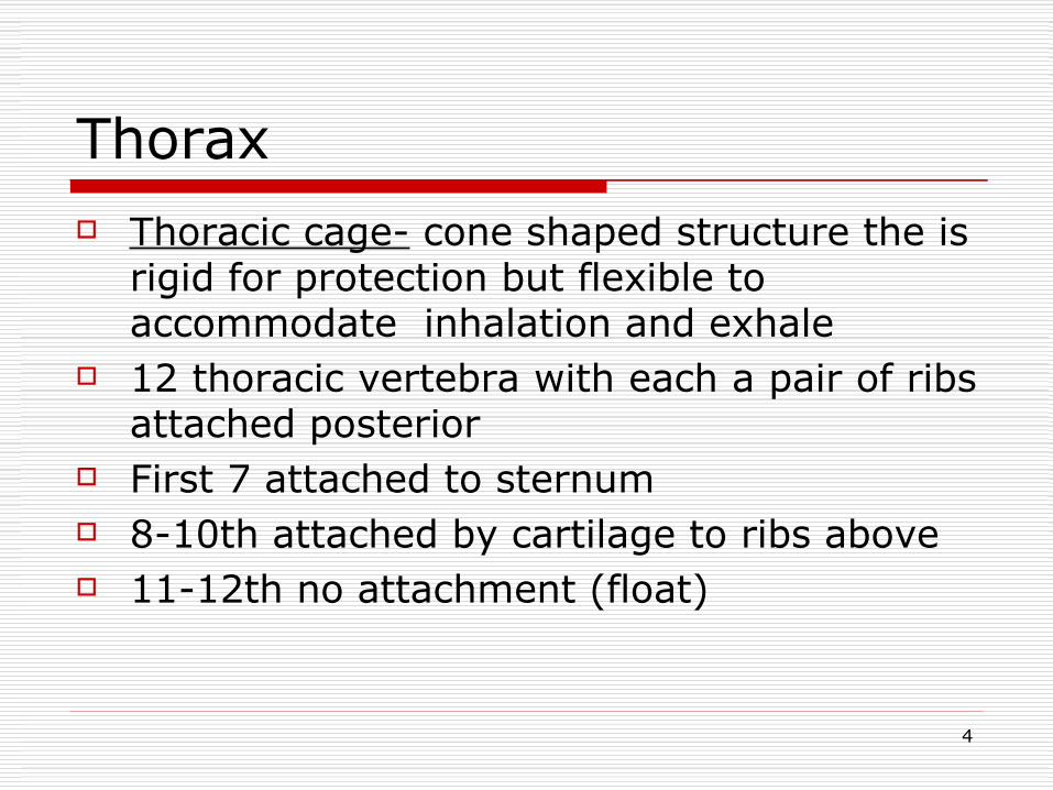
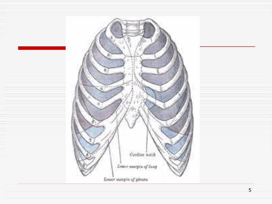
Thorax Thoracic cage- cone shaped structure the is
rigid for protection but flexible to accommodate inhalation and exhale
12 thoracic vertebra with each a pair of ribs attached posterior
First 7 attached to sternum 8-10th attached by cartilage to ribs above 11-12th no attachment (float)
5
6
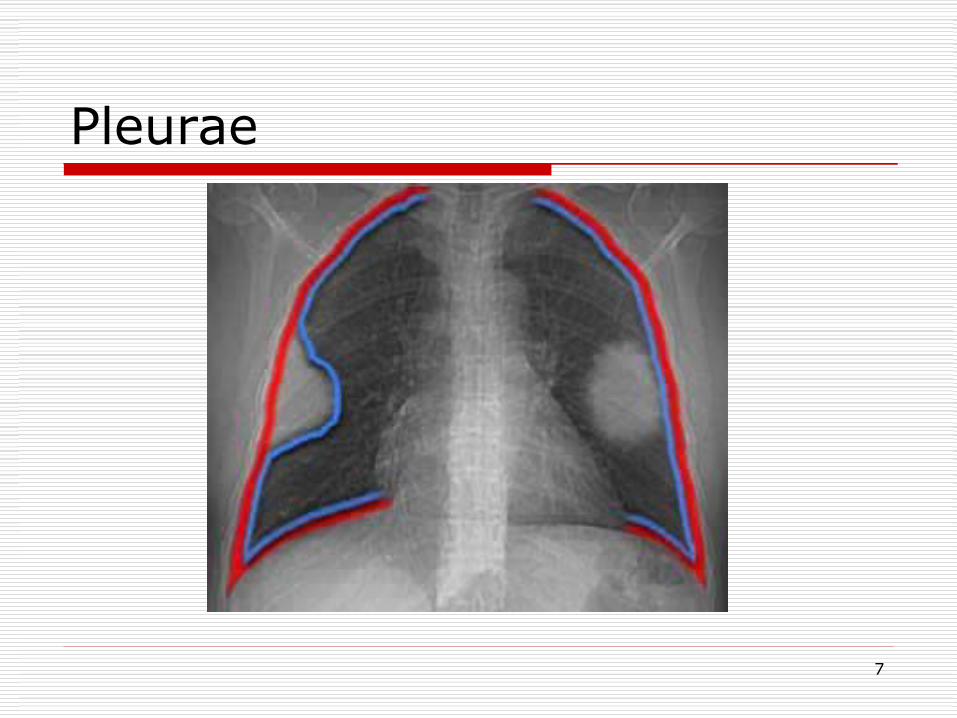
Pleurae Thin membrane that lines the outside of the
lungs and the inside of the chest wall Visceral pleura-adheres to the lungs, hilar
bronchi and major fissures Parietal pleura-lines the inner surface of
chest wall and mediastinum Separated by airtight space that contains a
lubricating fluid that allows the two pleura to glide against each other during breathing
7
Pleurae
8
Innervations of the pleura Parietal : Sensitive to pain
and temperature Innervated by the
intercostal nerves and phrenic nerve
Visceral : Not sensitive to
pain and temperature
Some sensory fibers travel with autonomic nerves that innervate the lungs
9
Vasculature Parietal
→ Intercostal→ Internal thoracic→ Pericardiacophereni
cs→ Anterior mediastinal
artery and veins
Visceral→ Bronchial vessels→ Pulmonary trunks
10
Lymphatic Parietal pleura
drain with those of the thoracic wall
Visceral pleura drain with lymphatics from the lung
11
Conducting Airways Upper airways-nasal and oral cavities, pharynx and
larynx Warms, Humidifies, and filters some irritants. Course
hairs in the nasal passage cleans the air of large inhaled particles during inhalation.
Epiglottis-Closes during swallowing of food or liquid to protect the trachea. Opens widely during inhalation to allow air to pass into the trachea onto the lower air passages
Trachea-hollow tube 11cm(4.5 in) in length and 2.5 cm (1in) in width Begins with the cricoid cartilage and ends at the bifurcation(major carina) from which the two mainstem bronchi arise.
12
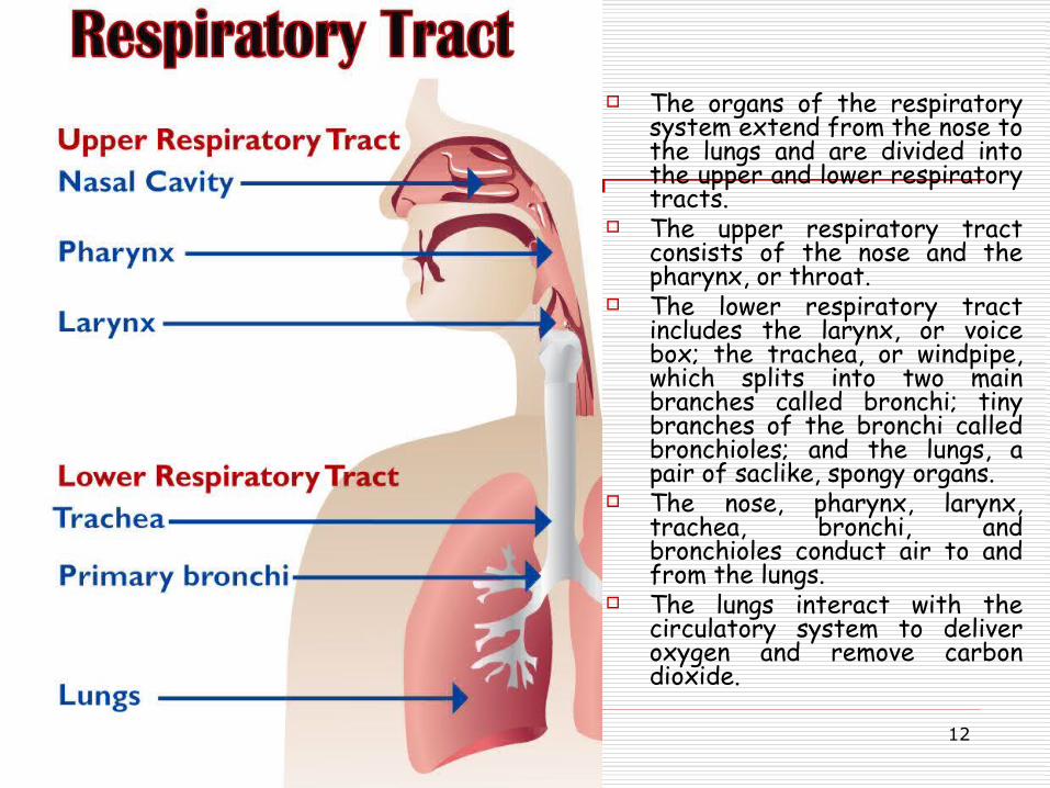
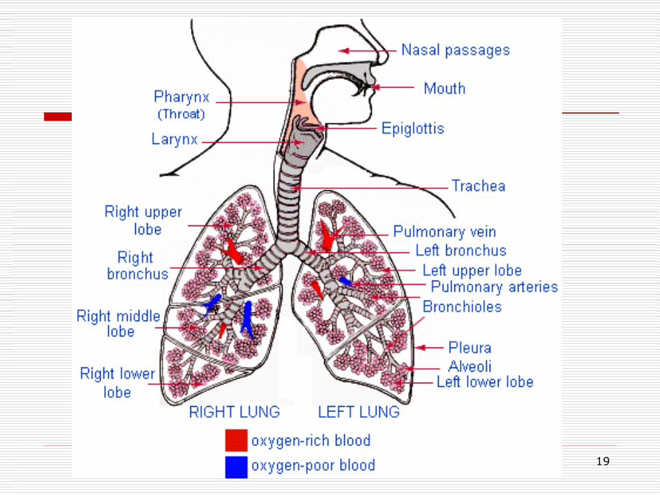
The organs of the respiratory system extend from the nose to the lungs and are divided into the upper and lower respiratory tracts.
The upper respiratory tract consists of the nose and the pharynx, or throat.
The lower respiratory tract includes the larynx, or voice box; the trachea, or windpipe, which splits into two main branches called bronchi; tiny branches of the bronchi called bronchioles; and the lungs, a pair of saclike, spongy organs.
The nose, pharynx, larynx, trachea, bronchi, and bronchioles conduct air to and from the lungs.
The lungs interact with the circulatory system to deliver oxygen and remove carbon dioxide.
13
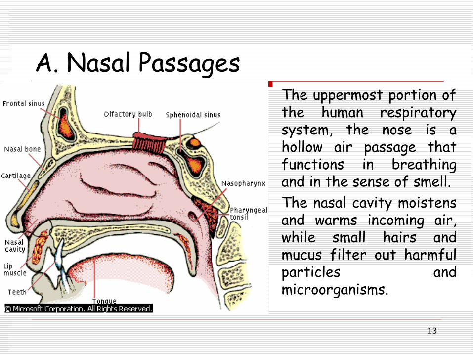
A. Nasal Passages The uppermost portion of
the human respiratory system, the nose is a hollow air passage that functions in breathing and in the sense of smell.
The nasal cavity moistens and warms incoming air, while small hairs and mucus filter out harmful particles and microorganisms.
14
B. Pharynx Air leaves the nasal passages and flows to the pharynx, a short, funnel-
shaped tube about 13 cm (5 in) long that transports air to the larynx. Like the nasal passages, the pharynx is lined with a protective mucous membrane and ciliated cells that remove impurities from the air.
The tonsils are strategically located to prevent these organisms from moving further into the body. One tonsil, called the adenoids, is found high in the rear wall of the pharynx. A pair of tonsils, the palatine tonsils, is located at the back of the pharynx on either side of the tongue.
Another pair, the lingual tonsils, is found deep in the pharynx at the base of the tongue. In their battles with disease-causing organisms, the tonsils sometimes become swollen with infection. When the adenoids are swollen, they block the flow of air from the nasal passages to the pharynx, and a person must breathe through the mouth.
15
C. Larynx Air moves from the pharynx to the larynx, a structure about
5 cm (2 in) long located approximately in the middle of the neck.
While the primary role of the larynx: Transport air to the trachea Producing sound Prevents food and fluid from entering the air passage to
cause choking Mucous membranes and cilia-bearing cells help filter air
When a person is breathing, the epiglottis is held in a vertical position, like an open trap door. When a person swallows, however, a reflex causes the larynx and the epiglottis to move toward each other, forming a protective seal, and food and fluids are routed to the esophagus.
16
D. Trachea, Bronchi, and Bronchioles
Air passes from the larynx into the trachea, a tube about 12 to 15 cm (about 5 to 6 in) long located just below the larynx. The trachea is formed of 15 to 20 C-shaped rings of cartilage. The sturdy cartilage rings hold the trachea open, enabling air to pass freely at all times. The open part of the C-shaped cartilage lies at the back of the trachea, and the ends of the “C” are connected by muscle tissue.
The base of the trachea is located a little below where the neck meets the trunk of the body. Here the trachea branches into two tubes, the left and right bronchi, which deliver air to the left and right lungs, respectively. Within the lungs, the bronchi branch into smaller tubes called bronchioles.
The trachea, bronchi, and the first few bronchioles contribute to the cleansing function of the respiratory system, for they, too, are lined with mucous membranes and ciliated cells that move mucus upward to the pharynx.
17
LUNGS
Cone shaped Total volume of 3.5-8.5 liters Superior portion is apex Inferior portion is base Apical portion rises above the clavicle Attached by hilum and pulmonary
ligament
18
Lobes and Segments Lungs are divided into lobes and segments Right lung is larger ad heavier: upper
middle and lower lobes Left lung divides into upper and lower
lobes. The lobes have a total of 18 segments:
10 on right and 8 on left - each has own bronchus
19
20
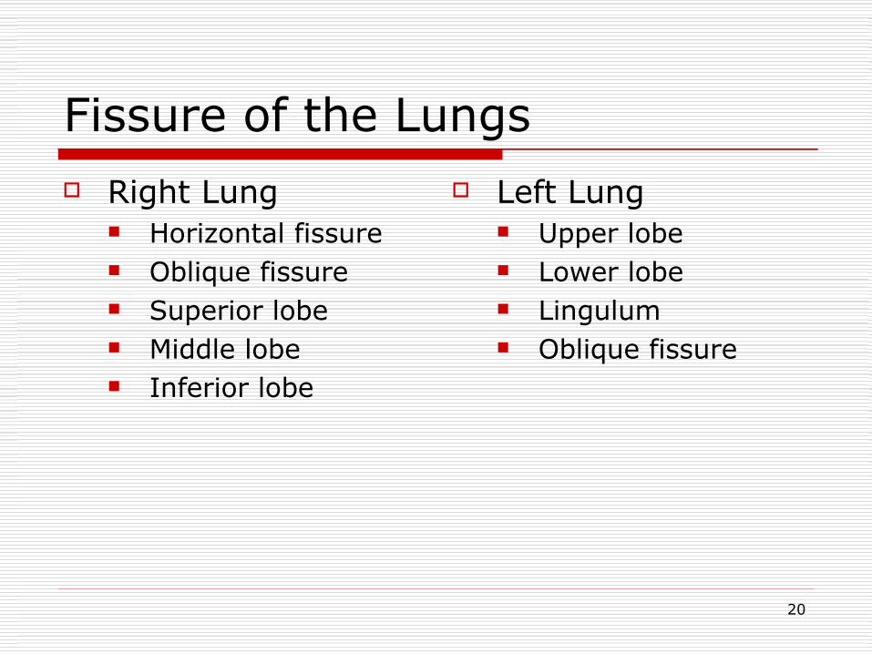
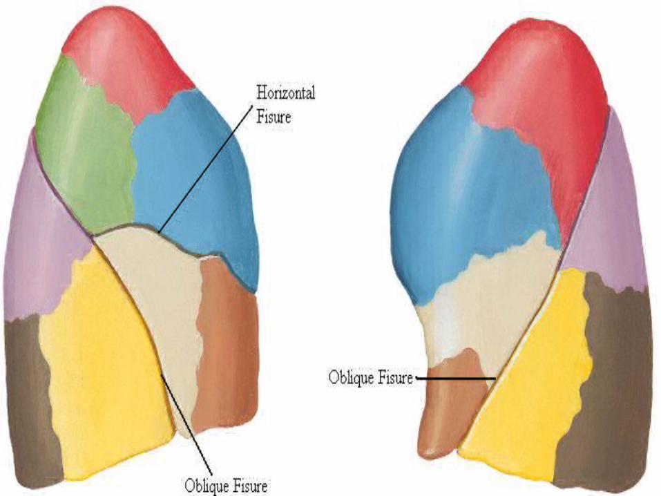
Fissure of the Lungs Right Lung
Horizontal fissure Oblique fissure Superior lobe Middle lobe Inferior lobe
Left Lung Upper lobe Lower lobe Lingulum Oblique fissure
21
22
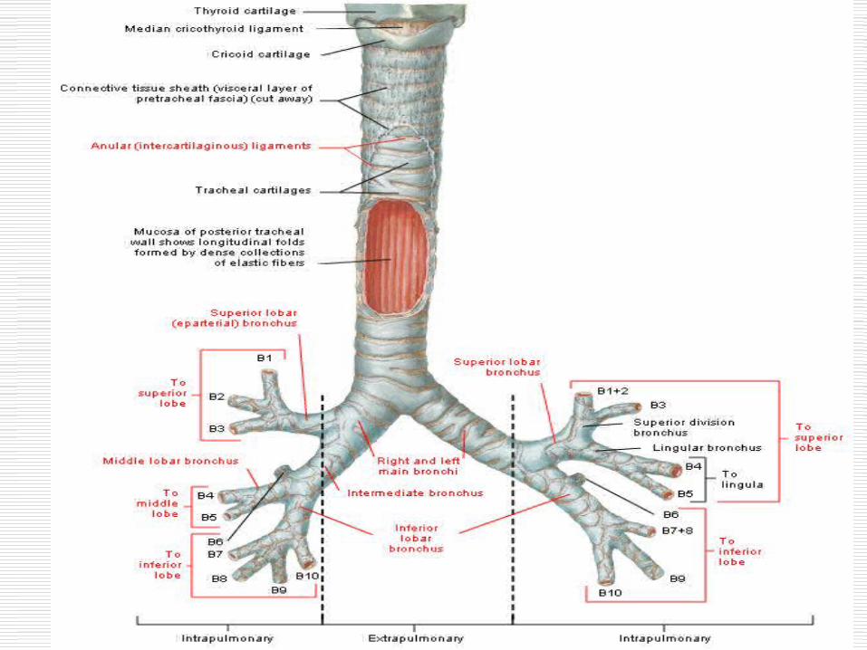
Bronchial Tree
The two main stem bronchi are different
Left bronchi-narrower then right, angles into left lung at approximately 45 to 55 degrees
Right bronchi- wider than left and inters right lung at a 20-30 degrees
23
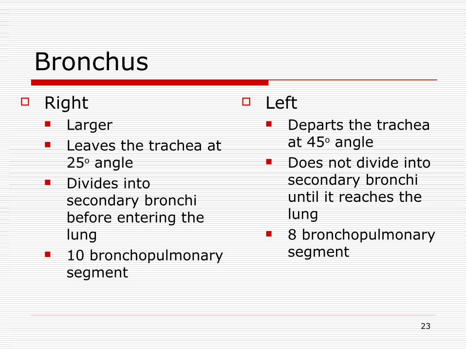
Bronchus Right
Larger Leaves the trachea at
25o angle Divides into
secondary bronchi before entering the lung
10 bronchopulmonary segment
Left Departs the trachea
at 45o angle Does not divide into
secondary bronchi until it reaches the lung
8 bronchopulmonary segment
24
25
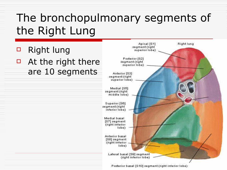
The bronchopulmonary segments of the Right Lung
Right lung At the right there
are 10 segments
26
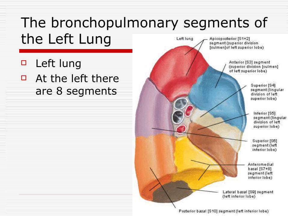
The bronchopulmonary segments of the Left Lung
Left lung At the left there
are 8 segments
27
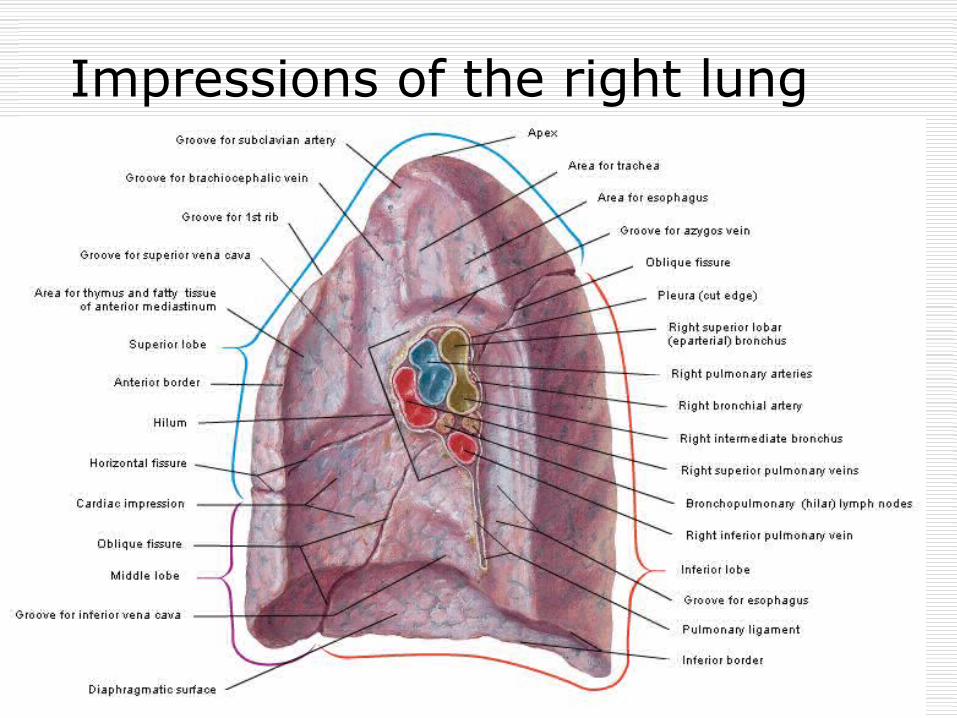
Impressions of the right lung
28
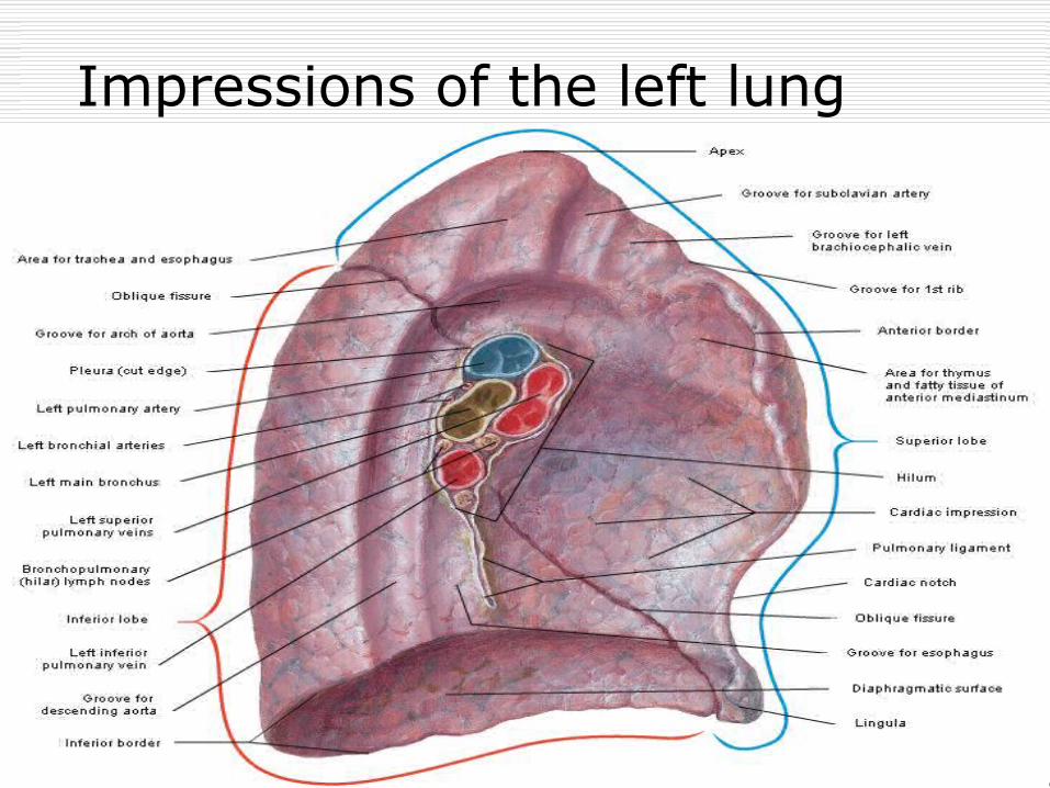
Impressions of the left lung
29
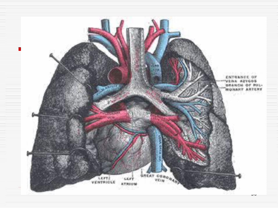
Blood circulation
Pulmonary Circulation Pulmonary artery
carry blood to the lungs for oxygenated. These vessels are central in each bronchopulmonary segment and branch in a manner similar to the segmental bronchi. They are considered segmental in nature.
Pulmonary vein at the periphery of each segment and are
considered intersegmental.
30
31
Innervations of the lungs (1)
Sensory fibers from the lungs and visceral pleura travel with the vagus nerve
Preganglionic parasympathetic fibers travel in the vagus nerve through the pulmonary plexus to the level of the tracheobronchial tree where they synapse with postganglionics.
32
Innervations of the lungs (2) Sympathetic preganglionic from T1-6
synapse with postganglionics in the cervical and upper thoracic ganglia
Postganglionics the descend to the cardiac and pulmonary plexuses
Pulmonary plexus lies on the anterior and posterior aspects of root structure of the lungs.
The anterior and posterior of the lungs receive fibers from vagus and cardiac plexus
Posterior also receive from the thoracic sympathetic ganglion
33
Parasympathetic and sympathetic fibers of the Lungs
Parasympathetic fibers of the lungs: Vasodilators Bronchoconstrictors Secremotor in
function
Sympathetic fibers of the lung: Vasoconstrictor Bronchodilator Decreased
secretory function of mucus bronchial glands
34
Surface projections of the lungs Right Lung
Apex – 1 inch above medial 1/3 of the clavicle
Anterior border – slopes toward the midline, curves out between 4th and 6th costal cartillage
Inferior border – 6th costal cartillage and the lower end of sternum, 6th rib at the midclavicular line, 8th rib at the midaxillary line, 10th thoracic vertebra at the paravertebral line
Left Lung Same as right, except
the cardiac notch cruves 11/2 inches lateral to the sternum in the 5th ICS
Fissures Oblique – extends from
the spine of the scapula to the 6th costal cartilage
Horizontal – posterior to the 4th rib

35
T h a n k Y o u