Embed Size (px)
Citation preview
Published in final edited form as:
ACS Applied Materials & Interfaces, 2018, doi:10.1021/acsami.8b12623
1
Antibacterial Surface Coating for Bone Scaffolds Based on the Dark Catalytic Effect of Titanium Dioxide
David Wiedmera, Chen Cuia, Florian Webera, Fernanda C. Petersenb, Hanna Tiainena,*
aDepartment of Biomaterials, Institute for Clinical Dentistry, University of Oslo, Norway
bDepartment of Oral Biology, Faculty of Dentistry, University of Oslo, Norway
*Corresponding author
Tel.: +47-22852354
Email: [email protected]
PO Box 1109 Blindern
0317 Oslo
Norway
Keywords: Titanium dioxide, porous scaffold, antibacterial coating, dark catalysis, sol-gel coating,
H2O2 decomposition
Wiedmer et al. ACS Applied Materials & Interfaces
2
Abstract
Biomaterials which promote tissue integration and resist microbial colonisation are required in
bone tissue engineering to prevent biomaterial-associated infections. Surface modification of
established materials for bone tissue engineering, such as TiO2, have emerged as promising anti-
infective strategies. Interestingly, the antibacterial activity of TiO2 in the form of particles can be
enhanced by combining it with H2O2, even in the absence of irradiation. However, it remains
unknown whether TiO2 surfaces elicit a similar effect. In this study, the antibacterial effect of
porous TiO2 scaffolds generated by the catalytic decomposition of H2O2 in the absence of light
(dark catalysis) was investigated. Porous ceramic foams were fabricated and sol-gel coated for high
catalytic activity. Degradation of methylene blue in the presence of 3% H2O2 increased by 80% for
the sol-gel coated surfaces. The degradation kinetics indicate that intermediate free radicals that
form at the liquid-TiO2 interface are responsible for the oxidative behaviour of the surface. TiO2
surfaces were further pre-treated with 30% H2O2 for prolonged oxidative behaviour. The biological
response towards such surfaces was assessed in vitro. S. epidermidis biofilms formed on modified
surfaces showed reduced viability compared to non-modified surfaces. Further, the same surface
modification showed no cytotoxic effects on MC3T3 pre-osteoblasts. However, the results from
the conducted genotoxicity assay were inconclusive and further studies are needed to exclude ROS-
mediated DNA damage. To conclude, this study provides evidence that a simple surface
modification based on the dark catalytic effect of TiO2 can be used to create antibacterial surface
properties for ceramic bone scaffolds.
Wiedmer et al. ACS Applied Materials & Interfaces
3
1. Introduction
The therapy of non-healing skeletal defects generally requires the support of a three-dimensional
framework to fill the defect volume and guide bone repair.1 Currently, the gold standard for the
surgical treatment of such defects is the augmentation of missing tissue with bone transplants
(autogeneous, allogeneous or xenogeneous). The use of these bone transplants is limited by the
scarcity of donor tissue, immunogenic reactions or pathogen transfer.2 Ceramic bone graft
substitutes may overcome these shortcomings and several scaffold-based treatments have shown
to result in the development of vascularised bone in vivo.3-5
However, the success of synthetic scaffolds in bone restauration may be jeopardised by the
occurrence of biomaterial-associated infections (BAI).6 The infection incidence for orthopaedic
devices is only 0.5% - 5% over the lifetime of the implant but often results in complicated revision
surgeries and high economic consequences.7 Peri- and postoperative contamination with Gram-
positive aerobes, particularly staphylococci, have been identified as the major cause for BAI in
orthopaedics.7-8 The initial contamination is defined by the attachment of planktonic bacteria onto
the biomaterial surface and the subsequent formation of a bacterial biofilm. Biofilms are
characterised by a three-dimensional structure of micro-organisms in a self-produced, extracellular
polymeric substance (EPS).9 Pathogens which are incorporated in protective EPS show low
metabolic activity and are generally less susceptible to host immune response or antibiotic
therapy.10 Therefore, many surface modifications under development aim at the prevention of
biofilm formation by creating an unfavourable environment for initial microbial adhesion.11
Commonly used scaffold materials in bone repair, such as ceramic calcium phosphates and various
polymers, lack intrinsic antimicrobial properties and need to be functionalised for antimicrobial
activity. Nonadhesive surfaces by e.g. hydrophilic polymer12 or polymer brush coatings13 are
generally excluded for applications which require tissue integration. Coatings which release
antimicrobial molecules, such as silver14 or antibiotics,15 or kill upon direct contact with bacteria,
e.g. immobilised antimicrobial peptides,16 have shown high antibacterial activity against several
pathogens in vitro. However, concerns remain regarding impaired host cell adhesion or cytotoxic
effects of the coatings17-18 and the development of resistant microbes.19 Therefore, multifunctional
coatings that combine antimicrobial activity and tissue integration have been suggested as the most
promising candidates to fight BAI.6
Wiedmer et al. ACS Applied Materials & Interfaces
4
Titanium dioxide (TiO2) is a well-established scaffold material in bone repair with excellent
biocompatibility and good osteoconductive properties.5, 20 Highly porous TiO2 scaffolds with
mechanical properties in the range of cancellous bone have been fabricated by the polymer sponge
method.21 The good mechanical stability of these scaffolds is related to high sintering temperature
applied which is accompanied by the phase transformation from anatase to rutile.22 Contrary to
most other biomaterials, TiO2 possesses intrinsic antimicrobial activity due to its photocatalytic
properties.23 In photocatalysis, strongly bactericidal reactive oxygen species (ROS) are formed
under the irradiation of the ceramic surface with UV light. Today, TiO2 photocatalysis is used in
water and air purification applications, while the potential as antimicrobial strategy in tissue
integrating applications is often limited due to the dependency on UV irradiation.24-25 However, it
has recently been shown that ROS can be formed in the absence of light during the catalytic
decomposition of hydrogen peroxide (H2O2) on TiO2 particles.26 The catalytic reaction was
favoured on anatase and the superoxide anion (O2-) was shown to be the predominant radical
species.26-27 O2- is highly toxic to a broad spectrum of pathogens and promising results for the
antibacterial effect of TiO2-H2O2 suspensions in vitro have been reported.28-29 Further, it has been
suggested that O2- can be stabilised on metal oxide surfaces with prolonged oxidative behaviour.30-
31 Therefore, TiO2 dark catalysis is an interesting approach in the search for antimicrobial surfaces.
So far, the dark catalytic behaviour has only been shown for TiO2 particle suspensions. The aim of
this study is to investigate the decomposition of H2O2 on porous TiO2 scaffolds. A thin film coating
is applied and characterised in order to change the surface crystal structure of the scaffolds from
rutile to anatase. The oxidative behaviour is assessed by the degradation of the model dye
methylene blue (MB). Further, the biological response towards oxidative TiO2 surfaces is assessed
against a S. epidermidis biofilm model and potential cyto- and genotoxic effects on the murine
osteoblastic cell line MC3T3 are evaluated.
2. Experimental section
2.1. Sample preparation
Porous TiO2 scaffolds were fabricated as described by Tiainen et al.32 In short, commercial TiO2
powder (Kronos 1171, Kronos International, Leverkusen, Germany) was used to produce a shear-
Wiedmer et al. ACS Applied Materials & Interfaces
5
thinning ceramic slurry. Cylindrical polyurethane foams ( = 10 mm) were infiltrated with the
TiO2 slurry and a custom-made rolling machine was used to remove excess slurry. Samples were
dried at room temperature (RT) for 24 h before a two-stage heat treatment was applied. First,
samples were heated to 1100C at 0.5 K min-1 and kept at this temperature for 1 h to burn out the
polymer template (HTC-08/16, Nabertherm GmbH, Bremen, Germany). The green bodies were
then heated to 1500C at 3 K min-1 and sintered for 20 h before cooling down to RT at 5 K min-1.
TiO2 discs ( = 14.6 mm) were fabricated to represent the surface of scaffolds where analytical
methods were not applicable to the porous 3D structure (topographical analysis, in vitro studies).
Discs were prepared by pressing TiO2 powder in a mould using a hydraulic press at 20 kN. The
heat treatment of discs and scaffolds was identical. In addition to TiO2 discs, silicon wafers (Si
(100), Sigma Aldrich, St. Louis Missouri, USA) were used as a coating substrate to determine the
thickness and crystal structure of the coating.
2.2. Surface modification
TiO2 scaffolds and discs were modified by sol-gel dip-coating as previously described.33 A
precursor sol was prepared by dissolving 6.00 ml titanium(IV) isopropoxide (TTIP) in 16.66 ml
isopropanol (iPrOH) under vigorous stirring. A solution of 0.47 ml dH2O, 0.41 ml 11.75 M HCl
and 16.66 ml iPrOH was added dropwise to hydrolyse the precursor sol under acidic conditions at
4C. The molar ratios of the derived TiO2 sol were TTIP:dH2O:iPrOH:HCl = 1:2:21.5:0.24. The
derived sol was aged for 24 h at 4C before being used in the coating process. All chemicals were
purchased from Sigma Aldrich (St. Louis, Missouri, USA).
Prior to dip-coating, the substrates (TiO2 scaffolds, TiO2 discs and Si wafers) were bath sonicated
in trichloroethylene, ethanol and dH2O for 10 min and dried at 37C for 24 h. The cleaned samples
were then dipped into the TiO2 sol and withdrawn at a constant speed of 10 cm min-1 using a
custom-made dip-coating machine. Samples were dried at 120C for 1 h before the deposited film
was calcined at 500C for 1 h. Heating and cooling rates were set to 1 K min-1 (HTC-08/16,
Nabertherm GmbH, Bremen, Germany).
‘H2O2-treated’ samples were immersed in 30% H2O2 for 1 h at RT and subsequently dried at 37C
for 24 h. Discs used in the in vitro assays of this study were steam sterilised at 120C for 20 min
before the H2O2 pre-treatment and afterwards handled aseptically.
Wiedmer et al. ACS Applied Materials & Interfaces
6
2.3. Surface characterisation
2.3.1. Coating quality
Coated scaffolds and discs were qualitatively compared to uncoated substrates by scanning electron
microscopy (SEM). Samples were sputter coated with platinum and two randomly chosen areas on
each sample were imaged with a SE microscope (TM-3030, Hitachi, Tokyo, Japan) at an
accelerating voltage of 15 kV. The visual examination focused on homogeneity of the coating,
changes in grain boundary appearances and blocked pore windows for TiO2 scaffolds. Additional
higher resolution imaging was performed using a field emission SEM (S-4800, Hitachi, Tokyo,
Japan).
2.3.2. Surface topography and coating thickness
The topography of coated and uncoated coins was visualised and quantified by profilometry
(PLspecs, Sensofar, Terrassa, Spain). An area of 0.29×0.29 mm2 was scanned at a random position
on each sample at a magnification of 150× in true colour mode at 12% light intensity. The scanning
depth was 14 µm and images were analysed using the integrated SensoMap software to determine
the surface roughness (Sa). In addition, coated Si wafers and polished TiO2 coins (Exakt 400 CS
microgrinder, Norderstedt, Germany; sequential polishing with abrasive SiC grinding papers of
gradually increasing grits from P600 to P4000) were used to assess the nanotopography of the
coating surface using atomic force microscope (MFP-3D-SA, Asylum Research, Santa Barbara,
CA, USA) in combination with OMCL-AC240TS cantilevers (Olympus, Shinjuku-ku, Japan). An
area of 20×20 µm2 was scanned at a random position on each sample and surface roughness
(Sa,polished) was determined using the open source software Gwyddion (n = 3).
The coating thickness was measured on Si (100) substrates by spectroscopic ellipsometry (alfa-SE,
J.A., Woolam, NE) in the wavelength range of 390-900 nm. The integrated software
CompleteEASE was used to quantify the thickness by fitting the data to a Cauchy function.
2.3.3. Crystal structure
The crystal phase of the calcined films was determined by X-ray diffraction (XRD) in grazing
incident configuration. Crystallinity was measured on Si wafers which were coated and calcined
three consecutive times. Measurements were performed with a Ge (111) monochrometer providing
CuK1 irradiation, LynxEye detector (Bruker, Karlsruhe, Germany) in Bragg-Brenato geometry
- 2.
Wiedmer et al. ACS Applied Materials & Interfaces
7
2.3.4. Water contact angle
Water contact angles (WCA) of coated and uncoated polished TiO2 coins were determined before
and after H2O2 exposure (H2O2-treated surface) using an optical contact angle measuring system
(OCA 20, DataPhysics Instruments, Filderstadt, Germany. Contour of a 2 µl sessile drop of
deionised water on TiO2 sample surfaces was recorded at room temperature and contact angle was
determined using the ellipse fitting for 30° < WCA < 90° and tangent fitting for WCA < 30° (n =
5).
2.3.5. Catalytic activity
The oxidative power of modified TiO2 scaffolds was quantified by the degradation of MB. Coated
and uncoated samples were investigated in two different experimental setups. In the first setup, the
degradation of MB was determined in the presence of 3% H2O2. For the second setup, scaffolds
were pre-treated with H2O2 as described above and H2O2 was not present during the degradation.
TiO2 scaffolds coated with an anatase thin film using atomic layer deposition (ALD) at 250°C as
described in detail by Müller et al.34 were used as additional control in the first setup in order to
determine the effect of crystal structure on MB degradation.
Six scaffolds per group were placed in six-well plates (NuncTM, Roskilde, Denmark). All wells
were filled with 10 ml 0.03 mM MB either with or without 3% H2O2 depending on which setup
was used. Well plates were placed on an orbital shaker at moderate shaking and immediately
covered with an aluminium cover to avoid irradiation with ambient light. The cover was shortly
removed to take samples of 1.5 ml at 0, 15, 30, 45, 60, 90, 120, 150 and 180 min. Samples were
transferred to disposable cuvettes and the absorbance was measured spectrophotometrically
(Lambda 25, Perkin Elmer instruments, MA, USA) for a wavelength range of 500-750 nm. No
peak shifts over time were detected in a preliminary study and the degradation of MB is given for
the peak absorbance at 664 nm. Samples were returned to each well after analysis. All groups were
tested in triplicates (n = 3).
2.4. Biological response: bacteria
2.4.1. Bacteria culture
The genetically modified strain S. epidermidis Xen43 was used as a model for the colonisation of
TiO2 surfaces. The insertion of the luxABCDE gene cassette into the clinical isolate S. epidermidis
1457 resulted in the constitutive luminescent derivative strain Xen43.35 S. epidermidis Xen43 has
Wiedmer et al. ACS Applied Materials & Interfaces
8
been shown to be phenotypically equal to its parental strain35 and was kept as frozen stock cultures
at -80C to prevent phenotypic changes due to passaging. The frozen stock cultures were thawed
and grown in tryptic soy broth (TSB) at 37C to an optical density at 600 nm (OD600) of 0.05.
Cultures were then incubated for 24 h and diluted 1:100 in TSB for overnight cultures. This step
was repeated to achieve double overnight (DO) cultures which were adjusted to OD600 = 0.05
(~1×10⁷ cells ml-1)36 and immediately used to contaminate TiO2 discs.
A modified direct contact test was used to assess the antibacterial activity of the surface
modification. Three coins per group were placed in a 24-well plate (NuncTM, Roskilde, Denmark)
and contaminated by placing a 20 µl droplet of DO cultures (~2×10⁵ cells) in the centre of the coin.
After an initial attachment phase of 15 min at RT wells were carefully filled with 500 µl pre-
warmed TSB before further analysis.
2.4.2. Metabolic activity
The colonisation of TiO2 surfaces by S. epidermidis was measured by monitoring luminescence
during incubation for 12 h. Therefore, plates were sealed (TopSealTM A-Plus, Perkin Elmer,
Waltham, MA, USA) and incubated at 37C in a multi-detection microplate reader (Synergy HT,
BioTek, Winooski, USA). Luminescence was measured as relative light units (RLU) every 15 min
after 10 s of moderate, orbital shaking. The maximum RLU peak intensities are referred to as
RLUmax in this study. Luminescence experiments were run in triplicates for test groups (n = 9) and
control surfaces which were not inoculated with bacteria (n = 3).
2.4.3. Viability
The viability of the biofilm was assessed by the number of colony forming units (CFU) on the TiO2
surface 4 h and 12 h after contamination. At each time point, one disc per group was carefully
placed in a new 24-well plate and washed with sterile phosphate buffered saline (PBS) twice to
remove non-adherent bacteria. Wells were filled 1 ml PBS and bath sonicated (37 kHz, 20 W) at
37C for 3 min to detach adherent bacteria. After sonication the bacterial suspensions was
collected, diluted in PBS and 3 × 20 µl droplets were plated on TSB agar plates. CFUs were
counted manually 12 h after incubation at 37C. Control discs which were not inoculated with
bacteria were plated accordingly to verify the absence of microbes for aseptically handled discs.
CFU experiments were performed in three independent (n = 9) runs and data is presented as the
mean of means.
Wiedmer et al. ACS Applied Materials & Interfaces
9
2.4.4. Bacteria morphology
Scanning electron microscopy (SEM) and fluorescence microscopy after live/dead staining were
performed 4 h and 12 h after contamination. Samples for SEM were transferred into a new 24-well
plate and fixated with 2.5% glutaraldehyde in 0.1 M Sørensen’s phosphate buffer for 24 h.
Subsequently, samples were washed with PBS twice, dried in ethanol (70 - 100%), sputter coated
with platinum and imaged (S-4800, Hitachi, Tokyo, Japan) at an accelerating voltage of 5 kV.
Samples for fluorescence microscopy were prepared using a Live/Dead BacLight bacterial viability
kit (Molecular Probes Inc., Eugene, USA). TiO2 discs were placed in a new 24-well plate and
stained with a 1:1 mixture of propidium iodide and SYTO9 (both in ultrapure milliQ water) at RT
for 15 min in darkness. Afterwards, samples were carefully washed with PBS twice to remove
excessive dye and non-adherent bacteria. A coverslip was fixated onto the TiO2 surface before
imaging. Images were taken as full projection of 30 µm z-stacks at a magnification of 40× with a
confocal fluorescence microscope (DM 6000 CFS, Leica, Wetzland, Germany). SEM and
fluorescence images were taken at two randomly chosen spots of each coin (n = 6).
2.5. Biological response: osteoblastic cells
2.5.1 Cell culture
The osteoblastic mouse cell line MC3T3-E1 (DSMZ, Braunschweig, Germany) was used as a
model for the biological response of cells towards modified and unmodified TiO2 surfaces. Cells
were cultured in alpha-MEM supplemented with 10% fetal bovine serum and an antibiotic mix of
50 U ml-1 penicillin and 50 µg ml-1 streptomycin at 37C in a humidified atmosphere of 5% CO2.
All experiments were carried out after 20 passages.
Six TiO2 discs per group were placed in a 24-well plate and a 20 µl droplet of cell suspension
(2×10⁴ cells) was carefully placed in the centre of each disc. Cells cultured on tissue culture plastic
(TCP) functioned as a control group. After an initial attachment phase of 15 min in RT, wells were
carefully filled with culture media and incubated for 24 h. Colonised discs were used for
genotoxicity (n = 3) and cell morphological analysis (n = 3). Culture media was collected for
cytotoxicity analysis (n = 6).
Wiedmer et al. ACS Applied Materials & Interfaces
10
2.5.2. Genotoxicity
An alkaline comet assay was used to quantify permanent DNA damage in MC3T3-E1 cells attached
to TiO2 surfaces. Cells were detached from discs after 24 h by trypsin/EDTA, re-suspended in
1.2 ml culture media and immediately put on ice. After centrifugation (5 min at 0.2 G) the
supernatant was removed and the cell pellet was mixed in 44 µl low melting point agarose. The
agarose-cell suspension was divided into two equal drops of 20 µl and placed on an agarose coated
microscope slide. Slides were stored at 4C for 10 min for complete gelation. A high control (cells
on TCP) was placed in a slide holder filled with cold 3% H2O2 for 5 min. All slides were then
placed in slide holders filled with a cold lysis solution (2.5 M NaCl, 0.1 M EDTA, 10 mM Tris-
HCl, pH 10, 1% triton X-100) and stored at 4C for 1 h. All slides were placed in alkaline
electrophoresis solution (0.3 M NaOH, 1 mM EDTA) for 20 min at 4C for DNA unwinding before
electrophoresis was run for 30 min at 25 V and 350 mA. Slides were neutralised with cold PBS
and dH2O for 10 min each and dried overnight at RT.
Nuclei were stained with SYBR gold (Thermo Scientific, Waltham, MA, USA) at RT for 15 min
in darkness shortly before imaging. Slides were analysed using a fluorescence microscope
(DM6000 FS, Leica, Wetzland, Germany). At least 50 nuclei per droplet (100 per sample) were
imaged and analysed using the OpenComet plugin for ImageJ. Results of the comet assay are given
as %DNA in tail.
2.5.3 Cytotoxicity
Lactate dehydrogenase (LDH) activity was measured as an indicator for membrane related cell
death. 100 µl of cell culture medium was collected after 24 h and mixed 1:1 with a reaction mixture
according to manufacturer instructions (Cytotoxicity detection kit, Roche Diagnostics, Mannheim,
Germany), and incubated at RT for 30 min. The oxidation of NADH was measured
spectrophotometrically (ELx800, BioTek Instruments, VT, USA) at 490 nm. Results for TiO2 test
groups are given relative to a low control (cells on TCP) and a high control (cells on TCP with 1%
triton X-100) according to the following equation:
Cytotoxicity (%) = (sample - low control) / (high control - low control) × 100
2.5.4. Cell morphology
MC3T3-E1 cells attached on TiO2 discs were stained with DAPI and Alexa Phalloidin 488 for
fluorescent imaging to assess differences in cell morphology. Cells were carefully washed with
Wiedmer et al. ACS Applied Materials & Interfaces
11
sterile, calcium and magnesium free PBS and fixated with 4% paraformaldehyde for 20 min at RT.
Afterwards, paraformaldehyde was removed and samples were washed with PBS before stained
with 5 µg ml-1 Alexa Phalloidin 488 (in PBS) for 30 min at RT in darkness. Samples were
subsequently washed with PBS and stained with 300 nM DAPI (in PBS + 1% triton X-100) for 15
min at RT in darkness. A cover slip was fixed onto the surface and samples were analysed using a
confocal fluorescence microscope (DM 6000 CFS, Leica, Wetzland, Germany). Images were taken
at two randomly chosen spots of the coin surface as full projections of a 150 µm z-stack.
Differences in cell morphology between the groups were assessed qualitatively in this study.
2.6. Statistical analysis
Normality (Shapiro-Wilk) and equal variance (Brown-Forsythe) was tested prior to further
statistical analysis. Datasets were compared using Student’s t-test or one-way analysis of variance
(ANOVA) test and subsequent post hoc analysis for pairwise comparison by the Holm-Sidak
method. Statistical significant differences were considered for p < 0.05 and highlighted with an
asterisk. All statistical analyses were performed using SigmaPlot 13.0 (Systat Software Inc.,
Chicago, IL, USA).
3. Results and discussion
3.1. Coating characteristics
The dark catalytic effect of TiO2 describes the decomposition of H2O2 on TiO2 via radical
intermediates in the absence of light. The radicals formed are highly oxidative and essential for the
high antibacterial activity found in TiO2-H2O2 suspensions.36 Since anatase has been suggested to
be the more active phase in the catalytic decomposition of H2O2 on TiO2 compared to rutile,26 the
aim of the applied surface modification was to deposit a thin anatase layer onto the underlying
rutile surface of the prepared porous TiO2 substrates. The rutile phase of the used TiO2 scaffolds
used in this study is an inevitable consequence of the high sintering temperatures required to
achieve adequate mechanical strength needed in load bearing applications.21
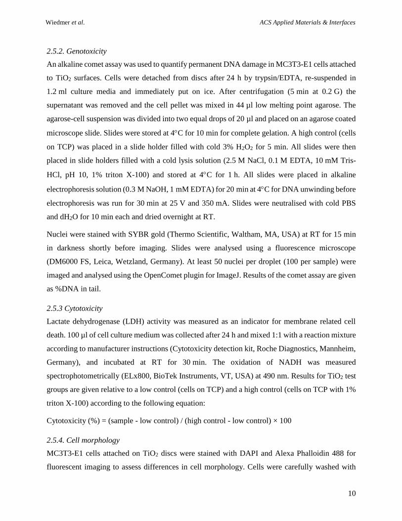
The dip-coating of TiO2 scaffolds in a TTIP-based sol and subsequent heat treatment resulted in a
thin film with a thickness of 42 12 nm that homogeneously covered the entire surface of the
substrate (Figure 1, Figure 2). Most importantly, the interconnective pore architecture, a key
Wiedmer et al. ACS Applied Materials & Interfaces
12
structural parameter for bone tissue engineering,32 remained intact after the coating process (Figure
1). Defect free dip-coating of porous substrates is generally challenging and the outcome is strongly
depending on the film thickness.37 A too thick coating may close pore windows or form flakes,
while complete coverage of the substrate is often not achieved for too thin coatings. The applied
coating layer was thin enough to uniformly coat the underlying grains of the substrate and even
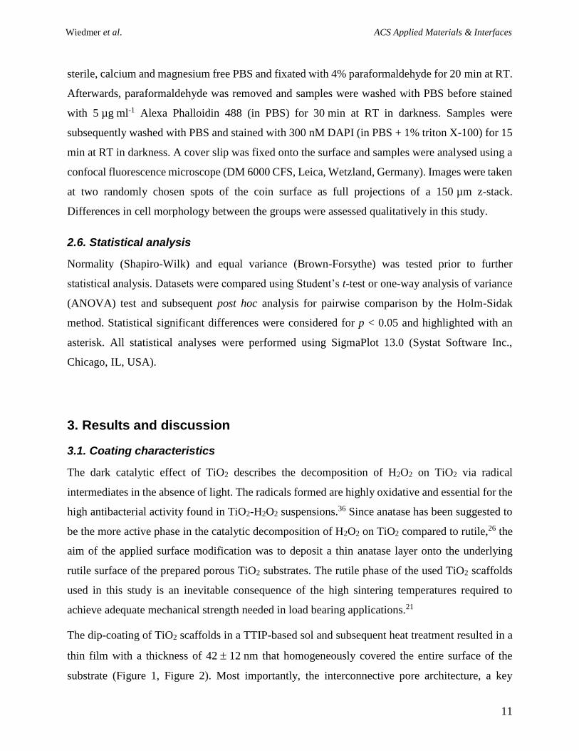
infiltrate the grain boundaries, which explains the significantly lower roughness measured on the
coated (Sa = 0.54 0.05 µm) than the uncoated (Sa = 1.15 0.22 µm) TiO2 surfaces. Further,
similar nanoscale surface roughness values with no statistically significant difference were
measured for both coated (Sa,polished = 25.6 23.2 nm) and uncoated (Sa,polished = 40.9 22.9 nm) on
polished TiO2 coins, and no distinct nanostructures were observed on sol-gel coated Si wafers as
shown in Figure 2. However, grain boundaries were found to be the areas that were most
susceptible to coating defects. Thin cracks were observed along the initial grain boundaries
(Figure1, Figure 2) which may appear upon internal stresses during calcination,37 and could
possibly be avoided by lower heating rates.
Figure 1. SEM images showing the porous microstructure of uncoated and sol-gel coated TiO2
scaffolds. Dashed circle highlights a minor delamination defect on a coated strut.
Wiedmer et al. ACS Applied Materials & Interfaces
13
Figure 2. Surface topography of uncoated and coated TiO2 discs. Thin cracks along original grain
boundaries of the TiO2 substrate were observed on the coated surfaces as seen in the highlighted
area. Nanoscale topography of the coating surface is illustrated on a sol-gel coated Si wafer.
While not altering the surface topography of the TiO2 substrate, the sol-gel coating was observed
to have a significant impact on the wettability of the TiO2 coin surfaces as the coated surfaces were
significantly more hydrophilic in comparison to the uncoated coin surfaces (WCAcoated = 9.7 ± 4.2°
vs. WCAuncoated = 79.2 ± 7.6°, p < 0.001). This significant increase in wettability of the sol-gel
coated surfaces maybe caused by the thin cracks observed along the grain boundaries of the
underlying substrate as these defects may wick the applied water droplet onto the surface of the
coin. However, a significant decrease in the wetting behaviour of the coated TiO2 surfaces was
observed following exposure to 30% H2O2 (WCAcoated,H2O2 = 42.4 ± 5.4°, p < 0.001), which
indicates that the capillary action of physical defects in the coating are not the reason for the very
hydrophilic surface of the sol-gel coating. Significant increase in WCA was also observed for the
uncoated H2O2-treated samples (WCAuncoated,H2O2 = 91.2 ± 7.4°, p < 0.01). Therefore, the presence
of more abundant surface hydroxyl groups on the sol-gel coated surfaces as a consequence of
higher number of oxygen vacancies present in the anatase coating than in the rutile substrate may
likely be responsible for the hydrophilicity of the coated TiO2 surfaces.38-39
Wiedmer et al. ACS Applied Materials & Interfaces
14
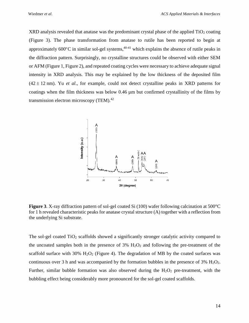
XRD analysis revealed that anatase was the predominant crystal phase of the applied TiO2 coating
(Figure 3). The phase transformation from anatase to rutile has been reported to begin at
approximately 600C in similar sol-gel systems,40-41 which explains the absence of rutile peaks in
the diffraction pattern. Surprisingly, no crystalline structures could be observed with either SEM
or AFM (Figure 1, Figure 2), and repeated coating cycles were necessary to achieve adequate signal
intensity in XRD analysis. This may be explained by the low thickness of the deposited film
(42 12 nm). Yu et al., for example, could not detect crystalline peaks in XRD patterns for
coatings when the film thickness was below 0.46 µm but confirmed crystallinity of the films by
transmission electron microscopy (TEM).42
Figure 3. X-ray diffraction pattern of sol-gel coated Si (100) wafer following calcination at 500°C
for 1 h revealed characteristic peaks for anatase crystal structure (A) together with a reflection from
the underlying Si substrate.
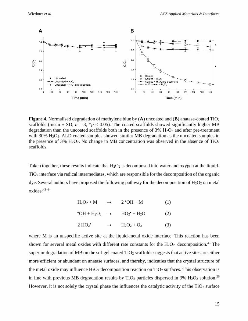
The sol-gel coated TiO2 scaffolds showed a significantly stronger catalytic activity compared to
the uncoated samples both in the presence of 3% H2O2 and following the pre-treatment of the
scaffold surface with 30% H2O2 (Figure 4). The degradation of MB by the coated surfaces was
continuous over 3 h and was accompanied by the formation bubbles in the presence of 3% H2O2.
Further, similar bubble formation was also observed during the H2O2 pre-treatment, with the
bubbling effect being considerably more pronounced for the sol-gel coated scaffolds.
Wiedmer et al. ACS Applied Materials & Interfaces
15
Figure 4. Normalised degradation of methylene blue by (A) uncoated and (B) anatase-coated TiO2
scaffolds (mean ± SD, n = 3, *p < 0.05). The coated scaffolds showed significantly higher MB
degradation than the uncoated scaffolds both in the presence of 3% H2O2 and after pre-treatment
with 30% H2O2. ALD coated samples showed similar MB degradation as the uncoated samples in
the presence of 3% H2O2. No change in MB concentration was observed in the absence of TiO2
scaffolds.
Taken together, these results indicate that H2O2 is decomposed into water and oxygen at the liquid-
TiO2 interface via radical intermediates, which are responsible for the decomposition of the organic
dye. Several authors have proposed the following pathway for the decomposition of H2O2 on metal
oxides:43-44
H2O2 + M 2 OH + M (1)
OH + H2O2 HO2 + H2O (2)
2 HO2 H2O2 + O2 (3)
where M is an unspecific active site at the liquid-metal oxide interface. This reaction has been
shown for several metal oxides with different rate constants for the H2O2 decomposition.45 The
superior degradation of MB on the sol-gel coated TiO2 scaffolds suggests that active sites are either
more efficient or abundant on anatase surfaces, and thereby, indicaties that the crystal structure of
the metal oxide may influence H2O2 decomposition reaction on TiO2 surfaces. This observation is
in line with previous MB degradation results by TiO2 particles dispersed in 3% H2O2 solution.26
However, it is not solely the crystal phase the influences the catalytic activity of the TiO2 surface
Wiedmer et al. ACS Applied Materials & Interfaces
16
as scaffolds coated by atomic layer deposited anatase thin film showed similarly low catalytic
activity for MB degradation as the uncoated rutile substrate (Figure 4B), despite their increased
surface area due to the distinct nanostructure of the deposited anatase film.34 As these surfaces also
featured similar wettability as the uncoated TiO2,34 the presence of more abundant oxygen
vacancies on the surface of the sol-gel coated scaffolds is a likely explanation for their significantly
stronger oxidative power in the presence of H2O2. Whereas the sequential film growth reaction in
ALD process is highly controlled,46 the hydrolysis and polymerisation steps in the sol-gel coating
process are far less controllable,47 which in combination with the relatively short sintering time in
low oxygen environment is likely to contribute to formation of surface point defects. Point defects
play an important role in heterogeneous catalysis, while reduced coordination of the metal ions has
been shown to accurately describe the catalytic decomposition of H2O2 on transition metal oxides
in theoretical models.44 Therefore, a more thorough chemical characterisation of the coated TiO2
surfaces is needed to clarify the role of the crystal phase and presence of lattice defects in the
decomposition of H2O2 on TiO2.
Nonetheless, the results of the degradation study indicate that the oxidative behaviour of TiO2-
H2O2 suspensions in the absence of light can also be observed for TiO2 surfaces. The reaction with
intermediate free radicals (reaction 1 & 2) are the most likely cause for complete decomposition of
the model dye. Hydroxyl radical (OH) is a powerful oxidant and capable of decomposing various
compounds in water. However, a previous study has pointed out the importance of superoxide (O2-
•) and hydroperoxyl radicals (HO2) in the destruction of organic dyes.26 HO2
is the protonated
form of the O2-• which has been shown to adsorb on the surface of several metal oxides.30 O2
-•
coordinated at a TiIV site have been shown to be stable in air for several days48 and may therefore
explain the oxidative effect of coated scaffolds also after the pre-treatment with H2O2 (Figure 4B).
The biomedical implications of stabilised superoxide radicals have been studied to some extent. In
their pioneering work, Tengvall et al. investigated the role of superoxide radicals bound in a
TiOOH matrix formed during the interaction of metallic titanium and H2O2.31, 49 The interaction of
host tissue cells with the TiOOH matrix has been suggested to be essential for the biocompatibility
of titanium in vivo.50 Further, several groups have utilised the stabilisation of free radicals for a
prolonged antibacterial effect of implant surfaces,25, 51 and therefore, the biological response
towards oxidative TiO2 scaffolds used in this study was assessed in two in vitro assays.
Wiedmer et al. ACS Applied Materials & Interfaces
17
3.2. Antibacterial activity
Staphylococci implant colonisation has been reported to be the most frequent cause for biomaterial-
associated infections related to orthopaedic devices.7, 52 Both S. epidermidis and S. aureus are
commensal bacteria which become pathogenic by passing the skin barrier and entering the
implantation site during surgery.52 While S. aureus is considered the more virulent strain, the
outstanding role of S. epidermidis in BAI is due to its ability to from a pathogenic biofilm on
implant surfaces.53-54 Bacterial biofilms show low susceptibility against antibiotic therapy and can
evade the host immune system, which make staphylococcal infections particularly difficult to treat.
The ability to form such biofilms on various substrates in a monoculture in vitro has made S.
epidermidis a valuable and well-established model strain in the assessment of antibacterial
surfaces. It was therefore selected as a biofilm model in the presented study. The use of the
constitutive luminescent strain Xen43 further allowed us to monitor luminescence as an indicator
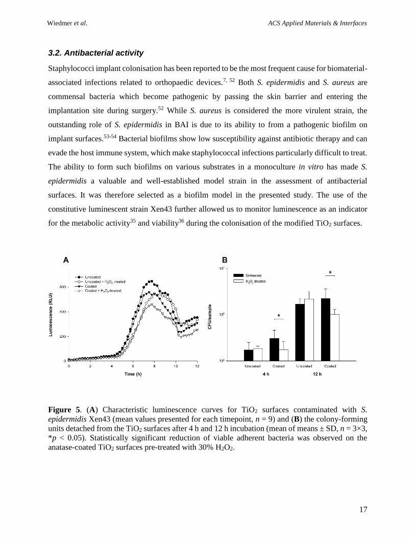
for the metabolic activity35 and viability36 during the colonisation of the modified TiO2 surfaces.
Figure 5. (A) Characteristic luminescence curves for TiO2 surfaces contaminated with S.
epidermidis Xen43 (mean values presented for each timepoint, n = 9) and (B) the colony-forming
units detached from the TiO2 surfaces after 4 h and 12 h incubation (mean of means ± SD, n = 3×3,
*p < 0.05). Statistically significant reduction of viable adherent bacteria was observed on the
anatase-coated TiO2 surfaces pre-treated with 30% H2O2.
Wiedmer et al. ACS Applied Materials & Interfaces
18
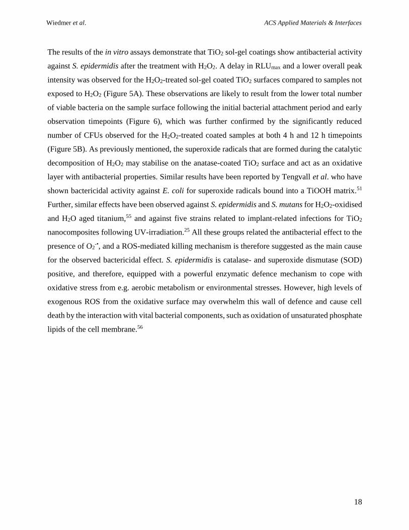
The results of the in vitro assays demonstrate that TiO2 sol-gel coatings show antibacterial activity
against S. epidermidis after the treatment with H2O2. A delay in RLUmax and a lower overall peak
intensity was observed for the H2O2-treated sol-gel coated TiO2 surfaces compared to samples not
exposed to H2O2 (Figure 5A). These observations are likely to result from the lower total number
of viable bacteria on the sample surface following the initial bacterial attachment period and early
observation timepoints (Figure 6), which was further confirmed by the significantly reduced
number of CFUs observed for the H2O2-treated coated samples at both 4 h and 12 h timepoints
(Figure 5B). As previously mentioned, the superoxide radicals that are formed during the catalytic
decomposition of H2O2 may stabilise on the anatase-coated TiO2 surface and act as an oxidative
layer with antibacterial properties. Similar results have been reported by Tengvall et al. who have
shown bactericidal activity against E. coli for superoxide radicals bound into a TiOOH matrix.51
Further, similar effects have been observed against S. epidermidis and S. mutans for H2O2-oxidised
and H2O aged titanium,55 and against five strains related to implant-related infections for TiO2
nanocomposites following UV-irradiation.25 All these groups related the antibacterial effect to the
presence of O2-•, and a ROS-mediated killing mechanism is therefore suggested as the main cause
for the observed bactericidal effect. S. epidermidis is catalase- and superoxide dismutase (SOD)
positive, and therefore, equipped with a powerful enzymatic defence mechanism to cope with
oxidative stress from e.g. aerobic metabolism or environmental stresses. However, high levels of
exogenous ROS from the oxidative surface may overwhelm this wall of defence and cause cell
death by the interaction with vital bacterial components, such as oxidation of unsaturated phosphate
lipids of the cell membrane.56
Wiedmer et al. ACS Applied Materials & Interfaces
19
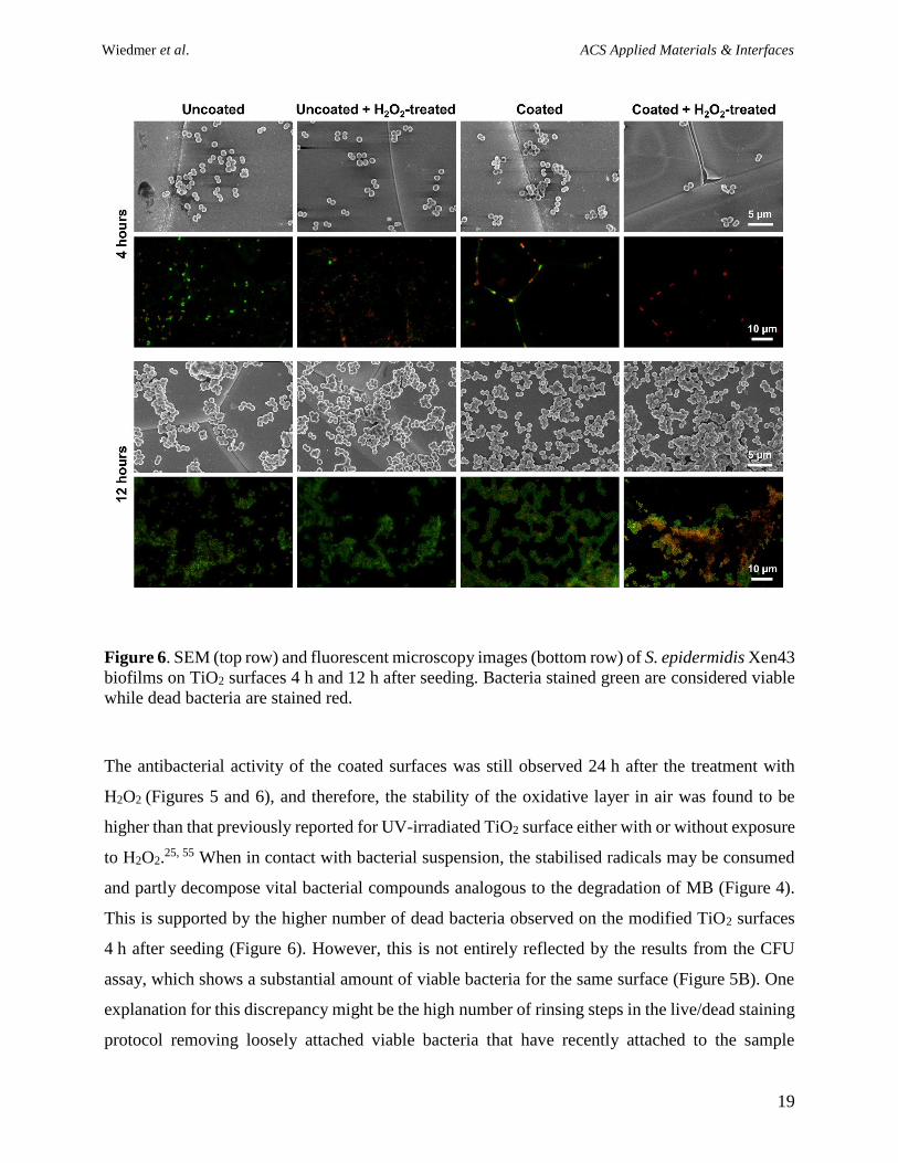
Figure 6. SEM (top row) and fluorescent microscopy images (bottom row) of S. epidermidis Xen43
biofilms on TiO2 surfaces 4 h and 12 h after seeding. Bacteria stained green are considered viable
while dead bacteria are stained red.
The antibacterial activity of the coated surfaces was still observed 24 h after the treatment with
H2O2 (Figures 5 and 6), and therefore, the stability of the oxidative layer in air was found to be
higher than that previously reported for UV-irradiated TiO2 surface either with or without exposure
to H2O2.25, 55 When in contact with bacterial suspension, the stabilised radicals may be consumed
and partly decompose vital bacterial compounds analogous to the degradation of MB (Figure 4).
This is supported by the higher number of dead bacteria observed on the modified TiO2 surfaces
4 h after seeding (Figure 6). However, this is not entirely reflected by the results from the CFU
assay, which shows a substantial amount of viable bacteria for the same surface (Figure 5B). One
explanation for this discrepancy might be the high number of rinsing steps in the live/dead staining
protocol removing loosely attached viable bacteria that have recently attached to the sample
Wiedmer et al. ACS Applied Materials & Interfaces
20
surface. Interestingly, adherent bacteria on the anatase-coated samples seemed to align along the
grain boundaries (Figure 6). These areas are particularly prone to coating defects and thin cracks
along the grain boundaries on coated TiO2 discs were observed frequently as shown in Figure 2.
Hence, these cracks may function as preferred attachment sites for bacteria and explain the overall
higher number of viable bacteria on the coated samples in comparison to the uncoated surfaces
(Figure 5B).
While the H2O2-treated anatase surfaces showed bactericidal activity after 4 h incubation time, they
were unable to prevent biofilm formation in the long term. A homogeneous biofilm of
predominantly viable bacteria covered most of the sample surface for all groups 12 h after seeding
(Figure 6). This lack of long-term antibacterial activity may be caused by the consumption of the
ROS adsorbed on the H2O2-exposed surfaces over time, and therefore, the formation of a less
hostile environment for viable planktonic bacteria to adhere onto the surface. The secretion of EPS
during biofilm maturation may further provide a physical barrier, which protects bacteria from
direct contact with the oxidative layer. Nevertheless, confocal microscopy revealed patches of high
toxicity against S. epidermidis even after 12 h of incubation on the H2O2-treated coatings (Figure
6), indicating some long-term effect of the surface modification.
3.3. Cyto- and genotoxicity
An antibacterial surface modification strategy based on ROS formation on TiO2 surfaces poses the
risk of causing unwanted damage to tissue cells. Adverse effects of the surface modification were
assessed for the murine osteoblast precursor cell line MC3T3, which has been used previously for
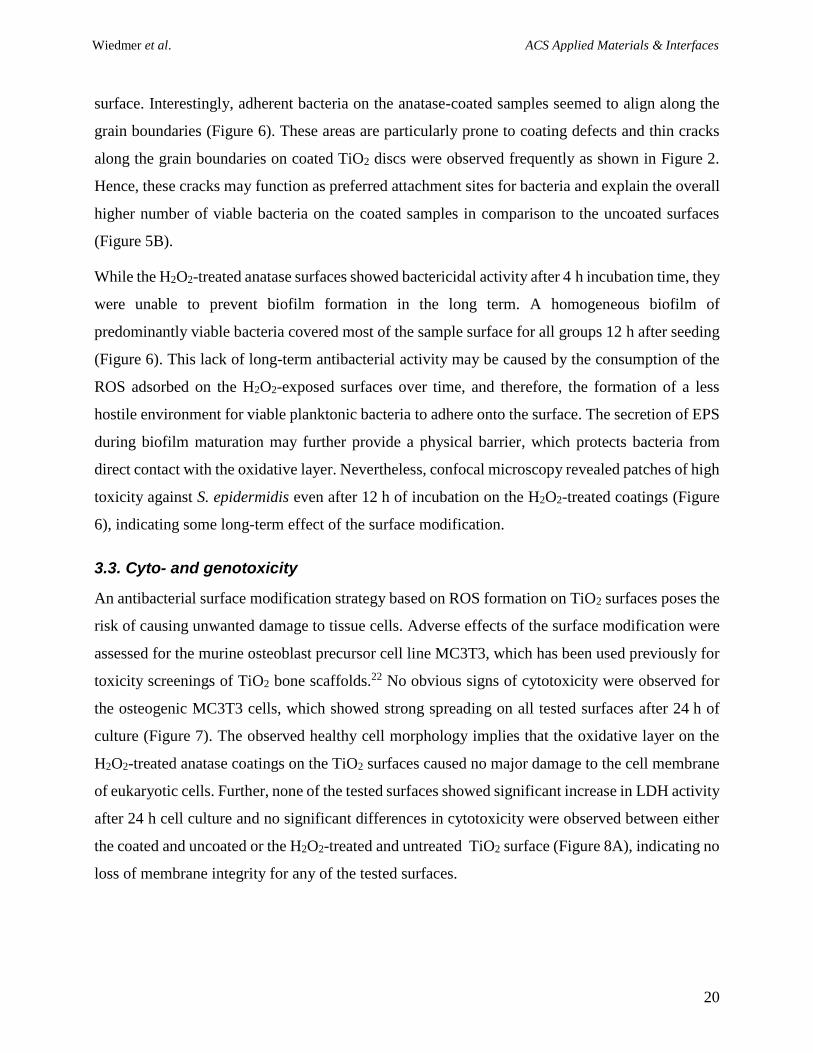
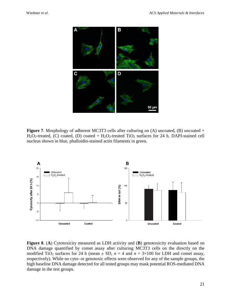
toxicity screenings of TiO2 bone scaffolds.22 No obvious signs of cytotoxicity were observed for
the osteogenic MC3T3 cells, which showed strong spreading on all tested surfaces after 24 h of
culture (Figure 7). The observed healthy cell morphology implies that the oxidative layer on the
H2O2-treated anatase coatings on the TiO2 surfaces caused no major damage to the cell membrane
of eukaryotic cells. Further, none of the tested surfaces showed significant increase in LDH activity
after 24 h cell culture and no significant differences in cytotoxicity were observed between either
the coated and uncoated or the H2O2-treated and untreated TiO2 surface (Figure 8A), indicating no
loss of membrane integrity for any of the tested surfaces.
Wiedmer et al. ACS Applied Materials & Interfaces
21
Figure 7. Morphology of adherent MC3T3 cells after culturing on (A) uncoated, (B) uncoated +
H2O2-treated, (C) coated, (D) coated + H2O2-treated TiO2 surfaces for 24 h. DAPI-stained cell
nucleus shown in blue, phalloidin-stained actin filaments in green.
Figure 8. (A) Cytotoxicity measured as LDH activity and (B) genotoxicity evaluation based on
DNA damage quantified by comet assay after culturing MC3T3 cells on the directly on the
modified TiO2 surfaces for 24 h (mean ± SD, n = 4 and n = 3×100 for LDH and comet assay,
respectively). While no cyto- or genotoxic effects were observed for any of the sample groups, the
high baseline DNA damage detected for all tested groups may mask potential ROS-mediated DNA
damage in the test groups.
Wiedmer et al. ACS Applied Materials & Interfaces
22
The potential toxic effects of oxidative TiO2 surfaces are likely to occur predominantly due to
extracellular cell damage, such as lipid peroxidation. The O2-• radicals, which are formed during
the interaction of H2O2 and TiO2 and create the oxidative layer on the metal oxide surface,26, 30 are
unable to cross cell membranes, thereby, suggesting extracellular damage as the most likely
damaging mechanism of oxidative TiO2 surfaces.57 The absence of any detectable membrane-
related damage observed on the MC3T3s may be based on a higher tolerance of MC3T3s against
lipid peroxidation compared to S. epidermidis. However, intracellular ROS damage may
nonetheless still occur in both mammalian and bacterial cells exposed to the oxidative layer on the
modified TiO2 surfaces. It is possible that uncharged reactive species such as H2O2 are formed by
secondary reactions of O2-•. H2O2 has been shown to cause severe DNA damage in various cells by
intracellular Fenton chemistry.57 Therefore, the genotoxicity of modified TiO2 surfaces was further
analysed by means of a comet assay. While the results of the comet assay showed no increase in
DNA damage of TiO2 surfaces after the pre-treatment with H2O2 (Figure 8B), high baseline
genotoxicity values were observed for all tested groups, including the unmodified TiO2 surface.
This may be related to the poor automatic shape detection of the used analysis software for small
comets or unwanted DNA damage during sample preparation.58 Further, a high overall damage of
TiO2 surfaces was not supported by the cytotoxicity assay, and therefore, further studies are needed
to verify the absence of irreparable DNA damage in osteogenic cells exposed to the bactericidal
H2O2-treated TiO2 surfaces.
4. Conclusions
In summary, this study showed the successful application of an oxidative thin-film coating for
porous ceramic scaffolds based on the dark catalytic effect of TiO2. Sol-gel derived anatase coating
was observed to exhibit strong oxidative behaviour in the presence of 3% H2O2, and the coating
maintained some of its oxidative characteristic in the absence of an oxidative agent when pre-
exposed to 30% H2O2. While the oxidative behaviour of the anatase-coated surfaces was related to
the decomposition of H2O2 at catalytically active sites on the sample surface and subsequent
adsorption of the formed O2-• on the TiO2 surface, the presence and identity of these radical species
on these surfaces is yet to be shown. Thus, a thorough characterisation of the chemical surface
properties of the oxidative anatase film is required to gain a better understanding of the molecular
mechanism behind TiO2 dark catalysis and to identify the crucial material properties responsible
Wiedmer et al. ACS Applied Materials & Interfaces
23
for the high catalytic activity of the sol-gel derived anatase coatings. The oxidative H2O2-treated
sol-gel coated surfaces showed no cytotoxic effects on MC3T3s but revealed antibacterial activity
particularly at the early stages of S. epidermidis biofilm development. Further, no increase in
genotoxicity was observed, and therefore, the applied surface modification provides a novel and
simple strategy to functionalise TiO2 surfaces for antibacterial activity.
Acknowledgments
This study was supported by Eureka-Eurostars Project (Application E!8320 NuGel) and the
Norwegian Research Council (Grant 257569). The authors acknowledge Dr Manuel Gomez-Florit,
Jonas Wengenroth, and Prof. Ola Nilsen for their technical assistance with the cell study, optical
profilometry, and XRD and ellipsometry.
The authors declare no competing financial interest.
References
(1) Cancedda, R.; Dozin, B.; Giannoni, P.; Quarto, R. Tissue Engineering and Cell Therapy of
Cartilage and Bone. Matrix Biol. 2003, 22 (1), 81-91.
(2) Hutmacher, D. W. Scaffolds in Tissue Engineering Bone and Cartilage. Biomaterials 2000,
21 (24), 2529-2543.
(3) Eniwumide, J. O.; Yuan, H.; Cartmell, S. H.; Meijer, G. J.; de Bruijn, J. D. Ectopic Bone
Formation in Bone Marrow Stem Cell Seeded Calcium Phosphate Scaffolds as Compared to
Autograft and (Cell Seeded) Allograft. Eur. Cell. Mater. 2007, 14, 30-39.
(4) Miguel, B. S.; Kriauciunas, R.; Tosatti, S.; Ehrbar, M.; Ghayor, C.; Textor, M.; Weber, F. E.
Enhanced Osteoblastic Activity and Bone Regeneration Using Surface-Modified Porous
Bioactive Glass Scaffolds. J. Biomed. Mater. Res. Part A 2010, 94A (4), 1023-1033.
(5) Tiainen, H.; Wohlfahrt, J. C.; Verket, A.; Lyngstadaas, S. P.; Haugen, H. J. Bone Formation
in TiO2 Bone Scaffolds in Extraction Sockets of Minipigs. Acta Biomater. 2012, 8 (6), 2384-
2391.
(6) Busscher, H. J.; van der Mei, H. C.; Subbiahdoss, G.; Jutte, P. C.; van den Dungen, J. J. A.
M.; Zaat, S. A. J.; Schultz, M. J.; Grainger, D. W. Biomaterial-Associated Infection: Locating the
Finish Line in the Race for the Surface. Sci. Transl. Med. 2012, 4 (153), 153rv10.
(7) Campoccia, D.; Montanaro, L.; Arciola, C. R. The Significance of Infection Related to
Orthopedic Devices and Issues of Antibiotic Resistance. Biomaterials 2006, 27 (11), 2331-2339.
(8) Urban, J. A.; Garvin, K. L. Prosthetic Joint Infection. Curr. Treat. Options Infect. Dis. 2003,
5, 309-321.
(9) Costerton, J. W.; Montanaro, L.; Arciola, C. R. Biofilm in Implant Infections: Its Production
and Regulation. Int. J. Artif. Organs 2005, 28 (11), 1062-1068.
Wiedmer et al. ACS Applied Materials & Interfaces
24
(10) Corbin, A.; Pitts, B.; Parker, A.; Stewart, P. S. Antimicrobial Penetration and Efficacy in an
in Vitro Oral Biofilm Model. Antimicrob. Agents Chemother. 2011, 10.1128/aac.00206-11.
(11) Campoccia, D.; Montanaro, L.; Arciola, C. R. A Review of the Biomaterials Technologies
for Infection-Resistant Surfaces. Biomaterials 2013, 34 (34), 8533-8554.
(12) Banerjee, I.; Pangule, R. C.; Kane, R. S. Antifouling Coatings: Recent Developments in the
Design of Surfaces That Prevent Fouling by Proteins, Bacteria, and Marine Organisms. Adv.
Mater. 2011, 23 (6), 690-718.
(13) Nejadnik, M. R.; van der Mei, H. C.; Norde, W.; Busscher, H. J. Bacterial Adhesion and
Growth on a Polymer Brush-Coating. Biomaterials 2008, 29 (30), 4117-4121.
(14) Jia, Z.; Xiu, P.; Xiong, P.; Zhou, W.; Cheng, Y.; Wei, S.; Zheng, Y.; Xi, T.; Cai, H.; Liu, Z.;
Wang, C.; Zhang, W.; Li, Z. Additively Manufactured Macroporous Titanium with Silver-
Releasing Micro-/Nanoporous Surface for Multipurpose Infection Control and Bone Repair – a
Proof of Concept. ACS Appl. Mater. Inter. 2016, 8 (42), 28495-28510.
(15) Wu, P.; Grainger, D. W. Drug/Device Combinations for Local Drug Therapies and Infection
Prophylaxis. Biomaterials 2006, 27 (11), 2450-2467.
(16) Costa, F.; Carvalho, I. F.; Montelaro, R. C.; Gomes, P.; Martins, M. C. L. Covalent
Immobilization of Antimicrobial Peptides (Amps) onto Biomaterial Surfaces. Acta Biomater.
2011, 7 (4), 1431-1440.
(17) Subbiahdoss, G.; Kuijer, R.; Grijpma, D. W.; van der Mei, H. C.; Busscher, H. J. Microbial
Biofilm Growth Vs. Tissue Integration: “The Race for the Surface” Experimentally Studied. Acta
Biomater. 2009, 5 (5), 1399-1404.
(18) Albers, C. E.; Hofstetter, W.; Siebenrock, K. A.; Landmann, R.; Klenke, F. M. In Vitro
Cytotoxicity of Silver Nanoparticles on Osteoblasts and Osteoclasts at Antibacterial
Concentrations. Nanotoxicology 2013, 7 (1), 30-36.
(19) Campoccia, D.; Montanaro, L.; Speziale, P.; Arciola, C. R. Antibiotic-Loaded Biomaterials
and the Risks for the Spread of Antibiotic Resistance Following Their Prophylactic and
Therapeutic Clinical Use. Biomaterials 2010, 31 (25), 6363-6377.
(20) Tsukimura, N.; Kojima, N.; Kubo, K.; Att, W.; Takeuchi, K.; Kameyama, Y.; Maeda, H.;
Ogawa, T. The Effect of Superficial Chemistry of Titanium on Osteoblastic Function. J. Biomed.
Mater. Res. Part A 2008, 84A (1), 108-116.
(21) Tiainen, H.; Wiedmer, D.; Haugen, H. J. Processing of Highly Porous TiO2 Bone Scaffolds
with Improved Compressive Strength. J. Eur. Ceram. Soc. 2013, 33 (1), 15-24.
(22) Tiainen, H.; Monjo, M.; Knychala, J.; Nilsen, O.; Lyngstadaas, S. P.; Ellingsen, J. E.;
Haugen, H. J. The Effect of Fluoride Surface Modification of Ceramic TiO2 on the Surface
Properties and Biological Response of Osteoblastic Cells in Vitro. Biomed. Mater. 2011, 6 (4),
045006.
(23) Visai, L.; De Nardo, L.; Punta, C.; Melone, L.; Cigada, A.; Imbriani, M.; Arciola, C. R.
Titanium Oxide Antibacterial Surfaces in Biomedical Devices. Int. J. Artif. Organs 2011, 34 (9),
929.
(24) Hashimoto, K.; Irie, H.; Fujishima, A. TiO2 Photocatalysis: A Historical Overview and
Future Prospects. Jpn. J. Appl. Phys. 2005, 44 (12R), 8269.
(25) Cai, Y.; Strømme, M.; Welch, K. Photocatalytic Antibacterial Effects Are Maintained on
Resin-Based TiO2 Nanocomposites after Cessation of UV Irradiation. PLOS ONE 2013, 8 (10),
e75929.
(26) Wiedmer, D.; Sagstuen, E.; Welch, K.; Haugen, H. J.; Tiainen, H. Oxidative Power of
Aqueous Non-Irradiated TiO2-H2O2 Suspensions: Methylene Blue Degradation and the Role of
Reactive Oxygen Species. Appl. Catal. B 2016, 198, 9-15.
Wiedmer et al. ACS Applied Materials & Interfaces
25
(27) Sánchez, L. D.; Taxt-Lamolle, S. F. M.; Hole, E. O.; Krivokapić, A.; Sagstuen, E.; Haugen,
H. J. TiO2 Suspension Exposed to H2O2 in Ambient Light or Darkness: Degradation of
Methylene Blue and EPR Evidence for Radical Oxygen Species. Appl. Catal. B 2013, 142–143
(0), 662-667.
(28) Gustumhaugen, E.; Lönn-Stensrud, J.; Scheie, A. A.; Lyngstadaas, S. P.; Ekfeldt, A.; Taxt-
Lamolle, S. Effect of Chemical and Mechanical Debridement Techniques on Bacterial Re-
Growth on Rough Titanium Surfaces: An in Vitro Study. Clin. Oral Implants Res. 2014, 25 (6),
707-713.
(29) Henderson, E.; Schneider, S.; Petersen, F. C.; Haugen, H. J.; Wohlfahrt, J. C.; Ekstrand, K.;
Ekfeldt, A. Chemical Debridement of Contaminated Titanium Surfaces: An in Vitro Study. Acta
Odontol. Scand. 2013, 71 (3-4), 957-964.
(30) Anpo, M.; Che, M.; Fubini, B.; Garrone, E.; Giamello, E.; Paganini, M. C. Generation of
Superoxide Ions at Oxide Surfaces. Top. Catal. 1999, 8 (3), 189.
(31) Tengvall, P.; Lundström, I.; Sjöqvist, L.; Elwing, H.; Bjursten, L. M. Titanium-Hydrogen
Peroxide Interaction: Model Studies of the Influence of the Inflammatory Response on Titanium
Implants. Biomaterials 1989, 10 (3), 166-175.
(32) Tiainen, H.; Lyngstadaas, S. P.; Ellingsen, J. E.; Haugen, H. J. Ultra-Porous Titanium Oxide
Scaffold with High Compressive Strength. J. Mater. Sci.: Mater. Med. 2010, 21 (10), 2783-2792.
(33) Verket, A.; Tiainen, H.; Haugen, H. J.; Lyngstadaas, S. P.; Nilsen, O.; Reseland, J. E.
Enhanced Osteoblast Differentiation on Scaffolds Coated with TiO2 Compared to SiO2 and CaP
Coatings. Biointerphases 2012, 10.1007/s13758-012-0036-8.
(34) Müller, B.; Haugen, H.; Nilsen, O.; Tiainen, H. Atomic Layer Deposited TiO2 Protects
Porous Ceramic Foams from Grain Boundary Corrosion. Corrosion Science 2016, 106, 35-42.
(35) Vuong, C.; Kocianova, S.; Yu, J.; Kadurugamuwa, J. L.; Otto, M. Development of Real-
Time in Vivo Imaging of Device-Related Staphylococcus epidermidis Infection in Mice and
Influence of Animal Immune Status on Susceptibility to Infection. J. Infect. Dis. 2008, 198 (2),
258-261.
(36) Wiedmer, D.; Petersen, F. C.; Lönn-Stensrud, J.; Tiainen, H. Antibacterial Effect of
Hydrogen Peroxide-Titanium Dioxide Suspensions in the Decontamination of Rough Titanium
Surfaces. Biofouling 2017, 33 (6), 451-459.
(37) Kueper, T. W.; Visco, S. J.; De Jonghe, L. C. Thin-Film Ceramic Electrolytes Deposited on
Porous and Non-Porous Substrates by Sol-Gel Techniques. Solid State Ion. 1992, 52 (1), 251-
259.
(38) Langlet, M.; Permpoon, S.; Riassetto, D.; Berthomé, G.; Pernot, E.; Joud, J. C.
Photocatalytic Activity and Photo-Induced Superhydrophilicity of Sol–Gel Derived TiO2 Films.
J. Photoch. Photobio. A 2006, 181 (2), 203-214.
(39) Simonsen, M. E.; Li, Z.; Søgaard, E. G. Influence of the Oh Groups on the Photocatalytic
Activity and Photoinduced Hydrophilicity of Microwave Assisted Sol–Gel TiO2 Film. Appl. Surf.
Sci. 2009, 255 (18), 8054-8062.
(40) Ohya, Y.; Mishina, J.; Matsuda, T.; Ban, T.; Takahashi, Y. Crystallization and
Microstructure Development of Sol–Gel-Derived Titanium Dioxide Thin Films with Single and
Multiple Layers. J. Am. Ceram. Soc. 1999, 82 (10), 2601-2606.
(41) Takahashi, Y.; Matsuoka, Y. Dip-Coating of Tio2 Films Using a Sol Derived from Ti(O-I-
Pr)4-Diethanolamine-H2O-I-PrOH System. J. Mater. Sci. 1988, 23 (6), 2259-2266.
(42) Yu, J.; Zhao, X.; Zhao, Q. Effect of Film Thickness on the Grain Size and Photocatalytic
Activity of the Sol-Gel Derived Nanometer TiO2 Thin Films. J. Mater. Sci. Lett. 2000, 19 (12),
1015-1017.
Wiedmer et al. ACS Applied Materials & Interfaces
26
(43) Hiroki, A.; LaVerne, J. A. Decomposition of Hydrogen Peroxide at Water−Ceramic Oxide
Interfaces. J. Phys. Chem. B 2005, 109 (8), 3364-3370.
(44) Lousada, C. M.; Johansson, A. J.; Brinck, T.; Jonsson, M. Mechanism of H2O2
Decomposition on Transition Metal Oxide Surfaces. J. Phys. Chem. C 2012, 116 (17), 9533-
9543.
(45) Lousada, C. M.; Yang, M.; Nilsson, K.; Jonsson, M. Catalytic Decomposition of Hydrogen
Peroxide on Transition Metal and Lanthanide Oxides. J. Mol. Catal. A: Chem. 2013, 379, 178-
184.
(46) George, S. M. Atomic Layer Deposition: An Overview. Chem. Rev. 2010, 110 (1), 111-131.
(47) Prusakova, V.; Armellini, C.; Carpentiero, A.; Chiappini, A.; Collini, C.; Dirè, S.; Ferrari,
M.; Lorenzelli, L.; Nardello, M.; Normani, S.; Vaccari, A.; Chiasera, A. Morphologic, Structural,
and Optical Characterization of Sol-Gel Derived TiO2 Thin Films for Memristive Devices. Phys.
Status Solidi C 2015, 12 (1‐2), 192-196.
(48) Antcliff, K. L.; Murphy, D. M.; Griffiths, E.; Giamello, E. The Interaction of H2O2 with
Exchanged Titanium Oxide Systems (TS-1, TiO2, [Ti]-APO-5, Ti-ZSM-5). Phys. Chem. Chem.
Phys. 2003, 5 (19), 4306-4316.
(49) Tengvall, P.; Wälivaara, B.; Westerling, J.; Lundström, I. Stable Titanium Superoxide
Radicals in Aqueous Ti-Peroxy Gels and Ti-Peroxide Solutions. J. Colloid Interface Sci. 1991,
143 (2), 589-592.
(50) Larsson, J.; Persson, C.; Tengvall, P.; Lundqvist-Gustafsson, H. Anti-Inflammatory Effects
of a Titanium-Peroxy Gel: Role of Oxygen Metabolites and Apoptosis. J. Biomed. Mater. Res.
Part A 2004, 68A (3), 448-457.
(51) Tengvall, P.; Hörnsten, E. G.; Elwing, H.; Lundström, I. Bactericidal Properties of a
Titanium-Peroxy Gel Obtained from Metallic Titanium and Hydrogen Peroxide. J. Biomed.
Mater. Res. 1990, 24 (3), 319-330.
(52) Otto, M. Staphylococcal Biofilms. In Bacterial Biofilms; Romeo, T., Ed.; Springer: Berlin
Heidelberg, 2008; pp 207-228.
(53) Otto, M. Staphylococcus epidermidis - the 'Accidental' Pathogen. Nat. Rev. Microbiol. 2009,
7 (8), 555-567.
(54) Mack, D.; Davies, A. P.; Harris, L. G.; Jeeves, R.; Pascoe, B.; Knobloch, J. K.-M.; Rohde,
H.; Wilkinson, T. S. Staphylococcus epidermidis in Biomaterial-Associated Infections. In
Biomaterials Associated Infection: Immunological Aspects and Antimicrobial Strategies;
Moriarty, T. F.; Zaat, S. A. J.; Busscher, H. J., Eds.; Springer New York: New York, NY, 2013;
pp 25-56.
(55) Unosson, E.; Tsekoura, E. K.; Engqvist, H.; Welch, K. Synergetic Inactivation of
Staphylococcus epidermidis and Streptococcus mutans in a TiO2/H2O2/UV System. Biomatter
2013, 3 (4), e26727.
(56) Kiwi, J.; Nadtochenko, V. New Evidence for TiO2 Photocatalysis During Bilayer Lipid
Peroxidation. J. Phys. Chem. B 2004, 108 (45), 17675-17684.
(57) Halliwell, B.; Gutteridge, J. M. C. Oxidative Stress and Redox Regulation: Damage, Repair,
Senescence, and Death. In Free Radicals in Biology and Medicine; Oxford University Press:
USA, 2015; pp 199-283.
(58) Speit, G.; Kojima, H.; Burlinson, B.; Collins, A. R.; Kasper, P.; Plappert-Helbig, U.; Uno,
Y.; Vasquez, M.; Beevers, C.; De Boeck, M.; Escobar, P. A.; Kitamoto, S.; Pant, K.; Pfuhler, S.;
Tanaka, J.; Levy, D. D. Critical Issues with the in Vivo Comet Assay: A Report of the Comet
Assay Working Group in the 6th International Workshop on Genotoxicity Testing (IWGT).
Mutat. Res. Genet. Toxicol. Environ. Mutagen. 2015, 783, 6-12.