Embed Size (px)
Citation preview
RSC Advances
PAPER
Ope
n A
cces
s A
rtic
le. P
ublis
hed
on 2
9 M
arch
201
7. D
ownl
oade
d on
3/2
2/20
22 6
:04:
58 A
M.
Thi
s ar
ticle
is li
cens
ed u
nder
a C
reat
ive
Com
mon
s A
ttrib
utio
n 3.
0 U
npor
ted
Lic
ence
.
View Article OnlineView Journal | View Issue
Long-term antib
Department of Biomedical Engineering, Sc
University, Wuhan 430071, P. R. China. E-m
2768759991; Tel: +86 2768759509
† Electronic supplementary informa10.1039/c7ra01464c
Cite this: RSC Adv., 2017, 7, 18917
Received 5th February 2017Accepted 21st March 2017
DOI: 10.1039/c7ra01464c
rsc.li/rsc-advances
This journal is © The Royal Society of C
acterial protected cotton fabriccoating by controlled release of chlorhexidinegluconate from halloysite nanotubes†
Yu Wu, Yongtao Yang, Haoyang Liu, Xihui Yao, Fan Leng, Yun Chenand Weiqun Tian *
In this work, an environmentally friendly antibacterial composite was prepared by loading natural halloysite
nanotubes (HNTs) with chlorhexidine gluconate (CG). Fourier transform-infrared spectroscopy and X-ray
diffraction demonstrated that CG could be successfully loaded on HNTs without changing the crystalline
structure of HNTs. In vitro drug release assay showed that CG could be released from HNTs in
a sustainable manner and could last as long as about ten days. Inhibition zone test suggested that the
new composite displayed significant bacterial killing activity. In addition, the composite was deposited on
the surface of cotton fabric by dip-coat technique. Our results showed that the HNTs/CG composite
could be distributed on the cotton fabric and possessed better thermal stability and hydrophilicity
properties than raw cotton fabric. HNTs/CG-coated cotton fabric exhibited over 98% bacterial reduction
of Staphylococcus aureus, Escherichia coli, and Pseudomonas aeruginosa, and still showed over 90%
antibacterial activity even after being washed 20 times. MTT test and animal skin irritation tests indicate
that HNTs/CG-coated cotton was nontoxic to mouse fibroblast cells (L929) and gave no irritation to the
rabbit's skin. Thus, HNTs/CG composite is a safe and promising antibacterial agent and has great
potential application in the textile field.
Introduction
Owing to their outstanding properties of air permeability,comfort and soness, cotton textiles are widely used as medicaland household products in our daily life. However, underappropriate temperature and humidity, the natural cotton beris benecial to the rapid growth of microorganisms.1 Thisbacterial pollution leads to the loss of mechanical strength ofthe textiles, and is also a threat to human health. Therefore,there are high demands for environmentally friendly, costeffective and biocompatible antibacterial agents to impartantibacterial and antifouling properties to cotton fabrics.
In recent years, lots of studies have been performed toimprove the antibacterial property of cotton fabrics bycombining with different kinds of metal nanoparticles, such asAg nanoparticles,2–5 TiO2,6,7 and ZnO.8,9 Although these nano-particles have good antibacterial effect, safety and environ-mental issues caused by the release of heavy metal ions areconcerned and limit their widespread use.10 Compared withthose metal nanoparticles, natural nanoparticles are desirable
hool of Basic Medical Sciences, Wuhan
ail: [email protected]; Fax: +86
tion (ESI) available. See DOI:
hemistry 2017
materials to use with the aim of developing new antibacterialtextile products without environmental hazards or biologicaltoxicity for the textile industry.
Halloysite nanotubes (HNTs) are a kind of hydrated claynanotubes, which have deserved considerable interest owing totheir low cost, environmentally friendly nature and readilyavailability.11 Owing to their peculiar hollow tubular shape, theyexhibit high aspect ratio, good adsorbing ability and cationexchange capacity. The biocompatibility of halloysite is an issueof concern for the application of HNTs.12,13 The low toxicity ofHNTs has been demonstrated by several studies. Ahmed et al.14
studied the cytotoxicity and cytogenetic toxicity of HNTs as oraldrug delivery system, indicating HNTs did not cause toxicity toHCT116, HepG2 cells and human peripheral blood lymphocytes.Vergaro et al.15 investigated the interaction between HNTs anddifferent cells for toxicity tests, and observed the uptake processof nanotubes into cells, representing a high level of biocompat-ibility of HNTs. Because of the high biocompatibility and lowtoxicity, HNTs can be used in food, biomedical and industrialelds, such as food packaging,16 tissue engineering scaffold,17
cancer cell isolation,18 bone implants,19 and cosmetics.20
It is identied that an ideal antimicrobial nishing agentshould have broad-spectrum antimicrobial activity, low toxicity,no negative effect on the original properties of fabric, and exhibitexcellent antimicrobial durability against washing.21,22 Asa natural claymineral, HNTs exhibit no antibacterial effect unless
RSC Adv., 2017, 7, 18917–18925 | 18917
RSC Advances Paper
Ope
n A
cces
s A
rtic
le. P
ublis
hed
on 2
9 M
arch
201
7. D
ownl
oade
d on
3/2
2/20
22 6
:04:
58 A
M.
Thi
s ar
ticle
is li
cens
ed u
nder
a C
reat
ive
Com
mon
s A
ttrib
utio
n 3.
0 U
npor
ted
Lic
ence
.View Article Online
antibacterial drugs are intercalated.23 Chlorhexidine gluconate(CG) is an efficient antibacterial agent, which has broad-spectrum antibacterial activity and low toxicity.24 As the positivecharge of the chlorhexidine molecule will interact with thenegatively charged phosphate groups on microbial cell walls, theelectrostatic interaction can lead to the disruption of cellularmembrane permeability.25 Some studies have demonstrated thatCG-impregnated cloth has an obvious long-lasting antisepticeffect, as CG can persist on skin and is not inactivated by blood orserum proteins.26 Thus, intercalating the organic CG into inor-ganic HNTs may be an efficient way to obtain a novel compositeantibacterial material. Moreover, the HNTs/CG composite as anantibacterial material coating on cotton fabric has not, to ourknowledge, been reported previously.
In this paper, HNTs/CG composite material was prepared byloading CG in HNTs by vacuum suction method and then wascoated on cotton fabric. The objectives of this research were toobtain an innovative cotton fabric which possesses long-termantibacterial properties. Fourier transform-infrared spectros-copy (FTIR) and X-ray diffraction (XRD) were used to charac-terize the new composite. In vitro drug release and inhibitionzone test were performed to detect the controlled releaseperformance and antibacterial activity of the composite mate-rial. To characterize the morphology of the cotton fabric,scanning electron microscopy (SEM) was employed. Finally, theeffects of the HNTs/CG-coated cotton content on the thermalstability, hydrophilicity, antibacterial activity and biocompati-bility are discussed in detail.
Experimental sectionMaterials
Halloysite was obtained from Jinyangguang Ceramic Co., Ltd(Zhengzhou, P. R. China). Chlorhexidine gluconate waspurchased from Jingchuchen Pharmaceutical Chemical Co., Ltd(Wuhan, P. R. China). The structural formula of chlorhexidinegluconate is shown in Fig. S1.† The test bacteria, Escherichiacoli, Pseudomonas aeruginosa and Staphylococcus aureus, usedfor this study were purchased from the China Center for TypeCulture Collection. The Gram-positive S. aureus strain selectedin the experiment was CCTCC AB 91093, the Gram-negative E.coli strain selected in the experiment was CCTCC AB 93154 andthe P. aeruginosa strain selected in the experiment was CCTCCAB 93066. All of the chemical reagents used were analyticalgrade.
Drug loading procedure
The purchased HNTs were pretreated according to the protocolsdescribed in the literature.27 An ethyl alcohol (absolute)suspension of 0.5 g HNT powder was mixed with a saturatedsolution of CG drug and then evacuated for 30 min usinga vacuum pump. Air bubbles were removed from the pores ofHNTs to ensure the antibacterial agents could enter the lumensunder negative pressure. Aer 30 min, the vacuum had van-ished and air was allowed into the container when back toatmospheric pressure. The process was repeated twice for the
18918 | RSC Adv., 2017, 7, 18917–18925
most efficient loading. Finally, the drug loaded samples wereseparated from the solution by centrifugation (9000 rpm) andwere washed three times with deionized water to removeunloaded drug. Finally, HNTs/CG composite was dried in anoven at 60 �C and milled to a ne powder.
Preparation of HNTs/CG-coated cotton fabric
The HNTs/CG composite was coated on fabric by using the dip-coat method. HNTs/CG powder was added to deionized waterand stirred vigorously. Then, the clean cotton fabric wasimpregnated in HNTs/CG suspension at room temperature, andthen the excess liquid was squeezed out of the fabric. Aerbeing dried in an oven at 60 �C, the HNTs/CG-coated cottonfabric was obtained.
Structural characterization
Transmission electron microscopy (TEM) images of HNTs wereobtained from a Tecnai 12 Transmission Electron Microscope(FEI, Eindhoven, The Netherlands) equipped with a MegaviewIII CCD camera and analysis camera control soware (Olympus)at an operating voltage of 120 kV.
Fourier transform-infrared (FTIR) spectra of samples wererecorded on an FTIR spectrometer (TNZI-5700, Nicolet, USA) withthe wavenumber range of 4000 to 400 cm�1 using KBr pellets.The crystal structure of HNT powder before and aer CG loadingwas analyzed by an X-ray diffractometer (D8 ADVANCE, Ger-many) with Cu Ka radiation (g ¼ 0.15406 nm). XRD data wererecorded from 4� to 80� (2q) at a scanning speed of 1� min�1.
In vitro drug release
Drug release from HNTs was investigated in deionized water atroom temperature. Three milliliters of pure CG drug (10 mgmL�1) was loaded into a dialysis bag and then dipped in 30 mLwater. The halloysite nanotubes were dispersed in 10 mL waterand constantly oscillated at room temperature. Then, thesuspension was centrifuged to separate the sample for analysis.The concentration of CG was determined by UV spectropho-tometry (PerkinElmer Lambda 25, USA). To estimate thecomplete release prole, halloysite samples were sonicated for1 h at the end of each release study.28 All experiments wereperformed in triplicate.
Inhibition zone test of HNTs/CG composite
The antibacterial activity of HNTs/CG powder against S. aureus,E. coli and P. aeruginosa was tested through inhibition zone test.One hundred microliters of bacterial suspension witha concentration of 105 CFU mL�1 was spread on Luria–Bertani(LB) agar plates, and then three holes were punched in eachplate. HNTs/CG powder was put into the holes and then theplates were incubated at 37 �C. The diameter of inhibition zonewas measured aer 24 h.
Characterization of HNTs/CG-coated cotton fabric
The morphology of the raw cotton fabric and the HNTs/CG-coated cotton fabric was characterized by scanning electron
This journal is © The Royal Society of Chemistry 2017
Paper RSC Advances
Ope
n A
cces
s A
rtic
le. P
ublis
hed
on 2
9 M
arch
201
7. D
ownl
oade
d on
3/2
2/20
22 6
:04:
58 A
M.
Thi
s ar
ticle
is li
cens
ed u
nder
a C
reat
ive
Com
mon
s A
ttrib
utio
n 3.
0 U
npor
ted
Lic
ence
.View Article Online
microscopy (SEM; VEGA3, TESCAN, Czech Republic) at a 20 kVaccelerating voltage. Thermogravimetric analysis (TGA) wascarried out on a Diamond TG/DTA instrument (PerkinElmer,USA) under nitrogen atmosphere with a ow capacity of 20 mLmin�1. The scan was performed over the temperature rangefrom 40 to 700 �C at a heating rate of 10 �C min�1.
Evaluation of hydrophilicity of HNTs/CG-coated cotton
The hydrophilicity of raw cotton textiles and HNTs/CG-coatedcotton was evaluated on the basis of textile-test method forcapillary effect (FZ/T 01071-2008). Three specimens were cutwith a size of 250 mm length and 30mmwidth. Thereaer, oneside of each specimen was xed on a bracket and the other sideof the specimen was vertically suspended 15 mm � 2 mmbelow the zero point of a ruler. Deionized water with red colorwas poured into the container below the zero point of ruler,and the bottom of the specimen was immersed in the water.The water wicking height of specimen was recorded atdifferent times. Each test was repeated in triplicate to calculatethe mean value.
Antibacterial tests of HNTs/CG-coated cotton fabric
The antimicrobial activity of the HNTs/CG-coated cotton fabricwas tested against the Gram-positive S. aureus and the Gram-negative E. coli and P. aeruginosa by the viable cell countingtechnique. In order to quantitatively analyze the antibacterialactivity of the HNTs/CG-coated cotton fabric, antibacterial ratewas used to represent the antibacterial effect. Bacteria wereincubated in Luria–Bertani (LB) liquid nutrient medium for 18 hat 37 �C, and the number of cells was diluted to 105 to 106 CFUmL�1 by standard serial dilution method. The coated cotton andthe raw cotton were cut into pieces with an area of 1 cm2, andthen dipped into the bacterial dilution mentioned above (5 mL).Aer incubation for 24 h, 100 mL bacterial suspension was takenout and diluted to appropriate concentration. The dilutionsuspension was transferred onto agar plates, and then incubatedat 37 �C for 24 h. The numbers of viable colonies were counted.The antibacterial rate was calculated by the following equation:
Antibacterial rate ¼ (1 � B/A) � 100%
where A is the number of bacterial colonies on the plates treatedwith the raw cotton fabric suspension and B is the number ofbacterial colonies on the plates treated with the suspensionfrom the cotton fabric nished with HNTs/CG.
The bacterial cell viability was detected based on uoresceinisothiocyanate (FITC) and propidium iodide (PI) double stain.FITC remains on the outermost intact cell walls and producesa green color, whereas PI cannot penetrate the membrane ofliving bacterials, and can only bind to the DNA of damagedcells, giving a red color. Five microliters of FITC and 10 mL PIstain were mixed to co-culture with a bacterial sample for10 min at room temperature. Then, the sample was rinsed toremove the uncombined stain and observed under a uores-cence microscope (Olympus IX 73 DP80).
This journal is © The Royal Society of Chemistry 2017
Durability test
The antibacterial rate was calculated aer 20 washing cycles toevaluate the durability of HNTs/CG-coated cotton fabric againstrepeated laundering. The textile sample was washed with 2 gL�1 non-ionic detergent at 40 �C for 10 min, then rinsed withclean water once and dehydrated, which was recorded asa washing cycle. The above steps were repeated to 20 washingtimes. To avoid the interference of washing agent, the textilesample was thoroughly washed with a large amount of waterand dried before the antibacterial test.
Skin irritation test
All experiments were performed in compliance with the rele-vant laws and institutional guidelines such as the Ministry ofScience and Technology of the People's Republic of China“Guidelines on the Treatment of Animals” (09/03/2006). Wetook great efforts to reduce the number of animals used inthese studies and also to reduce animals' suffering pain anddiscomfort. All procedures concerning animals were approvedby and conformed to the guidelines of the Animal Care &Welfare Committee of Wuhan University School of Medicine.The skin irritation test was carried out according to a technicalstandard for disinfection (Ministry of Health of P. R. China,2002). Three New Zealand rabbits were chosen as test animalsand their hair was removed before testing. HNTs/CG suspen-sion (0.5 mL) was added to the skin on one side of each rabbit,and covered with 4 layers of gauze. The skin on the other sideof the rabbit served as a control. The irritation response(erythema and edema) of the skin was observed aer 1 h, 24 h,48 h and 72 h. The score of skin irritation for each tested rabbitwas evaluated.
Cytotoxicity test
The cytotoxicity of the HNTs/CG-coated cotton was detected byMTT (3-(4,5-dimethylthiazol-2-yl)-2,5-diphenyltetrazoliumbromide) assay with L929 mouse broblast cells. Cells werecultured in MEM-Alpha medium supplemented with 10% fetalbovine serum (FBS), and incubated at 37 �C in a wet atmospherecontaining 5% CO2. The tested cotton fabric was cut into 2.5 cm� 2.5 cm pieces and soaked in 2 mL culture medium with noFBS for 3 days. The extracts were diluted to a concentration of25% with culture medium. The L929 cells were seeded in 96-well plates at a density of 2000 cells per well and incubated for24 h. Then, the medium was removed and replaced by preparedextracted dilutions. Aer incubation for 24, 48 and 72 h, thecells were treated with 20 mL per well MTT solution (5 mg mL�1
in phosphate buffered saline, PBS) and incubated for another4 h at 37 �C. Then, 200 mL per well dimethyl sulfoxide (DMSO)was added to dissolve the formazan crystals. The plates wereshaken for 15 min, and the optical density was detected ona multiwell microplate reader (Tecan GENios, Tecan AustriaGmbH, Salzburg, Austria) at 490 nm. Cell viability was calcu-lated by the following equation:
Cell viability ¼ ODsample/ODcontrol � 100%
RSC Adv., 2017, 7, 18917–18925 | 18919
RSC Advances Paper
Ope
n A
cces
s A
rtic
le. P
ublis
hed
on 2
9 M
arch
201
7. D
ownl
oade
d on
3/2
2/20
22 6
:04:
58 A
M.
Thi
s ar
ticle
is li
cens
ed u
nder
a C
reat
ive
Com
mon
s A
ttrib
utio
n 3.
0 U
npor
ted
Lic
ence
.View Article Online
where ODsample is the absorbance of the test sample andODcontrol is the absorbance for untreated control cells.
Results and discussionMorphology of HNTs observed by TEM
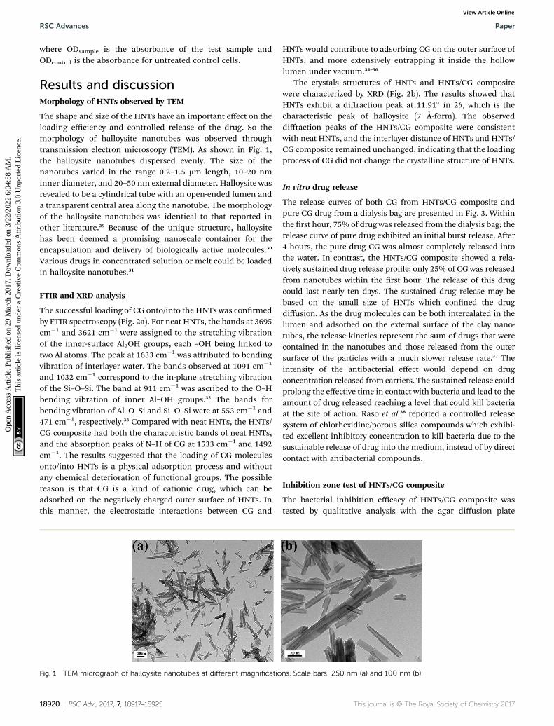
The shape and size of the HNTs have an important effect on theloading efficiency and controlled release of the drug. So themorphology of halloysite nanotubes was observed throughtransmission electron microscopy (TEM). As shown in Fig. 1,the halloysite nanotubes dispersed evenly. The size of thenanotubes varied in the range 0.2–1.5 mm length, 10–20 nminner diameter, and 20–50 nm external diameter. Halloysite wasrevealed to be a cylindrical tube with an open-ended lumen anda transparent central area along the nanotube. The morphologyof the halloysite nanotubes was identical to that reported inother literature.29 Because of the unique structure, halloysitehas been deemed a promising nanoscale container for theencapsulation and delivery of biologically active molecules.30
Various drugs in concentrated solution or melt could be loadedin halloysite nanotubes.31
FTIR and XRD analysis
The successful loading of CG onto/into the HNTs was conrmedby FTIR spectroscopy (Fig. 2a). For neat HNTs, the bands at 3695cm�1 and 3621 cm�1 were assigned to the stretching vibrationof the inner-surface Al2OH groups, each –OH being linked totwo Al atoms. The peak at 1633 cm�1 was attributed to bendingvibration of interlayer water. The bands observed at 1091 cm�1
and 1032 cm�1 correspond to the in-plane stretching vibrationof the Si–O–Si. The band at 911 cm�1 was ascribed to the O–Hbending vibration of inner Al–OH groups.32 The bands forbending vibration of Al–O–Si and Si–O–Si were at 553 cm�1 and471 cm�1, respectively.33 Compared with neat HNTs, the HNTs/CG composite had both the characteristic bands of neat HNTs,and the absorption peaks of N–H of CG at 1533 cm�1 and 1492cm�1. The results suggested that the loading of CG moleculesonto/into HNTs is a physical adsorption process and withoutany chemical deterioration of functional groups. The possiblereason is that CG is a kind of cationic drug, which can beadsorbed on the negatively charged outer surface of HNTs. Inthis manner, the electrostatic interactions between CG and
Fig. 1 TEM micrograph of halloysite nanotubes at different magnificatio
18920 | RSC Adv., 2017, 7, 18917–18925
HNTs would contribute to adsorbing CG on the outer surface ofHNTs, and more extensively entrapping it inside the hollowlumen under vacuum.34–36
The crystals structures of HNTs and HNTs/CG compositewere characterized by XRD (Fig. 2b). The results showed thatHNTs exhibit a diffraction peak at 11.91� in 2q, which is thecharacteristic peak of halloysite (7 �A-form). The observeddiffraction peaks of the HNTs/CG composite were consistentwith neat HNTs, and the interlayer distance of HNTs and HNTs/CG composite remained unchanged, indicating that the loadingprocess of CG did not change the crystalline structure of HNTs.
In vitro drug release
The release curves of both CG from HNTs/CG composite andpure CG drug from a dialysis bag are presented in Fig. 3. Withinthe rst hour, 75% of drug was released from the dialysis bag; therelease curve of pure drug exhibited an initial burst release. Aer4 hours, the pure drug CG was almost completely released intothe water. In contrast, the HNTs/CG composite showed a rela-tively sustained drug release prole; only 25% of CG was releasedfrom nanotubes within the rst hour. The release of this drugcould last nearly ten days. The sustained drug release may bebased on the small size of HNTs which conned the drugdiffusion. As the drug molecules can be both intercalated in thelumen and adsorbed on the external surface of the clay nano-tubes, the release kinetics represent the sum of drugs that werecontained in the nanotubes and those released from the outersurface of the particles with a much slower release rate.37 Theintensity of the antibacterial effect would depend on drugconcentration released from carriers. The sustained release couldprolong the effective time in contact with bacteria and lead to theamount of drug released reaching a level that could kill bacteriaat the site of action. Raso et al.38 reported a controlled releasesystem of chlorhexidine/porous silica compounds which exhibi-ted excellent inhibitory concentration to kill bacteria due to thesustainable release of drug into the medium, instead of by directcontact with antibacterial compounds.
Inhibition zone test of HNTs/CG composite
The bacterial inhibition efficacy of HNTs/CG composite wastested by qualitative analysis with the agar diffusion plate
ns. Scale bars: 250 nm (a) and 100 nm (b).
This journal is © The Royal Society of Chemistry 2017
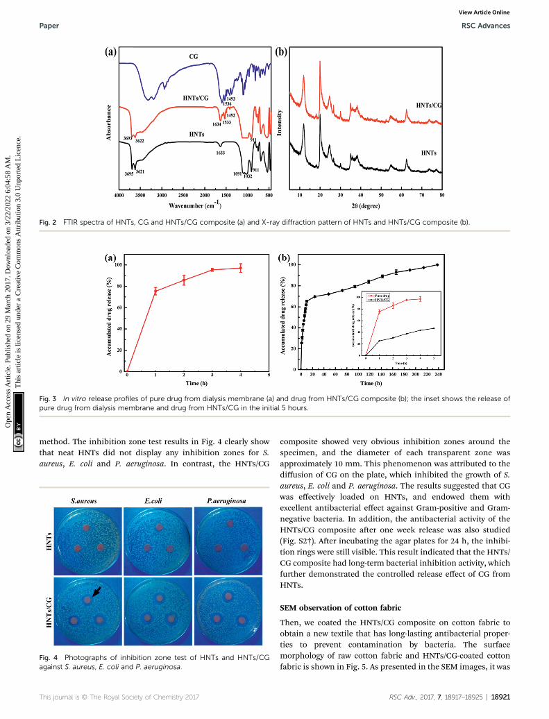
Fig. 2 FTIR spectra of HNTs, CG and HNTs/CG composite (a) and X-ray diffraction pattern of HNTs and HNTs/CG composite (b).
Fig. 3 In vitro release profiles of pure drug from dialysis membrane (a) and drug from HNTs/CG composite (b); the inset shows the release ofpure drug from dialysis membrane and drug from HNTs/CG in the initial 5 hours.
Paper RSC Advances
Ope
n A
cces
s A
rtic
le. P
ublis
hed
on 2
9 M
arch
201
7. D
ownl
oade
d on
3/2
2/20
22 6
:04:
58 A
M.
Thi
s ar
ticle
is li
cens
ed u
nder
a C
reat
ive
Com
mon
s A
ttrib
utio
n 3.
0 U
npor
ted
Lic
ence
.View Article Online
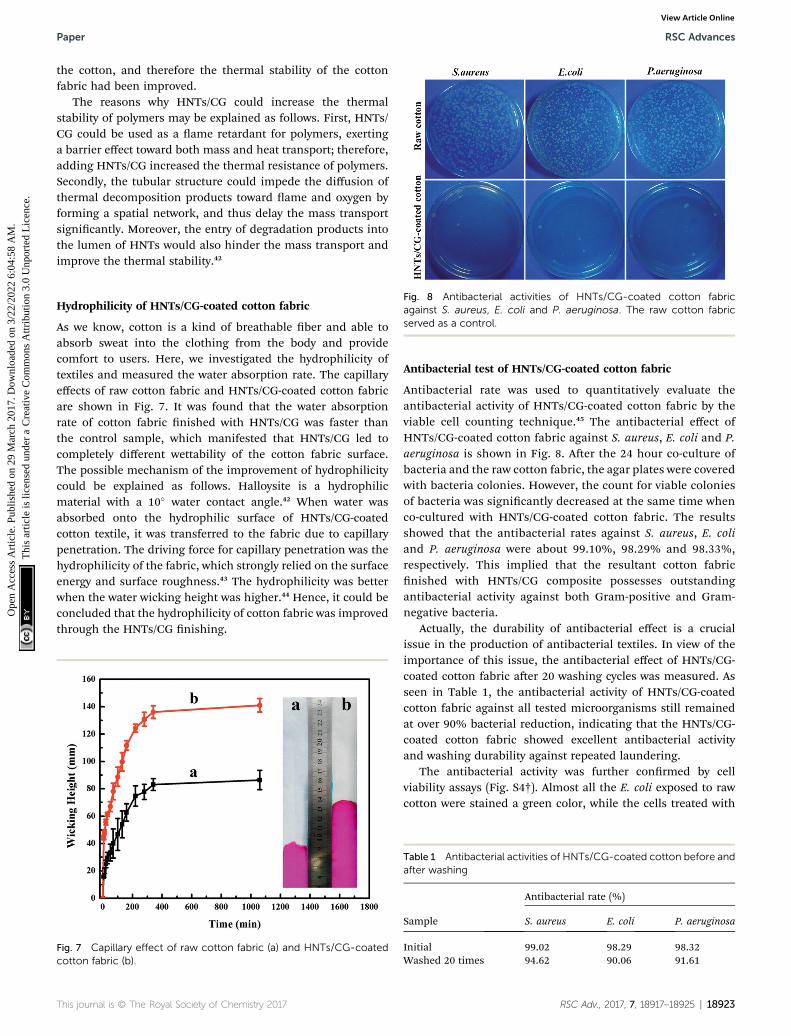
method. The inhibition zone test results in Fig. 4 clearly showthat neat HNTs did not display any inhibition zones for S.aureus, E. coli and P. aeruginosa. In contrast, the HNTs/CG
Fig. 4 Photographs of inhibition zone test of HNTs and HNTs/CGagainst S. aureus, E. coli and P. aeruginosa.
This journal is © The Royal Society of Chemistry 2017
composite showed very obvious inhibition zones around thespecimen, and the diameter of each transparent zone wasapproximately 10 mm. This phenomenon was attributed to thediffusion of CG on the plate, which inhibited the growth of S.aureus, E. coli and P. aeruginosa. The results suggested that CGwas effectively loaded on HNTs, and endowed them withexcellent antibacterial effect against Gram-positive and Gram-negative bacteria. In addition, the antibacterial activity of theHNTs/CG composite aer one week release was also studied(Fig. S2†). Aer incubating the agar plates for 24 h, the inhibi-tion rings were still visible. This result indicated that the HNTs/CG composite had long-term bacterial inhibition activity, whichfurther demonstrated the controlled release effect of CG fromHNTs.
SEM observation of cotton fabric
Then, we coated the HNTs/CG composite on cotton fabric toobtain a new textile that has long-lasting antibacterial proper-ties to prevent contamination by bacteria. The surfacemorphology of raw cotton fabric and HNTs/CG-coated cottonfabric is shown in Fig. 5. As presented in the SEM images, it was
RSC Adv., 2017, 7, 18917–18925 | 18921
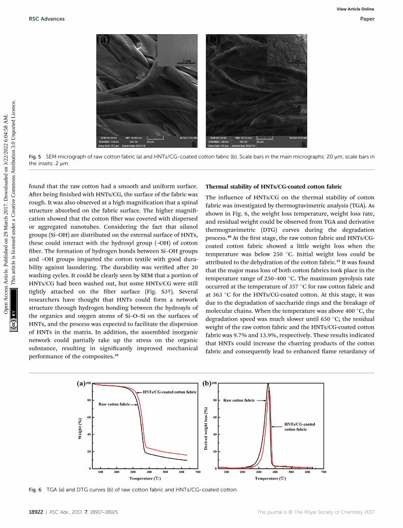
Fig. 5 SEMmicrograph of raw cotton fabric (a) and HNTs/CG-coated cotton fabric (b). Scale bars in the main micrographs: 20 mm; scale bars inthe insets: 2 mm.
RSC Advances Paper
Ope
n A
cces
s A
rtic
le. P
ublis
hed
on 2
9 M
arch
201
7. D
ownl
oade
d on
3/2
2/20
22 6
:04:
58 A
M.
Thi
s ar
ticle
is li
cens
ed u
nder
a C
reat
ive
Com
mon
s A
ttrib
utio
n 3.
0 U
npor
ted
Lic
ence
.View Article Online
found that the raw cotton had a smooth and uniform surface.Aer being nished with HNTs/CG, the surface of the fabric wasrough. It was also observed at a high magnication that a spinalstructure absorbed on the fabric surface. The higher magni-cation showed that the cotton ber was covered with dispersedor aggregated nanotubes. Considering the fact that silanolgroups (Si–OH) are distributed on the external surface of HNTs,these could interact with the hydroxyl group (–OH) of cottonber. The formation of hydrogen bonds between Si–OH groupsand –OH groups imparted the cotton textile with good dura-bility against laundering. The durability was veried aer 20washing cycles. It could be clearly seen by SEM that a portion ofHNTs/CG had been washed out, but some HNTs/CG were stilltightly attached on the ber surface (Fig. S3†). Severalresearchers have thought that HNTs could form a networkstructure through hydrogen bonding between the hydroxyls ofthe organics and oxygen atoms of Si–O–Si on the surfaces ofHNTs, and the process was expected to facilitate the dispersionof HNTs in the matrix. In addition, the assembled inorganicnetwork could partially take up the stress on the organicsubstance, resulting in signicantly improved mechanicalperformance of the composites.39
Fig. 6 TGA (a) and DTG curves (b) of raw cotton fabric and HNTs/CG-c
18922 | RSC Adv., 2017, 7, 18917–18925
Thermal stability of HNTs/CG-coated cotton fabric
The inuence of HNTs/CG on the thermal stability of cottonfabric was investigated by thermogravimetric analysis (TGA). Asshown in Fig. 6, the weight loss temperature, weight loss rate,and residual weight could be observed from TGA and derivativethermogravimetric (DTG) curves during the degradationprocess.40 At the rst stage, the raw cotton fabric and HNTs/CG-coated cotton fabric showed a little weight loss when thetemperature was below 250 �C. Initial weight loss could beattributed to the dehydration of the cotton fabric.41 It was foundthat the major mass loss of both cotton fabrics took place in thetemperature range of 250–400 �C. The maximum pyrolysis rateoccurred at the temperature of 357 �C for raw cotton fabric andat 363 �C for the HNTs/CG-coated cotton. At this stage, it wasdue to the degradation of saccharide rings and the breakage ofmolecular chains. When the temperature was above 400 �C, thedegradation speed was much slower until 650 �C; the residualweight of the raw cotton fabric and the HNTs/CG-coated cottonfabric was 9.7% and 13.9%, respectively. These results indicatedthat HNTs could increase the charring products of the cottonfabric and consequently lead to enhanced ame retardancy of
oated cotton.
This journal is © The Royal Society of Chemistry 2017
Paper RSC Advances
Ope
n A
cces
s A
rtic
le. P
ublis
hed
on 2
9 M
arch
201
7. D
ownl
oade
d on
3/2
2/20
22 6
:04:
58 A
M.
Thi
s ar
ticle
is li
cens
ed u
nder
a C
reat
ive
Com
mon
s A
ttrib
utio
n 3.
0 U
npor
ted
Lic
ence
.View Article Online
the cotton, and therefore the thermal stability of the cottonfabric had been improved.
The reasons why HNTs/CG could increase the thermalstability of polymers may be explained as follows. First, HNTs/CG could be used as a ame retardant for polymers, exertinga barrier effect toward both mass and heat transport; therefore,adding HNTs/CG increased the thermal resistance of polymers.Secondly, the tubular structure could impede the diffusion ofthermal decomposition products toward ame and oxygen byforming a spatial network, and thus delay the mass transportsignicantly. Moreover, the entry of degradation products intothe lumen of HNTs would also hinder the mass transport andimprove the thermal stability.42
Fig. 8 Antibacterial activities of HNTs/CG-coated cotton fabricagainst S. aureus, E. coli and P. aeruginosa. The raw cotton fabricserved as a control.
Hydrophilicity of HNTs/CG-coated cotton fabric
As we know, cotton is a kind of breathable ber and able toabsorb sweat into the clothing from the body and providecomfort to users. Here, we investigated the hydrophilicity oftextiles and measured the water absorption rate. The capillaryeffects of raw cotton fabric and HNTs/CG-coated cotton fabricare shown in Fig. 7. It was found that the water absorptionrate of cotton fabric nished with HNTs/CG was faster thanthe control sample, which manifested that HNTs/CG led tocompletely different wettability of the cotton fabric surface.The possible mechanism of the improvement of hydrophilicitycould be explained as follows. Halloysite is a hydrophilicmaterial with a 10� water contact angle.42 When water wasabsorbed onto the hydrophilic surface of HNTs/CG-coatedcotton textile, it was transferred to the fabric due to capillarypenetration. The driving force for capillary penetration was thehydrophilicity of the fabric, which strongly relied on the surfaceenergy and surface roughness.43 The hydrophilicity was betterwhen the water wicking height was higher.44 Hence, it could beconcluded that the hydrophilicity of cotton fabric was improvedthrough the HNTs/CG nishing.
Fig. 7 Capillary effect of raw cotton fabric (a) and HNTs/CG-coatedcotton fabric (b).
This journal is © The Royal Society of Chemistry 2017
Antibacterial test of HNTs/CG-coated cotton fabric
Antibacterial rate was used to quantitatively evaluate theantibacterial activity of HNTs/CG-coated cotton fabric by theviable cell counting technique.45 The antibacterial effect ofHNTs/CG-coated cotton fabric against S. aureus, E. coli and P.aeruginosa is shown in Fig. 8. Aer the 24 hour co-culture ofbacteria and the raw cotton fabric, the agar plates were coveredwith bacteria colonies. However, the count for viable coloniesof bacteria was signicantly decreased at the same time whenco-cultured with HNTs/CG-coated cotton fabric. The resultsshowed that the antibacterial rates against S. aureus, E. coliand P. aeruginosa were about 99.10%, 98.29% and 98.33%,respectively. This implied that the resultant cotton fabricnished with HNTs/CG composite possesses outstandingantibacterial activity against both Gram-positive and Gram-negative bacteria.
Actually, the durability of antibacterial effect is a crucialissue in the production of antibacterial textiles. In view of theimportance of this issue, the antibacterial effect of HNTs/CG-coated cotton fabric aer 20 washing cycles was measured. Asseen in Table 1, the antibacterial activity of HNTs/CG-coatedcotton fabric against all tested microorganisms still remainedat over 90% bacterial reduction, indicating that the HNTs/CG-coated cotton fabric showed excellent antibacterial activityand washing durability against repeated laundering.
The antibacterial activity was further conrmed by cellviability assays (Fig. S4†). Almost all the E. coli exposed to rawcotton were stained a green color, while the cells treated with
Table 1 Antibacterial activities of HNTs/CG-coated cotton before andafter washing
Sample
Antibacterial rate (%)
S. aureus E. coli P. aeruginosa
Initial 99.02 98.29 98.32Washed 20 times 94.62 90.06 91.61
RSC Adv., 2017, 7, 18917–18925 | 18923
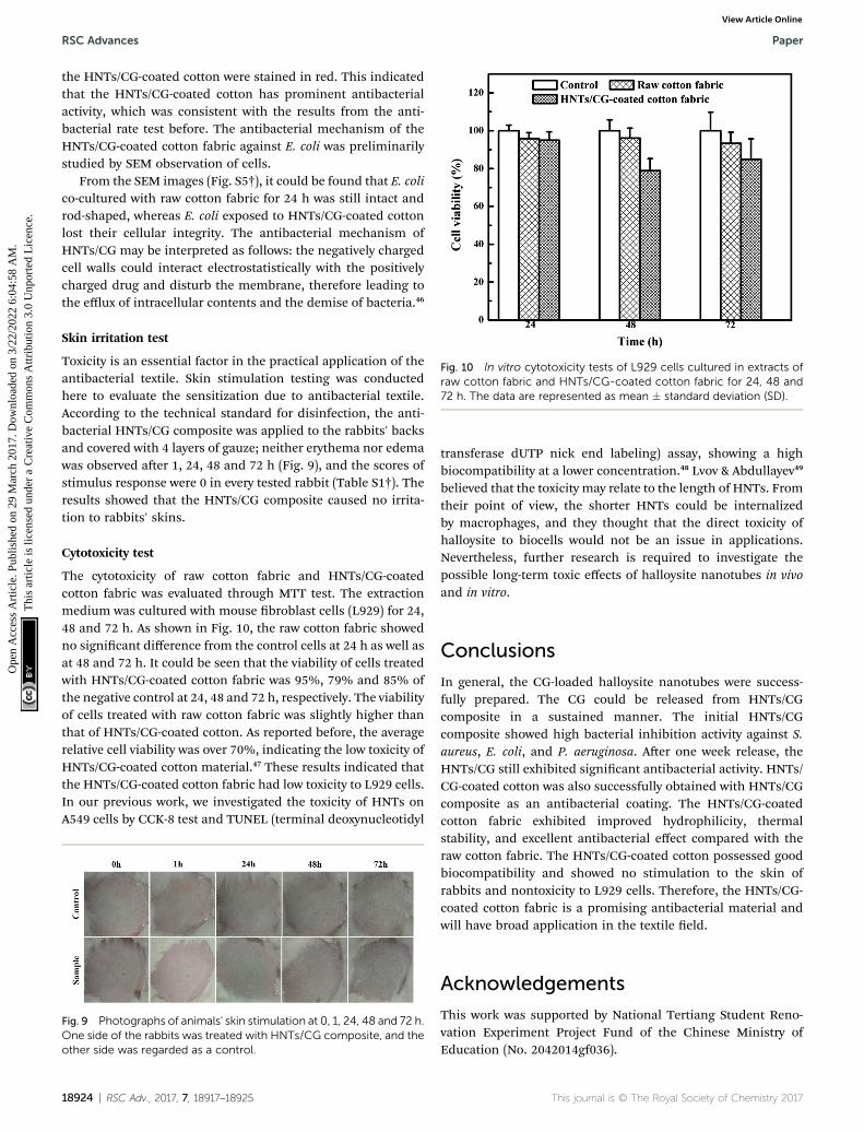
Fig. 10 In vitro cytotoxicity tests of L929 cells cultured in extracts ofraw cotton fabric and HNTs/CG-coated cotton fabric for 24, 48 and72 h. The data are represented as mean � standard deviation (SD).
RSC Advances Paper
Ope
n A
cces
s A
rtic
le. P
ublis
hed
on 2
9 M
arch
201
7. D
ownl
oade
d on
3/2
2/20
22 6
:04:
58 A
M.
Thi
s ar
ticle
is li
cens
ed u
nder
a C
reat
ive
Com
mon
s A
ttrib
utio
n 3.
0 U
npor
ted
Lic
ence
.View Article Online
the HNTs/CG-coated cotton were stained in red. This indicatedthat the HNTs/CG-coated cotton has prominent antibacterialactivity, which was consistent with the results from the anti-bacterial rate test before. The antibacterial mechanism of theHNTs/CG-coated cotton fabric against E. coli was preliminarilystudied by SEM observation of cells.
From the SEM images (Fig. S5†), it could be found that E. colico-cultured with raw cotton fabric for 24 h was still intact androd-shaped, whereas E. coli exposed to HNTs/CG-coated cottonlost their cellular integrity. The antibacterial mechanism ofHNTs/CG may be interpreted as follows: the negatively chargedcell walls could interact electrostatistically with the positivelycharged drug and disturb the membrane, therefore leading tothe efflux of intracellular contents and the demise of bacteria.46
Skin irritation test
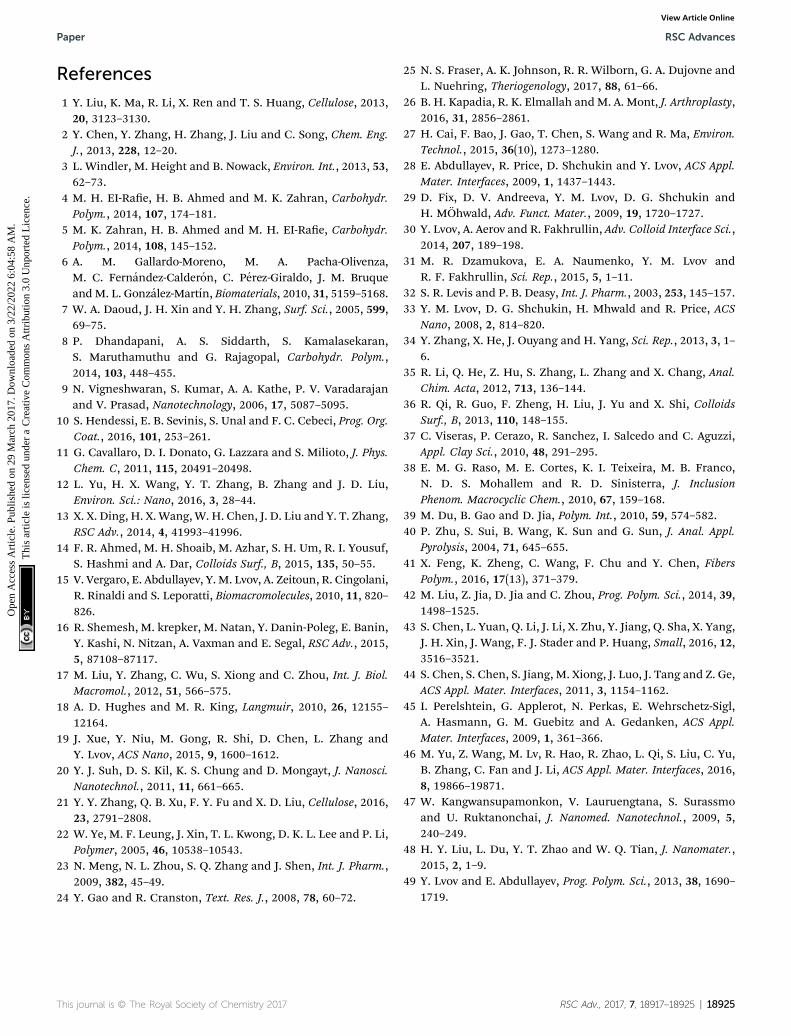
Toxicity is an essential factor in the practical application of theantibacterial textile. Skin stimulation testing was conductedhere to evaluate the sensitization due to antibacterial textile.According to the technical standard for disinfection, the anti-bacterial HNTs/CG composite was applied to the rabbits' backsand covered with 4 layers of gauze; neither erythema nor edemawas observed aer 1, 24, 48 and 72 h (Fig. 9), and the scores ofstimulus response were 0 in every tested rabbit (Table S1†). Theresults showed that the HNTs/CG composite caused no irrita-tion to rabbits' skins.
Cytotoxicity test
The cytotoxicity of raw cotton fabric and HNTs/CG-coatedcotton fabric was evaluated through MTT test. The extractionmedium was cultured with mouse broblast cells (L929) for 24,48 and 72 h. As shown in Fig. 10, the raw cotton fabric showedno signicant difference from the control cells at 24 h as well asat 48 and 72 h. It could be seen that the viability of cells treatedwith HNTs/CG-coated cotton fabric was 95%, 79% and 85% ofthe negative control at 24, 48 and 72 h, respectively. The viabilityof cells treated with raw cotton fabric was slightly higher thanthat of HNTs/CG-coated cotton. As reported before, the averagerelative cell viability was over 70%, indicating the low toxicity ofHNTs/CG-coated cotton material.47 These results indicated thatthe HNTs/CG-coated cotton fabric had low toxicity to L929 cells.In our previous work, we investigated the toxicity of HNTs onA549 cells by CCK-8 test and TUNEL (terminal deoxynucleotidyl
Fig. 9 Photographs of animals' skin stimulation at 0, 1, 24, 48 and 72 h.One side of the rabbits was treated with HNTs/CG composite, and theother side was regarded as a control.
18924 | RSC Adv., 2017, 7, 18917–18925
transferase dUTP nick end labeling) assay, showing a highbiocompatibility at a lower concentration.48 Lvov & Abdullayev49
believed that the toxicity may relate to the length of HNTs. Fromtheir point of view, the shorter HNTs could be internalizedby macrophages, and they thought that the direct toxicity ofhalloysite to biocells would not be an issue in applications.Nevertheless, further research is required to investigate thepossible long-term toxic effects of halloysite nanotubes in vivoand in vitro.
Conclusions
In general, the CG-loaded halloysite nanotubes were success-fully prepared. The CG could be released from HNTs/CGcomposite in a sustained manner. The initial HNTs/CGcomposite showed high bacterial inhibition activity against S.aureus, E. coli, and P. aeruginosa. Aer one week release, theHNTs/CG still exhibited signicant antibacterial activity. HNTs/CG-coated cotton was also successfully obtained with HNTs/CGcomposite as an antibacterial coating. The HNTs/CG-coatedcotton fabric exhibited improved hydrophilicity, thermalstability, and excellent antibacterial effect compared with theraw cotton fabric. The HNTs/CG-coated cotton possessed goodbiocompatibility and showed no stimulation to the skin ofrabbits and nontoxicity to L929 cells. Therefore, the HNTs/CG-coated cotton fabric is a promising antibacterial material andwill have broad application in the textile eld.
Acknowledgements
This work was supported by National Tertiang Student Reno-vation Experiment Project Fund of the Chinese Ministry ofEducation (No. 2042014gf036).
This journal is © The Royal Society of Chemistry 2017
Paper RSC Advances
Ope
n A
cces
s A
rtic
le. P
ublis
hed
on 2
9 M
arch
201
7. D
ownl
oade
d on
3/2
2/20
22 6
:04:
58 A
M.
Thi
s ar
ticle
is li
cens
ed u
nder
a C
reat
ive
Com
mon
s A
ttrib
utio
n 3.
0 U
npor
ted
Lic
ence
.View Article Online
References
1 Y. Liu, K. Ma, R. Li, X. Ren and T. S. Huang, Cellulose, 2013,20, 3123–3130.
2 Y. Chen, Y. Zhang, H. Zhang, J. Liu and C. Song, Chem. Eng.J., 2013, 228, 12–20.
3 L. Windler, M. Height and B. Nowack, Environ. Int., 2013, 53,62–73.
4 M. H. EI-Rae, H. B. Ahmed and M. K. Zahran, Carbohydr.Polym., 2014, 107, 174–181.
5 M. K. Zahran, H. B. Ahmed and M. H. EI-Rae, Carbohydr.Polym., 2014, 108, 145–152.
6 A. M. Gallardo-Moreno, M. A. Pacha-Olivenza,M. C. Fernandez-Calderon, C. Perez-Giraldo, J. M. Bruqueand M. L. Gonzalez-Martın, Biomaterials, 2010, 31, 5159–5168.
7 W. A. Daoud, J. H. Xin and Y. H. Zhang, Surf. Sci., 2005, 599,69–75.
8 P. Dhandapani, A. S. Siddarth, S. Kamalasekaran,S. Maruthamuthu and G. Rajagopal, Carbohydr. Polym.,2014, 103, 448–455.
9 N. Vigneshwaran, S. Kumar, A. A. Kathe, P. V. Varadarajanand V. Prasad, Nanotechnology, 2006, 17, 5087–5095.
10 S. Hendessi, E. B. Sevinis, S. Unal and F. C. Cebeci, Prog. Org.Coat., 2016, 101, 253–261.
11 G. Cavallaro, D. I. Donato, G. Lazzara and S. Milioto, J. Phys.Chem. C, 2011, 115, 20491–20498.
12 L. Yu, H. X. Wang, Y. T. Zhang, B. Zhang and J. D. Liu,Environ. Sci.: Nano, 2016, 3, 28–44.
13 X. X. Ding, H. X. Wang,W. H. Chen, J. D. Liu and Y. T. Zhang,RSC Adv., 2014, 4, 41993–41996.
14 F. R. Ahmed, M. H. Shoaib, M. Azhar, S. H. Um, R. I. Yousuf,S. Hashmi and A. Dar, Colloids Surf., B, 2015, 135, 50–55.
15 V. Vergaro, E. Abdullayev, Y. M. Lvov, A. Zeitoun, R. Cingolani,R. Rinaldi and S. Leporatti, Biomacromolecules, 2010, 11, 820–826.
16 R. Shemesh, M. krepker, M. Natan, Y. Danin-Poleg, E. Banin,Y. Kashi, N. Nitzan, A. Vaxman and E. Segal, RSC Adv., 2015,5, 87108–87117.
17 M. Liu, Y. Zhang, C. Wu, S. Xiong and C. Zhou, Int. J. Biol.Macromol., 2012, 51, 566–575.
18 A. D. Hughes and M. R. King, Langmuir, 2010, 26, 12155–12164.
19 J. Xue, Y. Niu, M. Gong, R. Shi, D. Chen, L. Zhang andY. Lvov, ACS Nano, 2015, 9, 1600–1612.
20 Y. J. Suh, D. S. Kil, K. S. Chung and D. Mongayt, J. Nanosci.Nanotechnol., 2011, 11, 661–665.
21 Y. Y. Zhang, Q. B. Xu, F. Y. Fu and X. D. Liu, Cellulose, 2016,23, 2791–2808.
22 W. Ye, M. F. Leung, J. Xin, T. L. Kwong, D. K. L. Lee and P. Li,Polymer, 2005, 46, 10538–10543.
23 N. Meng, N. L. Zhou, S. Q. Zhang and J. Shen, Int. J. Pharm.,2009, 382, 45–49.
24 Y. Gao and R. Cranston, Text. Res. J., 2008, 78, 60–72.
This journal is © The Royal Society of Chemistry 2017
25 N. S. Fraser, A. K. Johnson, R. R. Wilborn, G. A. Dujovne andL. Nuehring, Theriogenology, 2017, 88, 61–66.
26 B. H. Kapadia, R. K. Elmallah andM. A. Mont, J. Arthroplasty,2016, 31, 2856–2861.
27 H. Cai, F. Bao, J. Gao, T. Chen, S. Wang and R. Ma, Environ.Technol., 2015, 36(10), 1273–1280.
28 E. Abdullayev, R. Price, D. Shchukin and Y. Lvov, ACS Appl.Mater. Interfaces, 2009, 1, 1437–1443.
29 D. Fix, D. V. Andreeva, Y. M. Lvov, D. G. Shchukin andH. MOhwald, Adv. Funct. Mater., 2009, 19, 1720–1727.
30 Y. Lvov, A. Aerov and R. Fakhrullin, Adv. Colloid Interface Sci.,2014, 207, 189–198.
31 M. R. Dzamukova, E. A. Naumenko, Y. M. Lvov andR. F. Fakhrullin, Sci. Rep., 2015, 5, 1–11.
32 S. R. Levis and P. B. Deasy, Int. J. Pharm., 2003, 253, 145–157.33 Y. M. Lvov, D. G. Shchukin, H. Mhwald and R. Price, ACS
Nano, 2008, 2, 814–820.34 Y. Zhang, X. He, J. Ouyang and H. Yang, Sci. Rep., 2013, 3, 1–
6.35 R. Li, Q. He, Z. Hu, S. Zhang, L. Zhang and X. Chang, Anal.
Chim. Acta, 2012, 713, 136–144.36 R. Qi, R. Guo, F. Zheng, H. Liu, J. Yu and X. Shi, Colloids
Surf., B, 2013, 110, 148–155.37 C. Viseras, P. Cerazo, R. Sanchez, I. Salcedo and C. Aguzzi,
Appl. Clay Sci., 2010, 48, 291–295.38 E. M. G. Raso, M. E. Cortes, K. I. Teixeira, M. B. Franco,
N. D. S. Mohallem and R. D. Sinisterra, J. InclusionPhenom. Macrocyclic Chem., 2010, 67, 159–168.
39 M. Du, B. Gao and D. Jia, Polym. Int., 2010, 59, 574–582.40 P. Zhu, S. Sui, B. Wang, K. Sun and G. Sun, J. Anal. Appl.
Pyrolysis, 2004, 71, 645–655.41 X. Feng, K. Zheng, C. Wang, F. Chu and Y. Chen, Fibers
Polym., 2016, 17(13), 371–379.42 M. Liu, Z. Jia, D. Jia and C. Zhou, Prog. Polym. Sci., 2014, 39,
1498–1525.43 S. Chen, L. Yuan, Q. Li, J. Li, X. Zhu, Y. Jiang, Q. Sha, X. Yang,
J. H. Xin, J. Wang, F. J. Stader and P. Huang, Small, 2016, 12,3516–3521.
44 S. Chen, S. Chen, S. Jiang, M. Xiong, J. Luo, J. Tang and Z. Ge,ACS Appl. Mater. Interfaces, 2011, 3, 1154–1162.
45 I. Perelshtein, G. Applerot, N. Perkas, E. Wehrschetz-Sigl,A. Hasmann, G. M. Guebitz and A. Gedanken, ACS Appl.Mater. Interfaces, 2009, 1, 361–366.
46 M. Yu, Z. Wang, M. Lv, R. Hao, R. Zhao, L. Qi, S. Liu, C. Yu,B. Zhang, C. Fan and J. Li, ACS Appl. Mater. Interfaces, 2016,8, 19866–19871.
47 W. Kangwansupamonkon, V. Lauruengtana, S. Surassmoand U. Ruktanonchai, J. Nanomed. Nanotechnol., 2009, 5,240–249.
48 H. Y. Liu, L. Du, Y. T. Zhao and W. Q. Tian, J. Nanomater.,2015, 2, 1–9.
49 Y. Lvov and E. Abdullayev, Prog. Polym. Sci., 2013, 38, 1690–1719.
RSC Adv., 2017, 7, 18917–18925 | 18925