Embed Size (px)
Citation preview

Vol-7 Issue-1 2021 IJARIIE-ISSN(O)-2395-4396
13646 www.ijariie.com 1171
ANTIBIOGRAM PROPERTIES OF
ALOE VERA EXTRACTS AGAINST
FUNGAL ISOLATES FROM
AGRICULTURAL LAND SornambigaRavi
1, Archana S nair
2, Vishnu Priyanka K
3, Poovendiran P
4,
Dr. Mahenthiran R*
1,2, 3 &4 PG Students, Post graduate Research Department of Microbiology, Dr.N.G.P. Arts and Science
College, Tamilnadu, India. *Corresponding author, Assistant professor, Department of Microbiology, Dr.N.G.P. Arts and science
college, Tamilnadu, India.
ABSTRACT Pathogenic fungi are the main infectious agents in plants, causing alterations during developmental stages
including post-harvest. In soil and vegetables, there is a wide variety of fungal genera causing quality problems
related to aspect, nutritional value, organoleptic characteristics, and limited shelf life. In addition, in some
cases fungi are indirectly responsible for allergic or toxic disorders among consumers because of the
production of mycotoxins or allergens. Generally, phytopathogenic fungi are controlled by synthetic fungicides;
however, the use of these is increasingly restricted due to the harmful effects of pesticides on human health and
the environment. The increasing demand of production and regulations on the use of agrochemicals and the
emergence of pathogens resistant to the products employed, justifies the search for novel active molecules and
new control strategies. Currently, there is little evidence on the antimicrobial properties of the medicinal plants
under investigation against phytopathogen fungi. Fungi are ubiquitous in the environment, and infection due to
fungal pathogens has become more common. The genus Alternaria Nees is widely distributed in nature and its
species are among the most common fungi on the phyllo sphere. It includes both plant-pathogenic and plant-
saprophytic species that may damage crops in the field or cause post-harvest decays, causing considerable
economics losses for farmers and food industries. Soil samples were characterized for the incidence of fungal
strains from contaminated agricultural soils. A total of 5 fungal strains were isolated and 33 fungi were
characterized using various isolation and identification methods. Soil samples were also characterized for
physiochemical properties. There are limited reports on the antimicrobial effects of isolated Aloe Vera
components. Shamir were noted high zone of inhibition with ethanol extracted from Aloe Vera baradenisis
against Aspergillums spp, penicillium spp, macro spp, rhizobium spp, Fusarium spp, Candida spp.
Antimicrobial susceptibility test showed that both the gel and the leaf inhibited the growth of fungal spp.
Experiment has been carried out to define the effect if Aloe vera gels In vitro for the fungi.
Keyword: Aloe vera, Aspergillus Niger, Candida Albicans
1.INTRODUCTION Since antiquity, the plant kingdom has provided a variety of compounds of known therapeutic properties, like
analgesics, anti-inflammatory, medicines for asthma, and others. In recent years, antimicrobial properties of
plant extracts have been reported with increasing frequency from different parts of the world. For example, a
large proportion of the South American population use plant extracts obtained from traditional medicinal plants
as medicine for many infectious diseases. Plants from the genus Pterocaulon, known as “quitoco”, are
commonly used in veterinary medicine in southern Brazil to treat animal problems popularly diagnosed as
“mycoses”. Several works have demonstrated in laboratory trials that different plant tissues, such as roots,
leaves, seeds and flowers possess inhibitory properties against bacteria, fungi andinsects.
In addition, the genus produces mycotoxins and phytotoxins, and studies in the last decade have emphasized its
toxicogenic properties rather than simply those that cause spoilage. As well, the smallest concentration capable
of inhibiting or preventing growth was determined among the species and extracts that demonstrated
inhibitoryproperties.
Soil samples were characterized for the incidence of fungal strains from contaminated agricultural soils. A total
of 5 fungal strains were isolated and 33 fungi were characterized using various isolation and identification
methods. Soil samples were also characterized for physiochemical properties.
The isolated fungal strains were successfully identified belonging to the phylum Ascomycota, deuteromycota
and Zygomycota. Alternaria, Aspergillus, Drechslera and Fusarium were predominant genera. Curvularia,

Vol-7 Issue-1 2021 IJARIIE-ISSN(O)-2395-4396
13646 www.ijariie.com 1172
Exserohilum, Humicola, Rhizopus and Torula were the most frequently isolated genera. Rests of the strains were
not identified owing to the lack of sporulating structures under presently used incubation conditions. Such
strains were designated as Mycelia sterilia. Further, these species will be used in biodegradation of commonly
used pesticides.
Aloe vera belongs to the family Lilaceae, of which there are about 360 species Aloe Vera contain over 75
nutrient and 200 active compounds, including vitamins, enzymes, minerals, sugar, lignin, anthraquinones,
sapiens, salicylic acid and amino acids.
It is a cactus-like plant that grows readily in hot, dry climates and currently, because of demand, is cultivated in
large quantities 1. The gel of A. Vera was used to treat stomach ailments, gastrointestinal problems, skin
disease, constipation, radiation injury, inflammatory effect, healing wounds and burns, ulcer and diabetes.
Medicinal plants according to the World Health Organization (WHO) defines them as herbal preparations made
by introducing plant materials to extraction, fractionation, purification, concentration, or other physical or
biological processes, which may be produced as a basis for herbal products or for immediate consumption. Aloe
vera has modified thick fleshy leaves, it not only has cell wall carbohydrates such as cellulose and hemicellulose
but also storage carbohydrates such as acetylated mannans the polysaccharides found in the inner leaves
parenchymatous tissue have medicinal importance and also the biological activities are due to presence of large
number of compounds. The herb is used internally to combat most digestive problems, including constipation,
poor appetite, colitis, irritable bowel syndrome as well as, asthma, diabetes, immune system enhancement,
peptic ulcers. Aloe is used externally for the treatment of skin irritation, burns, scalds, sunburn wounds, eczema,
psoriasis, acne, dermatitis, ulcers, to stimulate cell regeneration. The plant is also used in the treatment of
healing properties, effects on skin exposure to UV and gamma radiation, anti-inflammatory, antiviral and
antitumor, moisturizing, anti-aging effect, antiseptic, enhance immunesystem.
The study showed that Aloe vera juice has antimicrobial activity against Candida albicans & Aspergilla’s spp,
penicilliumspp, macro spp, Fusarium spp, rhizobium spp. Caspian, found that hydro alcoholic extracts of fresh
leaves of Aloe vera have inhibitory effect against the mycelia growth of Fusarium oxysporum, and Penicillium
gladioli. Jasso also evaluated antifungal activity of pulp and liquid fraction of Aloe vera on the mycelium
development of Rhizoctonia solani, Fusarium oxysporum &Collectotrichum coccids and found positiveresults.
2. OBJECTIVE:
To isolate and identify the fungi spp, from the agricultural soilsample.
To study about the aloe vera activity against the isolatefungi.
Optimization of the aloe vera activity under different extract. (gel andpowder)
To comparison of gel extract and powder extract against isolated fungi.
To check the MIC of the aloe vera gel and powder against isolated fungi spp, (Well Diffusion
Method & Tube testmethod)
3.REVIEW OF LITERATURE:
3.1 SOIL MYCOBIOTA: ROLE AND IMPORTANCE
The saprophytic fungi, typically exemplified by the Phycomycetes, are pioneer colonizers of injured, moribund
and dead plant and animal tissues, for which role they are ecologically equipped by an exceptionally high
growth rate, and especially by a capacity for rapid germination of resting spores and mycelial cells. (Agrios et
al.., 2004)
A rapid 'flare up' in the presence of a suitable substrate, followed by a period of quiescence, appears to be
characteristic of such fungi, and it is suggested that the substrate comes to the dormant fungus at least as often as
the active fungus contacts fresh substrates by mycelial growth through the soil. (Harris et al..,2001)
Coleman et al…, (2001) discussed on the soil biota, soil systems and major soil processes occurring in those
ecosystems. The review describes how soils act as components of ecosystems, their role as organizing centers in
ecosystems, various major soil processes and biodiversity in soils. It is apparent that a large proportion of the
biota associated with soils are as yet undescribed, with the most extreme cases being the bacteria and fungi.
Therefore, it is premature to give even a rough estimate of the total numbers of species that occur in many of
these taxa, as such large percentages of the total number of organisms are still unknown. It is incumbent on the
rising generation of ecologists and biologists to develop more innovative ways to describe, catalog, and
understand the myriad patterns and processes in the biosphere, which are due in large part to the actions of
thebiota.

Vol-7 Issue-1 2021 IJARIIE-ISSN(O)-2395-4396
13646 www.ijariie.com 1173
Cowan et al.., (2001) made a review on fungi as life support for ecosystems. Fungi are fundamental to the
success and health of almost every ecosystem on the earth, both terrestrial and aquatic, and essential to the
sustainability ofbiodiversity.
Fungi are perhaps the most unappreciated, undervalued and unexplained organisms on earth. They perform a
crucial role in the transport, storage, release and recycling of nutrients.
Fig 1: Fungal spp, on the soil Jenkins et al., (2005) describes soil fungi as microscopic plant-like cells that grow in long threadlike structures
or hyphae that make a mass called mycelium. The mycelium absorbs nutrients from the roots it has colonized,
surface organic matter or the soil. It produces special hyphae that create the reproductive spores. Some fungi are
single celled (e.g. Yeast). Fungi have many different structures but they can act in similar ways and thus are not
as plant specific in their needs as some soil bacteria such as Rhizobia.
There are three functional groups of fungi: Pathogens, Mutualists and Decomposers that they perform important
functions within the soil in relation to nutrient cycling, disease suppression and water dynamics, all of which
help plants become healthier and more vigorous. (Demo and olive et al..,2008).
4.SOIL POLLUTION AND ITS IMPACT ON MICROBIOTA AND HUMAN HEALTH:
The antifungal activity and antioxidant activities of the leaf extract was determined by the agar-well diffusion
method against plant and human fungal pathogens. Phytopathogenic fungi were Fusarium oxysporum, Pythium
sp. and Rhizoctonia solani. The results of the antifungal activity of the Aloe Vera leaf extract were shown to
display antifungal activity against all tested fungi (Khaing et al..,2011)
Rabah and Ibrahim et al.., (2010) observed changes in microbial community content as well as physio-chemical
properties of soil contaminated with tannery effluents in Sokoto metropolis that were determined using standard
procedures. The results showed that the soil sample contained a variety of microorganisms which include
Bacillus cereus, Bacillus subtilis, Pseudomonas aeruginosa, Proteus mirabilis, Serratia marcensces,
Escherichia coli, pyogenes, Klebsiella pneumoniae, A. niger, A. flavus, A. fumigatus, Penicillium notatum,
Mucor pusillus and Fusarium sporotrichosis’s. It also revealed high counts of bacteria and fungi in all the
sampling sites. The viable count of bacteria was in the range of 8.60±1.80 – 8.70±0.52 ×10⁵ Cfu/g while that of
fungi was 1.70±0.30 – 2.0±0.10 × 10⁴ Cfu/g. Similarly, it revealed high levels of Sulphide (0.35-0.44 mg/g),
Ammonia (0.40-0.60 mg/g), and Chromium (Cr) (0.20-0.26 mg/g) in all the sampling sites. These levels
exceeded the tolerable levels set by the Federal Ministry of Environment. The presence of these microorganisms
and chemical substances pose a potential threat to the local inhabitants of these areas.
Despite their central role in ecosystems and their applications in biotechnology, knowledge about fungi remains
at a low level. It has been estimated that only 5% of the World's fungi have so far been discovered, and for most
of these, little is known about their biology. (cowan et al.., 1999).
5.ISOLATION OF SOIL SAPROPHYTIC FUNGI:
Commercial malt-extract (Bio-Malz), 20 g; K2HPO4, 0.5 g; Fe2+, Mn, Cu, Zn, Mo, B, Co, 1 ppm each
(added as soluble salts, not as nitrate); rose Bengal, 1 part in 15,000; agar, 20 g; tap water, 1 liter; pH 6.0 to 6.2.
The incorporation of rose Bengal in the medium suppressed and reduced the development of bacterial colonies,
which either remained white or were slightly dyed. Comparative results yielding highest counts of
actinomycetes and fungi were obtained on von Plotho's glycerol-glycine medium, closely followed by rose
Bengal-malt extract- agar with least colonies on oat meal agar.

Vol-7 Issue-1 2021 IJARIIE-ISSN(O)-2395-4396
13646 www.ijariie.com 1174
Fig2: fungal spp,
Khalil et al.., (2013) analyzed the microbiota composition of the mangrove soil in Egypt by collecting
24 soil samples. Almost all samples showed clay, sandy to sandy loam texture. pH of the soil samples ranged
from 7.5 to 8.9 and water content ranged from 8% to 9%. The filamentous fungi of each soil sample were
examined using two isolation methods. Fifteen fungal species belonging to nine genera were identified. Results
showed that most of the genera detected belonged to the Ascomycotina with fewer proportions belonging to the
Deuteromycotina and Zygomycotina. The frequent species were in decreasing order: Aspergillus spp,
Cladosporium sp, Alternaria sp, Penicillium sp, Rhizopus sp, Absidia sp, Acremonium sp,
andTrichodermasp.One-wayAnalysisofVariance(ANOVA)testshowedsignificant differences of richness and
diversity of microflora between sites likewise pH and Na ion of soil analysis. In Egypt as well as in other
developing countries, the information on diversity of fungi associated with mangrove soil is limited. Thus, this
study was conduct to elucidate the distribution and diversity of fungal species associated with selected
mangrove areas.
6. TURBIDOMETRIC GROWTH STUDIES OF FUNGI: Bajwa et al. (2007) carried out in-vitro studies to evaluate the antifungal activity of Aloe vera shoot extract in
aqueous (polar) and organic (non-polar) solvents against few pathogenic species of genus Alternaria viz., A.
alternata, A. citri and A. tenuissima. The assessments revealed that Aloe vera contained substantial
antimicrobial efficacy. The shoot aqueous extracts caused significant inhibition in growth and biomass
production of the three tested fungi.
Taiga et al.., (2011) the efficacy of three plants extracts (Azadirachta Indica, Nicotiana tabacum and Aloe
barbadensis) were tested in-vivo on the fungal pathogens, using four different concentrations of cold and hot
aqueous extracts. fungi (Fusarium oxysporium, Aspergillus niger, Rhizopus stolonifer, Penicillium oxalicum)
pathogens associated with dry rot of yam (Dioscorea rotundata) tubers were isolated.
7. ASPERGILLUS: DOMINANT SOIL GENERA: Iram et al.., (2011) studied the micro-fungal flora of heavy metals contaminated peri- urban agricultural fields of
Pakistan that were investigated in terms of their diversity by soil serial dilution method. A total of 30 micro-
fungi were isolated from 6 sampling sites. Of these isolates 24 belong to phylum Ascomycota, 3 to phylum
Zygomycota, 2 to phylum
Basidiomycotaand1tophylumDeuteromycota.ThemostwidespreadgenuswasAspergillusand common species A.
niger. Frequency percentage showed that Kasur is rich in fungal population as compared to other peri urban
areas while Wah Cantt showed maximum fungal Colony Forming Unit (CFU). The aim of present investigation
was to see the diversity of fungi in heavy metal contaminated soils of peri-urban agricultural areas and study
them in future for heavy metal tolerance and biosorption analysis in reference tobioremediation.
Wahegaonkar et al.., (2011) sampled twenty-three soil samples of three ecosystems in and around the city of
Aurangabad and were investigated for total number of organisms and the specific composition of
hyphomycetous fungi. Total 45 genera distributed in 85 species were isolated, maximum being in agricultural
soils. The relationship between the genera of fungi and different ecosystem type was analyzed. No obvious
variation was observed in the different soil types.
The dominant genera in all the ecosystem types were also studied. Aspergillus was dominant in all the three
types of soils followed by Alternaria, Cladosporium, Trichoderma, Gliocladium and Gloeosporium. Species
diversity and diversity indices of these soil types were calculated.

Vol-7 Issue-1 2021 IJARIIE-ISSN(O)-2395-4396
13646 www.ijariie.com 1175
8. CHARACTERISTICS OF ALOE VERA:
Fig 3: Aloevera
SCIENTIFIC CLASSIFICATION
Kingdom : plantae
Clade : angiosperms
Order :Asparagales
Family :Asphodelaceae
Subfamily :Asphodeloideae
Genus :Aloe
Species :A. Vera
8.1Active components with its properties:
Aloe vera contains 75 potentially active constituents: vitamins, enzymes, minerals, sugars, lignin,
saponins, salicylic acids and amino acids. (Atherton p etal..,1996)
1. Vitamins: It contains vitamins A (beta-carotene), C and E, which are antioxidants. It also contains
vitamin B12, folic acid, and choline. Antioxidant neutralizes freeradicals.
2. Enzymes: It contains 8 enzymes: aliiase, alkaline phosphatase, amylase, brady kinase,
carboxypeptidase, catalase, cellulase, lipase, and peroxidase. Brady kinase helps to reduce excessive
inflammation when applied to the skin topically, while others help in the breakdown of sugars andfats.
3. Minerals: It provides calcium, chromium, copper, selenium, magnesium, manganese, potassium,
sodium and zinc. They are essential for the proper functioning of various enzyme systems in different metabolic
pathways and few areantioxidants.
4. Sugars: It provides monosaccharides (glucose and fructose) and polysaccharides:
(glucomannans/polyominoes). These are derived from the mucilage layer of the plant and are known as
mucopolysaccharides. The most prominent monosaccharide is mannose-6- phosphate, and the most common
polysaccharides are called glucomannans [beta-(1,4)- acetylated mannan]. Acemannan, a prominent
glucomannan has also been found. Recently, a glycoprotein with antiallergic properties, called alprogen and
novel anti-inflammatory compound, C-glucosyl chromone, has been isolated from Aloe vera gel. (Hutter
JA,Salmon M etal.,)
5. Anthraquinones: It provides 12 anthraquinones, which are phenolic compounds traditionally
known as laxatives. Aloin and emodin act as analgesics, anti-bacterial and antivirals.
6. Fatty acids: It provides 4 plant steroids; cholesterol, campesterol, β-sisosterol and lupeol. All these
have anti-inflammatory action and lupeol also possesses antiseptic and analgesic properties.
7. Hormones: Auxins and gibberellins that help in wound healing and have anti-inflammatory action.
Others: It provides 20 of the 22-human required amino acids and 7 of the 8 essential amino acids. It also
contains salicylic acid that possesses anti-inflammatory and antibacterial properties. Lignin, an inert substance,
when included in topical preparations, enhances penetrative effect of the other ingredients into the skin.
Saponins that are the soapy substances form about 3% of the gel and have cleansing and antiseptic properties

Vol-7 Issue-1 2021 IJARIIE-ISSN(O)-2395-4396
13646 www.ijariie.com 1176
9. MATERIALS AND METHODS: 9.1. SAMPLES COLLECTION:
The soil sample 50 were collected from different agricultural land in around Tirupur, Coimbatore district and
transferred aseptically to the lab for further analysis.
9.2. SAMPLEANALYSIS:
9.2.1. Color of soil:
Different color soils were collected from agricultural land.
9.2.2. Temperature ofsoil:
The soil sample collecting areas where be prepared and insert the thermometer into the soil approximately 3-4
deep for 10 -15 mins
9.2.3. Moisture content:
10 g of soil samples collected from agricultural fields were dried at 600C for 72 h in oven and then the moisture
content was calculated. Dry weight of the sample was taken till it showed its constant weight.
The percent moisture was expressed as follows:
Moisture % = W1 − W2×100
100
Where, W1 = Weight of soil before oven drying W2 = Weight of soil after oven drying
9.2.4.pH
of soilsample:
Soil sample were dried at 60 0C for 72 hrs., and dissolved in distilled water (2.5w/v) and vertexing for 5 minutes
at 120 rpm then pH was measured by digital pH meter15.
9.3. ISOLATION OFFUNGUS:
Isolation of fungus spp, was done by spread plate method.
9.3.1. Procedure:
Collected the soil sample from agriculturalland
Each of the 50-soil sample ,1g of soil sample was taken and mixed with 9 ml of sterile distilled
water which gives10-1
Take 1ml from this and transferred to 9ml distilled water in a tube which gives 10-2
dilution,
Serial dilution was performed by transferring 1ml of this to the subsequenttubes,
Poured Potato Dextrose Agar medium into the petri plate, and allowed tosolidify,
After, Transferred 1ml of the desired soil suspension to sterile petridish,
To spread the sample by using of “L” rod and rotatingtable,
After, allowed to incubation at370,
Observed the development of colonies after 4-7day.
9.4. IDENTIFICATION OF FUNGUS LACTOPHENOL COTTON BLUESTAINING:
The staining technique used for the identification of fungi and to study the morphological characteristics was
lactophenol cotton blue staining.
Lactophenol cotton blue stain is recommended for mounting and staining yeast and molds. Itconsists of phenol
crystal, lactic acid, glycerin, cotton blue and distilled water. The phenol crystal helps in the penetration of the
fungal cell wall and lactic acid helps in fixing the fungal cells to the slides. Cotton blue is an acid dye that stains
the chitin present in the cell walls of fungi.
9.4.1. Procedure
A drop of the fungal culture was taken on a clean glass slide and teased with the aid of
sterilizedneedle.
A drop of lactophenol cotton blue was poured over theculture.
A cover slip was then placed over it, taking care to prevent the formation of air bubbles.
The excess stain was removed by means of a blottingpaper.
The slide was viewed under themicroscope.

Vol-7 Issue-1 2021 IJARIIE-ISSN(O)-2395-4396
13646 www.ijariie.com 1177
9.5. SUBCULTURING:
Most of the plates show the development of a particular fungal colony. The fungal colony was sub cultured to
Czapek dox agar (CDA) plates and incubated at 370c for 4-7 days and pure cultures were obtained.
9.6. DETERMINTION OF ANTIFUNGALACTIVITY:
9.6.1. Preparation of aqueous extract:
The above dried residue was extracted with 100 ml of distilled water by occasional shaking for 24 hours. The
extract was filtered through Whatman No.1 filter paper and then concentrated to one fourth of the original
volume by evaporation at roomtemperature.
9.6.2. Preparation of brothinoculum:
A part of the fungal colony was then transferred into the Potato Dextrose Broth (PDA) by aseptically punching
out 5 mm of the agar plate culture with a cutter. A shake flask culture was carried out in 250 ml flasks
containing 50 ml of the medium at 130 rpm and incubated at room temperature over a period of time.
9.6.3. Agar well diffusionmethod:
Antifungal assay was carried out with standard fungal cultures such as Aspergillus spp, Penicillium spp,
Rhizopus spp, Fusarium spp, and Mucorspp, by well diffusion method.
Czapek Dox Agar - 49 g
Sucrose -30.0g
DipotassiumPhosphate - 1.00g
Magnesium sulphate - 0.50g
Potassium chloride - 0.50g
Ferrous sulphate -0.01g
Agar -15.00g
The test fungus was grown on CDA plate at 28ºC and maintained with periodic sub culturing and storing at 4ºC.
The fungal spore suspension was prepared by flooding the surface of a two-week-old culture of the individual
test fungi with 10 mL of sterile phosphate buffered saline, (PBS; 10 mM phosphate buffer, 2.7 mM potassium
chloride, 137 mM sodium chloride, pH 7.4). The spore suspensions were stored at 4ºC for furtheruse.
CDA medium was poured in to a Petri dish, allowed for solidification and after that the fungal broth culture was
swabbed uniformly with fungal spore suspension of different fungal cultures. The appropriate wells were made
on agar plate by using well cutter and loaded with different concentration of powder and gel extracts like 100
µg/ml, 50µg/ml, 25 µg/ml and 12.5 µg/ml and antifungal agent clotrimazole was also loaded in the center well
of the agar plate that served as a control. Incubation period of 24-28 hours at 28ºC was maintained for
observation of antifungal activity of Aloe Vera aqueous extract and gel extracts. The antifungal activity was
evaluated by measuring zones of inhibition of fungal growth surrounding the aloe vera extracts. The complete
antifungal analysis was carried out under strict aseptic conditions. The zones of inhibition were measured with
antibiotic zone scale in mm. (This procedure similar for both powder aqueous extract and gel extract)
9.7.DETERMINATION OF MINIMUM INHIBITORY CONCENTRATION (MIC):
9.7.1. Broth dilution method:
Minimum inhibitory concentration (MIC) are defined as the lowest concentration of an
antimicrobial that will inhibit the visible growth of a microorganisms after overnight incubation, as minimum
fungicidal concentration (MFC) as the lowest concentration of antimicrobial that will prevent the growth of
an organisms after subculture on to antibiotic free media.
9.7.2. Procedure
The plant extract was serially diluted(twofold) in 2ml of potato dextrose broth to obtain a concentration range of
512 mg/ml, 256 mg/ml, 128 mg/ml, 64 mg/ml, 32 mg/ml, 16 mg/ml, 8 mg/ml, 4 mg/ml, 2 mg/ml, 1 mg/ml, and
0.5mg/ml.
A fungal inoculum, equal to the volume of the broth was added to each test tube. Controls were equally set up
by using gentamycin and test organisms withoutextract.
The test tube was incubated at 370c for 24hours.
The lowest concentration (highest dilutions) of the extract preventing the visible bacterial growth was recorded
as theMIC.
(This procedure carried for both gel and powder extract).

Vol-7 Issue-1 2021 IJARIIE-ISSN(O)-2395-4396
13646 www.ijariie.com 1178
10. RESULTS: 10.1. SAMPLES COLLECTION:
Fig5: agricultural land
The soil samples were collected from agricultural land. 50 soil samples were collected in and around Tirupur
and Coimbatore area.
10.2. COLOUR, TEMPERATURE,MOISTUR
Fig 6: Soil from sulurFig 7:Different soil sample
TABLE 1: CHARACTERISTICS OF SOIL SAMPLE
SAMPLE
COLOUR
TEMP0C
PH
%MOISTURE
1 Black 30.2 8.26 0.62
2 Gray 30.5 8.56 0.37
3 Black 32.4 8.55 0.51
4 Dark gray 30.3 8.43 0.62
5 Gray 31.0 8.56 0.52
6 Black 32.4 8.37 0.39
7 Brown 30.5 7.96 0.42
8 Brown 32.6 8.57 0.70
9 Gray 31.2 8.55 0.59
10 Black 30. 8.57 0.61
11 Dark gray 30.6 8.57 0.51
12 Gray 31.1 8.57 0.60
13 Gray 30.7 8.43 0.43
14 Brown 30.4 8.18 0.96
15 Brown 31.3 8.25 0.58
Vol-7 Issue-1 2021 IJARIIE-ISSN(O)-2395-4396
13646 www.ijariie.com 1179
16 Black 30.7 8.35 0.36
17 Brown 31.1 8.44 0.62
18 Dark gray 30.5 8.26 0.37
19 Black 31.2 8.56 0.52
20 Gray 33.3 8.56 0.62
21 Black 33.4 8.55 0.51
22 Brown 32.2 8.43 0.69
23 Dark gray 33.1 7.96 0.42
24 Black 32.3 7.63 0.38
25 Brown 33.5 8.55 0.61
26 Dark gray 30.6 8.65 0.51
27 Gray 33.1 8.57 0.63
28 Brown 32.5 8.63 0.36
29 Black 31.1 8.26 0.58
30 Brown 34.0 8.20 0.95
31 Dark gray 30.6 8.26 0.90
32 Gray 31.6 8.82 0.78
33 Black 31.3 8.43 0.35
34 Brown 31.3 0.46 0.69
35 Dark gray 30.5 7.02 0.80
36 Gray 33.1 8.26 0.46
37 Black 30.3 8.56 0.73
38 Brown 29.4 0.04 0.62
39 Gray 30.1 8.55 0.37
40 Brown 32.2 8.43 0.62
41 Gray 32.3 8.76 0.51
42 Black 28.9 8.65 0.70
43 Black 30.5 8.40 0.42
44 Brown 37.2 0.69 0.95
45 Gray 34.0 7.29 0.90
46 Dark gray 31.5 6.00 0.78
47 Black 32.4 8.39 0.35
48 Brown 31.2 8.29 0.69
49 Black 32.3 0.45 0.46
50 Brown 31.0 0.78 0.80
10.3. ISOLATION OFFUNGI:
Isolation of fungus was done by spread plate method. Different fungal colonies were observed on the CDA
plate, the colonies that are different colors and they are selected and sub cultured on SDA plates.
10.4. IDENTIFICATION OFFUNGUS
From the collected 50 soil sample, five types of fungi were repeated and isolated. The colonies morphology of
the isolated fungal mycelia was observed as Black, Light green, Brown, dark green color fungi respectively.

Vol-7 Issue-1 2021 IJARIIE-ISSN(O)-2395-4396
13646 www.ijariie.com 1180
Table 2: Types Of isolated fungi and their features:
S.NO
ISOLATE
COLONY MORPHOLOGY
NAME OF THE
STAIN
Macroscopic feature Microscopic feature
1
Strain 1
Brown-black, downy to
powdery. Growth rate varies
from slow
to rapid.
Septate hyphae,
conidiophores are biseriate,
flask shaped
phialides.
Aspergillus
spp,
2
Strain 2
Initially white and fluffy
colonies, later turns to
greenish colour.
Septate hyaline hyphae,
simple or branched
conidiophores,
phialides observed.
Penicilliumspp,
3
Strain 3
Its fluffy appearance with a
height of several cm
resembles cotton candy.
White initial and become
grayish brown in
time.
Non-septate or sparsely
septate, broad (6-15 µm)
hyphae, sporangiophores,
sporangia, and spores
are visualized.
Mucor spp,
4 Strain 4 The color of the colony may
be white, cream, tan, salmon,
cinnamon, yellow,
Hyaline septate hyphae,
conidiophores,
phialides,
Fusarium spp,
5 Strain 5 The texture is typically
cotton candy like. From the
front the color of the colony
is white initially and turns
gray to yellowish brown in
time.
Non-septate or sparsely
septate broad hyphae
sporangiophores, rhizoids
sporangia, sporangiospores
are visualized.
Rhizopus
spp,
Based on staining and microscopic observations among these five cultures were identified as Aspergillus spp,
Penicillium spp, Mucor spp, Fusarium spp, and Rhizopus spp,
Strain 1
1.subculturing 2.Aspergillus spp,

Vol-7 Issue-1 2021 IJARIIE-ISSN(O)-2395-4396
13646 www.ijariie.com 1181
Strain 2
1.sub culturing2.penicilliumspp
Strain 3
1.subculturing 2.Mucorspp,
Strain 4
1.subculturing 2.fusariumspp
Vol-7 Issue-1 2021 IJARIIE-ISSN(O)-2395-4396
13646 www.ijariie.com 1182
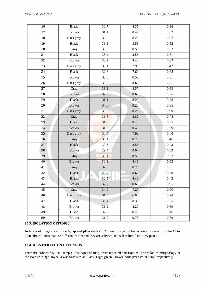
Strain 5
1.subculturing 2.Rhizopusspp,
TABLE3: ANTIFUNGAL ACTIVITY OF ALOE VERA AGAR WELL DIFFUSION (GEL
EXTRACT)METHOD
FUNGAL
STRAINS
1.00 g 0.500 g 0.250 g 0.125 g CONTROL
Aspergillusspp, No zone 0.1 0.5 1.0 No zone
Fusariumspp, No zone No zone 0.4 0.9 No zone
Mucorspp, No zone 0.1 0.3 0.7 No zone
Penicilumspp, No zone No zone 0.4 0.9 No zone
Rhizopusspp, No zone 0.2 0.6 1.1 No zone
TABLE 4: ANTIFUNGAL ACTIVITY OF ALOE VERA AGAR WELL DIFFUSION METHOD(Aqueous extract)
FUNGAL STRAINS 1.00 g 0.500 g 0.250 g 0.125 g CONTROL
Aspergillusspp, No zone 0.1 0.4 0.9 No zone
Fusariumspp, No zone No zone 0.3 0.7 No zone
Mucorspp, 0.1 0.3 0.7 0.9 No zone
Penicilumspp, No zone No zone 0.4 0.9 No zone
Rhizopusspp, No zone No zone 0.2 0.7 No zone
TABLE 5: DETERMINATION OF MINIMUM INHIBITORY
CONCENTRATION (MIC)
BROTH DILUTION METHOD (GEL EXTRACT)
Vol-7 Issue-1 2021 IJARIIE-ISSN(O)-2395-4396
13646 www.ijariie.com 1183
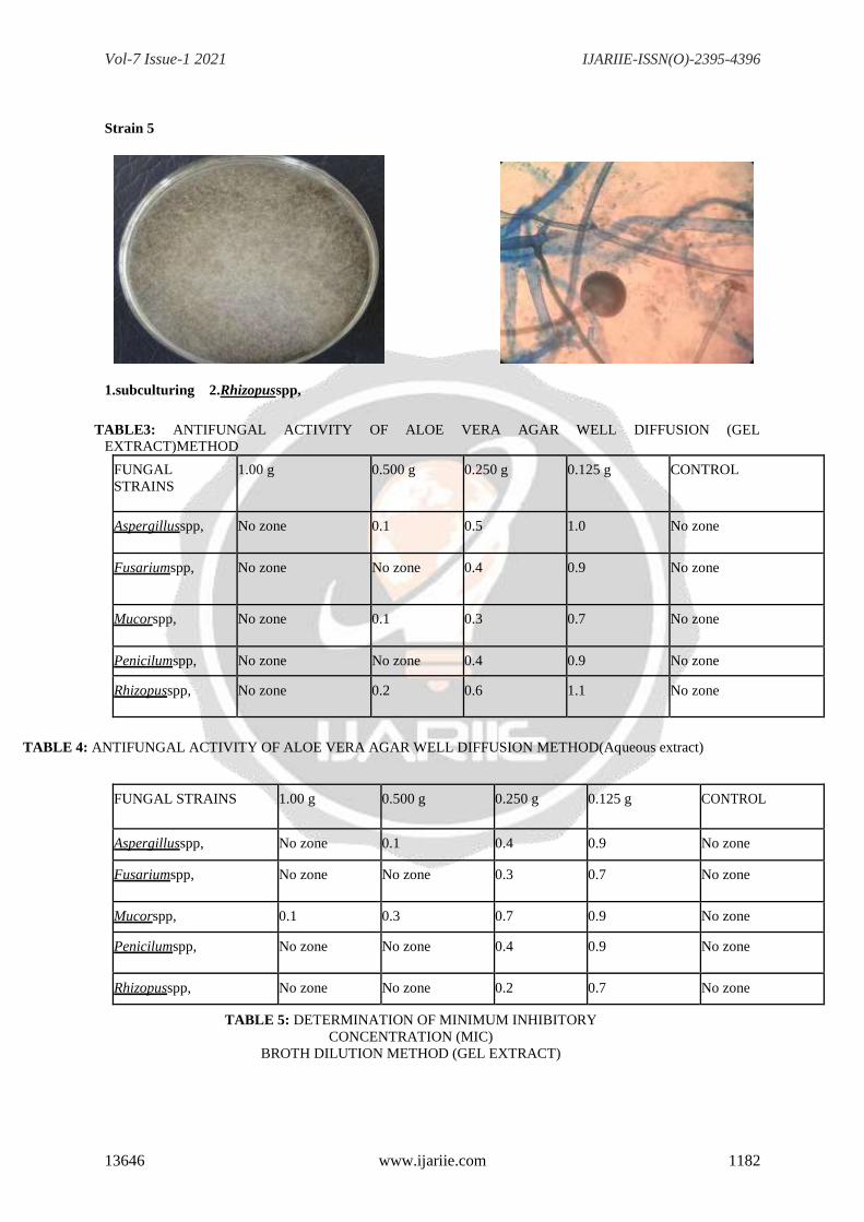
TABLE 6: DETERMINATION OF MINIMUM INHIBITORY CONCENTRATION (MIC)BROTH
DILUTION METHOD (AQUEOUS EXTRACT -POWDER)
FUNGAL STRAINS
512
256
128
64
32
16
8
4
2
1 contro l
Aspergillusspp,
-
-
-
-
-
+
+
+
+
+
+
Fusariumspp,
-
-
-
-
-
-
+
+
+
+
+
Mucorspp,
-
-
-
-
-
-
+
+
+
+
+
Penicilliumspp,
-
-
-
-
-
-
-
+
+
+
+
Rhizopusspp, -
-
-
-
-
+
+
+
+
+
+
11.DISCUSSION:
Antifungal activity of Aloe vera gel was determined against five plant pathogenic fungi viz., Aspergillus spp,
Penicilliumspp, Mucor spp, Fusarium spp, and Rhizopus spp,
The Aloe vera gel @ 0.125g,0.250g,0.500g & 1.00g concentration tested by agar diffusion plate method caused
significantly reduction in the growth of above-mentioned fungi. The rate of growth reduction was directly
proportional to the concentration of tested material in the medium.
Result showed that Aloe vera gel significantly inhibited the growth of all tested fungi. 0.15% concentration of
Aloe vera gel posses’ remarkable antifungal activity toward all fungi compared to control except Aspergillus
niger; whereas &Penicillium spp, showed moderate antifungal activity at this concentration.
Antifungal activity of Aloe vera gel was determined against five plant pathogenic fungi viz., Aspergillus spp,
Penicillium spp, Mucor spp, Fusarium spp, and Rhizopus spp,
The Aloe vera gel @ 0.125g,0.250g,.500g, and 1.00g concentration tested by agar diffusion plate method caused
significantly reduction in the growth of above-mentioned fungi. The rate of growth reduction was directly
proportional to the concentration of tested material in the medium. Result showed that Aloe vera gel
significantly inhibited the growth of all tested fungi. 0.15% concentration of Aloe vera gel posses’ remarkable
antifungal activity toward all fungi compared to control except Aspergillus niger; Rhizopus spp, whereas
Penicillium spp, Mucor spp, Fusarium spp, showed moderate antifungal activity at this concentration. Only two
fungal species viz., Mucor spp and Fusarium spp, had strong antifungal properties towards Aloe vera gel at
same concentration. The result in close conformity with the tested antifungal activity of natural Aloe vera gel on
four plant pathogenic fungi viz.,Mucor spp, Aspergillus niger; Rhizopus spp, the result showed that natural gel
suppresses the mycelial growth of Penicillium spp, and Fusarium spp,
The in vitro antifungal effect of Malaysian Aloe vera leaf extracts in alcohol and aqueous solutions on two
common pathogenic penicilliumspecies, A. niger and C. albicans, using the zone of inhibition and MIC to
determine antimicrobial activity. We found that both alcohol and aqueous extracts demonstrated notable
antifungal properties against A. niger. The antifungal effect of this study was solvent dependent. The highest
concentrations of alcohol and aqueous extracts displayed the maximum zone of inhibition. Fusarium spp,
showed resistance to both. The no zonesof inhibition of Aspergillus spp, for both the gel and aqueous extracts
for all five concentrations tested.
The used the agar diffusion method and crude Aloe vera leaf extracts in methanol, ethanol, and ethyl acetate and
found that all three extracts had no zone of inhibition for the Fusarium spp, As for A. niger, the zone of
inhibition was highest for the methanol extract followed by gel and aqueous extracts. According to Aqueous
extract was effective secondary to its constituent of extraction. The resistance of Rhizopus spp, towards Aloe
vera leaf extracts in our study compared to other regions may be due to geographical and climatic conditions,
which may affect the phytochemical composition of the plant and its antifungalactivity

Vol-7 Issue-1 2021 IJARIIE-ISSN(O)-2395-4396
13646 www.ijariie.com 1184
12.CONCLUSION:
Fungi have many different structures but they can act in similar ways and thus are not as plant specific in their
needs as some soil bacteria such as Rhizobia.
The total hydroalcoholic plant extract obtained from Aloe vera fresh gel and powder extract had antifungal
activity against the mycelial growth of Aspergillus spp, Fusariumspp,Penicillium spp, Mucor spp, and Rhizopus
spp, compared to the control (Gentamycin).
Results bring new information to the literature data about the antifungal activity of A. vera plant extract against
the mycelial growth, on Czapek-agar medium, of phytopathogenic fungi isolated from ornamental plants.
These isolated fungal spp, are sensitive to maximum amount of ALOE VERA extract the zone were observed in
1.00g and 0.500g of concentration of A. Vera.
REFERANCES:
1. Agrios et al.., (2004) Antifungal activity of Allium, Aloe, and Solanumspecies.
Pharmaceutical Biology, 42(7): 491-498.
2. Ahmad et al.., (2005) Antifungal effects of different plant extracts and their major components of
selected Aloe species. Phytotherapy Research, 13(5): 401-407.
3. Athertnop et al.., (1996) Inhibitory effect of botanical extracts against Alternariaalternata
of Aloe vera dry rot. Archives of Phytopathology and Plant Protection, 44(15): 1462-1466.
4. Bajwa et al.., (2007) For Aloe vera - A Semi Finished Products like gel powder, like Aloe vera
Drink or Fizzy Tablet. Technology Transfer and Project Management Network. Ensymm Consulting
forBiotechnology.
5. Bills et al.., (2004) Appraisal of antifungal activity of Aloe vera. Mycopathology, 5(1):5-9.
6. Byeons S and Pelley RE et al., (1998) Antisporulant activity of watery extracts of plants against
Sclerospora graminicola causing Downy mildew disease of Pearl Millet. American Journal of Agricultural and
Biological Science, 2(1):36-42.
7. Chandrakasan G et al.., (1998) Illustrated handbook of succulent plants: Monocotyledons, Springer.
Reynolds T., Bot. J. Linn. Soc., 19:157.
8. Chandramohan and Bharathi et al.., (2009) In vitro evaluation of crude extracts of Catharanthus
roseus for potential antifungal activity. International Journal of Green Pharmacy, 2(3):176-181.
9. Coleman et al.., (2001) The A. vera phenomenon: a review of the properties and modern uses of the
leaf parenchyma gel. J. Ethnopharm, 16:117–151.
10. Cowan et al.., (1999) A drug for all seasons: medical and pharm. History of Aloe. Bull. NY Acad.
Med., 66:647–659.
11. Cowan et al.., (2001) Evaluation of Plant Extracts, Bio-agents and Fungicides Against Exserohilum
turcicum (Pass.) Leonard and Suggs. Causing Turcicum Leaf Blight of Maize. Karnataka J. Agric. Sci.,
20(3):541-544.
12. Demo and Olive et al.., (2008) Antifungal Effect of Aloe-Vera Gel on Fungal Organisms
Associated with Yam (Dioscorea Rotundata, Poir) Rot. Journal of Molecular Genetics, 1(1):11-17.
13. Division et al.., (2007) Evaluation of the antifungal and antioxidant activities of the leaf extract of
Aloe vera (Aloe barbadensis Miller). World Acad. of Sci., Engg. and Techno., 75: 610-612.
14. Gaddeyyaetal..,(2012)ReviewonantifungalactivitiesofAyurvedicMedicinalPlants.
Drug Invention Today, 2(2): 146-148.
15. Garret et al.., (1950) Antifungal activity of medicinal plants against plant pathogenic fungus
Fusarium oxysporum. J. Pharmacy Res., 4(3):843-844.
16. Harris et al.., (2001) Antifungal Activity of Some Extractives and Constituents of Aloe vera.
Research Journal of Medicinal Plant, 5:196-200.
17. Hutter JA and Salmon M et al.., (1996) Comparative efficacy of fungicides and biological control
agents against fungal rots in vanilla - an in vitro study. Karnataka Journal of Agricultural Sciences, 19(1):170-
173.
18. Iram et al ..., (2011) Antifungal activity of medicinal plants against plant pathogenic fungus
Colletotrichum falcatum. Asian J. Pl. Sci. and Res., 1(1):84-87.
19. Ishiiy and Tanizawa H et al..,(1994)
20. Aloe vera leaf gel: a review update. J. Ethnopharm, 68: 3–37.
Vol-7 Issue-1 2021 IJARIIE-ISSN(O)-2395-4396
13646 www.ijariie.com 1185
21. Jenkins et al.., (2005) Antifungal activity in vitro of Aloe vera pulp and liquid fraction against plant
pathogenic fungi. Industrial Crops Products, 21: 81–87.
22. Khaing et al.., (2011) Antifungal activity of Aloe vera leaves. Fitoterapia, 78(3):219-222.
23. Khalil et al.., (2013) Antifungal activity of Aloe vera gel against plant pathogenicfungi.
Pakistan. J. Bot.,
24. LopesandMartinsetal..,(2008)Aloeveragelactivityagainstplantpathogenicfungi.
Postharvest Biology and Technology, 6(1&2): 159-165.
25. Kim HS and Lee BM et al.., (1997) Management of alternaria blight of mustard through botanicals.
Flora and Fauna, 11(2):234-235.
26. Larsson et al.., (2007) Efficacy of selected plant extracts in the control of fungal dry rot of white
yam (Dioscorea rotundata) tubers in Kogi State. American-Eurasian Journal of Sustainable Agriculture,
3(3):310-313.
27. Larsson et al.., (2007) Comparative studies of the efficacy of some selected fungicidal aqueous
plant extracts on yam tuber dry rot disease. Annals of Biological Research, 2(2): 332-336.
28. Meletiadis et al.., (2003) Inhibition activities of extracts from Aloe spp. with different solvents
against Alternaria brassicae. Southwest China Journal of Agricultural Sciences, 22(5):1336-1340.
29. Meletiadis et al.., (2001) Phytochemical screening and comparative study of antimicrobial activity
of Aloe vera various extracts. African Journal of Microbiology Research, 5(10): 1182-1187.
30. Okafor et al.., (2009) Aloe vera gel activity against plant pathogenic fungi. Post-Harvest Bio. and
Techn., 6:159-165.
31. OttowandGlatheetal..,(1968)IdentificationofsomeproteinoidsinAloeveraextracts.
Planta Media., 57: 38-40.
32. Rabah and Ibrahim et al.., (2010) Comparative antimicrobial activities of Aloe vera gel and leaf.
African Journal of Biotechnology., 4(12): 1413-1414.
33. Robert DB and Travis EL et al.., (1995) Analysis of phytochemical constituents and antimicrobial
activities of Alvera L. against clinical pathogens. World Journal of Agricultural Sciences, 5(5):572-576.
34. Roy and Lee B et al.., (2000) Appraisal of antifungal activity of Aloe vera. Myopathy., 5(1):5-9
35. Rosca – Casian et al.., (2007) Antifungal activity of Aloe vera leaves. Fitoterapia., 78(3): 219-222.
36. Santos et al.., (2007) Antifungal activity of Aloe vera leaves. Fitoterapia, 78(3):219-222.
37. Saraswathy et al.., (2010) Antimicrobial activity of Aloe barbadensis Miller leaf gel components.
The International Journal of Microbiology, 4(2)-ISSN:1937-8289.
38. Saravana Kumar and kaviyarasan et al.., (2010) Traditional use, antibacterial activity and antifungal
activity of crude extract of Aloe excelsa. African Journal of Biotechnology, 6(20): 2406-2410.
39. Sharma et al.., (2010) Chrysophanol, an antimicrobial anthraquinone from the root extract of
Colubrina gregii. J. Mex. Chem. Soc., 50(2):76-78.
40. Swer et al.., (2011) Antifungal activity In vitro of Aloe vera pulp and liquid fraction against plant
pathogenic fungi. Industrial Crops and Products, 21(1):81-87.
41. Wahegaonkar et all., (1011) Perspective of industrial application of Aloe vera. In: New Perspective
on Aloe. (Eds.): Y.I. Park and S.K. Lee. Springer Verlag, New York, USA, pp: 191-200. ISBN:0387317996.
42. West DP and Zhu YF et al.., (2003) Aloe vera leaf gel: or review update. J. Ethnopharmacology,
68:3-37


![Research Article Extracellular Biosynthesis of …downloads.hindawi.com/journals/jnm/2015/789089.pdfe synthesis has been reported mainly using plant extracts, such as soya [ ], Aloe](https://img.pdfslide.net/doc/110x75/5f7fa750068d7a347951c3df/research-article-extracellular-biosynthesis-of-e-synthesis-has-been-reported-mainly.jpg)