Embed Size (px)
Citation preview

Research ArticleAntioxidant and Antimicrobial Properties of CactusPear (Opuntia) Seed Oils
Esther Ramírez-Moreno,1 Raquel Cariño-Cortés,2 Nelly del Socorro Cruz-Cansino,1
Luis Delgado-Olivares,1 José Alberto Ariza-Ortega,1 Vanessa Yelina Montañez-Izquierdo,3
María Manuela Hernández-Herrero,3 and Tomás Filardo-Kerstupp4
1Academic Area of Nutrition, Health Sciences Institute, Autonomous University of Hidalgo State, 42160 Pachuca, HGO, Mexico2Academic Area of Medicine, Autonomous University of Hidalgo State, Eliseo Ramırez Ulloa 400, 42090 Pachuca, HGO, Mexico3Department of Food Hygiene, Faculty of Veterinary, Autonomous University of Barcelona, 08193 Bellaterra, Spain4Academic Area of Chemistry, Basic Science and Engineering Institute, Autonomous University of Hidalgo State,Carretera Pachuca-Tulancingo Km. 4.5, Mineral de la Reforma, 42183 Pachuca, HGO, Mexico
Correspondence should be addressed to Nelly del Socorro Cruz-Cansino; [email protected]
Received 23 January 2017; Accepted 5 April 2017; Published 26 April 2017
Academic Editor: Andrea Laukova
Copyright © 2017 Esther Ramırez-Moreno et al.This is an open access article distributed under the Creative CommonsAttributionLicense, which permits unrestricted use, distribution, and reproduction in anymedium, provided the originalwork is properly cited.
Seed oils from two Mexican varieties of cactus pear (green: Opuntia albicarpa and red: Opuntia ficus indica) were extracted withdifferent solvents (hexane, ethanol, and ethyl acetate) to evaluate their antioxidant activity. The seed oil with higher antioxidantactivitywas selected to evaluate antimicrobial activity.The fatty acid profilewas analyzed by gas chromatography-mass spectrometry(GC-MS). Oil from green cactus pear seeds obtained with ethanol and ethyl acetate exhibited higher antioxidant activity (𝑝 < 0.05)of 323 and 316𝜇mol TE/20mg (p < 0.05), respectively, compared to red cactus pear seed oil (≈274 and 247 𝜇mol TE/20mgwith ethyl acetate and ethanol, resp.). The oil obtained with ethanol and higher antioxidant activity was used to determine theantimicrobial activity. Both cactus pear oils produced a microbial inhibition zone in most of the microorganisms evaluated,particularly Saccharomyces cerevisiae which had similar diameter (38–40mm). The oil fatty acids profiles of both varieties weresimilar and exhibited a high content of linoleic acid, while two fatty acids (linolenic and behenic) found in red cactus pear were notobserved in the green variety.
1. Introduction
A relatively untapped source of lipid and protein rawmaterialis the by-product of fruit-processing plants. Millions ofpounds of fruit seeds are discarded yearly causing disposalproblems, while proper utilization of these waste productscould lead to an important new source of oil and meal [1].Seeds of fruits collect at least some cytoplasmic lipid bodies asmajor storage reserve for lipid accumulation [2]. Fruit seedsoils are of great interest because they are edible oils with highdegree of unsaturation, antioxidant radical scavenging prop-erties [3–8], and a broad spectrum of antimicrobial activity[9–15]. Therefore, the oil from plants can be potentially usedby the food industry for the manufacturing of “natural” or“green” safe foods [16] and extend shelf-life [17, 18].
The oil from cactus pear seed has been found to havean appreciable amount of oil with high levels of unsaturatedfatty acids [19], with antioxidant [20, 21] and antimicrobialactivity [22], as well as cardioprotective, antithrombotic, anti-inflammatory, antiarrhythmic, hypolipidemic, and antihy-perglycemic effect [23, 24]. These properties are of interestfor the pharmaceutical and food industry. However, theconcentration and effectiveness of these oils may vary amongcultivars or varieties, crop environmental factors (e.g., light,temperature, and type of soil nutrients), or methods andsolvents used for their extraction. Therefore, the purpose ofthis research was to determine the antioxidant and antimi-crobial activity, and fatty acid profile of the oil obtained fromtwo Mexican varieties of cactus pear (Opuntia albicarpa andOpuntia ficus indica) seeds extracted with different solvents.
HindawiJournal of Food QualityVolume 2017, Article ID 3075907, 8 pageshttps://doi.org/10.1155/2017/3075907

2 Journal of Food Quality
2. Materials and Methods
2.1. Plant Material. Two Mexican varieties of cactus pear(Opuntia albicarpa and Opuntia ficus indica) fruit, green (cv.Reyna) and red (cv. Rojo Pelon), respectively, were providedby theMexican association CoMeNTuna (ConsejoMexicanodel Nopal y la Tuna, A.C.; Actopan, Hidalgo, Mexico). Fruitsfree of external injuries were selected, washed, and manuallypeeled. Cactus pear seeds were obtained after juice wasextracted stirring the pulpwith an industrial blender (38BL52LBC10, Waring Comercial�, USA) and passing it through aconventional strainer. The seeds retained were washed in thestrainer with water until pulp residues were removed.
2.2. Powder Seed and Oil Extraction. Green cactus pear seeds(GCPS) and red cactus pear seeds (RCPS)were sun-dried andthen grounded (Cyclotec 1093, Tecator Sweden) to a 1mmdiameter mesh and stored at −32∘C until further analysis.Theseed oil was extracted as follows: 25 g of powdered seeds wasmixed with 500mL of solvents with varying polarities (hex-ane, ethanol, and ethyl acetate) and the obtained residue wasreextracted until extraction solvents become colourless. Allthe extracts were filtered through filtration paper Whatmannumber 1 and the filtered extracts were collected for furtherdrying and removal of the remaining solvent at 50∘C using arotary evaporator (BUCHI, R-200, Switzerland). All extractswere placed in plastic bottles and then stored at −20∘C untilused. The oils obtained were used to further analysis.
2.3. Free Radical Scavenging Assay. The free radical scaven-ging activity was measured using 1,1-diphenyl-2-picrylhy-drazyl (DPPH∙) radical as described byMorales and Jimenez-Perez [25]. A volume of 500 𝜇L of ethanolic DPPH∙ solution(7.4mg/100mL) was added to a sample aliquot of 100 𝜇Lplaced in vials.Themixturewas left to sit at room temperaturefor 1 h and then was vortexed and centrifuged at 3000 rpm for10 minutes.The absorbance of the supernatant was measuredat 520 nm in amicroplate reader (PowerWave XSUV-Biotek,software KC Junior, USA), and 𝜇mol of Trolox equivalentsper 20 milligram (𝜇mol TE/20mg) of sample was obtained.Oil samples with best antioxidant capacity obtained from thedifferent solvents were used for the antimicrobial analysis.
2.4. Antimicrobial Activity. Eight standard freeze-dried cul-tures of bacteria, Candida albicans (ATCC 10231), Escherichiacoli O58:H21 (ATCC 10536), Escherichia coli O157:H7(CCUG 44857), Staphylococcus aureus (ATCC 13565), Liste-ria monocytogenes (CCUG 15526), Pseudomonas aeruginosa(ATCC 15442), Saccharomyces cerevisiae (CECT 1942), andSalmonella Typhi (CCUG 29478) were obtained in ther-mosealed vials from the Spanish Type Culture Collection(Autonomous University of Barcelona, Barcelona, Spain).Freeze-dried cultures were rehydrated in tryptone soy brothat 37∘C for 18 h and then were used to inoculate tryptone soyagar and malt extract agar plates; all microorganisms wereincubated at 37∘CexceptCandida albicans and Saccharomycescerevisiae which were incubated at 25∘C. Individual colonies
were maintained on specific agar slants, stored at 4∘C, andsubcultured every 15 days.
Disc Diffusion Assay. Antimicrobial activity of oil extractedfromGCPS andRCPSwas carried out using the disc diffusionmethod [26]. Petri plates were filled with ∼20mL of steriletryptone soy agar for bacteria and malt extract agar for fungi.The test cultures were swabbed on the top of the solidifiedmedia and allowed to dry for 10 minutes. Serial dilutions(10–50 𝜇g/mL) of the seed oil from a stock solution (1mg/mL)were prepared in 20% DMSO and 10 𝜇L loaded onto thesterile blank discs (BBL� Sensi-Disc�) of 6 millimetersof diameter. On the media surface the loaded disks wereplaced and left for 30 minutes at room temperature toallow compound diffusion. The seed oil was serially dilutedin Mueller–Hinton broth medium and duplicate tubes ofeach dilution (10–100 𝜇g/mL) were inoculated with 5 ×10⁶ cells of the test bacteria strain and cultures.The antibioticagents Sensi-Disc streptomycin, ampicillin, and sulfamethox-azole/trimethoprim (BBL Sensi-Disc) were used as positivecontrols at the same concentration level. After plates wereincubated at 37∘C for 24 h, the diameters of the inhibitionzones were recorded in millimeters. Three independentrepetitions were performed and tests were made in triplicate.
2.5. GC-MS Analysis. The GC-MS analysis was performedwith a GC-MS HP-5890 (Hewlett-Packard Company, PaloAlto, California, USA) equipped with a Flame IonizationDetector (FID), and a ZB-WAX fused silica capillary column(60m × 0.25mm i.d. × 0.25mm film thickness) packedwith 5% phenylmethylpolysiloxane (Phenomenex, Torrance,CA). To obtain the methyl esters, the cactus pear seed oilswere saponified and derivatized using KOH 1N (IUPAC,1969). Changes in the fatty acids of the oils samples werecompared against a standard mixture of 37 components offatty acids methyl esters (FAMEs) (Food Industry FAMEsMix, Restek) comprised by methyl esters with chains C4:0,C6:0, C8:0, C10:0, C11:0, C12:0, C13:0, C14:0, C14:1, C15:0,C15:1, C16:0, C16:1, C17:0, C17:1, C18:0, C18:1n9c, C18:1n9t,C18:2n6c, C18:2n6t, C18:3n6, C18:3n3, C20:0, C20:1n9, C20:2,C20:3n6, C20:3n3, C20:4n6, C20:5n3, C21:0, C22:0, C22:1n9,C22:2, C22:6n3, C23:0, C24:0, and C24:1n9. The samplevolume injected was of 2 𝜇L (split ratio 20 : 2) at an injectorand detector temperatures of 225 and 225∘C, respectively. N2was used as carrier gas at a flow rate of 1.2mL⋅min−1. Fattyacids were calculated as percentage of total FAMEs.
2.6. Statistical Analysis. All values were obtained by triplicateand expressed as means ± standard deviations (SD). Datawere analyzed using the SPSS V.15 software (SPSS InstituteInc., Cary, NC). An ANOVA was carried out to determinedifferences between oils extracted as well as its antimicrobialactivity that were significant at the 5% level of probability anda Tukey test was used for comparison of data.
3. Results and Discussion
3.1. Yield Comparison between Extraction Solvents. Hexane,ethanol, and ethyl acetate were used to extract the oil from

Journal of Food Quality 3
11.83
6.69
10.13
5.11
10.6
3.81
Gre
en/h
exan
e
Red/
hexa
ne
Gre
en/e
than
ol
Red/
etha
nol
Gre
en/e
thyl
acet
ate
Red/
ethy
l ace
tate
0
2
4
6
8
10
12
14
Oil
yiel
d (%
)
Figure 1: Oil yields (%) extracted from GCPS and RCPS withhexane, ethanol, and ethyl acetate.
cactus pear seeds. The extraction yields are compared inFigure 1, which shows that the higher amount of oil (%) wasobtained from the green cultivar and that yield depended onthe solvent used. Oil extraction with hexane was higher forboth fruit varieties (11.83% for GCPS and 6.89% for RCPS),followed by ethanol, which reached the same yield as ethylacetate for GCPS (≈10%). Ethyl acetate was the least effectivesolvent for RCPS. The extraction yields were similar to thosereported (≈7 to 11%) for several varieties of Opuntia ficusindica [27–29]. This extraction yields will vary depending onseveral factors as fruit variety, harvest period, maturation,geographic region, percentage of oil in the seed, and chemicalcompounds found in the source and by the extractionmethod[30]. Researchers have determined that solvent extractioncombinedwith othermethods could increase oil yield, as highpressure or supercritical fluid combined with solvent reacheda yield of 9.33% from tobacco seeds (Nicotiana tabacum L.)while sonication and Soxhlet reached a 7.75% and 13.72%,respectively [31].
3.2. Free Radical Scavenging Activity. Solvent extraction isusually used for isolation of antioxidants; the extractiondepends on the solvent selected based on the different antiox-idant compounds with varying polarity [32, 33]. DPPH is astable free radical that accepts an electron or hydrogen radicalto become a stable diamagnetic molecule [34, 35].The DPPHassay has also been used to predict the oxidative stability ofedible oils [36, 37]. The antioxidant activity determined byDPPH of the oil extracted from RCPS and GCPS is shownin Figure 2. Oil from the GCPS extracted with ethanol andethyl acetate exhibited the higher antioxidant activity (𝑝 <0.05) of 323 and 316 𝜇mol TE/20mg extract, respectively,followed by RCPS oil extracted with ethyl acetate (274𝜇molTE/20mg extract) and ethanol (247 𝜇mol TE/20mg extract).These results demonstrate that the extraction solvent hada significant effect on the free radical scavenging capacityof the oil, where the hexane had the lower values. In ourstudy, the green variety exhibited a higher antioxidant activityregardless of solvent. Different results may depend mainly
e
d
a
c
ab
Gre
en/h
exan
e
Red/
hexa
ne
Gre
en/e
than
ol
Red/
etha
nol
Gre
en/e
thyl
acet
ate
Red/
ethy
l ace
tate
0
50
100
150
200
250
300
350
�휇m
ol T
E/20
mg
extr
act
Figure 2: Antioxidant activity of GCPS and RCPS oils extractedwith different solvents. a–dDifferent letters above bars indicate thatsamples are significantly different (𝑝 < 0.05).
on the content and concentration of bioactive compounds inthe oil, but other factors such as solvent polarity, solubilityof the extracts in different testing systems, stereoselectivityof the radicals [38], and strong synergism between fattyacids [6] may affect antioxidant activities. Other studies havealso reported diverse antioxidant activity among oils fromdifferent Opuntia varieties [20, 39, 40].
3.3. Antibacterial and Antifungal Activity. The most recom-mended way to prevent or inhibit microbial growth in foodsis the use of food preservatives. Essential oils are secondarymetabolites of plants that have wide applications in thefood flavoring and preservative industry [41]. Six differentbacterium and two fungi species were used to screen theantimicrobial potential of the oils extracted from the twovarieties of cactus pear seeds. Oil extracted with ethanolexhibited the highest antioxidant activity and therefore itwas used to evaluate the antibacterial and antifungal activity.Figure 3 shows the results from the microbial assay wheremost microorganisms showed an inhibition zone whenexposed to GCPS and RCPS oils, except Salmonella Typhiand Escherichia coli O157:H7 (image not shown). From thesetwo microorganisms, the first showed an inhibition zonein the presence of antibiotic agents streptomycin (S), ampi-cillin (AMP), and sulfamethoxazole/trimethoprim (STX) indiameters of 14.6, 11.3 and 27.3mm, respectively (Table 1),while Escherichia coli O157:H7 was only inhibited by SXT(25.3mm), which agrees with other reports of multiantibi-otic resistance of E. coli O157:H7 due to the presence ofthe gene cluster AMR-SSuT [42] and production of beta-lactamase [43]. On the other hand, Saccharomyces cerevisiaewas highly inhibited (38–40mm) by the extracted oils butgrew in presence of the antimicrobial agents (Figure 3).Similar results were observed for Candida albicans, althoughinhibition zones were smaller and similar for both oils.These observations demonstrate that certain compounds inthe cactus pear seed oil have antimicrobial activity. Otherresearchers also reported similar observations for cactus

4 Journal of Food Quality
Table 1: Diameters of growth inhibition zones (mm) in the presence of oil extracted from cactus pear seeds and conventional antimicrobialsA.
Microbial cultures Extract Antimicrobial agentGCPS RCPS S AMP SXT
Saccharomyces cerevisiae (CECT1942) 38.3 ± 4.2a 40.3 ± 4.5a ND ND NDEscherichia coli O58:H21 (ATCC 10536) 11.9 ± 0.7d 11.4 ± 0.9d 19.5 ± 1.4b 17.8 ± 1.5c 29.9 ± 0.9a
Escherichia coli O157:H7 (CCUG 44857) NDB ND ND ND 25.3 ± 1.0Staphylococcus aureus (ATCC 13565) 12.1 ± 1.0c 11.1 ± 1.1c 18.3 ± 0.9b 28.1 ± 1.5a 33.3 ± 1.9a
Listeria monocytogenes (CCUG15526) 13.3 ± 1.5c 11.4 ± 0.9d 21.3 ± 1.2a 17.3 ± 0.8c 37.6 ± 1.8a
Pseudomonas aeruginosa (ATCC15442) 16.4 ± 2.1c 15.1 ± 2.0c 20.1 ± 1.5b 24.1 ± 2.5a 36.6 ± 2.9a
Salmonella typhi (CCUG29478) ND ND 14.6 ± 0.4b 11.3 ± 1.8c 27.3 ± 0.8a
Candida albicans (ATCC 10231) 11.1 ± 1.0a 11.0 ± 1.8a ND ND NDAInhibition zone diameters for oil and reference antibiotics are means ± SE of three replicas. GCPS: green cactus pear seed oil extract, RCPS: red cactus pearseed oil extract, S: streptomycin (10𝜇g/disc), AMP: ampicillin (10 𝜇g/disc), and SXT: sulfamethoxazole/trimethoprim (10𝜇g/disc). BND: not detected activity.a–dDifferent letters in the same row indicate significant differences.
RG
SXT
S
AMP
(a)
RG
SXT
S
AMP
(b)
R G
SXTS
AMP
(c)
RG
SXTS
AMP
(d)
RG
SXT
S
AMP
(e)
R G
SXT
S
AMP
(f)
Figure 3: Antimicrobial activity of oil extracted from green cactus pear (G); oil extracted from red cactus pear seeds (R); streptomycin(S); ampicillin (AMP); and sulfamethoxazole/trimethoprim (SXT). Candida. albicans (ATCC 10231) (a); Escherichia. coli O58:H21 (ATCC10536) (b); Staphylococcus aureus (ATCC 13565) (c); Listeria monocytogenes (CCUG15526) (d); Pseudomonas aeruginosa (ATCC15442) (e);Saccharomyces cerevisiae (CECT1942) (f).
pear fruit cv. Opuntia stricta [44] and for other plants asfennel (Foeniculum vulgare L.) and chamomile (Matricariachamomilla L.) [5]. Differences in the levels of antimicrobialactivity may be partially attributed to variable chemicalcomposition of the oils [45]. Mnayer et al. [46] suggestedthat oil compounds can act on different bacterial structures,while Gill et al. [47] mentioned that whole oils have a greater
antibacterial activity than the major component mixed, sothat minor components are critical for the activity and exerta synergistic effect [16, 48, 49].
In the present study, the antimicrobial activity of cactuspear seed oil was more effective against fungi comparedto bacteria cultures. These interesting results suggest thatthere is a link between the oil chemical contents and the
Journal of Food Quality 5
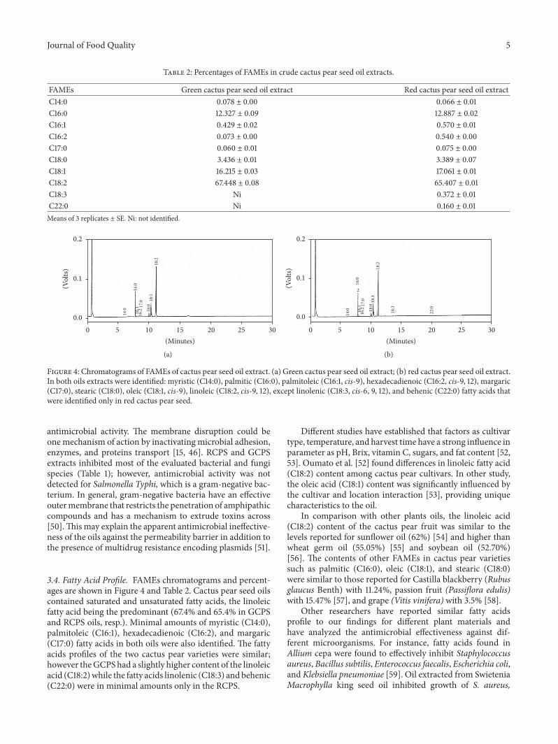
Table 2: Percentages of FAMEs in crude cactus pear seed oil extracts.
FAMEs Green cactus pear seed oil extract Red cactus pear seed oil extractC14:0 0.078 ± 0.00 0.066 ± 0.01C16:0 12.327 ± 0.09 12.887 ± 0.02C16:1 0.429 ± 0.02 0.570 ± 0.01C16:2 0.073 ± 0.00 0.540 ± 0.00C17:0 0.060 ± 0.01 0.075 ± 0.00C18:0 3.436 ± 0.01 3.389 ± 0.07C18:1 16.215 ± 0.03 17.061 ± 0.01C18:2 67.448 ± 0.08 65.407 ± 0.01C18:3 Ni 0.372 ± 0.01C22:0 Ni 0.160 ± 0.01Means of 3 replicates ± SE. Ni: not identified.
14:0
16:0
18:1
18:2
18:017
:016
:116
:2
0.0
0.1
0.2
(Vol
ts)
5 10 15 20 25 300(Minutes)
(a)14
:0 16:1
16:2
17:0
18:0
18:1
18:2
18:3
22:0
16:0
2
0.0
0.1
0.2
(Vol
ts)
5 10 15 20 25 300(Minutes)
(b)
Figure 4: Chromatograms of FAMEs of cactus pear seed oil extract. (a) Green cactus pear seed oil extract; (b) red cactus pear seed oil extract.In both oils extracts were identified: myristic (C14:0), palmitic (C16:0), palmitoleic (C16:1, cis-9), hexadecadienoic (C16:2, cis-9, 12), margaric(C17:0), stearic (C18:0), oleic (C18:1, cis-9), linoleic (C18:2, cis-9, 12), except linolenic (C18:3, cis-6, 9, 12), and behenic (C22:0) fatty acids thatwere identified only in red cactus pear seed.
antimicrobial activity. The membrane disruption could beone mechanism of action by inactivating microbial adhesion,enzymes, and proteins transport [15, 46]. RCPS and GCPSextracts inhibited most of the evaluated bacterial and fungispecies (Table 1); however, antimicrobial activity was notdetected for Salmonella Typhi, which is a gram-negative bac-terium. In general, gram-negative bacteria have an effectiveoutermembrane that restricts the penetration of amphipathiccompounds and has a mechanism to extrude toxins across[50].Thismay explain the apparent antimicrobial ineffective-ness of the oils against the permeability barrier in addition tothe presence of multidrug resistance encoding plasmids [51].
3.4. Fatty Acid Profile. FAMEs chromatograms and percent-ages are shown in Figure 4 and Table 2. Cactus pear seed oilscontained saturated and unsaturated fatty acids, the linoleicfatty acid being the predominant (67.4% and 65.4% in GCPSand RCPS oils, resp.). Minimal amounts of myristic (C14:0),palmitoleic (C16:1), hexadecadienoic (C16:2), and margaric(C17:0) fatty acids in both oils were also identified. The fattyacids profiles of the two cactus pear varieties were similar;however theGCPS had a slightly higher content of the linoleicacid (C18:2)while the fatty acids linolenic (C18:3) and behenic(C22:0) were in minimal amounts only in the RCPS.
Different studies have established that factors as cultivartype, temperature, and harvest time have a strong influence inparameter as pH, Brix, vitamin C, sugars, and fat content [52,53]. Oumato et al. [52] found differences in linoleic fatty acid(C18:2) content among cactus pear cultivars. In other study,the oleic acid (C18:1) content was significantly influenced bythe cultivar and location interaction [53], providing uniquecharacteristics to the oil.
In comparison with other plants oils, the linoleic acid(C18:2) content of the cactus pear fruit was similar to thelevels reported for sunflower oil (62%) [54] and higher thanwheat germ oil (55.05%) [55] and soybean oil (52.70%)[56]. The contents of other FAMEs in cactus pear varietiessuch as palmitic (C16:0), oleic (C18:1), and stearic (C18:0)were similar to those reported for Castilla blackberry (Rubusglaucus Benth) with 11.24%, passion fruit (Passiflora edulis)with 15.47% [57], and grape (Vitis vinifera) with 3.5% [58].
Other researchers have reported similar fatty acidsprofile to our findings for different plant materials andhave analyzed the antimicrobial effectiveness against dif-ferent microorganisms. For instance, fatty acids found inAllium cepa were found to effectively inhibit Staphylococcusaureus, Bacillus subtilis, Enterococcus faecalis, Escherichia coli,and Klebsiella pneumoniae [59]. Oil extracted from SwieteniaMacrophylla king seed oil inhibited growth of S. aureus,

6 Journal of Food Quality
S. Typhimurium, and P. aeruginosa [60]. These studies de-monstrate that seed oil can inhibit fungi and bacteria, buttheir efficacy would depend on their concentration levels andspecific pathogen [15].
4. Conclusions
Oil yield from the green cactus pearwas higher in comparisonto the red cultivar andwas also influenced by the solvent used.Hexane exhibited high extraction yield while oils extractedwith ethanol had the better antioxidant activity. The resultsdemonstrated that oil extracts from both varieties have anoticeable antimicrobial activity against gram-positive andgram-negative bacteria comparable to antimicrobial com-pounds such as ampicillin, streptomycin, and sulfamethoxa-zole/trimethoprim.This research provides further incentivesto develop additives for the food, cosmetic, and pharmaceu-tical sectors seeking natural compounds with antimicrobialactivity. Further studies are needed to determine the specificcomponent responsible for the antimicrobial activity in cac-tus pear seeds oil and determine the optimum levels of oilextract and the antimicrobial effectiveness in the foodmatrix.
Additional Points
Practical Application. Our results suggest that the oilsextracted from cactus pear seeds have the potential to be usedas a natural antioxidant and antimicrobial agents by the food,cosmetic, and pharmaceutical sectors.
Conflicts of Interest
The authors have declared that no conflicts of interest exist.
Acknowledgments
This study was possible thanks to the financial supportfrom the Programa Integral de Fortalecimiento Institucional(PIFI 2013–2015), Mexico. This research project was partiallysupported by the Food Hygiene Department, Faculty ofVeterinary from the Autonomous University of Barcelona,Spain. The authors acknowledge the Mexican associationCoMeNTuna (Consejo Mexicano del Nopal y la Tuna,Hidalgo, Mexico) for providing the plant materials.
References
[1] B. S. Kamel and Y. Kakuda, “Fatty acids in fruits and fruit prod-ucts,” in Acids in Foods and Their Health Implications, pp. 263–301, CRC Press, 3rd edition, 2007.
[2] D. J. Murphy, I. Hernandez-Pinzon, and K. Patel, “Role oflipid bodies and lipid-body proteins in seeds and other tissues,”Journal of Plant Physiology, vol. 158, no. 4, pp. 471–478, 2001.
[3] A. Kedia, B. Prakash, P. K.Mishra, andN. K. Dubey, “Antifungaland antiaflatoxigenic properties of Cuminum cyminum (L.)seed essential oil and its efficacy as a preservative in storedcommodities,” International Journal of Food Microbiology, vol.168-169, pp. 1–7, 2014.
[4] S. Singh, S. S. Das, G. Singh, C. Schuff,M. P. de Lampasona, andC. A. Catalan, “Composition, in vitro antioxidant and antimi-crobial activities of essential oil and oleoresins obtained fromblack cumin seeds (Nigella sativa L.).,” BioMed Research Inter-national, vol. 2014, Article ID 918209, 10 pages, 2014.
[5] M. H. H. Roby, M. A. Sarhan, K. A.-H. Selim, and K. I. Khalel,“Antioxidant and antimicrobial activities of essential oil andextracts of fennel (Foeniculum vulgare L.) and chamomile(Matricaria chamomilla L.),” Industrial Crops and Products, vol.44, pp. 437–445, 2013.
[6] G. Kavoosi, A. Tafsiry, A. A. Ebdam, and V. Rowshan, “Evalua-tion of antioxidant and antimicrobial activities of essential oilsfrom carum copticum seed and ferula assafoetida latex,” Journalof Food Science, vol. 78, no. 2, pp. T356–T361, 2013.
[7] S. Lalas, O. Gortzi, V. Athanasiadis, J. Tsaknis, and I. Chinou,“Determination of antimicrobial activity and resistance tooxidation of Moringa peregrina seed oil,”Molecules, vol. 17, no.3, pp. 2330–2334, 2012.
[8] B. Ozcan, M. Esen, M. K. Sangun, A. Coleri, and M. Caliskan,“Effective antibacterial and antioxidant properties of methano-lic extract of Laurus nobilis seed oil,” Journal of EnvironmentalBiology, vol. 31, no. 5, pp. 637–641, 2010.
[9] R. S. Bhat and S. Al-daihan, “Antimicrobial activity of Litchichinensis and Nephelium lappaceum aqueous seed extractsagainst some pathogenic bacterial strains,” Journal of King SaudUniversity—Science, vol. 26, no. 1, pp. 79–82, 2014.
[10] J. M. Silvan, E. Mingo, M. Hidalgo, S. de Pascual-Teresa, A. V.Carrascosa, and A. J. Martinez-Rodriguez, “Antibacterial activ-ity of a grape seed extract and its fractions against Campylobac-ter spp.,” Food Control, vol. 29, no. 1, pp. 25–31, 2013.
[11] K. Rakholiya, M. Kaneria, D. Desai, and S. Chanda, “Antimi-crobial activity of decoction extracts of residual parts (seedand peels) ofMangifera indica L. var. Kesar against pathogenicand food spoilage microorganism,” in Microbial pathogens andstrategies for combating them: science, technology and education,A. Mendez-Vilas, Ed., vol. 2, pp. 850–856, 2013.
[12] N. A. Hasan, M. Z. Nawahwi, and H. Ab Malek, “Antimicrobialactivity ofNigella sativa seed extract,” Sains Malaysiana, vol. 42,no. 2, pp. 143–147, 2013.
[13] Z. I. Sajid, F. Anwar, G. Shabir, G. Rasul, K.M. Alkharfy, and A.-H. Gilani, “Antioxidant, antimicrobial properties and phenolicsof different solvent extracts from bark, leaves and seeds ofPongamia pinnata (L.) pierre,”Molecules, vol. 17, no. 4, pp. 3917–3932, 2012.
[14] M. Khoobchandani, B. K. Ojeswi, N. Ganesh et al., “Antimicro-bial properties and analytical profile of traditional Eruca sativaseed oil: comparisonwith various aerial and root plant extracts,”Food Chemistry, vol. 120, no. 1, pp. 217–224, 2010.
[15] V. Kesari, A. Das, and L. Rangan, “Physico-chemical charac-terization and antimicrobial activity from seed oil of Pongamiapinnata, a potential biofuel crop,” Biomass and Bioenergy, vol.34, no. 1, pp. 108–115, 2010.
[16] K. Carovic-Stanko, S. Orlic, O. Politeo et al., “Composition andantibacterial activities of essential oils of seven Ocimum taxa,”Food Chemistry, vol. 119, no. 1, pp. 196–201, 2010.
[17] M.M. Tajkarimi, S. A. Ibrahim, andD.O. Cliver, “Antimicrobialherb and spice compounds in food,” Food Control, vol. 21, no. 9,pp. 1199–1218, 2010.
[18] F. Solorzano-Santos andM. G.Miranda-Novales, “Essential oilsfrom aromatic herbs as antimicrobial agents,” Current Opinionin Biotechnology, vol. 23, no. 2, pp. 136–141, 2012.

Journal of Food Quality 7
[19] N. Chougui, A. Tamendjari, W. Hamidj et al., “Oil compositionand characterisation of phenolic compounds of Opuntia ficus-indica seeds,” Food Chemistry, vol. 139, no. 1–4, pp. 796–803,2013.
[20] W. Liu, Y.-J. Fu, Y.-G. Zu et al., “Supercritical carbon dioxideextraction of seed oil from Opuntia dillenii Haw. and itsantioxidant activity,” Food Chemistry, vol. 114, no. 1, pp. 334–339,2009.
[21] B. Matthaus and M. M. Ozcan, “Habitat effects on yield, fattyacid composition and tocopherol contents of prickly pear(Opuntia ficus-indica L.) seed oils,” Scientia Horticulturae, vol.131, pp. 95–98, 2011.
[22] P. Zito, M. Sajeva, M. Bruno, S. Rosselli, A. Maggio, and F.Senatore, “Essential oils composition of two Sicilian cultivars ofOpuntia ficus-indica (L.)Mill. (Cactaceae) fruits (prickly pear),”Natural Product Research, vol. 27, no. 14, pp. 1305–1314, 2013.
[23] K.Mobraten, T.M.Haug, C. R. Kleiveland, and T. Lea, “Omega-3 and omega-6 PUFAs induce the same GPR120-mediatedsignalling events, but with different kinetics and intensity inCaco-2 cells,” Lipids in Health and Disease, vol. 12, no. 1, ArticleID 101, 2013.
[24] A. Berraaouan,A. Ziyyat,H.Mekhfi et al., “Evaluation of antidi-abetic properties of cactus pear seed oil in rats,” PharmaceuticalBiology, vol. 52, no. 10, pp. 1286–1290, 2014.
[25] F. J. Morales and S. Jimenez-Perez, “Free radical scavengingcapacity of Maillard reaction products as related to colour andfluorescence,” Food Chemistry, vol. 72, no. 1, pp. 119–125, 2001.
[26] P. R. Murray, E. J. Baron, M. A. Pfaller, F. C. Tenover, and R. H.Yolke,Manual of Clinical Microbiology, 1995, Washington, DC,USA.
[27] M. Ennouri, H. Fetoui, E. Bourret, N. Zeghal, and H. Attia,“Evaluation of some biological parameters of Opuntia ficusindica. 1. Influence of a seed oil supplemented diet on rats,”Bioresource Technology, vol. 97, no. 12, pp. 1382–1386, 2006.
[28] Y. Habibi, L. Heux, M. Mahrouz, and M. R. Vignon, “Morpho-logical and structural study of seed pericarp of Opuntia ficus-indica prickly pear fruits,” Carbohydrate Polymers, vol. 72, no. 1,pp. 102–112, 2008.
[29] M.Mouden,M. Boujnah, S. Salmaoui, S. Zantar, and A. Douira,“Effect of two extraction methods and harvest period andperformance there statement of fatty oils of figs pear seed,”International Journal of Pure & Applied Bioscience, vol. 4, no.1, pp. 1–8, 2016.
[30] A. El Finti, M. Belayadi, R. El Boullani, F. Msanda, and A. ElMousadik, “Assessment of some agro-technological parametersof cactus pear fruit (Opuntia ficus-indica Mill.) in Moroccocultivars,” Journal of Medicinal Plants Research, no. 7, pp. 2574–2583, 2013.
[31] S. Majdi, M. Barzegar, A. Jabbari, and M. Agha Alikhani,“Supercritical fluid extraction of tobacco seed oil and itscomparison with solvent extraction methods,” Journal of Agri-cultural Science and Technology, vol. 14, no. 5, pp. 1043–1051,2012.
[32] E. M. Marinova and N. V. Yanishlieva, “Antioxidative activity ofextracts from selected species of the family Lamiaceae insunflower oil,” Food Chemistry, vol. 58, no. 3, pp. 245–248, 1997.
[33] Y.-Y. Soong and P. J. Barlow, “Antioxidant activity and phenoliccontent of selected fruit seeds,” Food Chemistry, vol. 88, no. 3,pp. 411–417, 2004.
[34] J. R. Soares, T. C. P. Dinis, A. P. Cunha, and L. M. Almeida,“Antioxidant activities of some extracts of Thymus zygis,” FreeRadical Research, vol. 26, no. 5, pp. 469–478, 1997.
[35] I. Gulcin,M.Oktay, E. Kirecci, and O. I. Kufrevioglu, “Screeningof antioxidant and antimicrobial activities of anise (Pimpinellaanisum L.) seed extracts,” Food Chemistry, vol. 83, pp. 371–382,2003.
[36] J. Lee, H. Chung, P.-S. Chang, and J. Lee, “Development of amethod predicting the oxidative stability of edible oils using 2,2-diphenyl-1-picrylhydrazyl (DPPH),” Food Chemistry, vol. 103,no. 2, pp. 662–669, 2007.
[37] A. Fazio, P. Plastina, J. Meijerink, R. F. Witkamp, and B.Gabriele, “Comparative analyses of seeds of wild fruits ofRubus and Sambucus species from Southern Italy: fatty acidcomposition of the oil, total phenolic content, antioxidant andanti-inflammatory properties of the methanolic extracts,” FoodChemistry, vol. 140, no. 4, pp. 817–824, 2013.
[38] R. L. Prior, X. Wu, and K. Schaich, “Standardized methods forthe determination of antioxidant capacity and phenolics infoods and dietary supplements,” Journal of Agricultural andFood Chemistry, vol. 53, no. 10, pp. 4290–4302, 2005.
[39] A. Cardador-Martınez, C. Jimenez-Martınez, and G. Sandoval,“Revalorization of cactus pear (Opuntia spp.) wastes as a sourceof antioxidants,” Ciencia e Tecnologia de Alimentos, vol. 31, no.3, pp. 782–788, 2011.
[40] Z. Ghazi, M. Ramdani, M. Tahri et al., “Chemical compositionand antioxidant activity of seeds oils and fruit juice,” Journalof Materials and Environmental Science, vol. 6, no. 8, pp. 2338–2345, 2015.
[41] V. K. Bajpai, K.-H. Baek, and S. C. Kang, “Control of Salmonellain foods by using essential oils: a review,” Food ResearchInternational, vol. 45, no. 2, pp. 722–734, 2012.
[42] K. Ziebell, R. P. Johnson, A. M. Kropinski et al., “Genecluster conferring streptomycin, sulfonamide, and tetracyclineresistance in Escherichia coli O157:H7 phage types 23, 45, and67,” Applied and Environmental Microbiology, vol. 77, no. 5, pp.1900–1903, 2011.
[43] O. Chandra, K. J. Putra, and I. Wayan, “Antibiotic resistanceprofiles of Escherichia coli O157:H7 in cattle at Suth-Kuta,Badung Regency. Bali, Indonesia.,” Global Veterinaria, vol. 15,no. 5, pp. 480–484, 2015.
[44] E. Moosazadeh, M. R. Akhgar, and A. Kariminik, “Chemicalcomposition and antimicrobial activity of Opuntia stricta F.essential oil,” Journal of Biodiversity and Environmental Sciences,vol. 4, pp. 94–101, 2014.
[45] S. Burt, “Essential oils: their antibacterial properties and poten-tial applications in foods - a review,” International Journal ofFood Microbiology, vol. 94, no. 3, pp. 223–253, 2004.
[46] D. Mnayer, A.-S. Fabiano-Tixier, E. Petitcolas et al., “Chemicalcomposition, antibacterial and antioxidant activities of sixessentials oils from the Alliaceae family,” Molecules, vol. 19, no.12, pp. 20034–20053, 2014.
[47] A. O. Gill, P. Delaquis, P. Russo, and R. A. Holley, “Evaluationof antilisterial action of cilantro oil on vacuum packed ham,”International Journal of FoodMicrobiology, vol. 73, no. 1, pp. 83–92, 2002.
[48] M. Turgis, K. D. Vu, C. Dupont, and M. Lacroix, “Combinedantimicrobial effect of essential oils and bacteriocins againstfoodborne pathogens and food spoilage bacteria,”FoodResearchInternational, vol. 48, no. 2, pp. 696–702, 2012.
[49] H. Zengin and A. H. Baysal, “Antibacterial and antioxi-dant activity of essential oil terpenes against pathogenic andspoilage-forming bacteria and cell structure-activity relation-ships evaluated by SEM microscopy,” Molecules, vol. 19, no. 11,pp. 17773–17798, 2014.

8 Journal of Food Quality
[50] G. Tegos, F. R. Stermitz, O. Lomovskaya, and K. Lewis, “Mul-tidrug pump inhibitors uncover remarkable activity of plantantimicrobials,” Antimicrobial Agents and Chemotherapy, vol.46, no. 10, pp. 3133–3141, 2002.
[51] M. Baltazar, A. Ngandjio, K. E. Holt et al., “Multidrug-resistantSalmonella enterica serotype typhi, Gulf of Guinea Region,Africa,” Emerging Infectious Diseases, vol. 21, no. 4, pp. 655–659,2015.
[52] J. Oumato, S. Zrira,M. Boujnah, andB. Saidi, “Effect ofmaturitystage onmorphological and chemical characteristics ofOpuntiaficus indica from Morocco,” International Journal of Innovationand Applied Studies, vol. 20, no. 1, pp. 400–410, 2017.
[53] M. deWit, A. Hugo, N. Shongwe, and R. van derMerwe, “Effectof cultivar, season and locality on lipid content and fatty acidcomposition of cactus pear seed oil,” South African Journal ofPlant and Soil, no. 33, pp. 279–288, 2016.
[54] Australian Oilseeds Federation Incorporated 2015 Section 1,“Quality standards, technical information & typical analysis,”no. 14, pp. 31–40, 2016, (Accessed 1 November 2015).
[55] A. Awad, A. Adel, A. A. M., Nady, and A.A.E., “Wheat germ: anoverview on nutritional value, antioxidant potential andantibacterial characteristics,” Food and Nutrition Sciences, vol.6, no. 2, pp. 265–277, 2015.
[56] P. Pumrojana, S. Terapuntuwat, and P. Pakdee, “Influence offatty acid composition of soybean oil vs. beef tallow on egg yolkfatty acid profiles of laying hens,” Pakistan Journal of Nutrition,vol. 14, no. 7, pp. 383–387, 2015.
[57] A. F. Ceron, M. O. Osorio, and B. A. Hurtado, “Identificacionde acidos grasos contenidos en los aceites extraıdos a partir desemillas de tres diferentes especies de frutas,” Acta Agronomica,vol. 61, no. 2, pp. 126–132, 2012.
[58] J. Orsavova, L. Misurcova, J. Vavra Ambrozova, R. Vicha, andJ. Mlcek, “Fatty acids composition of vegetable oils and itscontribution to dietary energy intake and dependence of car-diovascular mortality on dietary intake of fatty acids,” Interna-tional Journal of Molecular Sciences, vol. 16, no. 6, pp. 12871–12890, 2015.
[59] M. Derbali, A. Elaissi, I. Cheraief, and M. Aouni, “Fatty acidcomposition and antimicrobial activity against sensitive andmulti-drug resistant bacteria of Tunisian Allium cepa seedextract,” Research & Reviews: Journal of Hospital and ClinicalPharmacy, vol. 2, no. 2, pp. 30–34, 2016.
[60] M. B. Suliman, A. H. Nour, M. M. Yusoff, P. Kuppusamy, A. R.Yuvaraj, and S. A. Mazza, “Fatty acid composition and antibac-terial activity of Swietenia macrophylla king seed oil,” AfricanJournal of Plant Science, vol. 7, no. 7, pp. 300–303, 2013.

Submit your manuscripts athttps://www.hindawi.com
Hindawi Publishing Corporationhttp://www.hindawi.com Volume 2014
Anatomy Research International
PeptidesInternational Journal of
Hindawi Publishing Corporationhttp://www.hindawi.com Volume 2014
Hindawi Publishing Corporation http://www.hindawi.com
International Journal of
Volume 201
Hindawi Publishing Corporationhttp://www.hindawi.com Volume 2014
Molecular Biology International
GenomicsInternational Journal of
Hindawi Publishing Corporationhttp://www.hindawi.com Volume 2014
The Scientific World JournalHindawi Publishing Corporation http://www.hindawi.com Volume 2014
Hindawi Publishing Corporationhttp://www.hindawi.com Volume 2014
BioinformaticsAdvances in
Marine BiologyJournal of
Hindawi Publishing Corporationhttp://www.hindawi.com Volume 2014
Hindawi Publishing Corporationhttp://www.hindawi.com Volume 2014
Signal TransductionJournal of
Hindawi Publishing Corporationhttp://www.hindawi.com Volume 2014
BioMed Research International
Evolutionary BiologyInternational Journal of
Hindawi Publishing Corporationhttp://www.hindawi.com Volume 2014
Hindawi Publishing Corporationhttp://www.hindawi.com Volume 2014
Biochemistry Research International
ArchaeaHindawi Publishing Corporationhttp://www.hindawi.com Volume 2014
Hindawi Publishing Corporationhttp://www.hindawi.com Volume 2014
Genetics Research International
Hindawi Publishing Corporationhttp://www.hindawi.com Volume 2014
Advances in
Virolog y
Hindawi Publishing Corporationhttp://www.hindawi.com
Nucleic AcidsJournal of
Volume 2014
Stem CellsInternational
Hindawi Publishing Corporationhttp://www.hindawi.com Volume 2014
Hindawi Publishing Corporationhttp://www.hindawi.com Volume 2014
Enzyme Research
Hindawi Publishing Corporationhttp://www.hindawi.com Volume 2014
International Journal of
Microbiology


![In Vitro Screening of Cactus [Opuntia ficus-indicia (L.) Mill ...cactus-pear plant is available [6] [7], very limited studies have been done on Cactaceae roots. They certainly differ](https://img.pdfslide.net/doc/110x75/60a39cc8476a880326684fb3/in-vitro-screening-of-cactus-opuntia-ficus-indicia-l-mill-cactus-pear-plant.jpg)
![Procempalproweb.procempa.com.br/pmpa/prefpoa/smam/usu_doc/cartilhalami_baixa[1].pdfinsetos, moluscos e peixes. Cactus arumbeva (no epppular) Opuntia monacantha (nome científico](https://img.pdfslide.net/doc/110x75/6087cad1a5cafe63b045f68a/1pdf-insetos-moluscos-e-peixes-cactus-arumbeva-no-epppular-opuntia-monacantha.jpg)


![Antioxidant and Antimicrobial Properties of Cactus Opuntia ...2017/01/23 · (C18:2) content of the cactus pear fruit was similar to the levelsreportedforsunfloweroil(62%)[54]andhigherthan](https://img.pdfslide.net/doc/110x75/60a3a539cf33ed732e71bca2/antioxidant-and-antimicrobial-properties-of-cactus-opuntia-20170123-c182.jpg)