Embed Size (px)
Citation preview
Saudi Journal of Biological Sciences (2013) 20, 213–225
King Saud University
Saudi Journal of Biological Sciences
www.ksu.edu.sawww.sciencedirect.com
ORIGINAL ARTICLE
Antioxidative, antimicrobial and cytotoxic effects
of the phenolics of Leea indica leaf extract
* Corresponding author at: Department of Biochemistry, School of
Biochemistry, Genetics and Microbiology, University of KwaZulu-
Natal (Westville Campus), Durban 4000, South Africa. Tel.: +27 31
260 8982; fax: +27 31 260 7942.E-mail addresses: [email protected], [email protected]
(M.A. Rahman).
Peer review under responsibility of King Saud University.
Production and hosting by Elsevier
1319-562X ª 2012 King Saud University. Production and hosting by Elsevier B.V. All rights reserved.
http://dx.doi.org/10.1016/j.sjbs.2012.11.007
Md. Atiar Rahman a,b,*, Talha bin Imran b, Shahidul Islam a
a Department of Biochemistry, School of Biochemistry, Genetics and Microbiology, University of KwaZulu-Natal(Westville Campus), Durban 4000, South Africab Department of Biochemistry and Molecular Biology, University of Chittagong, Chittagong-4331, Bangladesh
Received 8 June 2012; revised 17 November 2012; accepted 24 November 2012Available online 20 December 2012
KEYWORDS
Leea indica;
Radical scavenging;
Antibacterial;
Cytotoxic;
Probit
Abstract This study investigated the phytochemical, antioxidative, antimicrobial and cytotoxic
effects of Leea indica leaf ethanol extract. Phytochemical values namely total phenolic and flavo-
noid contents, total antioxidant capacity, DPPH radical scavenging effect, FeCl3 reducing power,
DMSO superoxide scavenging effect and Iron chelating effects were studied by established methods.
Antibacterial, antifungal and cytotoxic effects were screened by disk diffusion technique, food poi-
son technique and brine shrimp bioassay, respectively. Results showed the total phenolic content
24.00 ± 0.81 g GAE/100 g, total flavonoid content 194.68 ± 2.43 g quercetin/100 g and total anti-
oxidant capacity 106.61 ± 1.84 g AA/100 g dry extract. Significant (P < 0.05) IC50 values com-
pared to respective standards were recorded in DPPH radical scavenging (139.83 ± 1.40 lg/ml),
FeCl3 reduction (16.48 ± 0.64 lg/ml), DMSO superoxide scavenging (676.08 ± 5.80 lg/ml) and
Iron chelating (519.33 ± 16.96 lg/ml) methods. In antibacterial screening, the extract showed sig-
nificant (P < 0.05) zone of inhibitions compared to positive controls Ampicillin and Tetracycline
against Gram positive Bacillus subtilis, Bacillus cereus, Bacillus megaterium, and Staphylococcus
aureus and Gram negative Salmonella typhi, Salmonella paratyphi, Pseudomonas aeroginosa, Shi-
gella dysenteriae, Vibrio cholerae, and Escherichia coli. Significant minimum inhibitory concentra-
tions compared to tetracycline were obtained against the above organisms. In antifungal assay,
the extract inhibited the growth of Aspergillus flavus, Candida albicans and Fusarium equisetii by
38.09 ± 0.59, 22.58 ± 2.22, and 22.58 ± 2.22%, respectively. The extract showed a significant
LC50 value compared to vincristine sulfate in cytotoxic assay. The results evidenced the potential
214 M.A. Rahman et al.
antioxidative, antimicrobial and cytotoxic capacities of Leea inidica leaf extract to be processed for
pharmaceutical use.
ª 2012 King Saud University. Production and hosting by Elsevier B.V. All rights reserved.
1. Introduction
The investigation of medicinal properties of various plants at-
tracted an increasing interest since last couple of decades dueto their potent pharmacological activities, convenience tousers, economic viability and low toxicity (Chew et al., 2012;
Prashant et al., 2008). Recently, there has been an upsurgeof finding natural antioxidants, from plant materials to replacesynthetic antioxidants because the former ones are accepted as
green medicine to be safe (Chanwitheesuk et al., 2005) forhealth management whereas the latter ones are quite unsafeand their toxicity is a problem of concern (Vinay et al.,
2010). Natural antioxidants belonging to the higher plantsespecially the typical compounds, such as vitamins, carote-noids and phenolics exhibit antioxidant activity and theyreduce disease-associated chronic health problems (Duarte
et al., 2005). It has been reported that there is an inverse rela-tionship between antioxidative status and incidence of humandiseases such as cancer, aging, neurodegenerative disease, and
atherosclerosis (Morales et al., 2008).In recent years, multiple drug resistance in human patho-
genic microorganisms has developed due to indiscriminate
use of commercial antimicrobial drugs commonly used in thetreatment of infectious diseases (Janovska et al., 2003). Apartfrom this, most of the synthetic antimicrobial agents have var-
ious adverse effects on human health. On the contrary, theplant-derived antimicrobial agents are not associated with sideeffects and they have a prospective therapeutic benefit to healmany infectious diseases (Gulcin et al., 2004). This situation
forced scientists to search for new antimicrobial agents fromvarious sources like medicinal plants which are good sourcesof novel antimicrobial drugs (Karaman et al., 2003). For the
same, current global populations are as well turned to plantmedicines as their first line therapy for combating diseasesand for routine health management (Perumal Samy et al.,
2008).Leea indica (Burm.f.) Merr (Leeaceae), an evergreen large
shrub growing up to 2–3 m in height, is locally known as Ku-kur jiwa, Achila gach or Arengi. They grow in disturbed areas
of lowland and upland rain forest in Asia–Pacific islands. InBangladesh, it grows in hilly forests of Chittagong and Sylhet.L. indica has a long history of traditional medications by the
tribes of Bangladesh. They prescribed the use in a combinationof root paste of L. indica, Oreocnide integrifolia, and Cissus re-pens for bubo and boils (Yusuf et al., 2008). L. indica flowers
have also been studied for anti-microbial, anti-oxidant, anti-inflammatory, hypo-glycemic, and phosphodiesterase inhibi-tory activities (Srinivasan et al., 2009). Recently researchers
have reported the sedative and anxiolytic effect (Raihanet al., 2011), mitochondria mediated apoptosis effect of cancercells (Wong and Abdul Kadir, 2012), growth inhibitory effectof Ca Ski cervical cancer cells (Wong and Abdul Kadir, 2011),
and nitric oxide inhibitory effects (Saha et al., 2004) of L. indi-ca. It is also used as an ingredient in the preparation of leucor-rhea, intestinal cancer and uterus cancer treatment. The leaf
decoction is consumed by women during pregnancy and deliv-ery, for birth control or to treat obstetric diseases, and body
pain (Srithi et al., 2009). Numbers of known chemical com-pounds including phthalic acid, palmitic acid, 1-eicosanol,solanesol, farnesol, three phthalic acid esters, gallic acid, lupeol
and ursolic acid were identified from the leaves of L. indica.In this study, we reported the further progress whereby the
ethanol extract of L. indica was subjected to an analysis ofphytochemical status, total phenolic content, total flavonoid,
total antioxidant capacity, DPPH radical scavenging effect,FeCl3 reducing effect, DMSO superoxide scavenging effectand Iron chelating effect. This study also reported the antibac-
terial, antifungal, and cytotoxic activities of the leaf extractusing reference standards in each case.
2. Materials and methods
2.1. Chemicals and reagents
Absolute ethanol (99.50% v/v), 1,1-diphenyl-2-picrylhydrazyl(DPPH), Folin–Ciocalteu reagent, and NBT were purchased
from Sigma–Aldrich, Munich, Germany. Ascorbic acid(BDH, England), tetracycline disk (50 lg/disk), and ampicillindisks (50 lg/disks) were procured from Oxoid, England. Quer-
cetin, curcumin, and vincristine sulfate were purchased fromMerck, Germany.
2.2. Collection and identification of plant
The plant L. indica was selected by Md. Atiar Rahman, Assis-tant Professor, Department of Biochemistry & Molecular Biol-ogy, University of Chittagong and collected from Chittagong
University hilly forest. The plant was identified by Dr. ShaikhBokhtear Uddin, Taxonomist and Associate Professor,Department of Botany, University of Chittagong. A voucher
specimen (Accession No. 36789) that contains the identifica-tion characteristics of the plant has been preserved in the Ban-gladesh National Herbarium for future reference.
2.3. Preparation of plant extract
The fresh leaves of L. indica were washed immediately aftercollection and chopped into small pieces, air dried at room
temperature (25 ± 2 �C) and ground (Moulinex Blender AK-241, Moulinex, France) into powder (40–80 mesh). A 1475 gpowder was let to soak in 6 L pure ethanol for 7 days at room
temperature with occasional stirring. The extract was filteredthrough a cheese cloth followed by filter paper (WhatmanNo. 1). The whole filtrate was concentrated under reduced
pressure at 50–55 �C through a rotatory vacuum evaporator(RE200, Bibby Sterling Ltd., England). The concentrated ex-tract (79.0 g blackish-green crude, yield 5.4% w/w) was col-lected in a plastic Petri dish (90 · 15 mm) and allowed to air
dry for the complete evaporation of solvent.

Phytochemical and biological effects of the phenolics of Leea indica 215
2.4. Phytochemical group tests of extract
The freshly prepared crude extract was qualitatively tested forthe presence of chemical constituents. These were identified bycharacteristic color changes using standard procedures de-
scribed by Ghani (2003), Sofowara (1993), Trease and Evans(1989) and Harborne (1973). In each test 10% (w/v) solutionof the extract was taken unless otherwise mentioned in theindividual test.
2.5. Determination of total phenolic content (TPC)
TPC of the leaf extract was determined spectrophtometrically
following the Folin–Ciocalteu method described previouslywith a minor modification (Iqbal et al., 2005). Briefly, 20 llof sample or standard (2.5–50 mg/L gallic acid) plus 150 llof diluted Folin–Ciocalteu reagent (1:4 reagent: water) wasplaced in each well of a 96-well plate, and incubated at roomtemperature for 3 min. Following an addition of 300 ll of so-dium carbonate (2:3, saturated sodium carbonate: water) and afurther incubation for 2 h at room temperature, the absor-bance was read at 765 nm using a spectrophotometer (UV-1601 Shimadzu Corporation, Kyoto, Japan). The phenolic
compound content was determined as gallic acid equivalentsusing the linear equation based on the calibration curve:C = (c · V)/m, where, C = total content of phenolic com-
pounds (mg/g plant extract in GAE), c = concentration of gal-lic acid obtained from calibration curve (mg/ml), V = thevolume of the sample solution (ml), m = weight of the sample
(g). All tests were conducted in triplicate.
2.6. Determination of total flavonoid content (TFC)
TFC of the leaf extract was determined using the method de-scribed by Kumaran and Karunakaran (2007) with slight mod-ification. Briefly, 1.0 ml of extract solution (200 lg/ml) andstandard (quercetin) at different concentrations were taken in
test tubes. 3.0 ml of methanol followed by 200 ll of 10% alu-minum chloride solution was added into the test tubes. Twohundred microliters of 1 M potassium acetate solution was
added to the mixtures in the test tubes. Furthermore, eachreaction test tube was then immediately diluted with 5.6 mlof distilled water and mixed to incubate for 30 min at room
temperature to complete reaction. The absorbance of pink col-ored solution was noted at 415 nm using a spectrophotometeragainst blank methanol. TFC of the extract was expressed asquercetin equivalents (QE) after calculation using the follow-
ing equation: C = (c · V)/m, where, C = total flavonoid con-tents, mg/g plant extract in QE, c = concentration ofquercetin obtained from calibration curve (mg/ml), V = the
volume of the sample solution (ml), m = weight of the sample(g). All tests were conducted in triplicate.
2.7. Determination of total antioxidant capacity (TAC)
TAC of leaf extract was determined following the method de-scribed by Prieto et al. (1999). Three hundred microliters of ex-
tract (200 lg/ml) and standard (ascorbic acid) at differentconcentrations was taken in test tubes. 3.0 ml of reagent solu-tion (0.6 M sulfuric acid, 28 mM sodium phosphate and 4 mM
ammonium molybdate) was added into the test tubes. The testtubes were incubated at 95 �C for 90 min to complete reaction.The absorbance of the solution was read at 695 nm using a
spectrophotometer against blank after cooling to room tem-perature. Blank solution contained 3 ml of reagent solutionand the appropriate volume (300 ll) of the same solvent was
used for the sample, and it was incubated under the same con-ditions as for the rest of the sample solution. TAC is expressedas the number of equivalents of ascorbic acid following the
equation below: A = (c · V)/m, where, A = total antioxidantcapacity, mg/g plant extract, in ascorbic acid equivalents,c = concentration of ascorbic acid obtained from calibrationcurve (mg/ml), V = the volume of the sample solution (ml)
m = weight of the sample (g).
2.8. Assay of DPPH radical scavenging effect
The free radical scavenging effect of L. indica extract was as-sessed with the stable scavenger DPPH with slight modifica-tions of the method described by Brand-Williams et al.
(1995). Briefly, the concentrations (25, 50, 100, 200, 400, and800 lg/ml) of extracts were prepared in ethanol. Positive con-trol ascorbic acid solution was made with the concentration
between 1 and 100 lg/ml. DPPH solution (0.004%) was pre-pared in ethanol and 5 ml of this solution was mixed withthe same volume of extract and standard solution separately.These solutions were kept in dark for 30 min to read absor-
bance at 517 nm. The degree of DPPH-purple decolorizationto DPPH-yellow indicated the scavenging efficiency of the ex-tract. Lower absorbance of the reaction mixture indicated
higher free radical-scavenging activity. The scavenging activityagainst DPPH was calculated using the equation.
Percent of scavenging activity = [(A � B)/A] · 100, where,
A was the absorbance of control (DPPH solution withoutthe sample), B was the absorbance of DPPH solution in thepresence of the sample (extract/ascorbic acid).
2.9. Assay of FeCl3 reducing power
The reducing power of L. indica extract was determinedaccording to the method of Oyaizu (1986). 1.0 ml of extract
solution (final concentration 50–500 lg/ml) was mixed with2.5 ml of phosphate buffer (0.2 M, pH 6.6) and 2.5 ml ofpotassium ferricyanide (10 g/L), and then the mixture was
incubated at 50 �C for 20 min. Two and one-half, 2.5 ml of tri-chloro acetic acid (100 g/L) was added to the mixture, whichwas then centrifuged at 3000 rpm for 10 min. Finally, 2.5 ml
of the supernatant solution was mixed with 2.5 ml of distilledwater and 0.5 ml of FeCl3 (1.0 g/L) and absorbance measuredat 700 nm in UV–Visible sspectrophotometer. Increased absor-
bance of the reaction mixture indicates an increase in reducingpower. The increase of reducing power by the extract and stan-dard was calculated as follows.
Percentage of increase of reducing power
¼ Atest
Acontrol
� 1
� �� 100
where, Atest is absorbance of test solution; Acontrol is absor-bance of control. The antioxidant activity of the L. indica ex-tract was compared with the standard ascorbic acid. A
typical blank solution contained the same solution mixture
216 M.A. Rahman et al.
without plant extract or standard and it was incubated under
the same conditions as for the rest of the sample solution.
2.10. Assay of superoxide radical scavenging activity by alkalineDMSO method
Superoxide scavenging activity of L. indica extract was deter-mined by the alkaline DMSO method described by Madanet al. (2005) with slight modification. In this method, the con-
centration of superoxide in the alkaline DMSO system corre-sponds to the concentration of oxygen dissolved in DMSO.Briefly, superoxide radical was generated in non-enzymatic
system. To the reaction mixture containing 0.1 ml of NBT(1.0 mg/ml solution in DMSO) and 0.3 ml of the extract(100–600 lg/ml) and standard (curcumin 5–50 lg/ml) in
DMSO, 1.0 ml of alkaline DMSO (1.0 ml DMSO containing,5 mM NaOH in 0.1 ml water) was added to give a final volumeof 1.4 ml and the absorbance was measured at 560 nm. Controlwas prepared by mixing 300 ll of plain DMSO, 0.1 ml NBT
solution and 1.0 ml alkaline DMSO. The decrease in the absor-bance at 560 nm with antioxidants indicated the consumptionof generated superoxide (Srinisavan et al., 2007; Reddy et al.,
2008). The percentage of super oxide radical scavenging by theL. indica extract and standard scavenger curcumin were calcu-lated as follows:
Percentage of superoxide scavenging activity
Test absorbance� control absorbance
Test absorbance� 100
2.11. Assay of iron chelating activity
Iron chelating activity of L. indica extract was determinedaccording to the method described by Benzie and strain(1996). The reaction mixture containing 1.0 ml of O-Phenan-
throline, 2.0 ml of FeCl3 and 2.0 ml of extract at various con-centrations ranging from 2 to 1000 lg/ml in a final volume of5.0 ml was incubated for 10 min at ambient temperature. The
absorbance at 510 nm was recorded. Ascorbic acid was addedinstead of extract and the absorbance obtained was taken asequivalent to 100% reduction of all ferric ions. Blank was car-ried out without extract. Experiment was performed in tripli-
cate. The percentage of iron chelating activity by the L.indica extract and standard compound ascorbic acid was calcu-lated as follows:
Percent of iron chelating activity
¼ Test absorbance� Control
Test absorbance� 100
2.12. IC50 value of the extract
Based on the screening results of the triplicate measurementof the extract, the inhibition concentration (IC50) value wasdetermined from the plotted graph of scavenging activity ver-
sus the concentration of extract (using linear regression anal-ysis), which is defined as the amount of antioxidant necessaryto reduce the initial radical concentration by 50%. LowerIC50 value indicates the higher scavenging effect (Chew
et al., 2012).
2.13. Antimicrobial (antibacterial and antifungal) activity of L.indica extract
2.13.1. Bacterial strains with stock ID
Gram positive Bacillus subtilis (B. subtilis; BTCC 18), Staphy-lococcus aureus (S. aureus; ATCC 6538) Bacillus cereus (B. cer-eus; BTCC 19), Bacillus megaterium (B. megaterium; BTCC17), and Gram negative Salmonella typhi (S. typhi;
AE14296), Salmonella paratyphi (S. paratyphi; CRL-ICDDR,B), Pseudomonas aeruginosa (P. aeroginosa; CRL-ICDDR,B), Vibrio cholerae (V. cholera; AE14748), Shigella
dysenteriae (S. dysenteriae; AE14612) and Escherichia coli(E. coli; ATCC25922) were used for antimicrobial screening.All the stock cultures were collected from ICDDR, B and
the Department of Microbiology, University of Chittagong,Bangladesh.
2.13.2. Media preparation and maintenance of bacteria sample
concentration
All of the bacterial strains were grown and maintained onMuller Hinton agar (Hi media, India) media at 37 �C and
pH 7.3 ± 0.2. The bacteria were subcultured overnight in Mul-ler Hinton broth which was further adjusted to obtain turbid-ity comparable to McFarland (0.5) standard when required(Sein et al., 2008). Three different concentrations 1.0, 2.0,
and 3.0 mg/disk of the extract were used for antibacterialassay.
2.13.3. Antibacterial screening by disk diffusion technique
The antibacterial activity of the extract was determined by diskdiffusion technique (National Committee for Clinical Labora-tory Standards, NCCLS, 2002). The test microbes were taken
from the broth culture with inoculating loop and transferred totest tubes containing 5.0 ml of sterile distilled water. Theinoculums were added until the turbidity was equal to 0.5
McFarland standards. Cotton swab was then used to inoculatethe test tube suspension onto the surface of Muller Hintonagar plate and the uniformly swabbed plates were then allowed
to dry. On the dry inoculated surfaces were placed disks pre-pared as follows. Sterilized Whatman paper disks (6 mm indiameter) were prepared by placing 0.5 ml of the desired solu-tion (1, 2 and 3 mg/disk) of the extract on (6 mm diameter)
disks in 0.01- or 0.02-ml increments (4) and allowing the disksto dry at 40 �C after each application. The disks containingplant extract were placed with blunt-nosed thumb forceps on
the inoculated plates at equidistance in a circle. These plateswere kept for 4–6 h at a low temperature (<8 �C) to allowfor diffusion of the extract from the disk into the medium.
The same was done for negative control (ethanol). Referenceantibiotic disks, tetracycline and ampicillin (positive control),were purchased as ready disks (30 lg/disk, Oxoid, England).
The plates were incubated at 37 �C for 24 h. The experimentwas conducted in triplicates. Antimicrobial activity was deter-mined by a measurement of the inhibition zone diameter (mm)around each test organism.
2.13.4. Minimum inhibitory concentration (MIC) determination
MIC was determined by the micro-dilution method using seri-
ally diluted (2 folds) plant extract according to the NationalCommittee for Clinical Laboratory Standards (NCCLS) (Na-
Phytochemical and biological effects of the phenolics of Leea indica 217
tional Committee for Clinical Laboratory Standards, 2000).MIC of the extract was determined by the dilution of L. indicaof various concentrations of 0.0–25, 0.0–50, 0.0–75, 0.0–100,
0.0–125, and 0.0–150 lg/ml respectively. Equal volume of eachextract and nutrient broth was mixed in a test tube. Specifically0.1 ml of standardized inoculum (1–2 · 107 cfu/ml) was added
in each tube. The tubes were incubated aerobically at 37 �C for18–24 h. Two control tubes were maintained for each testbatch. These included antibiotic control (tube containing ex-
tract and growth media without inoculum) and organism con-trol (tube containing the growth medium, saline and theinoculum). The lowest concentration (highest dilution) of theextract that produced no visible bacterial growth (no turbidity)
when compared with the control tubes was regarded as MIC.
2.14. Fungal strains and stock ID
Four human pathogenic fungal strains namely Aspergillus fla-vus (A. flavus; UCFT 02), Aspergillus oryzae (A. oryzae; UCFT03), Candida albicans (C. albicans; UCFT 06) and Fusarium
equisetii (F. equisetii; UCFT 08) were used for antifungal as-say. The strains were collected from the Department of Micro-biology, University of Chittagong, Bangladesh.
2.14.1. Determination of antifungal activity of L. aspera extract
The poisoned food technique (Grover and Moore, 1962; Mish-ra and Tiwari, 1992; Nene and Thapilyal, 2002) was used to
screen for anti-fungal activity of the extract. Potato dextroseagar was used as a culture medium. For this, the required con-centration of extract (10% sample solution) was taken in a
sterilized pipette in a sterilized petriplate and then 15 ml ofmedium was poured into the petriplate to mix well and allowedto solidify. Inoculation was done at the center of each platewith 5 mm of mycelium block for each fungus. The mycelium
block was prepared with the help of cork-borer from the grow-ing area of a 5 day old culture of the test fungi on PDA. Theblocks were placed at the center of each petriplate in an in-
verted position to get greater contact of the mycelium withthe culture medium. The inoculated plates were incubated at(25 ± 1 �C). The experiment was repeated three times. Proper
control (PDA without extract) was also maintained. The diam-eters of fungal colonies were measured after 5 days of incuba-tion. The average of three measurements was taken as colonydiameter of the fungus in millimeters. The percentage inhibi-
tion of mycelial growth of the test fungus was calculated bythe formula: I = {(C � T)/C} · 100, where, I = percent ofinhibition; C = diameter of the fungal colony in control;
T = diameter of the fungal colony in treatment. The anti-fungal effect was compared with the standard antifungal drugfluconazole (100 lg/disk).
2.15. Brine shrimp lethality bioassay
Brine shrimp lethality bioassay was carried out according to
Meyer et al. (1982) to investigate the cytotoxicity of the ex-tract. The dried extract preparation was redissolved in DMSOto obtain a solution of 10 mg/ml of the extract for toxicity test.Serial dilution was then carried out in order to obtain the con-
centration 20–1000 lg/ml of the extract. 5.0 ml of artificial seawater was added into all test tubes. Simple zoological organism(Artemia salina) was used as a convenient monitor for cyto-
toxic screening. The eggs of the brine shrimps were collectedfrom the Institute of Marine Science and Fisheries, Universityof Chittagong, Bangladesh and hatched in artificial seawater
(prepared by using sea salt 38 g/L and adjusted to pH 8.5 using1 N NaOH) under constant aeration for 24 h under the light.The hatched shrimps were allowed to grow by 48 h to get
shrimp larvae called nauplii. After 48 h, active nauplii were at-tracted to one side in a glass petri dish by using a micropipette.The nauplii were then separated from the eggs by aliquoting
them into another glass petri dish containing artificial seawater and used for the assay. Suspension containing 20 naupliiwas added into each test tube and was incubated at room tem-perature (25 ± 1 �C) for 12 h under the light. The tubes were
then examined after 24 h and the number of surviving larvaein each tube was counted with the aid of a 3· magnifying glass.Experiments were conducted along with control in a set of
three tubes per dose. The percentage of mortality was plottedagainst the logarithm of concentration. The concentration thatwould kill 50% of the nauplii (LC50) was determined from
Probit analysis as well as linear regression equation using thesoftware ‘‘BioStat-2009’’.
2.16. Statistical analysis
All data are presented as mean ± standard deviation (SD).
The data were analyzed by a statistical software package(SPSS, version 19.0, IBM Corporation, NY, USA) using Tu-key’s multiple range post hoc tests. The values were consideredsignificantly different at P < 0.05.
3. Results
3.1. Phytochemical screening of L. indica extract
Phytochemical screening of L. indica leaf extract under this
study showed the presence of medicinally active secondarymetabolites alkaloid, glycoside, cardiac glycoside, terpenoids,flavonoids, steroid and tannin. Anthraquinones, glycosides,
carbohydrates, phlobatanins and saponins were not detectedin the extract (Table 1).
3.2. Determination of total phenolic content(TPC), total
flavonoid content (TFC) and total antioxidant capacity (TAC)of L. indica extract
The total phenolic contents, total flavonoid content and totalantioxidant capacity in the examined plant extract are ex-pressed in terms of gallic acid, quercetin and ascorbic acid
equivalent respectively. The values obtained for the concentra-tion of total phenol, total flavonoid and total antioxidantcapacity were measured as 24.00 ± 0.81 g GAE/100 g,
194.68 ± 2.43 g quercetin/100 g and 106.61 ± 1.84 g AA/100 g of dry extract (Table 2).
3.3. Antioxidant activity of L. indica extract
3.3.1. Assay of DPPH free radical scavenging effect
The results for DPPH free radical scavenging effect of the ex-tract shown in Fig. 1(A) indicated that there was a significant
(P< 0.05) difference of mean percentage scavenging effect be-tween all the tested concentrations of the extract and reference

Table
1Observationonphytochem
icalscreeningofL.indicaextract.
Phytochem
icals
Nameofthetest
Observation
Result
Alkaloids
Mayer’stest
Creamywhiteprecipitate
++
Hager’stest
Yellow
crystallineprecipitate
++
Wagner’stest
Deepbrownprecipitate
++
Glycosides
Generaltest
Yellow
color
++
Cardiacglycosides
Legal’stest
Pinkto
redcolor
++
Baljet’stest
Yellow
orangecolor
++
Anthraquinoneglycoside
ForO-glycoside
Norose
pink,redorvioletcolorin
theaqueouslayer
��
ForC-glycoside
Norose
redcolorin
aqueouslayer
��
Terpenoids
Salkowskytest
Areddishbrowncoloration
++
Carbohydrates
MolischTest
Nored-violetlayer
attheinterface
ofacid(bottom)andaqueous(upper)layers
��
Fehling’sTest
Noredprecipitate
��
Flavonoids
Specifictest
Orangeto
redcolor
++
Steroids
Libermann-Burchard’stest
Greenishcolor
++
Tannins
FeC
l 3test
Brownishgreen
color
++
Phlobatanins
Generaltest
Noredprecipitate
form
ation
��
Saponins
Frothingtest
Nochangeisobserved
��
Double
plus(+
+)andminus(��)ensuresthepresence
andabsence,respectively.
218 M.A. Rahman et al.
antioxidant ascorbic acid. The extract showed the highly sig-nificant radical scavenging activity (80.98 ± 0.42%) comparedto that (98.36 ± 0.45%) of ascorbic acid. The inhibition con-
centration (IC50) of the extract was determined by plotting agraph (Fig. 1E) of scavenging activity against the log concen-tration. The IC50 value of the extract (139.83 ± 1.40 lg/ml)
was statistically significant (P < 0.05) compared to that(1.46 ± 0.06 lg/ml) of ascorbic acid. This value suggested thatthe radical scavenging activity of L. indica leaf extract was very
high because the cutoff value is 1000 lg/ml. The value higherthan this indicates that the extract or other synthetic antioxi-dant is not effective as radical scavenger.
3.3.2. FeCl3 reducing power
L. indica extract and ascorbic acid showed a dose dependent
reducing activity (Fig. 1B) in FeCl3 assay. Highest reductionwas achieved 85.01 ± 0.22% by ascorbic acid and27.25 ± 0.25% by L. indica extract at the concentration of
50 lg/ml. Regression analysis for reducing activity versus logconcentration showed the IC50 value for L. indica was16.48 ± 0.64 lg/ml, which was close to that (14.04 ±1.20 lg/ml) of ascorbic acid indicating the potent FeCl3reducing power of L. indica extract (Fig. 1F).
3.3.3. Superoxide radical scavenging activity by alkaline DMSO
method
Super oxide radical was formed by alkaline DMSO which re-acted with NBT to produce colored diformazan. The ethanolicextract of L. indica scavenges super oxide radical and thus
inhibits formazan formation. Super oxide scavenging activityof L. indica extract and reference compound curcumin showeda dose dependent activity (Fig. 1C). L. indica extract showed
the largest superoxide scavenging effect 49.65 ± 0.51% whichwas significant to that (60.48 ± 0.53%) of curcumin, a stan-dard superoxide scavenging agent. IC50 value
(676.08 ± 5.80 lg/ml) of L. indica extract was highly signifi-cant compared to that of curcumin (Fig. 1G).
3.3.4. Iron chelating activity
The iron chelating effect of L. indica extract and standard anti-oxidant ascorbic acid is shown in Fig. 1D. However, regressionanalysis showed the IC50 value of L. indica extract was
519.33 ± 16.96 lg/ml which is not as promising a chelatingagent like ascorbic acid which had the IC50 value of8.81 ± 0.90 lg/ml (Fig. 1H).
3.4. Antibacterial activity of L. indica extract
Antibacterial activity results of L. indica ethanolic extract aregiven in Fig. 2. The mean zone of inhibition produced by the
reference antibiotics, tetracycline and ampicillin was between16 and 20 mm which was larger than that (9.0–12.0 mm) pro-duced by the extract. The extract at three different concentra-
tions 1, 2, and 3 mg/disk showed significant (P < 0.05) zone ofinhibitions against Gram positive B. subtilis (9 ± 0.50,11 ± 0.25, 12 ± 1.00 mm), B. cereus (9 ± 0.6, 10 ± 1.4,
11 ± 1.0) B. megaterium (15 ± 2.0, 16 ± 1.0, 19 ± 1.0 mm),and S. aureus (9 ± 1.0, 11 ± 2.0, 11 ± 1.0) and Gramnegative S. typhi (10 ± 0.0, 10 ± 1.0, 10 ± 1.0 mm),S. paratyphi (8 ± 2.0, 10 ± 1.0, 11 ± 1.5 mm), P. aeroginosa
(9 ± 1.5, 10 ± 1.0 10 ± 0.0 mm), S. dysenteriae

Table 2 Total phytochemical content in L. indica leaf extract.
Phytochemical Regression equation Content
Total phenolic Y = 45.2x + 0.026 24.00 ± 0.81 g of GAE/100 g dry extract
Total antioxidant Y = 0.004x + 0.131 194.68 ± 2.43 g of AA/100 g dry extract
Total flavonoid content Y = 0.009x � 0.075 106.61 ± 1.84 g Quercetin/100 g dry weight
Iron
che
latin
g ef
fect
(%
)
Supe
roxi
de s
cave
ngin
g ef
fect
(%
) D
PPH
Sca
veng
ing
effe
ct (
%)
FeC
l 3 r
educ
ing
effe
ct (
%)
(A) Concentration µg/ml (B) Concentration µg/ml
b
b
b
bb
b
a
aa a
aa
0
10
20
30
40
50
60
70
0 20 40 60
CurcuminL. indica
bbb
b
bb
b
b b
a
a a a a a
a
a
a
0
20
40
60
80
0 200 400 600 800
Ascorbic acid L. indica
(C) Concentration µg/ml (D) Concentration µg/ml
Figure 1 Comparative antioxidative effect (A–D) and IC50 values (e–h) of L. indica extract. Data are presented as mean ± SD for
triplicate. Data leveled letters a and b shown on the graph lines indicate that the values are significantly different (Tukey’s post hoc test for
multiple comparisons, SPSS for windows, version 18.0, P < 0.05) from each other.
Phytochemical and biological effects of the phenolics of Leea indica 219
(10 ± 1.5,11 ± 1.0,11 ± 2.0 mm), and V. cholerae (9 ± 0.5,10 ± 3.0, 10 ± 0.25 mm). Thus the extract showed the largestzone of inhibition against the Gram positive B. megaterium
and Gram negative E. coli at 3 mg/disk. Gram positive strainswere found more sensitive than Gram negative organisms tothe extract on average. However, S. typhi showed the lowest
antibacterial activity to the extract.
3.5. Minimum inhibitory concentration
The minimum inhibitory concentrations of L. indica leaf ex-tract for different bacterial strains ranged from 25 to 100 ll/ml. The arbitrary MIC trend to Gram positive bacteria (startat 25) is lower than that to Gram negative bacteria (start at
50). Bacillus cereus and Bacillus megaterium of the four grampositive bacteria and Shigella dysenteria and Vibrio choleraeof the six gram negative bacteria showed promising MIC val-
ues with L. indica extract.
3.6. Antifungal activity of L. indica extract
Antifungal activity results of L. indica ethanolic extract are gi-ven in Figs. 3 and 4. The percent inhibition achieved by the ex-tract at 10 mg/disk was compared with that of standardantifungal drug Fluconazole at 100 lg/disk. The extract
showed 38.09 ± 0.5, 22.58 ± 2.2, and 61.82 ± 2.7% ofgrowth inhibition against A. flavus, C. albicans, and F. equis-etii, respectively whereas fluconazole showed 67.01 ± 1.8,
40.00 ± 2.5, and 72.32 ± 2.3% of inhibition against thosefungal strains. These inhibitions by the extract were significant(P< 0.05) compared to those by standard drug fluconazole.
The extract showed no growth inhibition of A. oryzae.
3.7. Cytotoxic activity of L. indica extract
The results of brine shrimp nauplii testing are presented inTable 3 and Fig. 5. The LC50 value indicated the concentration

Figure 2 Comparative IC50 values (A–D) of L. indica extract. Data are presented as mean ± SD for triplicate. Data leveled letters a and
b shown on the graph lines indicate that the values are significantly different (Tukey’s post hoc test for multiple comparisons, SPSS for
windows, version 18.0, P < 0.05) from each other.
a a a
a
aa a a a a
b b b
b
b b b b b
b
c c c
c
c c c c c
cd
d
d
d
d
d d d
d
de
e e
e
e
e e
e
e
e
0
10
20
30
40
50
L. indica 1 mg/disc L. indica 2 mg/disc L. indica 3 mg/disc
Tetracycline 50 µg/disc Ampicillin 50 µg/disc
Figure 3 Comparative antibacterial effect of L. indica (three
different concentrations), tetracycline and ampicillin expressed as
zone of inhibition (diameter in mm). Data are shown as
mean ± SD for triplicate of concentration. Superscript letters (a
and b) shown in the bar line indicate that the values are
significantly different (Tukey’s post hoc test for multiple compar-
isons, SPSS for windows, version 18.0, P < 0.05) from each other.
220 M.A. Rahman et al.
by which 50% of the shrimps were killed. The effect of the ex-tract was compared with vincristine sulfate (positive control).
The L. indica extract had a LC50 value of 2.65 ± 0.16 lg/mlwhich was significantly (P < 0.05) different from that(0.76 ± 0.04 lg/ml) of positive control vincristine sulfate was(Fig. 5). Probit analysis (Table 3) showed that the ‘‘Chi
square’’ value was 1.70 for the extract and 0.63 for vincristinesulfate (see Fig. 6 and Table 4).
4. Discussion
4.1. Phytochemical group tests
The secondary metabolites existing in the plant extract play akey role in the pharmacological actions of any plant or plant
parts. This study was conducted to make an evidential ap-proach in ascertaining the mentioned biological functions ofL. indica extract. Alkaloids, flavonoids, terpenoids, steroids,
tannins, phlobatannins, saponins and glycosides were presentin the studied extract. These screened results were consistentwith the previously conducted partial studies (Srinivasanet al., 2008).
4.2. DPPH radical scavenging effect
To evaluate the scavenging effect of the extract in this study,
DPPH reduction was investigated against positive controlascorbic acid. The DPPH-stable free radical method is a sensi-tive way to determine the antioxidant activity of plant extracts
(Koleva et al., 2002; Suresh et al., 2008). The odd electron in
the DPPH free radical gives a strong absorption maximumat 517 nm and is purple in color (Sarla et al., 2011). The colorturns from purple to yellow when the odd electron of DPPH
radical becomes paired with hydrogen from a free radical scav-enging antioxidant to form the reduced DPPH-H. The result-ing decolorization is stoichiometric with respect to the number
of electrons captured. The more antioxidants occurred in theextract, the more DPPH reduction occurs.

A. flavus C. albicans
F. equiseti A. oryzae
Growth inhibition zone Growth inhibition zone
Growth inhibition zone No growth inhibition
Figure 4 In vitro antifungal effect of L. indica leaf extract.
Photographs shows the growth inhibitions of different fungal
strains.
Table 3 Minimum inhibitory concentrations (MICs) of L.
indica and tetracycline against tested bacterial strains.
Test organisms MIC of L. indica
extract (lg/ml)
MIC of tetracycline
(lg/ml)
Gram positive bacteria
B. subtilis P50 P4
S. aureus P75 P16
B. cereus P25 P4
B. megaterium P25 P4
Gram negative bacteria
S. typhi P75 P8
S. paratyphi P100 P16
P. aeroginosa P100 P16
S. dysenteriae P50 P4
V. cholerae P50 P8
E. Coli P75 P8
Gro
wth
inhi
bitio
n (%
)
a
a
a
a
b
b
b
b
0
10
20
30
40
50
60
70
80
A. flavus F. equiseti Candia albicans A. oryzae
Growth inhibition by L. indica Growth inhibition by Fluconazole
Figure 5 In vitro antifungal effect of L. indica leaf extract. Bar
graph shows the comparative growth inhibitions by L. indica
extract and atifungal drug fluconazole. Data are shown as
mean ± SD for triplicate. Superscript letters a and b indicate
that the values are significantly different (Tukey’s post hoc test for
multiple comparisons, SPSS for windows, version 18.0, P < 0.05)
from each other.
Mor
talit
y (%
)
Concentration µg/ml
a a a
aa
aa
aa
b
b
bb
b
b
bb
b
0
20
40
60
80
100
120
20 40 60 80 100 200 400 600 800
Figure 6 Lethality of L. indica (*) against 24-h-age brine shrimp
(A. Salina) in comparison to standard vincristine sulfate (n) with
the plotted concentration. Data are shown as mean ± SD of
twenty shrimps for each concentration. Letters (a and b) shown in
the plot indicate that the values are significantly different (Tukey’s
post hoc test for multiple comparisons, SPSS for windows, version
18.0, P < 0.05) from each other.
Phytochemical and biological effects of the phenolics of Leea indica 221
The quantification of antioxidant in the extract is made bycalculating the IC50 value. This study showed that the IC50 va-
lue of leaf extract, 39.83 ± 1.4 lg/ml, was statistically signifi-cant to that of ascorbic acid 1.46 ± 0.06, suggesting a highradical scavenging activity of L. indica leaf extract because
the cutoff value of IC50 is 1000 lg/ml. The value higher thanthis indicates that the extract or other synthetic antioxidantis not effective as radical scavenger (Chew et al., 2012). How-
ever, the scavenging effects of different parts of a plant mightvary from each other due to the varied concentrations of activephytochemicals responsible for antioxidants in those parts(Chew et al., 2012). Ascorbic acid is used as reference standard
because ascorbic acid acts as a chain breaking scavengingagent that impairs the formation of free radicals in the processof intracellular substance formation throughout the body,
including collagen, bone matrix and tooth dentine (Aqilet al., 2006).
4.3. FeCl3 reducing activity
The reducing capacity of a compound may serve as a signifi-
cant indicator of its potential antioxidant activity (Meiret al., 1995). The reducing power of L. indica ethanol extractalong with that of ascorbic acid at concentrations between
50–500 lg/ml showed that high absorbance indicates highreducing power (Roy et al., 2012). The reducing power ofthe plant extract was increased as the amount of extract con-centration increases. This is because the presence of reductants
such as antioxidant substances in the samples causes the reduc-tion of the Fe3+/ferricyanide complex to the ferrous form(Chung et al., 2002). In our study, the reducing power of ex-
tract was lower than that of ascorbic acid but the IC50 value
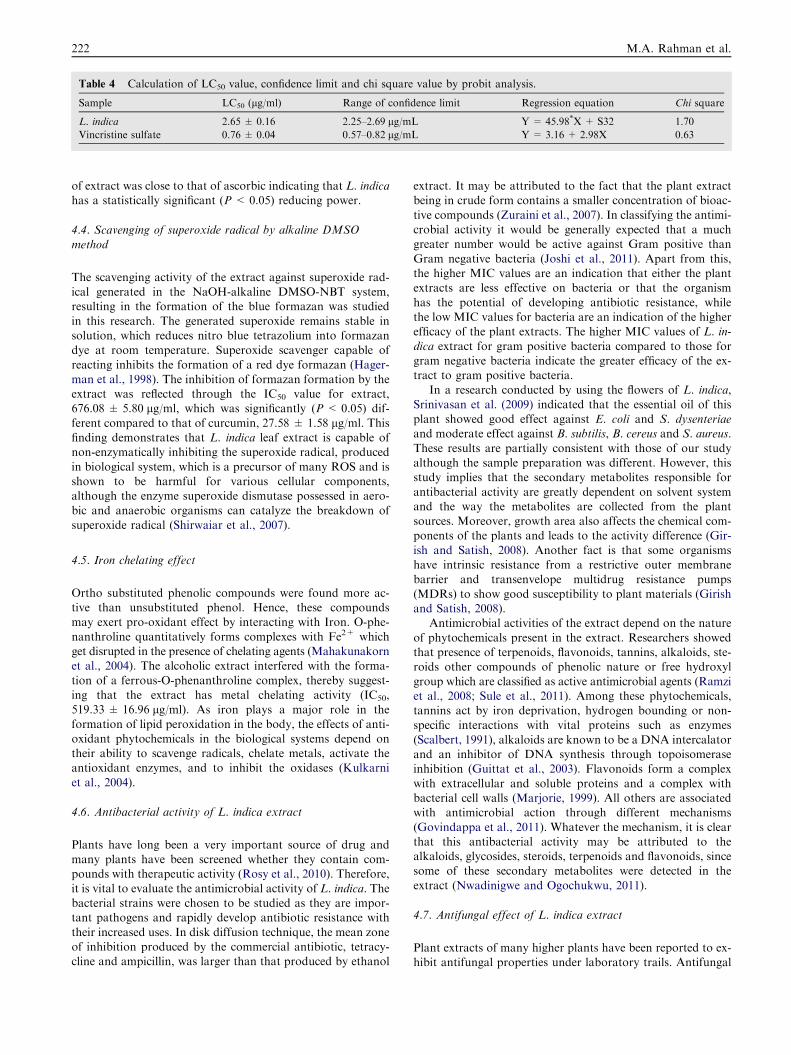
Table 4 Calculation of LC50 value, confidence limit and chi square value by probit analysis.
Sample LC50 (lg/ml) Range of confidence limit Regression equation Chi square
L. indica 2.65 ± 0.16 2.25–2.69 lg/mL Y= 45.98*X + S32 1.70
Vincristine sulfate 0.76 ± 0.04 0.57–0.82 lg/mL Y= 3.16 + 2.98X 0.63
222 M.A. Rahman et al.
of extract was close to that of ascorbic indicating that L. indicahas a statistically significant (P < 0.05) reducing power.
4.4. Scavenging of superoxide radical by alkaline DMSO
method
The scavenging activity of the extract against superoxide rad-ical generated in the NaOH-alkaline DMSO-NBT system,resulting in the formation of the blue formazan was studied
in this research. The generated superoxide remains stable insolution, which reduces nitro blue tetrazolium into formazandye at room temperature. Superoxide scavenger capable of
reacting inhibits the formation of a red dye formazan (Hager-man et al., 1998). The inhibition of formazan formation by theextract was reflected through the IC50 value for extract,676.08 ± 5.80 lg/ml, which was significantly (P < 0.05) dif-
ferent compared to that of curcumin, 27.58 ± 1.58 lg/ml. Thisfinding demonstrates that L. indica leaf extract is capable ofnon-enzymatically inhibiting the superoxide radical, produced
in biological system, which is a precursor of many ROS and isshown to be harmful for various cellular components,although the enzyme superoxide dismutase possessed in aero-
bic and anaerobic organisms can catalyze the breakdown ofsuperoxide radical (Shirwaiar et al., 2007).
4.5. Iron chelating effect
Ortho substituted phenolic compounds were found more ac-tive than unsubstituted phenol. Hence, these compoundsmay exert pro-oxidant effect by interacting with Iron. O-phe-
nanthroline quantitatively forms complexes with Fe2+ whichget disrupted in the presence of chelating agents (Mahakunakornet al., 2004). The alcoholic extract interfered with the forma-
tion of a ferrous-O-phenanthroline complex, thereby suggest-ing that the extract has metal chelating activity (IC50,519.33 ± 16.96 lg/ml). As iron plays a major role in the
formation of lipid peroxidation in the body, the effects of anti-oxidant phytochemicals in the biological systems depend ontheir ability to scavenge radicals, chelate metals, activate theantioxidant enzymes, and to inhibit the oxidases (Kulkarni
et al., 2004).
4.6. Antibacterial activity of L. indica extract
Plants have long been a very important source of drug andmany plants have been screened whether they contain com-pounds with therapeutic activity (Rosy et al., 2010). Therefore,
it is vital to evaluate the antimicrobial activity of L. indica. Thebacterial strains were chosen to be studied as they are impor-tant pathogens and rapidly develop antibiotic resistance with
their increased uses. In disk diffusion technique, the mean zoneof inhibition produced by the commercial antibiotic, tetracy-cline and ampicillin, was larger than that produced by ethanol
extract. It may be attributed to the fact that the plant extractbeing in crude form contains a smaller concentration of bioac-
tive compounds (Zuraini et al., 2007). In classifying the antimi-crobial activity it would be generally expected that a muchgreater number would be active against Gram positive than
Gram negative bacteria (Joshi et al., 2011). Apart from this,the higher MIC values are an indication that either the plantextracts are less effective on bacteria or that the organism
has the potential of developing antibiotic resistance, whilethe low MIC values for bacteria are an indication of the higherefficacy of the plant extracts. The higher MIC values of L. in-
dica extract for gram positive bacteria compared to those forgram negative bacteria indicate the greater efficacy of the ex-tract to gram positive bacteria.
In a research conducted by using the flowers of L. indica,
Srinivasan et al. (2009) indicated that the essential oil of thisplant showed good effect against E. coli and S. dysenteriaeand moderate effect against B. subtilis, B. cereus and S. aureus.
These results are partially consistent with those of our studyalthough the sample preparation was different. However, thisstudy implies that the secondary metabolites responsible for
antibacterial activity are greatly dependent on solvent systemand the way the metabolites are collected from the plantsources. Moreover, growth area also affects the chemical com-ponents of the plants and leads to the activity difference (Gir-
ish and Satish, 2008). Another fact is that some organismshave intrinsic resistance from a restrictive outer membranebarrier and transenvelope multidrug resistance pumps
(MDRs) to show good susceptibility to plant materials (Girishand Satish, 2008).
Antimicrobial activities of the extract depend on the nature
of phytochemicals present in the extract. Researchers showedthat presence of terpenoids, flavonoids, tannins, alkaloids, ste-roids other compounds of phenolic nature or free hydroxyl
group which are classified as active antimicrobial agents (Ramziet al., 2008; Sule et al., 2011). Among these phytochemicals,tannins act by iron deprivation, hydrogen bounding or non-specific interactions with vital proteins such as enzymes
(Scalbert, 1991), alkaloids are known to be a DNA intercalatorand an inhibitor of DNA synthesis through topoisomeraseinhibition (Guittat et al., 2003). Flavonoids form a complex
with extracellular and soluble proteins and a complex withbacterial cell walls (Marjorie, 1999). All others are associatedwith antimicrobial action through different mechanisms
(Govindappa et al., 2011). Whatever the mechanism, it is clearthat this antibacterial activity may be attributed to thealkaloids, glycosides, steroids, terpenoids and flavonoids, sincesome of these secondary metabolites were detected in the
extract (Nwadinigwe and Ogochukwu, 2011).
4.7. Antifungal effect of L. indica extract
Plant extracts of many higher plants have been reported to ex-hibit antifungal properties under laboratory trails. Antifungal

Phytochemical and biological effects of the phenolics of Leea indica 223
activity of the crude extract was tested using poisoned foodtechnique in our study (Grover and Moore, 1962; Mishraand Tiwari, 1992; Nene and Thapilyal, 2002). Percent inhibi-
tion of fungal mycelia growth showed that the extract had avery promising inhibitory effect on C. albicans compared tothe reference antifungal drug fluconazone, while the extract
showed moderate antifungal effect against A. flavus and F.equisetii. Usually the crude extract has a wide range of physi-ological activity of alkaloid, terpenoids, flavonoid, anthraqui-
none, steroid, and tannin. Of all these, flavonoid was found tobe the biochemical constituent responsible for the antifungalaction (Sule et al., 2011). Steroids were reported as a majorcomponent acting as antifungal secondary metabolite (Onwuliri
and Wonang, 2005). These observations are also consistentwith our findings suggesting that the antifungal effect of L. in-dica extract is probably due to the individual or synergistic
effect of the secondary metabolites present in the extract.
4.8. Cytotoxic activity of L. indica extract
Brine shrimp lethality is a general bioassay which is indicativeof cytotoxicity, antibacterial activities, pesticidal effects andvarious pharmacologic actions (Meyer et al., 1982). Therefore,
the isolation of bioactive compounds from natural sources andthe use of plant extracts require toxicity information on theconstituent of interest in order to delineate the effect of toxicityon both the host cells and target cells of pharmacological uses.
Lethal concentration (LC50) from the regression and probitanalysis (Bliss, 1934) in 24 h of our study showed that LC50 va-lue of L. indica extract was 2.65 ± 0.16 lg/ml (confidence limit
95%) where the lower and upper limits were 2.2561, and2.6981 lg/ml respectively. Comparison of this result with thestandard vincristine sulfate (0.76 ± 0.04 lg/ml) indicated that
the lethality of L. indica extract is statistically significant(P < 0.05) suggesting the notable clinical importance of theextract against tumor cells, pesticides etc. because the brine
shrimp assay is considered as a convenient probe for a preli-minary assessment of toxicity, detection of fungal toxins, pes-ticidal and anti-tumor effect and other pharmacologicalactions (Meyer et al., 1982). Apart from this, the LC50 value
of the extract was less than 1000 lg/mL, which is the cutoffpoint in detecting cytotoxicity ascertaining that the extract isconcluded to be very important to use in above mentioned ac-
tions. The ‘‘Chi square’’ value of the extract (0.76) rejects thenull hypothesis making the result statistically more significantto remove any discrepancy between expected mortality rates
and the actual mortality rates of freshly hatched A. salina.The cytotoxic effect of plants is principally contributed by
the presence of secondary metabolites like alkaloid, glycoside,steroid, tannin, phlobatannin, terpenoid and flavonoid in their
extract (Ozcelik et al., 2011). This is also consistent with ourobservation because the phytochemical group analysis of theextract showed the presence of alkaloid, terpenoids, tannins,
phlobatannins, steroids and flavonoids. Further toxicity stud-ies could be conducted on individual cell lines to confirm thetoxic effect of phytochemical groups.
5. Conclusion
In conclusion, the study demonstrates that ethanolic extract of
whole L. indica has promising antioxidant and antimicrobial
effects on human health. The extract also has very prominentcytotoxic effects to be used in many pharmacological as well asbiological actions. However, further studies are suggested to
investigate these effects for the isolated and identified purecompounds from L. indica.
Acknowledgements
Authors are grateful to the Chittagong University ResearchCell for providing the research Grant (Ref No. 5193/Res/Dir/CU/2011) to conduct the research. The authors are alsothankful to the Taxonomist and Associate professor, Dr. Sha-
ikh Bokhtear Uddin, Department of Botany, University ofChittagong, for identifying all the plants.
References
Aqil, F., Ahmed, I., Mehmood, Z., 2006. Antioxidant and free radical
scavenging properties of twelve traditionally used Indian medicinal
plants. Turk. J. Biol. 30, 177–183.
Benzie, I.F.F., Strain, J.T., 1996. The ferric reducing ability of plasma
(FRAP) as a measure of antioxidant power, the FRAP assay. Anal.
Biochem. 239, 70–76.
Bliss, C.I., 1934. The method of probits. Science 79, 38–39.
Brand-Williams, W., Cuvelier, M.E., Berset, C., 1995. Use of a free
radical method to evaluate antioxidant activity. LWT-Food Sci.
Technol. 28, 25–30.
Chanwitheesuk, A., Teerawutgulrag, A., Rakariyatham, N., 2005.
Screening of antioxidant activity and antioxidant compounds of
some edible plants of Thailand. Food Chem. 92, 491–497.
Chew, A.L., Jessica, J.J.A., Sasidharan, S., 2012. Antioxidant and
antibacterial activity of different parts of Leucas aspera. Asian Pac.
J. Trop. Biomed. 2, 176–180.
Chung, Y.C., Chang, C.T., Chao, W.W., Lin, C.F., Chou, S.T., 2002.
Antioxidative activity and safety of the 50% ethanolic extract from
red bean fermented by Bacillus subtilis IMR-NK1. J. Agric. Food
Chem. 50, 2454–2458.
Duarte, M.C.T., Figueira, G.M., Sartoratto, A., Rehder, V.L.G.,
Delarmelina, C., 2005. Anti-Candida activity of Brazilian medicinal
plants. J. Ethnopharmacol. 97, 305–311.
Ghani, A., 2003. Medicinal plant of Bangladesh:Chemical constituents
and uses. Asiatic Society of Bangladesh, Dhaka.
Girish, H.V., Satish, S., 2008. Antibacterial activity of important
medicinal plants on human pathogenic bacteria – a comparative
analysis. World Appl. Sci. J. 5, 267–271.
Govindappa, M., Naga Sravya, S., Poojashri, M.N., Sadananda, T.S.,
Chandrappa, C.P., 2011. Antimicrobial, antioxidant and in vitro
anti-inflammatory activity of ethanol extract and active phyto-
chemical screening of Wedelia trilobata (L.) Hitchc. J. Pharmacog.
Phytother. 3, 43–51.
Grover, R.K., Moore, J.D., 1962. Toximetric studies of fungicides
against brown rot organisms, Sclerotia fructicola and S. laxa.
Phytopathology 52, 876–880.
Guittat, L., Alberti, P., Rosu, F., Van Miert, S., Thetiot, E., Pieters,
L., Gabelica, V., De Pauw, E., Ottaviani, A., Roiu, J.-F., Mergny,
J.L., 2003. Interaction of cryptolepine and neocryptolepine with
unusual DNA structures. Biochimie 85, 535–541.
Gulcin, I., Uguz, M.T., Oktay, M., Beydemir, S., Kufrevioglu, O.I.,
2004. Evaluation of the antioxidant and antimicrobial activities of
clary sage (Salvia sclarea L.). Turk. J. Agric. For. 28, 25–33.
Hagerman, A.E., Riedel, K.M., Jones, G.A., Sovik, K.N., Ritchard,
N.T., Hartzfeld, P.W., 1998. High molecular weight plant poly-
phenolics (tannins) as biological antioxidants. J. Agric. Food
Chem. 46, 1887–1892.

224 M.A. Rahman et al.
Harborne, J.B., 1973. Phytochemical methods: a guide to modern
techniques of plant analysis. Chapman and Hall Ltd., London,
UK.
Iqbal, S., Bhanger, M.I., Anwar, F.A., 2005. Antioxidant properties
and components of some commercially varieties of rice in Pakistan.
Food Chem. 93, 265.
Janovska, D., Kubıkova, K., Kokoska, L., 2003. Screening for
antimicrobial activity of some medicinal plants species of tradi-
tional Chinese medicine. Czech J. Food Sci. 21, 107–110.
Joshi, B., Sah, G.P., Basnet, B.B., Bhatt, M.R., Sharma, D., Subedi,
K., Pandey, J., Malla, R., 2011. Phytochemical extraction and
antimicrobial properties of different medicinal plants: Ocimum
sanctum (Tulsi), Eugenia caryophyllata (Clove), Achyranthes biden-
tata (Datiwan) and Azadirachta indica (Neem). J. Microbiol.
Antimicrob. 3, 1–7.
Koleva, I.I., Van Beek, T.A., Linssen, J.P.H., de Groot, A., Evstatieva,
L.N., 2002. Screening of plant extracts for antioxidant activity: a
comparative study on three testing methods. Phytochem. Ana. 13,
8–17.
Karaman, I., Sahin, F., Gulluce, M., Ogutcu, H., Sengul, M.,
Adiguzel, A., 2003. Antimicrobial activity of aqueous and meth-
anol extracts of Juniperus oxycedrus L.. J. Ethnopharmacol. 85,
231–235.
Kulkarni, A.P., Aradhya, S.M., Divakar, S., 2004. Isolation and
identification of a radical scavenging antioxidant–punicalagin from
pith and carpellary membrane of pomegranate fruit. Food Chem.
87, 551–557.
Kumaran, A., Karunakaran, R.J., 2007. In vitro antioxidant activities
of methanol extracts of five Phyllanthus species from India. LWT -
Food Sci. Technol. 40, 344–352.
Madan, M.P., Raghavan, G., Ajay Kumar Singh, R., Palpu, P., 2005.
Free radical scavenging potential of Saussarea costus. Acta Pharm.
55, 297–304.
Mahakunakorn, P., Tohda, M., Murakami, Y., Matsumoto, K.,
Watanabe, H., 2004. Antioxidant and free radical scavenging
activity of Choto-san and its related constituents. Biol. Pharm.
Bull. 27, 38–46.
Marjorie, C., 1999. Plant products as antimicrobial agents. Clin.
Microb. Rev. 12, 564–582.
Meir, S., Kanner, J., Akiri, B., Hadas, S.P., 1995. Determination and
involvement of aqueous reducing compounds in oxidative defense
systems of various senescing leaves. J. Agric. Food. Chem. 43,
1813–1815.
Meyer, B.N., Ferrigni, N.R., Putnam, J.E., Jacobsen, L.B., Nichols,
D.E., McLaughlin, J.L., 1982. Brine shrimp: a convenient general
bioassay for active plant constituents. Planta Med. 45, 31–34.
Mishra, M., Tiwari, S.N., 1992. Toxicity of Polyalthia longifolia
against fungal pathogens of rice. Indian Phytopath. 45, 56–61.
Morales, G., Paredes, A., Sierra, P., Loyola, L.A., 2008. Antioxidant
activity of 50% aqueous-ethanol extract from Acantholippia
deserticola. Biol. Res. 41, 151–155.
NCCLS, 2002. Methods for dilution antimicrobial susceptibility
tests for bacteria that grow aerobically. Third ed. NCCLS, Wayne,
PA.
Nene, Y., Thapilyal, L., 2002. Poisoned food technique of fungicides in
plant disease control, third ed. Oxford and IBH Publishing
Company, New Delhi.
Nwadinigwe, Ogochukwu, A., 2011. Antimicrobial activities of meth-
anol and aqueous extracts of the stem of Bryophyllum pinnatum
Kurz (Crassulaceae). Afr. J. Biotechnol. 10, 16342–16346.
Onwuliri, F.C., Wonang, D.L., 2005. Studies on the combined
antibacterial action of Ginger (Zingiber officinale L) and Garlic
(Allium sativum L.) on some bacteria. Nig. J. Bot. 18, 224–228.
Oyaizu, M., 1986. Studies on product of browning reaction prepared
from glucoseamine. Jpn. J. Nutr. 44, 307–315.
Ozcelik, B., Kartal, M., Orhan, I., 2011. Cytotoxicity, antiviral and
antimicrobial activities of alkaloids, flavonoids, and phenolic acids.
Pharm. Biol. 49, 396–402.
Perumal Samy, R., Ignacimuthu, S., Patric Raja, D., 2008. Preliminary
screening of ethnomedicinal plants from India. Eur. Rev. Med.
Pharmacol. Sci. 12, 1–7.
Prashant, K.R., Dolly, J., Singh, K.R., Gupta, K.R., Watal, G., 2008.
Glycemic properties of Trichosanthes dioica leaves. Pharm. Biol. 46,
894–899.
Prieto, P., Pineda, M., Aguilar, M., 1999. Spectrophotometric quan-
titation of antioxidant capacity through the formation of a
phosphomolybdenum complex: specific application to the determi-
nation of vitamin E. Anal. Biochem. 269, 337–341.
Raihan, M.O., Habib, M.R., Brishti, A., Rahman, M.M., Saleheen,
M.M., Manna, M., 2011. Sedative and anxiolytic effects of the
methanolic extract of Leea indica (Burm. f.) Merr. leaf. Drug
Discov. Ther. 5, 185–189.
Ramzi, A.A.M., Salah, A.A.A., Hasson, S., Althawab, F.M.N.,
Alaghbari, S.A.Z., Lindequist, U., 2008. Antimicrobial, Antioxi-
dant and Cytotoxic Activities and Phytochemical Screening of
Some Yemeni Medicinal Plants. eCAM, 0, 1–8.
Reddy, B.S., Reddy, R.K.K., Reddy, B.P., Ramakrishna, S., Diwan,
P.V., 2008. Potential in vitro antioxidant and protective effects of
Soymida febrifuga on ethanol induced oxidative damage in HepG2
cells. Food Chem. Toxicol. 46, 3429–3442.
Rosy, B.A., Joseph, H., Rosalie, 2010. Phytochemical, pharmacog-
nostical, antimicrobial activity of Indigofera spalathoids Vahl.
(Fabaceae). Int. J. Biol. Technol. 1, 12–15.
Roy, A., Khanra, K., Mishra, A., Bhattacharyya, N., 2012. General
analysis and antioxidant study of traditional fermented drink
Handia, its concentrate and volatiles. Adv. Life Sci. Appl. 1, 54–57.
Saha, K., Lajis, N.H., Israf, D.A., Hamzah, A.S., Khozirah, S.,
Khamis, S., Syahida, A., 2004. Evaluation of antioxidant and nitric
oxide inhibitory activities of selected Malaysian medicinal plants. J.
Ethnopharmacol. 92, 263–267.
Sarla, S., Prakash, M.A., Apeksha, R., Subhash, C., 2011. Free radical
scavenging (DPPH) and ferric reducing ability (FRAP) of Apha-
namixis polystachya (Wall) Parker. Int. J. Drug Dev. Res. 3, 271–
274.
Scalbert, A., 1991. Antimicrobial properties of tannins. Phytochemis-
try 30, 3875–3883.
Sein, T.T., Spurio, R., Cecchini, C., Cresci, A., 2008. Screening for
microbial strains degrading glass fiber acrylic composite filters. Int.
Biodeterior. Biodegradation 63, 901–905.
Shirwaiar, A., Shirwaikar, A., Punitha, I.S.R., 2007. Antioxidant
studies on the methanol stem extract of Coscinium fenestratum.
Nat. Prod. Sci. 13, 40–45.
Sofowara, A., 1993. Medicinal plants and traditional medicine in
Africa. Spectrum Books Ltd., Ibadan, Nigeria.
Srinisavan, R., Chandrasekar, M.J.N., Nanjan, M.J., Suresh, B., 2007.
Antioxidant activity of Caesalpinia digyna root. J. Ethnopharma-
col. 113, 284–291.
Srinivasan, G.V., Ranjith, C., Vijayan, K.K., 2008. Identification of
chemical compounds from the leaves of Leea indica. Acta Pharm.
58, 207–214.
Srinivasan, G.V., Sharanappa, P., Leela, N.K., Sadashiva, C.T.,
Vijayan, K.K., 2009. Chemical composition and antimicrobial
activity of the essential oil of Leea indica (Burm. f.) Merr. flowers.
Nat. Prod. Rad. 8, 488–493.
Srithi, K., Balslev, H., Wangpakapattanawong, P., Srisanga, P.,
Trisonthi, C., 2009. Medicinal plant knowledge and its erosion
among the Mien (Yao) in northern Thailand. J. Ethnopharmacol.
123, 335–342.
Sule, W.F., Okonko, I.O., Omo-Ogun, S., Nwanze, J.C., Ojezele,
M.O., Ojezele, O.J., Alli, J.A., Soyemi, E.T., Olaonipekun, T.O.,
2011. Phytochemical properties and in vitro antifungal activity of
Senna alata Linn. crude stem bark extract. J. Med. Plants. Res. 5,
176–183.
Suresh, P.K., Sucheta, S., Sudarshana, V.D., Selvamani, P., Latha, S.,
2008. Antioxidant activity in some selected Indian medicinal plants.
Afr. J. Biotechnol. 7, 1826–1828.

Phytochemical and biological effects of the phenolics of Leea indica 225
Trease, G.E., Evans, W.C., 1989. Pharmacognosy, 11th ed. Brailliar
Tiridel and Macmillian Publishers, London, UK.
Vinay, R.P., Prakash, R.P., Sushil, S.K., 2010. Antioxidant activity of
some selected medicinal plants in western region of India. Adv.
Biol. Res. 4, 23–26.
Yusuf, M., Wahab, M.A., Yousuf, M.D., Chowdhury, J.U., Begum,
J., 2008. Some tribal medicinal plants of Chittagong Hill Tracts,
Bangladesh. Bangladesh J. Plant. Tax. 14, 117–128.
Wong, Y.H., Abdul Kadir, H., 2012. Induction of mitochondria-
mediated apoptosis in Ca Ski human cervical cancer cells triggered
by Mollic Acid Arabinoside isolated from Leea indica. eCAM,
684740.
Wong, Y.H., Abdul Kadir, H., 2011. Leea indica Ethyl acetate fraction
induces growth-inhibitory effect in various cancer cell lines and
apoptosis in Ca Ski human cervical epidermoid carcinoma cells.
eCAM, 293060.
Zuraini, Z., Yoga Latha, L., Suryani, S., Sasidharan, S., 2007.
Anticandida albicans activity of crude extract of the local plant,
winged beans leaf. Plant Kuala Lumpur 80, 653–657.