Embed Size (px)
Citation preview
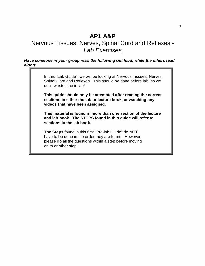
1
AP1 A&P Nervous Tissues, Nerves, Spinal Cord and Reflexes -
Lab Exercises Have someone in your group read the following out loud, while the others read along:
In this "Lab Guide", we will be looking at Nervous Tissues, Nerves, Spinal Cord and Reflexes. This should be done before lab, so we don't waste time in lab! This guide should only be attempted after reading the correct sections in either the lab or lecture book, or watching any videos that have been assigned. This material is found in more than one section of the lecture and lab book. The STEPS found in this guide will refer to sections in the lab book. The Steps found in this first "Pre-lab Guide" do NOT have to be done in the order they are found. However, please do all the questions within a step before moving on to another step!
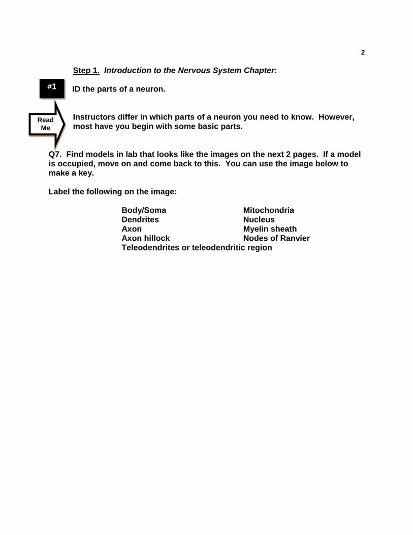
2
#1
Step 1. Introduction to the Nervous System Chapter: ID the parts of a neuron.
Instructors differ in which parts of a neuron you need to know. However, most have you begin with some basic parts.
Q7. Find models in lab that looks like the images on the next 2 pages. If a model is occupied, move on and come back to this. You can use the image below to make a key. Label the following on the image:
Body/Soma Mitochondria Dendrites Nucleus Axon Myelin sheath Axon hillock Nodes of Ranvier
Teleodendrites or teleodendritic region
Read Me
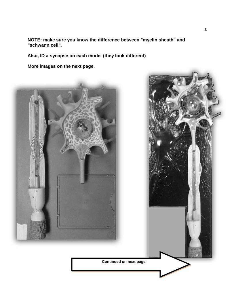
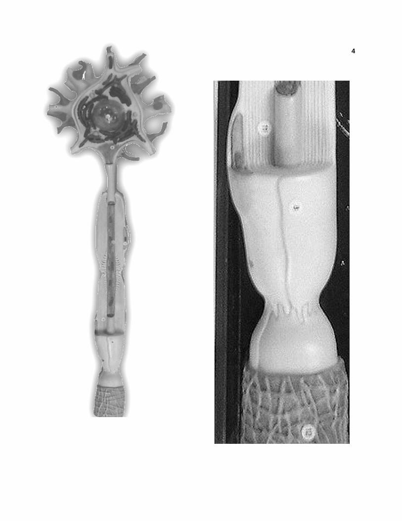
3 NOTE: make sure you know the difference between "myelin sheath" and "schwann cell". Also, ID a synapse on each model (they look different) More images on the next page.
Continued on next page
4
5
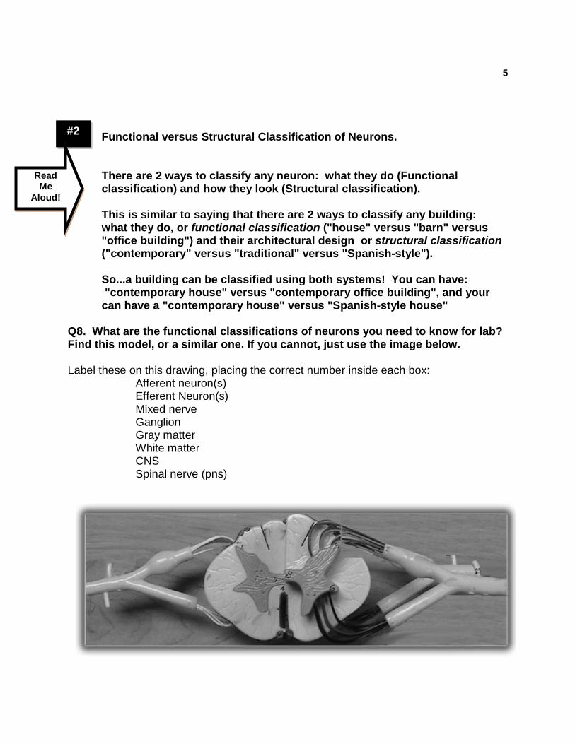
#2
Functional versus Structural Classification of Neurons. There are 2 ways to classify any neuron: what they do (Functional classification) and how they look (Structural classification). This is similar to saying that there are 2 ways to classify any building: what they do, or functional classification ("house" versus "barn" versus "office building") and their architectural design or structural classification ("contemporary" versus "traditional" versus "Spanish-style"). So...a building can be classified using both systems! You can have: "contemporary house" versus "contemporary office building", and your can have a "contemporary house" versus "Spanish-style house"
Q8. What are the functional classifications of neurons you need to know for lab? Find this model, or a similar one. If you cannot, just use the image below. Label these on this drawing, placing the correct number inside each box:
Afferent neuron(s) Efferent Neuron(s) Mixed nerve Ganglion Gray matter White matter CNS Spinal nerve (pns)
Read Me
Aloud!
6 Q9. What are the synonyms for:
Afferent neuron: Efferent neuron:
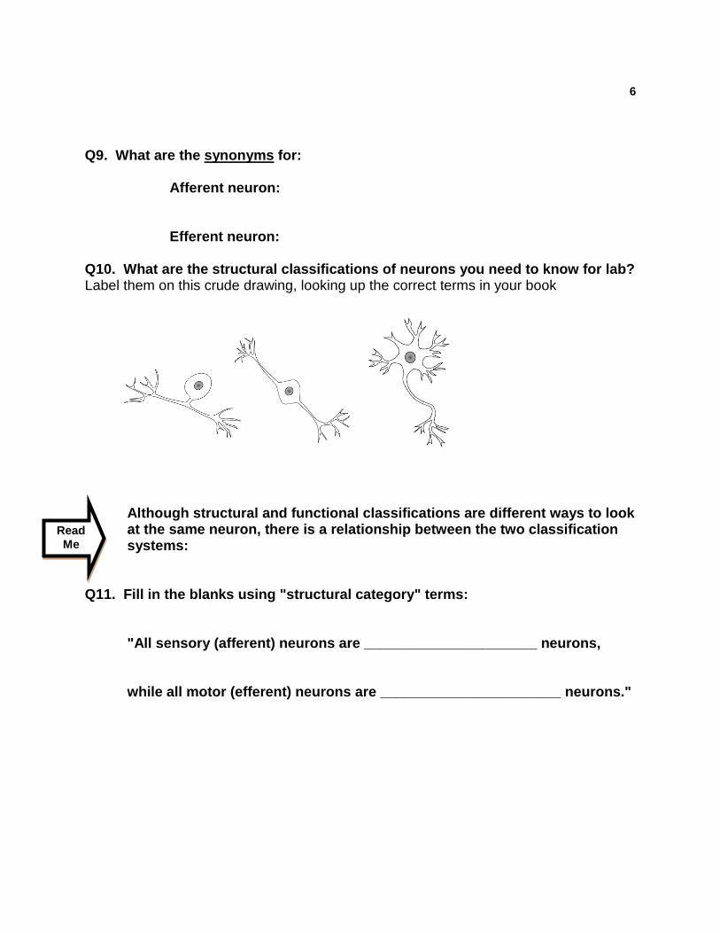
Q10. What are the structural classifications of neurons you need to know for lab? Label them on this crude drawing, looking up the correct terms in your book
Although structural and functional classifications are different ways to look at the same neuron, there is a relationship between the two classification systems:
Q11. Fill in the blanks using "structural category" terms:
"All sensory (afferent) neurons are ______________________ neurons, while all motor (efferent) neurons are _______________________ neurons."
Read Me
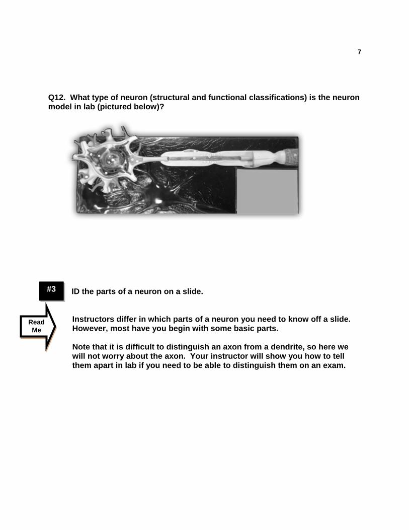
7
#3
Q12. What type of neuron (structural and functional classifications) is the neuron model in lab (pictured below)?
ID the parts of a neuron on a slide.
Instructors differ in which parts of a neuron you need to know off a slide. However, most have you begin with some basic parts. Note that it is difficult to distinguish an axon from a dendrite, so here we will not worry about the axon. Your instructor will show you how to tell them apart in lab if you need to be able to distinguish them on an exam.
Read Me
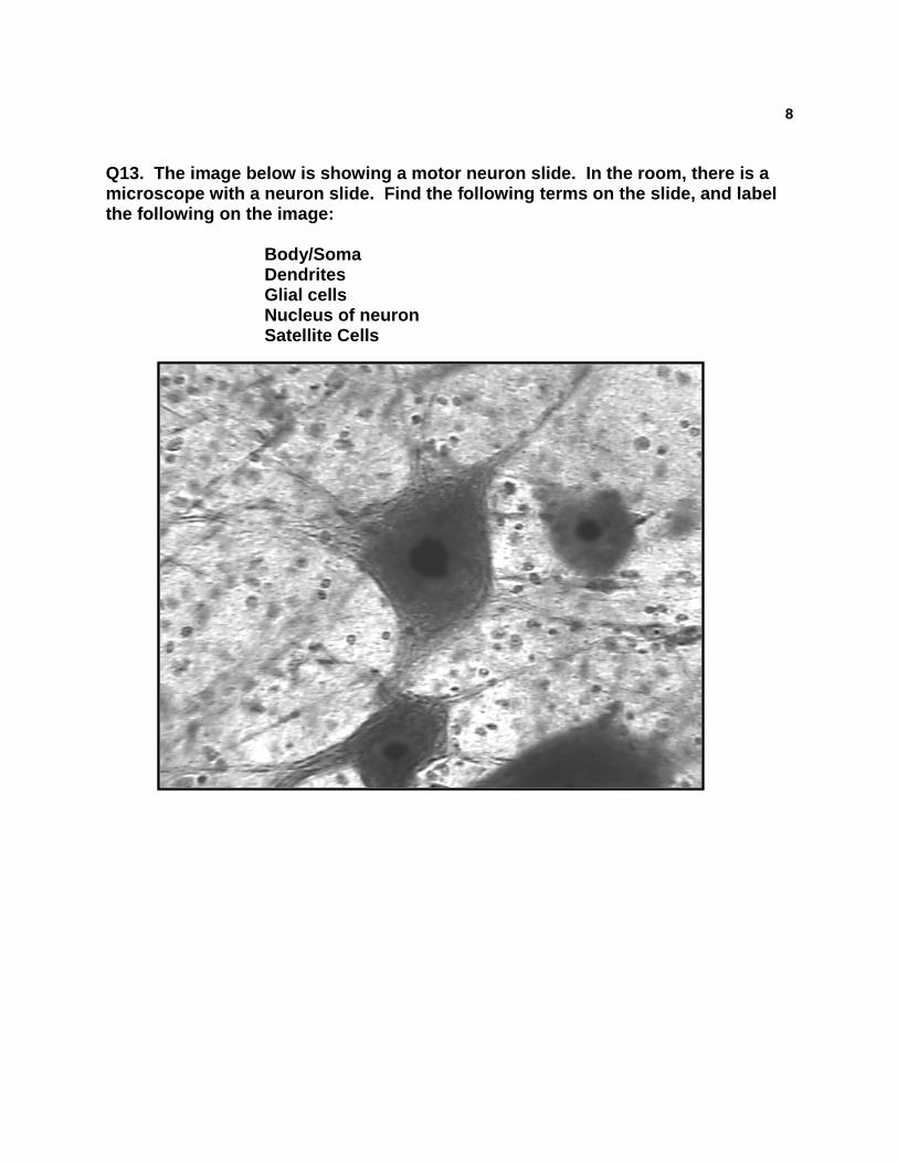
8
Q13. The image below is showing a motor neuron slide. In the room, there is a microscope with a neuron slide. Find the following terms on the slide, and label the following on the image:
Body/Soma Dendrites Glial cells Nucleus of neuron Satellite Cells
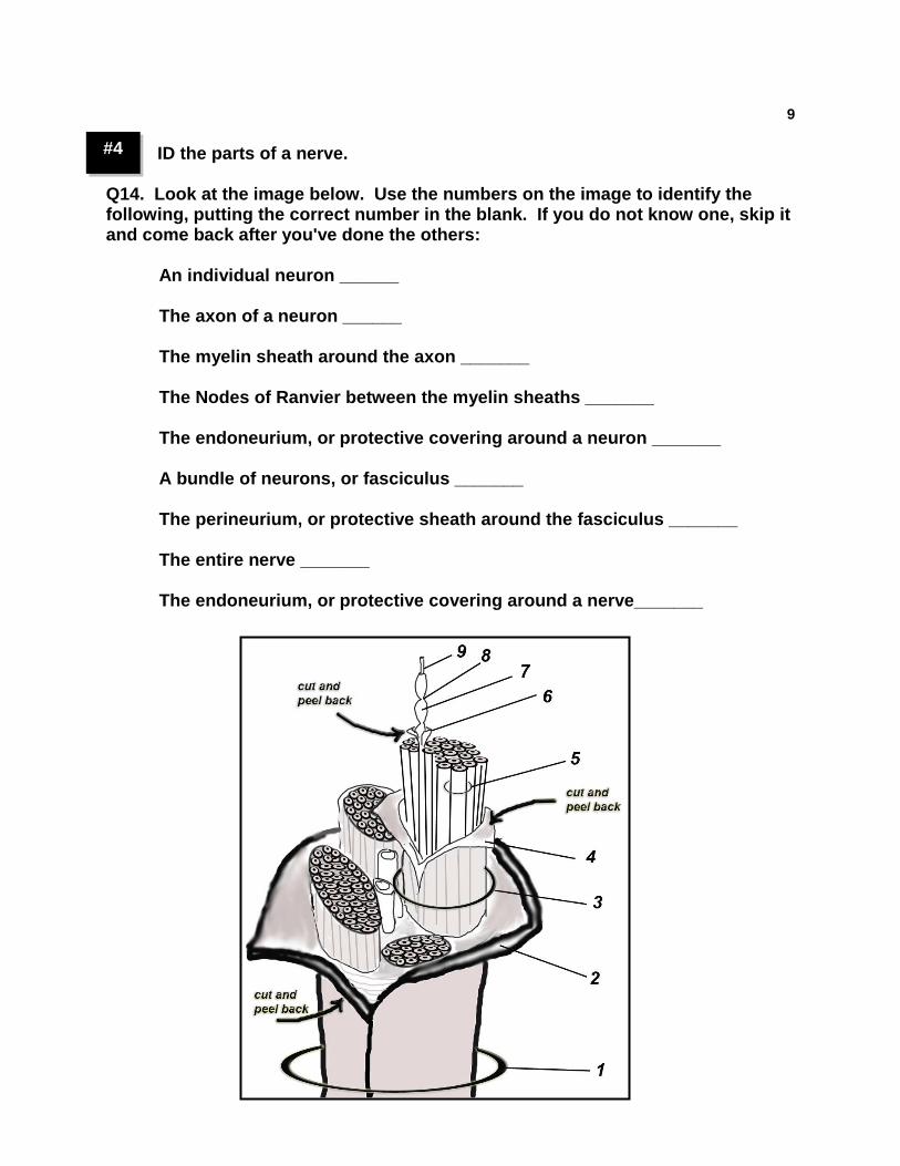
9 #4 ID the parts of a nerve.
Q14. Look at the image below. Use the numbers on the image to identify the following, putting the correct number in the blank. If you do not know one, skip it and come back after you've done the others:
An individual neuron ______ The axon of a neuron ______ The myelin sheath around the axon _______ The Nodes of Ranvier between the myelin sheaths _______ The endoneurium, or protective covering around a neuron _______ A bundle of neurons, or fasciculus _______ The perineurium, or protective sheath around the fasciculus _______ The entire nerve _______ The endoneurium, or protective covering around a nerve_______
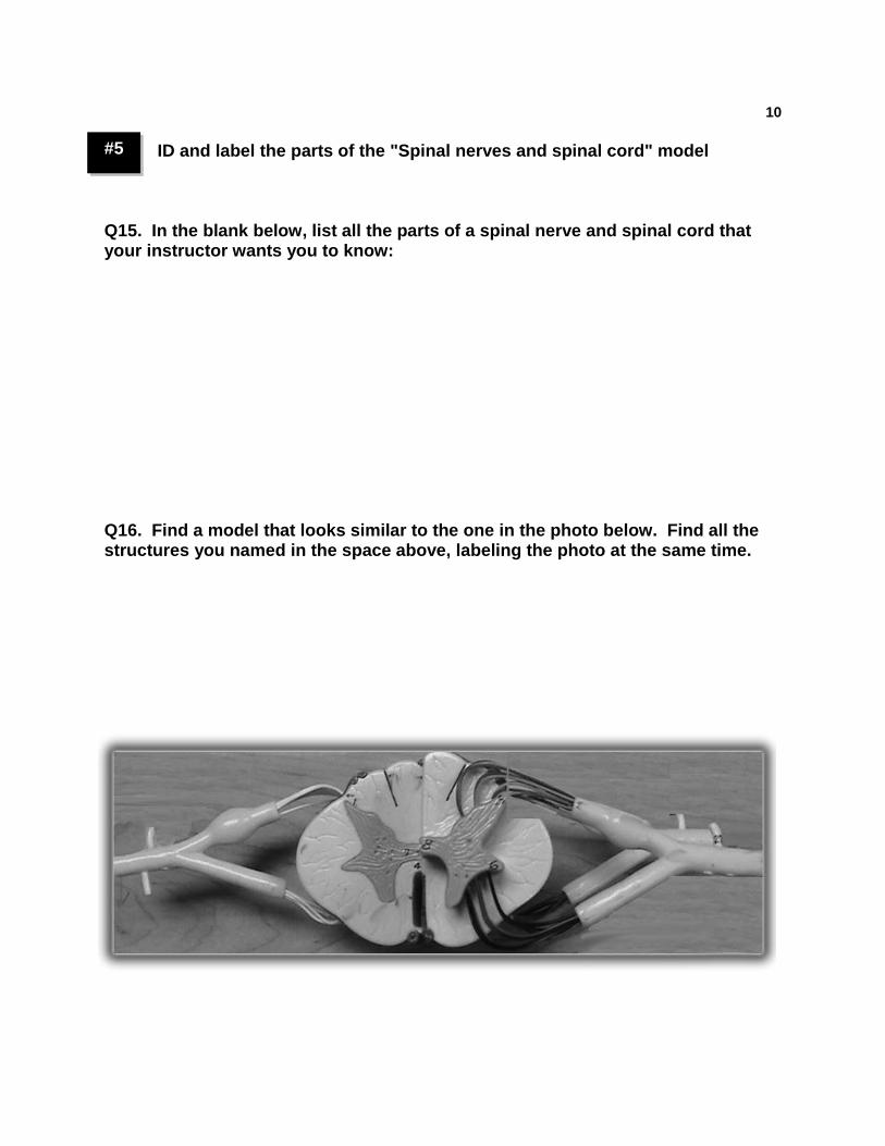
10 #5 ID and label the parts of the "Spinal nerves and spinal cord" model
Q15. In the blank below, list all the parts of a spinal nerve and spinal cord that your instructor wants you to know: Q16. Find a model that looks similar to the one in the photo below. Find all the structures you named in the space above, labeling the photo at the same time.
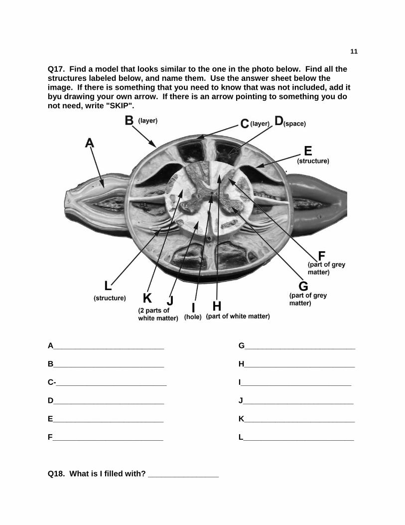
11 Q17. Find a model that looks similar to the one in the photo below. Find all the structures labeled below, and name them. Use the answer sheet below the image. If there is something that you need to know that was not included, add it byu drawing your own arrow. If there is an arrow pointing to something you do not need, write "SKIP".
A_________________________ G_________________________ B_________________________ H_________________________ C-_________________________ I_________________________ D_________________________ J_________________________ E_________________________ K_________________________ F_________________________ L_________________________ Q18. What is I filled with? ________________
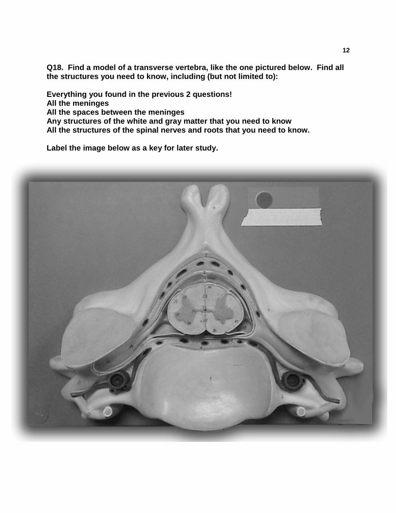
12 Q18. Find a model of a transverse vertebra, like the one pictured below. Find all the structures you need to know, including (but not limited to): Everything you found in the previous 2 questions! All the meninges All the spaces between the meninges Any structures of the white and gray matter that you need to know All the structures of the spinal nerves and roots that you need to know. Label the image below as a key for later study.
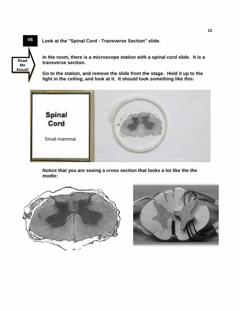
13 #6 Look at the "Spinal Cord - Transverse Section" slide.
In the room, there is a microscope station with a spinal cord slide. It is a transverse section. Go to the station, and remove the slide from the stage. Hold it up to the light in the ceiling, and look at it. It should look something like this:
Notice that you are seeing a cross section that looks a lot like the the modle:
Read Me
Aloud!
Small mammal
14
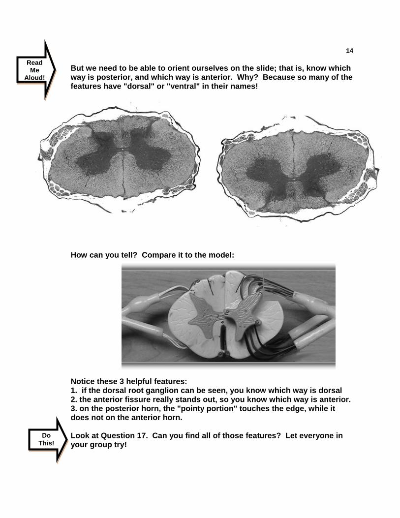
But we need to be able to orient ourselves on the slide; that is, know which way is posterior, and which way is anterior. Why? Because so many of the features have "dorsal" or "ventral" in their names!
How can you tell? Compare it to the model: Notice these 3 helpful features: 1. if the dorsal root ganglion can be seen, you know which way is dorsal 2. the anterior fissure really stands out, so you know which way is anterior. 3. on the posterior horn, the "pointy portion" touches the edge, while it does not on the anterior horn. Look at Question 17. Can you find all of those features? Let everyone in your group try!
Read Me
Aloud!
Do This!
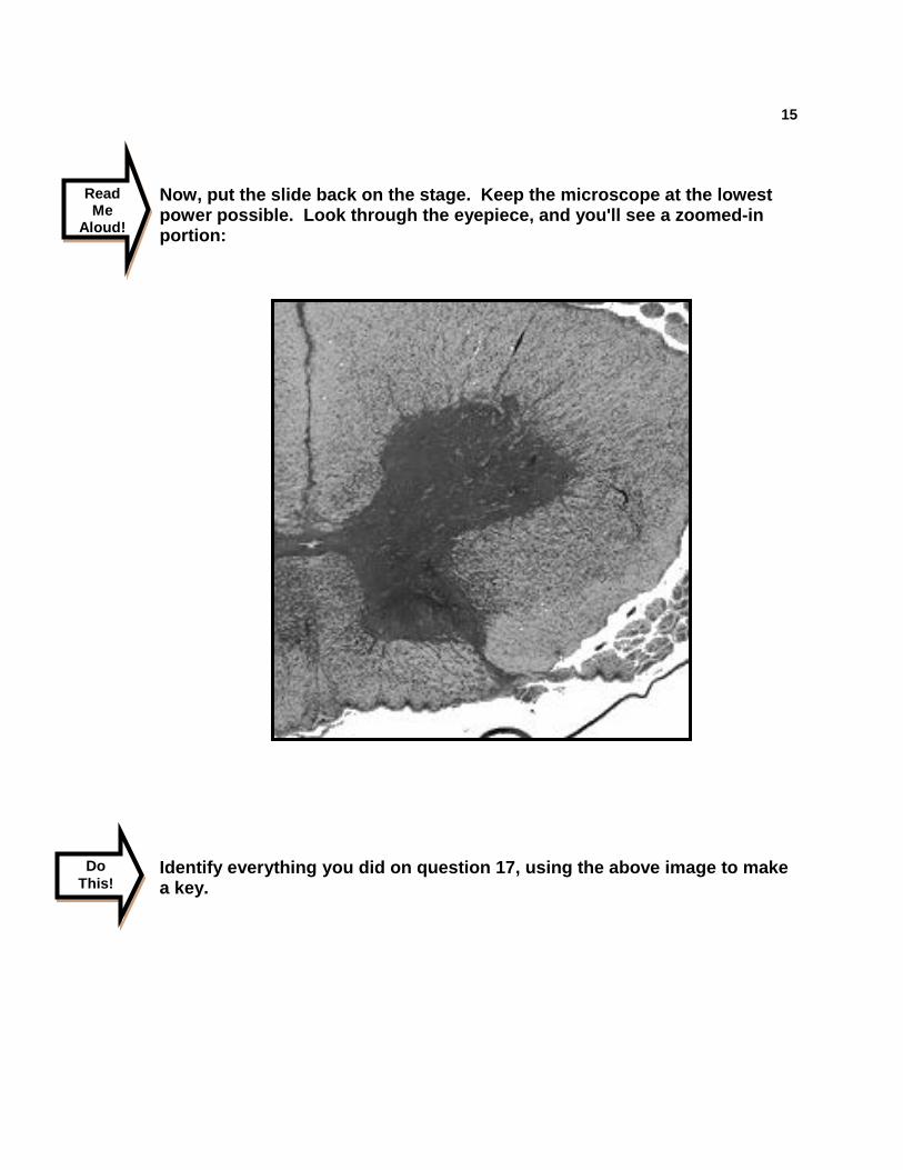
15
Now, put the slide back on the stage. Keep the microscope at the lowest power possible. Look through the eyepiece, and you'll see a zoomed-in portion: Identify everything you did on question 17, using the above image to make a key.
Read Me
Aloud!
Do This!
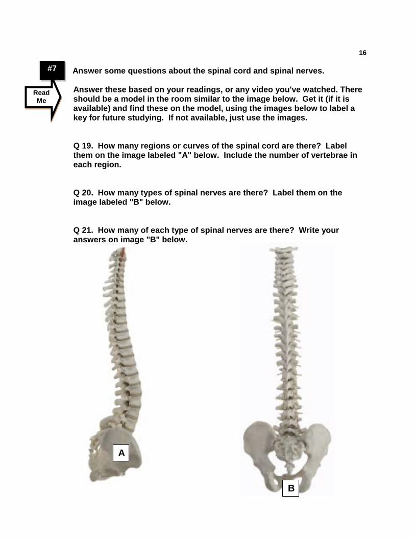
16 #7 Answer some questions about the spinal cord and spinal nerves.
Answer these based on your readings, or any video you've watched. There should be a model in the room similar to the image below. Get it (if it is available) and find these on the model, using the images below to label a key for future studying. If not available, just use the images. Q 19. How many regions or curves of the spinal cord are there? Label them on the image labeled "A" below. Include the number of vertebrae in each region. Q 20. How many types of spinal nerves are there? Label them on the image labeled "B" below. Q 21. How many of each type of spinal nerves are there? Write your answers on image "B" below.
Read Me
A
B
17
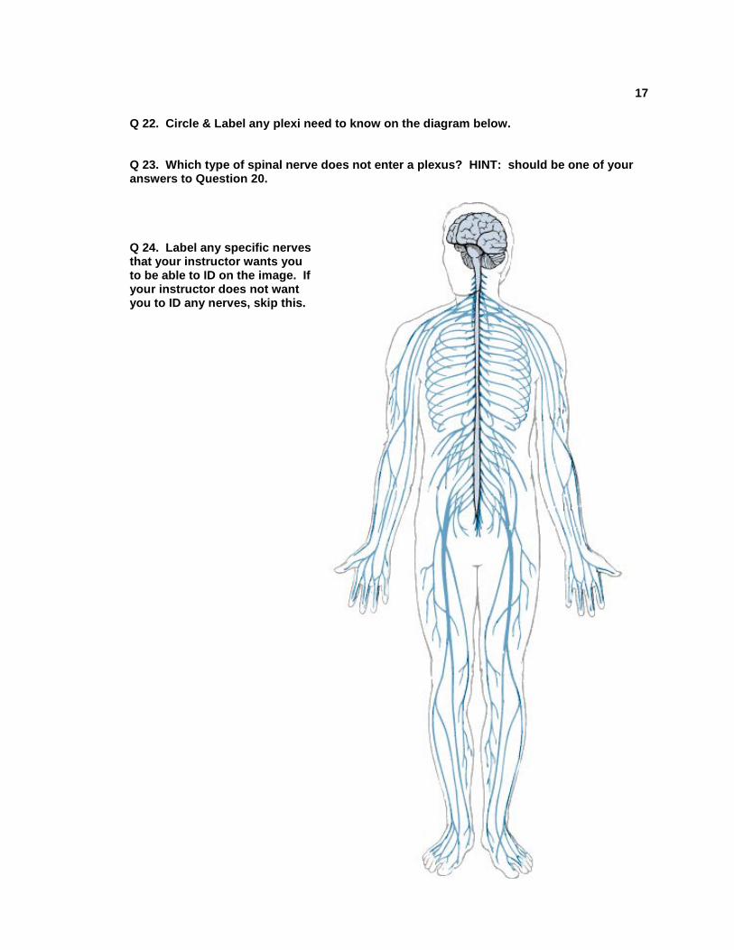
Q 22. Circle & Label any plexi need to know on the diagram below. Q 23. Which type of spinal nerve does not enter a plexus? HINT: should be one of your answers to Question 20.
Q 24. Label any specific nerves that your instructor wants you to be able to ID on the image. If your instructor does not want you to ID any nerves, skip this.
18
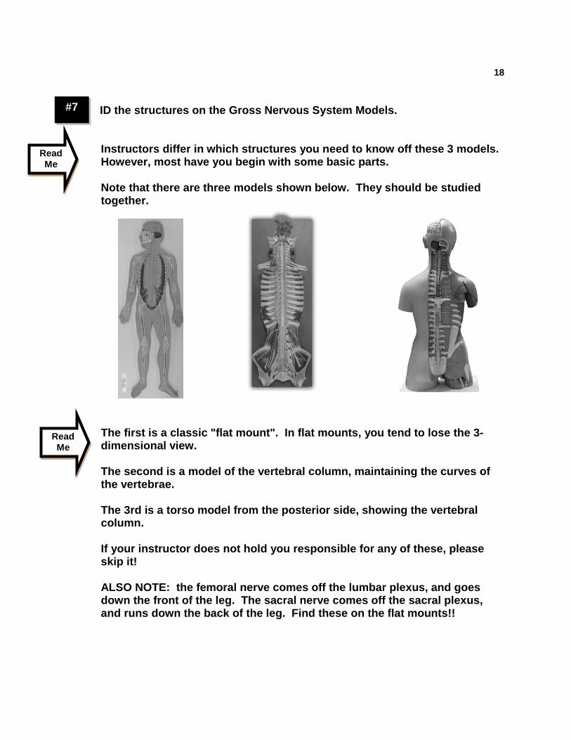
#7 ID the structures on the Gross Nervous System Models.
Instructors differ in which structures you need to know off these 3 models. However, most have you begin with some basic parts. Note that there are three models shown below. They should be studied together. The first is a classic "flat mount". In flat mounts, you tend to lose the 3-dimensional view. The second is a model of the vertebral column, maintaining the curves of the vertebrae. The 3rd is a torso model from the posterior side, showing the vertebral column. If your instructor does not hold you responsible for any of these, please skip it! ALSO NOTE: the femoral nerve comes off the lumbar plexus, and goes down the front of the leg. The sacral nerve comes off the sacral plexus, and runs down the back of the leg. Find these on the flat mounts!!
Read Me
Read Me
19 Q 25. Identify What You Need To Know. On the next few pages are images you can use for labelling a key. They all have basic structures labeled. Your instructor may have more. Start by listing all the structures you need to know off the flat mount in the space below. If you are unsure, ask your instructor.
Parts of the spinal cord: Plexi, roots and ganglia: Nerves of the arm/shoulder: Nerves of the leg/hip: Other nerves:
Q 26. Now, label the diagrams on the following pages (for the models you are responsible for). If there is something that you need to know that was not included, add it by drawing your own arrow. If there is an arrow pointing to something you do not need, write "SKIP".
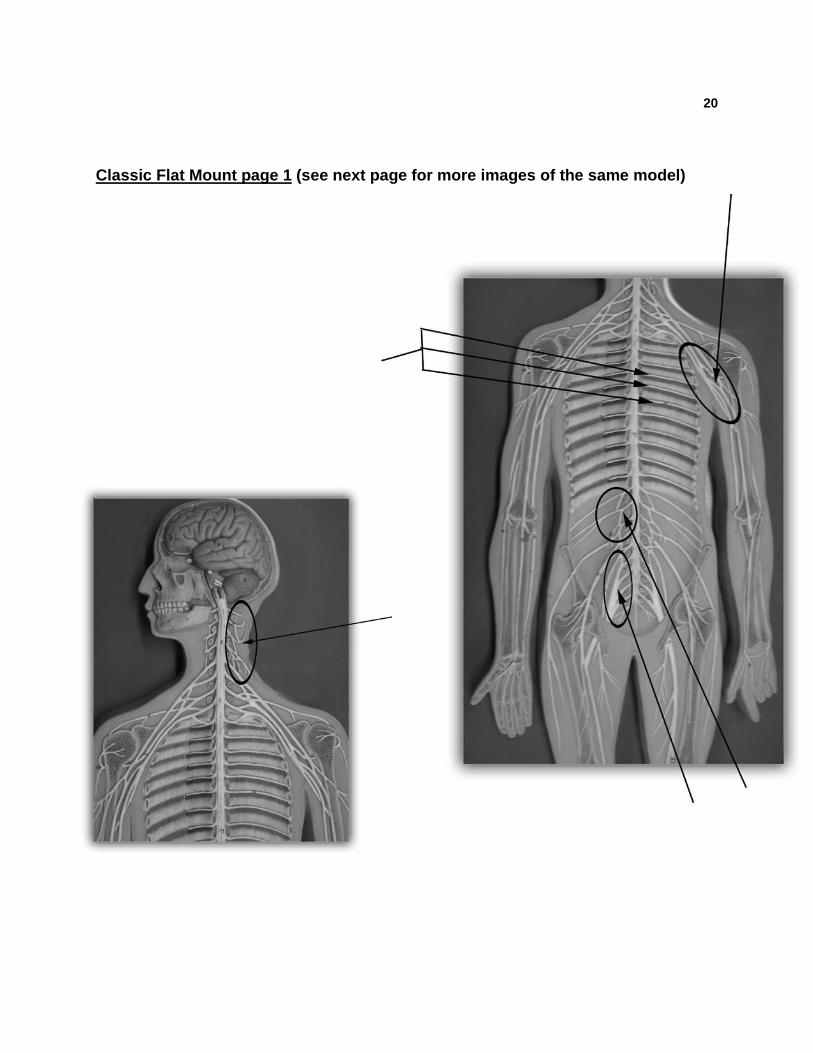
20 Classic Flat Mount page 1 (see next page for more images of the same model)
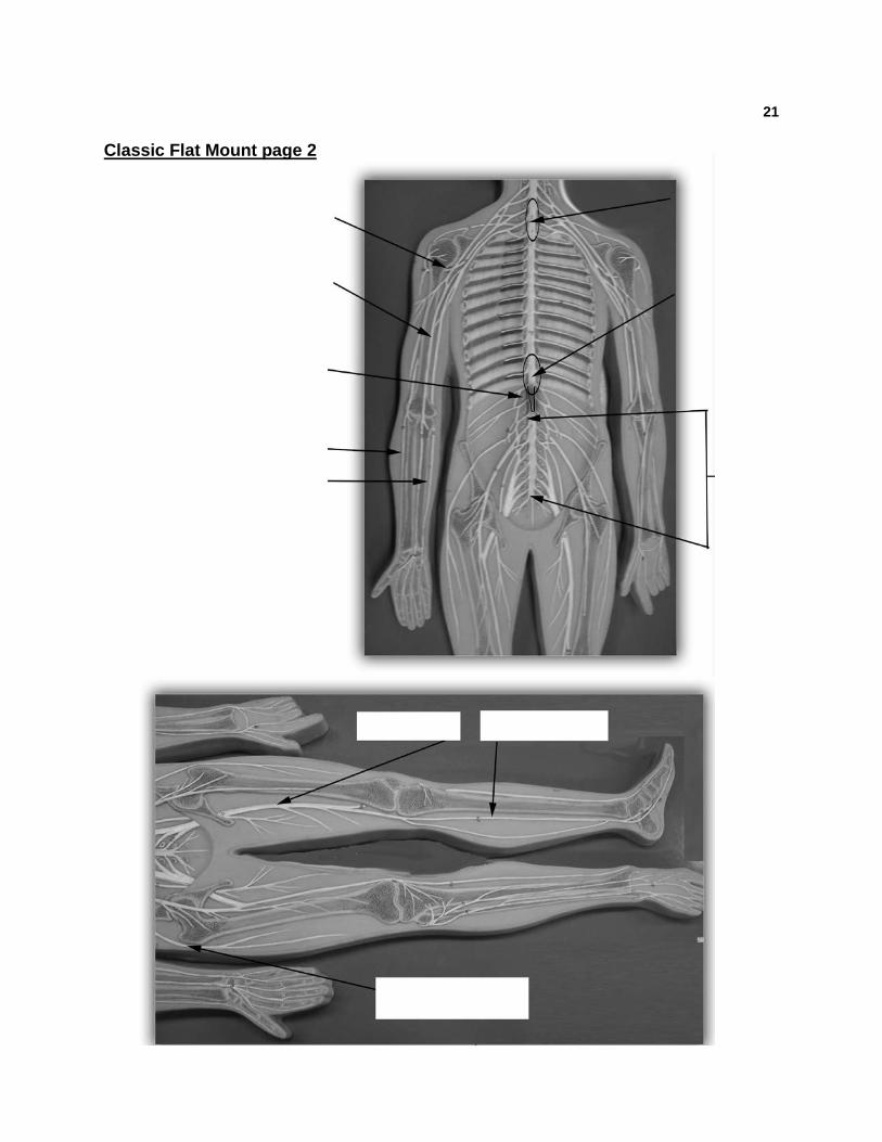
21 Classic Flat Mount page 2
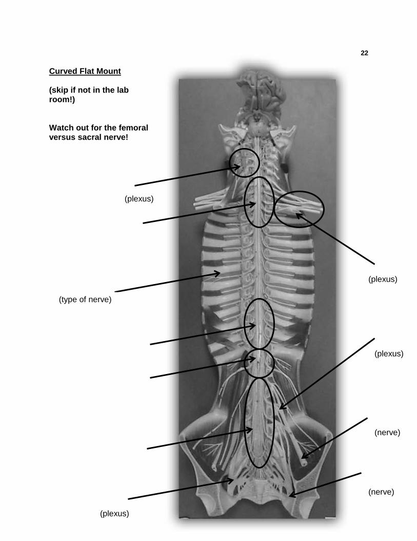
22 Curved Flat Mount (skip if not in the lab room!) Watch out for the femoral versus sacral nerve! (plexus)
(plexus)
(plexus)
(plexus)
(nerve)
(nerve)
(type of nerve)
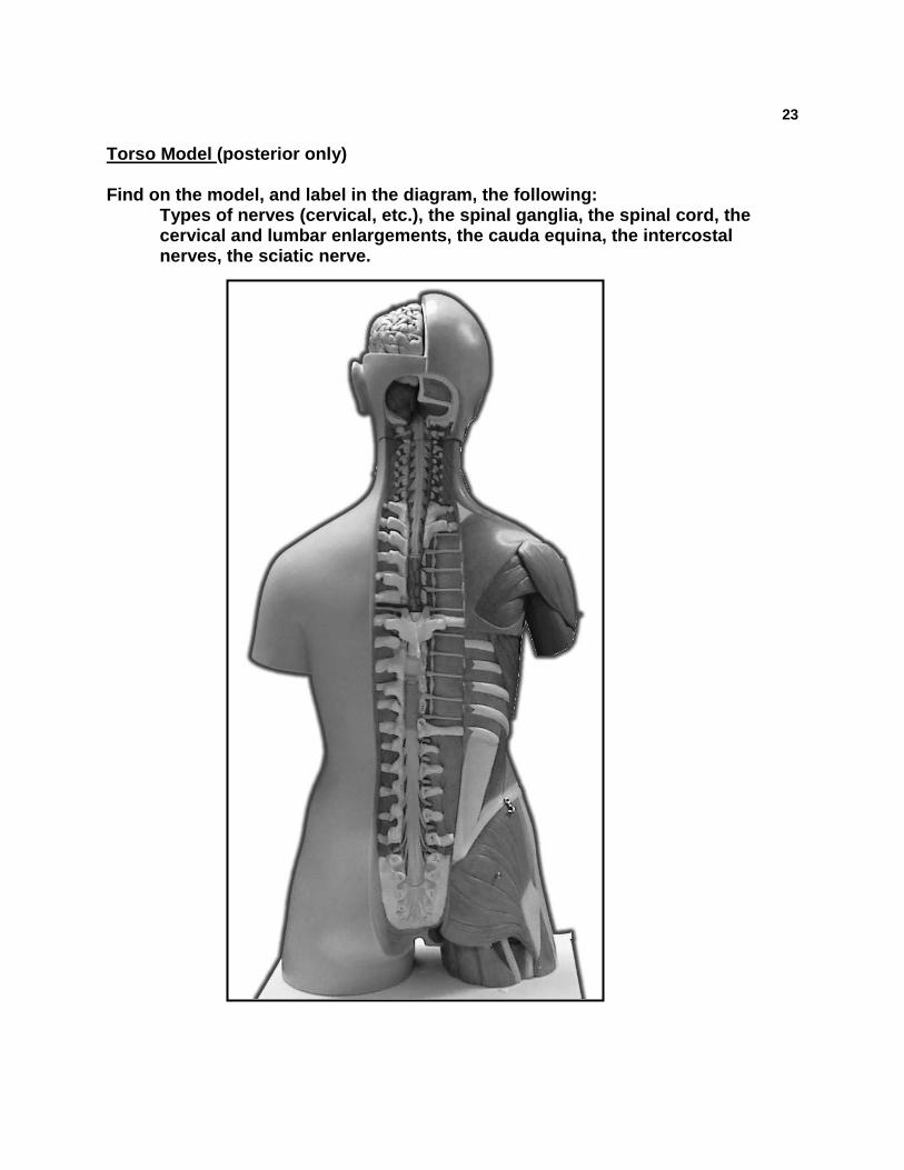
23 Torso Model (posterior only) Find on the model, and label in the diagram, the following:
Types of nerves (cervical, etc.), the spinal ganglia, the spinal cord, the cervical and lumbar enlargements, the cauda equina, the intercostal nerves, the sciatic nerve.
24 #8 Study the Reflexes.
Reflexes can be categorized in several ways. We will first divide them based on the effectors .... that is, somatic (skeletal muscle) versus autonomic (all others...gland, smooth muscle, etc.). Here is a categorization system commonly used:
1. Somatic Reflexes (all control skeletal muscle)
a. Spinal reflexes i. Stretch (example: Patellar) ii. Superficial (examples: Plantar) iii. Special cases: (example: Babinski’s sign)
b. Cranial i. Corneal ii. Gag
2. Autonomic Reflexes: control smooth muscle or glands
* Examples in lab book: papillary reflex
Another categorization system looks at where the response is: Understand the terms contralateral vs. ipsilateral
You lab book describes several examples of reflexes. There are various instruments available to you that can be used to replicate the reflexes outlined in your lab book:
Percussion hammers-reflex Tongue depressors Penlights Meter sticks/yardsticks 6” & 12” rulers Absorbent cotton Autoclave bag (for disposal of tongue depressors and cotton)
However, you will not be tested on your ability to actually perform the reflexes. You should do as many as you have time for. However, this is the information you will need for the lab practical
Read Me
Know this!
Read this!
Important point!
25
For each reflex your instructor wants you to know, know the following: 1. Be able to ID the reflex based on a description or an image of the reflex being performed. Examples:
Which reflex is tested by hammering just below someone's knee? Which reflex is tested by hammering just behind someone's elbow? Which reflex is tested by rubbing a sharp object along the sole of the foot? What is a special case of this? Which reflex is tested by rubbing a piece of cotton on someone's eye? Which reflex is tested by shining a light in someone's eye? Which reflex is tested by hammering on the subject's Achilles' tendon?
2. for each of the reflexes, be able to categorize them. Examples:
What sort of reflex is the Patellar reflex? Give both the general category, and the specific category. What sort of reflex (general category) is the Pupillary reflex? What sort of reflex is the Gag reflex? Give both the general category, and the specific category.
Know this!