Embed Size (px)
Citation preview
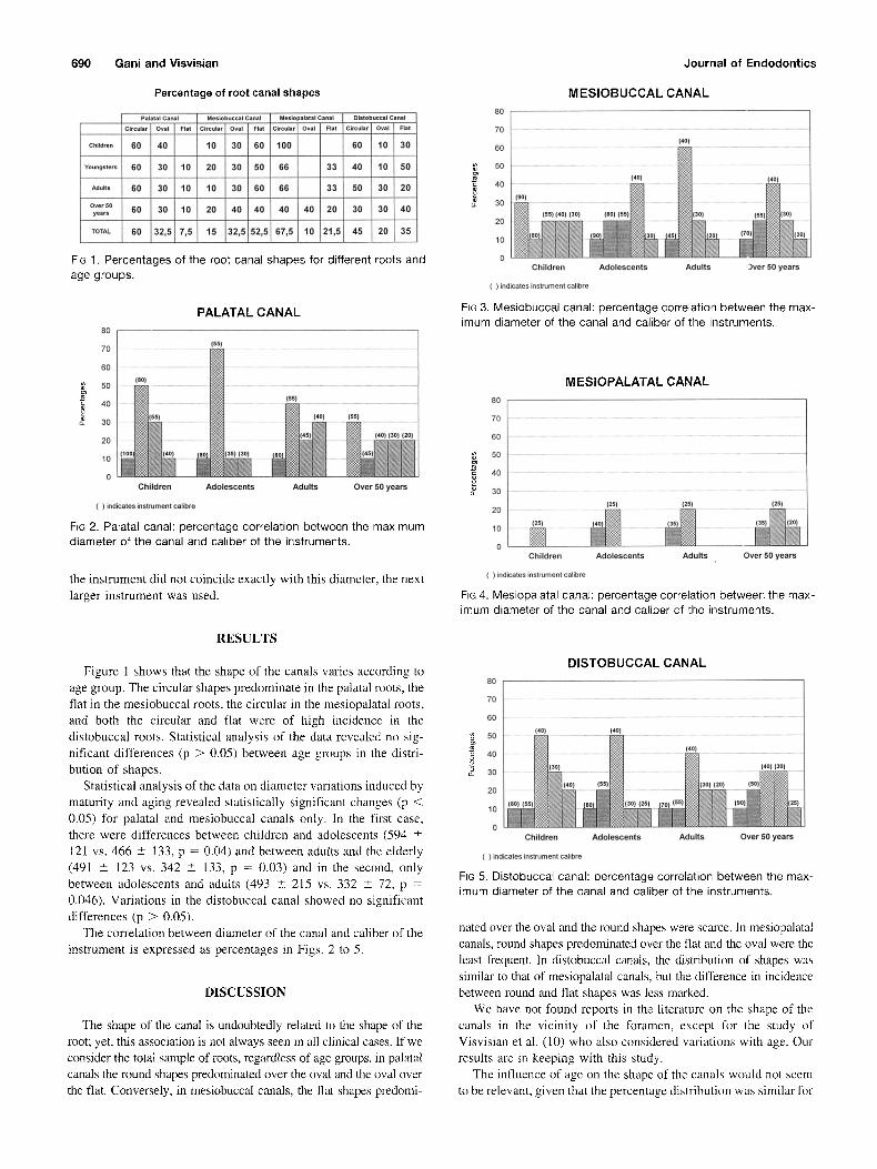
JOURNAL OF ENDODONTFCS Printed in U.S.A. Copyright © 1999 by The American Association of Endodontists VOL. 25, No, 10, OCTOBER 1999
Apical Canal Diameter in the First Upper Molar at Various Ages
Omar Gani, DDS, and Carmen Visvisian, DDS
The shape of root canals cross-sectioned through their roots at 2 mm from their apices and its cor- relation with the DO diameter of endodontic instru- ments was evaluated in 40 first upper molars. The molars were grouped according to age" under 13 yr (children), 18 to 20 yr (adolescents), 30 to 40 yr (adults), and over 50 yr. Evaluation of the root canal diameters revealed that the shapes were predom- inantly circular in the palatal canal, mostly flat in the mesiobuccal canal, and circular or flat in equal proportions in the distobuccal root. Age does not seem to affect the shape of the canals. Narrowing with age was statistically significant (p < 0.05) for palatal and mesiobuccal canals only. Correlation between the maximum diameter of the canals and the instruments was varied. Even in old age, diam- eters were observed that would require instru- ments of a size that would be impossible to use, because one internal diameter would exceed the root's external diameter in a different direction (i.e. intimal buccolingual diameter of #80 and external mesiodistal diameter of #70).
Current recommendations for endodontic treatment require that both instrumentation and obturation do not go beyond the dentin-cement constriction (1). However, this constriction may not exist at all or may be difficult to identify clinically (2). Thus, a conventional limit has been set at 1, 1.5, or even 2 nml from the root apex (3).
The 2 or 3 final millimeters of the canal are of vital importance, and their preparation must be meticulous to achieve a conical or cylindri- cal configuration of circular cross-section (4-6) , allowing apical sealing and preventing leakage to or from the canal. A frequent question is how far instrumentation must proceed. Some authors have attempted, without success, to establish rules to limit apical widening (7-9). Furthermore, recommendations for curved canals indicate that apical instrumentation should not go beyond #25 or #30 (9).
Because the question remains as to whether or not apical anat- omy allows for such attempts, the objectives of this study are to assess the root canals of human first upper molars of known age at - 2 mm from their apices in terms of diameter and shape and compare the diameters to the DO point of the endodontic instru- ments that were used.
M A T E R I A L S AND M E T H O D S
Forty permanent first upper molars of known age extracted for caries from patients free from advanced periodontal disease were thoroughly cleaned and disinfected, and divided into groups of 10 as follows:
• C h i l d r e n . " molars with developed apices extracted from patients under 13 yr old
• A d o l e s c e n t s : patients 18 to 20 yr old • A d u l t s : patients 30 to 40 yr old • E l d e r l y : patients over 50 yr old.
The apical 2 mm of the root were removed with a diamond disk. At this point, a cross-section of --2 mm in thickness was obtained. The section was washed, dried, and fixed on its larger surface with liquid, instant-drying glue to a celluloid slide. The surface of each section was stained with PeIikan blue ink to improve observation of the canals. Excess ink was removed with a damp swab. The mesiodistal and buccolingual diameters of all of the root canals were measured by means of a light microscope (DRC Zeiss), with a magnification of ×6.3; the results were tabulated.
If more than one canal was detected on the section surface, a radiographic analysis was performed to determine whether both canals were, in effect, separate and unrelated or were branches of a single canal. If the canals proved to be branches, only the larger of the two was considered.
The shape of the canals was determined in keeping with the following classification based on mesiodistal and buccolingual diameters:
• C i r c u l a r : when both diameters were equal • O v a l : when the largest diameter exceeded the smallest by less
than a radius • F l a t : when the largest diameter exceeded the smallest by more
than a radius; this category included tear-shaped, hourglass- shaped, ribbon-shaped canals, etc.
To establish the correlation between "diameter of canal-caliber of instrument," the largest diameter was compared with the diam- eter of standard endodontic instruments at the DO point. This point, according to ISO standards, corresponds to the smallest diameter of the instrument located in the extreme point of the active or cutting part of the instrument and its measurement (expressed in hundredths of millimeters, with a tolerance of _+0.02 ram), which corresponds to the number of the instrument (8). If the DO point of
689
690 Gani and Visvisian
Percentage of root canal shapes
Palatal Canal Mesiobuccal Canal Meslopalatal Canal Distobuccal Canal Circular Oval Flat Circular Oval Flat Circular Oval Flat Circular Oval Flat
Children 60 40 10 30 60 100 60 10 30
Youngsters 60 30 10 20 30 50 66 33 40 10 50
Aaolts 60 30 10 10 30 60 66 33 50 30 20
Over 50 60 30 10 20 40 40 40 40 20 30 30 40 years
TOTAL 60 32,5 7,5 15 32,5 52,51 67,5 t 0 21,5 45 20 35
FIG 1. Percentages of the root canal shapes for different roots and age groups.
80
PALATAL CANAL
70
60
50
E 40 e
g_ 30 20
10
O C h i l d r e n
( ) indicates instrumenl calibre
A d o l e s c e n t s A d u l t s O v e r 50 y e a r s
FIG 2. Palatal canal: percentage correlation between the maximum diameter of the canal and caliber of the instruments.
the instrument did not coincide exactly with this diameter, the next larger instrument was used.
RESULTS
Figure 1 shows that the shape of the canals varies according to age group. The circular shapes predominate in the palatal roots, the flat in the mesiobuccal roots, the circular in the mesiopalatal roots, and both the circular and flat were of high incidence in the distobuccal roots. Statistical analysis of the data revealed no sig- nificant differences (p > 0.05) between age groups in the distri- bution of shapes.
Statistical analysis of the data on diameter variations induced by maturity and aging revealed statistically significant changes (p < 0.05) for palatal and mesiobuccal canals only. In the first case, there were differences between children and adolescents (594 _+ 121 vs. 466 _+ 133, p = 0.04) and between adults and the elderly (491 _ 123 vs. 342 _+ 133, p = 0.03) and in the second, only between adolescents and adults (493 ± 215 vs. 332 -- 72, p = 0.046). Variations in the distobuccal canal showed no significant differences (p > 0.05).
The correlation between diameter of the canal and caliber of the instrument is expressed as percentages in Figs. 2 to 5.
DISCUSSION
The shape of the canal is undoubtedly related to the shape of the root; yet, this association is not always seen in all clinical cases. If we consider the total sample of roots, regardless of age groups, in palatal canals the round shapes predominated over the oval and the oval over the flat. Conversely, in mesiobuccal canals, the flat shapes predomi-
80
Journal of Endodontics
MESIOBUCCAL CANAL
70
60
5O
40
30
20
10
0 C h i l d r e n
( } indicates instrument calibre
A d o l e s c e n t s A d u l t s : )ver 5 0 y e a r s
FIG 3. Mesiobuccal canal: percentage correlation between the max- imum diameter of the canal and caliber of the instruments.
80
MESIOPALATAL CANAL
70
60
5O
40 o~ g_ 30
125}
10 (zs)
o N C h i l d r e n A d o l e s c e n t s
( ) indicates instrument calibre
(zs) 125}
Adul ts Over 50 y e a r s
FIG 4. Mesiopalatal canal: percentage correlation between the max- imum diameter of the canal and caliber of the instruments.
80
DISTOBUCCAL CANAL
7 0 •
60 (40) (40)
50 . . . . . . . 1401
40a0 _ 1301 (40) (30)
20 (401 (55) 1301 (20) 1501
10
0 C h i l d r e n A d o l e s c e n t s A d u l t s O v e r 50 y e a r s
( ) indica{es instrument calibre
FiG 5. Distobuccal canal: percentage correlation between the max- imum diameter of the canal and caliber of the instruments.
nated over the oval and the round shapes were scarce. In mesiopalatal canals, round shapes predominated over the flat and the oval were the least frequent. In distobuccal canals, the distribution of shapes was similar to that of mesiopalatal canals, but the difference in incidence between round and flat shapes was less marked.
We have not found reports in the literature on the shape of the canals in the vicinity of the foramen, except for the study of Visvisian et al. (10) who also considered variations with age. Our results are in keeping with this study.
The influence of age on the shape of the canals would not seem to be relevant, given that the percentage distribution was similar for
Vol. 25, No. 10, October 1999
all age groups, and the statistical analysis revealed that there were no significantly statistical differences. It is noteworthy, however, that fiat shapes and ribbon shapes persist near the apex, even in elderly patients, particularly in the mesiovestibular canal. This finding may be due to the fact that concentric narrowing of ribbon canals with age is statistically significant only along the smallest diameter and not along the largest diameter. In oval canals, nar- rowing is only statistically significant along the largest diameter. Thus, ribbon canals retain their shape even in elderly patients, whereas oval canals tend to turn circular.
The correlation of the largest diameter of the canal with the DO point of the instrument was individually analyzed for each root.
Palatal Canal
Round canals could be prepared in children with a #80 instru- ment or larger. This caliber may be insufficient; in some cases, a #100 instrument was necessary. Kerekes and Tronstad (1 l) pre- sented cases in which instruments of calibers over 140 were necessary, regardless of age.
In the groups of adolescents, adults, and elderly patients, the predominant caliber to achieve apical preparation of this canal was #55. However, this caliber may be insufficient (i.e. some cases required as much as #80).
The shape and size of the palatal canal would allow for endodontic treatment involving marked apical widening. This canal, however, exhibits many curvatures toward the buccal that cannot be detected radiographically (12) and that would preclude the use of instruments of a large caliber. Tronstad (6) states that all canals should be treated as if they were curved. This recommendation would hold particularly true for the palatal canal of the upper molars. The type of apical preparation to be used in such wide canals treated with techniques for curved canals (step-back or crown-down or their variants), with re- duced apical widening is still a matter of study.
Mesiobuccal Canal
The buccolingual width of this canal in the vicinity of the apex at all ages is noteworthy. Tear-shaped canals were very frequent, possibly due to the process of dentin deposition that occurs with age and that, in some cases, would lead to the formation of the fourth canal (13). Gutierrez and Gigoux (12) report on the presence of prolongations of the canals of the first upper molar; in particular, in the case of the mesiobuccal canal, which impairs cleaning and shaping. These prolongations would correspond to tear-shaped canals in cross-section.
Our data reveal that apical preparation in children would require a caliber of at least 90. These results are in fairly good agreement with those of Kerekes and Tronstad (11), who also report cases in which a #140 instrument was necessary.
In the groups of adolescent, adult, and elderly patients the greatest correlation was with the #40 instrument. However, all groups exhib- ited a percentage of cases that correspond to a greater caliber, reaching values as large as 90. Morphologically, the mesiobuccal root does not allow the use of instruments of such caliber, because they would exceed the smallest diameter of the root (13).
Characteristics of the mesiobuccal canal allow for clinical in- strumentation, with techniques designed for curved canals. The question remains, as for the palatal canal, what the results will be when techniques that involve minimal apical widening are applied to such wide canals.
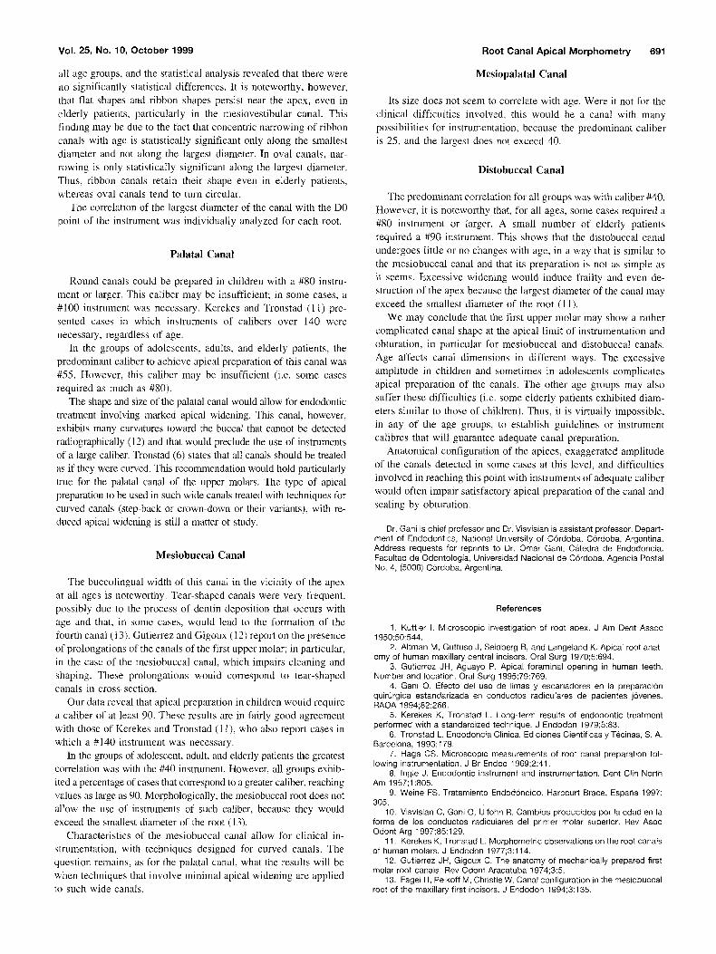
Root Canal Apical Morphometry 691
Mesiopalatal Canal
Its size does not seem to correlate with age. Were it not lbr the clinical difficulties involved, this would he a canal with many possibilities for instrumentation, because the predominant caliber is 25, and the largest does not exceed 40.
Distobuccal Canal
The predominant correlation for all groups was with caliber #40. However, it is noteworthy that, for all ages, some cases required a #80 instrument or larger. A small number of elderly patients required a #90 instrument. This shows that the distobuccal canal undergoes little or no changes with age, in a way that is similar to the mesiobuccal canal and that its preparation is not as simple as it seems. Excessive widening would induce frailty and even de- struction of the apex because the largest diameter of the canal may exceed the smallest diameter of the root (11).
We may conclude that the first upper molar may show a rather complicated canal shape at the apical limit of instrumentation and obturation, in particular for mesiobuccal and distobuccal canals. Age affects canal dimensions in different ways. The excessive amplitude in children and sometimes in adolescents complicates apical preparation of the canals. The other age groups may also suffer these difficulties (i.e. some elderly patients exhibited diam- eters similar to those of children). Thus, it is virtually impossible, in any of the age groups, to establish guidelines or instrument calibres that will guarantee adequate canal preparation.
Anatomical configuration of the apices, exaggerated amplitude of the canals detected in some cases at this level, and difficulties involved in reaching this point with instruments of adequate caliber would often impair satisfactory apical preparation of the canal and sealing by obturation.
Dr. Gani is chief professor and Dr. Visvisian is assistant professor, Depart- ment of Endodontics, National University of Cdrdoba, C&doba, Argentina. Address requests for reprints to Dr. Omar Gani, C&tedra de Endodoncia, Facultad de Odontologia, Universidad Nacional de Cordoba, Agencia Postal No. 4, (5000) Cordoba, Argentina.
References
1. Kuttler I. Microscopic investigation of root apex. J Am Dent Assoc 1950;50:544.
2. Altman M, Guttuso J, Seidberg B, and Langeland K. Apical root anat- omy of human maxillary central incisors. Oral Surg 1970;5:694.
3. Gutierrez JH, Aguayo P. Apical foraminal opening in human teeth. Number and location. Oral Surg 1995;79:769.
4. Gani O. Efecto del uso de limas y escariadores en la preparacion quirt]rgica estandarizada en conductos radiculares de pacientes jdvenes. RAOA 1994;82:266.
5. Kerekes K, Tronstad L. Long-term results of endodontic treatment performed with a standardized technique. J Endodon 1979;5:83.
6. Tronstad L. Endodoncia Clinic& Ediciones CientMcas y Tecinas, S. A. Barcelona, 1993:179.
7. Haga CS. Microscopic measurements of root canal preparation fol- lowing instrumentation, d Br Endod 1969;2:41.
8. Ingle J. Endodontic instrument and instrumentation. Dent Clin North Am 1957;1:805.
9. Weine FS. Tratamiento Endod6ncico. Harcourt Brace, Espafia 1997: 305.
10. Visvisian C, Gani O, Ulfohn R. Cambios producidos por la edad en la forma de los oonductos radiculares del primer molar superior. Rev Asoc Odont Arg 1997;85:129.
11. Kerekes K, Tronstad L. Morphometric observations on the root canals of human molars. J Endodon 1977;3:114.
12. Gutierrez JH, Gigoux C. The anatomy of mechanically prepared first molar root canals. Rev Odont Aracatuba 1974;3:5.
13. Fagel H, Peikoff M, Christie W. Canal configuration in the mesiobuccal root of the maxillary first incisors. J Endodon 1994;3:135.