Embed Size (px)
Citation preview
293Vol. 46, Nº3, 2011Revista de Biología Marina y Oceanografía
Revista de Biología Marina y OceanografíaVol. 46, Nº3: 293-302, diciembre 2011Article
Pathology caused by adult Pseudochondracanthusdiceraus (Copepoda: Chondracanthidae), a parasite
of bullseye puffer fish Sphoeroides annulatusPatología causada por adultos de Pseudochondracanthus diceraus (Copepoda:
Chondracanthidae) parásito del botete diana Sphoeroides annulatus
Emma Josefina Fajer-Ávila1, Leslie Guzman-Beltran2, Walter Camilo Zárate-Rodríguez2,Oscar Basilio Del Río-Zaragoza1 and Pablo Almazan-Rueda1
1Centro de Investigación en Alimentación y Desarrollo, A.C., Unidad Mazatlán en Acuicultura y Manejo Ambiental,Av. Sábalo Cerritos s/n, Estero del Yugo, C.P. 82010, Mazatlán, Sinaloa, México. [email protected] de La Salle, Facultad de Medicina Veterinaria, Sede La Floresta, Carretera 7a No. 172-85, Bogotá DC, Colombia
Resumen.- El copépodo condracántido Pseudochondracanthus diceraus es un parásito frecuente en las branquiasdel botete diana silvestre, Sphoeroides annulatus en Sinaloa, México. El presente estudio constituye la primeradescripción de algunos parámetros sanguíneos y la histología branquial de S. annulatus infestados por hembrasadultas de P. diceraus. Se aclimataron sesenta y dos peces silvestres en tanques experimentales por 15 días y a los16 y 21 días se tomaron muestras para la realización de los análisis. Se observó un 100% de prevalencia y unaintensidad media de 7,16 (1-17) parásitos por pez. P. diceraus prefirió los sectores anteriores y posteriores decada arco branquial. Los porcentajes de leucocitos fueron significativamente menores en peces con las más altasintensidades de infestación. La respuesta branquial del hospedero fue más pronunciada cerca del sitio de fijacióndel parásito y estuvo caracterizada por hiperplasia y fusión de las lamelas secundarias, hiperplasia condrocíticade las lamelas primarias, atrofia del epitelio lamelar e incremento del número de células mucosas. Los daños en eltejido branquial del botete diana son causados por las antenas grandes y robustas modificadas en forma de grapaque emplea el parásito para fijarse al pez. Esto indica que altas prevalencia e intensidades de infestación por P.diceraus representan un riesgo de salud potencial para el cultivo de esta especie de pez.
Palabras clave: Condracántidos, peces, hematología, histopatología, México
Abstract.- The chondracanthid copepod Pseudochondracanthus diceraus is a parasite commonly found on the gillsof wild bullseye puffer fish, Sphoeroides annulatus, in Sinaloa, Mexico. This paper provides the first description ofsome blood parameters and gill histology of wild bullseye puffer fish infected by an adult parasitic copepod P.diceraus. Sixty two wild fish were acclimatized for 15 days in experimental tanks, and then on days 16 and 21,samples were taken. Results showed a 100% prevalence and a mean intensity of 7.16 (1-17) copepods per fish. P.diceraus preferred the anterior and posterior sector of each gill arch. The percentage of leukocytes wassignificantly lower in fish with the highest intensity of infection. The gill response of the host was most evidentnear the parasite’s attachment site. It was characterised by hyperplasia and fusion of secondary lamellae,chondrocyte hyperplasia of primary lamellae, atrophy of the lamellar epithelium and an increase in the number ofmucous cells. The damaged gill tissue of the bullseye puffer fish was caused by the large and strong antennae of P.diceraus that is modified as a claw to attach to the host gills. Thus, a high prevalence and intensity of this copepodcould become a potential health risk to the bullseye puffer fish culture.
Key words: Chondracanthid, fish, haematology, histopathology, Mexico
INTRODUCTION
The bullseye puffer fish Sphoeroides annulatus (Jenyns,1842) is a tetraodontid species inhabiting the Gulf ofCalifornia, extending along the Eastern Pacific coast fromSan Diego (USA) to Peru (Thomson et al. 2000).Biotechnological advances have made it possible to
cultivate the bullseye puffer in Sinaloa, Mexico (Alvarez-Lajonchere et al. 2007). However, under culture conditions,juvenile fish have recurrent infections by ectoparasiticcopepods that have resulted in high mortalities (Fajer-Ávila et al. 2008).
294 Fajer-Ávila et al.Pathology of chondracanthid on Sphoeroides annulatus
Gill pathology caused by ectoparasitic copepods hasgreat economic importance for some species (Lin et al.1994, Wu et al. 1997, Bennett & Bennett 2001, Smith et al.2007, Tang et al. 2007). Three copepods belonging tothe families Lernaeopodidae, Bomolochidae andChondracanthidae were found on gills and in the branchialcavity of wild bullseye puffer fish from Mazatlán, Sinaloa,Mexico (Fajer-Ávila et al. 2004). The Chondracanthidaewas represented by Pseudochondracanthus dicerausWilson, 1908 and it was the most abundant copepod(Morales-Serna 2010). However, there is a lack ofinformation on the pathology associated with this parasiticcopepod.
High abundances of parasites on fish usually causealterations in several blood parameters because some ofthem feed on host blood (Chavez et al. 2006, Sitjà-Bobadilla& Alvarez-Pellitero 2009). Hematological analysis couldtherefore be used as a direct or inferential indicator of thetolerance to a stressor agent or/and health of the fish(Schreck & Moyle 1990, Del Rio-Zaragoza et al. 2008). Inorder to address such an approach, it is necessary tothoroughly characterize the blood cells of the organismsand use this knowledge to quantify normal bloodparameters (Shigdar et al. 2007). This paper provides thefirst description of the pathology caused by adult femalesof the copepod Pseudochondracanthus diceraus on thegills of Sphoeroides annulatus using histopathologicaland haematological analyses.
MATERIALS AND METHODS
EXPERIMENTAL DESIGN
Sixty two bullseye puffer fish, Sphoeroides annulatus,with an average weight of 223.2 ± 35.8 g and an averagetotal length of 21 ± 1.2 cm, were caught from Santa Maríade La Reforma Bay (25°10´N, 108°20´W and 24°50´N,107°55´W) Sinaloa, Mexico and were transported alive in450 L circular tanks to CIAD’s laboratory facilities. Uponarrival, six fish were randomly selected, anaesthetised with2 phenoxyethanol 0.75 mL L-1 (Sigma, St. Louis) for 3 minand gills were excised to be analysed for adult copepods.All fish were only infected by the chondracanthidPseudochondracanthus diceraus with an initial meanintensity of 4.16 parasites per fish. The rest of the fishwere randomly distributed into four 400 L circular tanks(14 fish per tank) with mesh-filtered (20 μm) flow throughseawater (SW) and aeration. Fish were fed a mixture offrozen tilapia fillet, squid and shrimp at 5% of their bodyweight twice per day. Fish were acclimatized during 15
days. After this period, on days 16 and 21, three fish pertank were sampled for parasites, blood parameters andgill histology (24 fish in total). During the trial fish did notshow signs of viral/bacterial disease. The rest of the fish(26) were used for other studies.
HAEMATOLOGICAL PARAMETERS
Fish were anaesthetised and in less than three min, bloodsamples were collected from the caudal vein using 3-mlnon-anticoagulant syringes (Terumo Mexico, DF).Immediately after sampling, one blood drop was used forblood smears. Then, the blood sample was placed intotubes coated with anticoagulant (K2EDTA BD Microtainer)for haematocrit and haemoglobin. Haematocrit (HCT) wascalculated after using a CORNING heparinised tube, filledto 2/3rds, and was placed for 10 min in a SOL-BAS P600microhaematocrit centrifuge. The packed cell percentageversus the total volume was measured using amicrohaematocrit reader and reported as a percentage.Haemoglobin (HGB) concentration in the blood wasdetermined by the cyanomethaemoglobin method with ahemogloWiener® kit and the aid of an absorbancespectrophotometer (Agilent® 8453) λ = 540 nm. Meancorpuscular haemoglobin concentration (MCHC) wascalculated using the following standard formula: MCHC(g dL-1) = Hb (g 100 ml-1) x 100 / Ht (%). For each fish,triplicates of all blood parameters were completed.Leukocyte cellular differentiation was observed in bloodsmears treated with Wright-Giemsa staining in an opticalmicroscope (LEICA DMLB-10, Guadalajara). The imageswere processed using Image-Pro Plus 3 software (MediaCybernetics, Bethesda). Thirty digital images with anaverage of 214.58 ± 55.33 leukocytes at 600X were capturedfrom each blood smear and the percentages of each celltype were calculated. An OLYMPUS BX-51 opticalmicroscope was used to measure the erythrocyte,thrombocyte and leucocyte diameters (n = 30 cells).
PARASITOLOGICAL AND HISTOPATHOLOGICAL
EXAMINATION
After blood collection, the gill arches were dissected,placed in Petri dishes with drops of filtered seawater (34psu) and the parasites from each gill arch were countedusing a Leica MZ 9.5 stereomicroscope (LeicaMicrosystems Wetzlar, Germany). The bullseye puffer fishis an exceptional teleost fish with only three gill arches ineach side. The gill arches were numbered I to III fromexternal to internal arch, on each side of the fish. Also,each arch was arbitrarily divided into three sectors
295Vol. 46, Nº3, 2011Revista de Biología Marina y Oceanografía
approximately equal in surface area: anterior (A), central(B) and posterior (C). To analyse the gills for parasites,each gill arch was individually placed in a Petri dish withdrops of seawater and observed under a stereomicroscope,and then the number of parasites per sector (anterior toposterior) was counted. Each gill arch was fixed in 10%neutral buffered formalin. Twenty four h later the gillswere removed from formalin and preserved in ethanol(70%).
Prevalence (%) and mean intensity ofPseudochondracanthus diceraus in the fish were definedaccording to Bush et al. (1997). The condition factor (K),was calculated according to Ricker (1975).
Small pieces of gill arch infected and uninfected withcopepods were embedded in paraffin wax forhistopathological analysis. Tissue sections of 3-5 μmthicknesses were obtained using a conventionalmicrotome. Each sample was dehydrated and stained withhaematoxylin-eosin-phloxin. Samples were observed andphotomicrography was carried out using an OLYMPUSBX51 microscope. Evaluation of severity and extent ofinjury on gill arch tissue was recorded according to Bernetet al. (1999). A brief description is as follows: Low level:cell and tissue structure slightly injured and visible.Moderate level: reduced activity of the injured cells andtissue, and High level: high cell activity and loss of tissuefunction and structure. The gill arch is severely damaged,making identification very difficult.
STATISTICAL ANALYSIS
Results were expressed as means ± standard deviation(SD) or standard error (SE). Normality tests wereperformed for all data. For the distribution of P. dicerauson the gill arch of the fish, data was analysed by theKruskal-Wallis test. To determine significant differencesamongst groups, a multiple comparison analysis ‘aposteriori’ Tukey test was carried out and for the bloodparameters a Student´s t-Test (P < 0.05) was applied. Allpercentage data were arcsine transformed beforestatistical comparisons. Non-normal data were analyzedby the Mann-Whitney Rank Sum Test (U). Spearman’scorrelation coefficient between blood parameters and levelof infestation was calculated. All analysis was realizedusing SigmaPlot 9 software (SYSTAT Software, Inc., SanJose).
RESULTS
NUMBERS AND DISTRIBUTION OF PARASITES
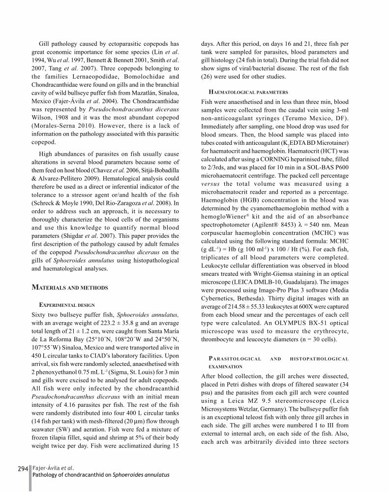
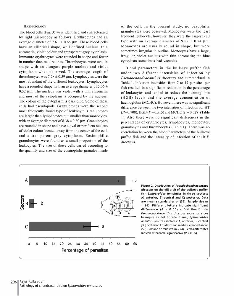
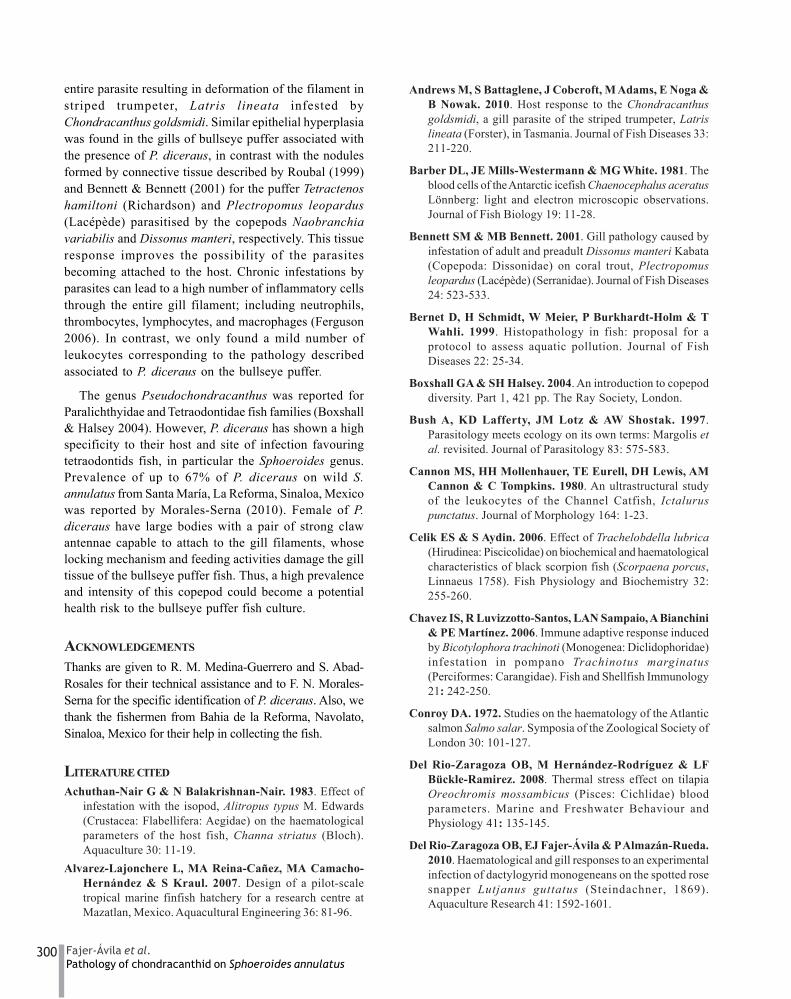
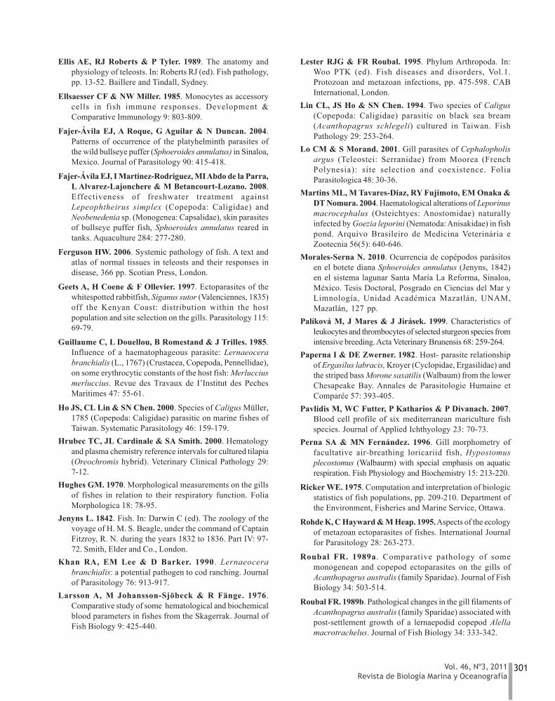
In total 24 fish were analysed and 208Pseudochondracanthus diceraus adults were found (Fig.1), with a prevalence of 100% and a mean intensity of 7.16parasites per fish, with a range from 1 to 17. P. diceraushad an average length of 3 mm and average width of 1mm. Significantly more P. diceraus adults were found inthe anterior and posterior sectors of the gill arch (Fig. 2)compared to the central sector (P < 0.05), and nopreference for any particular gill arch (P > 0.05) wasobserved. No differences for sampling days 16 and 21were found in respect to the numbers of parasites andcondition factor. So, independent of sampling time, anarbitrary categorization of two levels of infection wascarried out according to the total number of parasitesfoun on the three gill arches: 1 (1-6 parasites) and 2 (7-17parasites). Blood parameters were also analyzed basedon this categorization.
Figure 1. Ventral view of a Pseudochondracanthus dicerausfemale adult with eggs. Scale bar = 2 mm / Vista ventral de lahembra adulta ovígera de Pseudochondracanthus diceraus. Barra deescala = 2 mm
296 Fajer-Ávila et al.Pathology of chondracanthid on Sphoeroides annulatus
HAEMATOLOGY
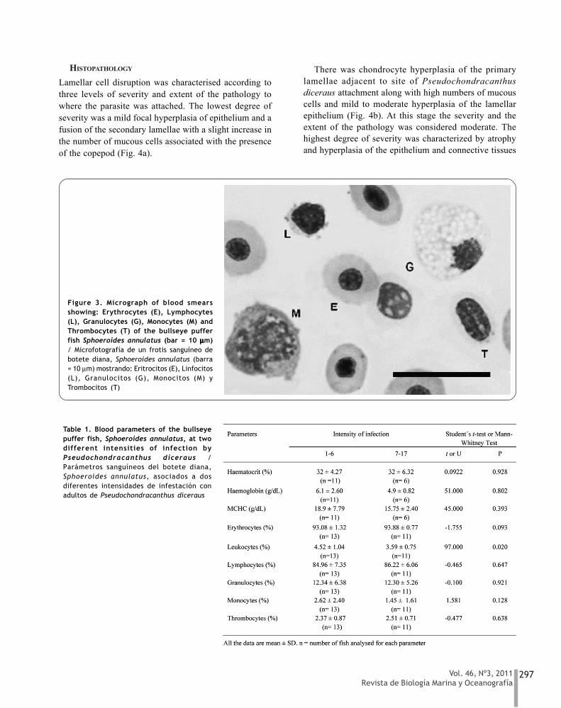
The blood cells (Fig. 3) were identified and characterizedby light microscopy as follows: Erythrocytes had anaverage diameter of 7.61 ± 0.66 μm. These blood cellshave an elliptical shape, well defined nucleus, thinchromatin, violet colour and transparent-grey cytoplasm.Immature erythrocytes were rounded in shape and fewerin number than mature ones. Thrombocytes were oval inshape with an elongate purple nucleus and violetcytoplasm when observed. The average length ofthrombocytes was 7.28 ± 0.59 μm. Lymphocytes were themost abundant of the different leukocytes. Lymphocyteshave a rounded shape with an average diameter of 5.06 ±0.52 μm. The nucleus was violet with a thin chromatinand most of the cytoplasm is occupied by the nucleus.The colour of the cytoplasm is dark blue. Some of thesecells had pseudopods. Granulocytes were the secondmost frequently found type of leukocyte. Granulocytesare larger than lymphocytes but smaller than monocytes,with an average diameter of 8.38 ± 0.80 μm. Granulocytesare rounded in shape and have a oval or reniform nucleusof violet colour located away from the center of the cell,and a transparent grey cytoplasm. Eosinophilicgranulocytes were found as a small proportion of theleukocytes. The size of these cells varied according tothe quantity and size of the eosinophilic granules inside
of the cell. In the present study, no basophilicgranulocytes were observed. Monocytes were the leastfrequent leukocyte, however, they were the largest celltype with an average diameter of 9.82 ± 0.74 μm.Monocytes are usually round in shape, but weresometimes irregular in outline. Monocytes have a large,irregular, violet nucleus with thin chromatin; the bluecytoplasm sometimes had vacuoles.
Blood parameters in the bullseye puffer fishunder two different intensities of infection byPseudochondracanthus diceraus are summarised inTable 1. Infection intensities from 7 to 17 parasites perfish resulted in a significant reduction in the percentageof leukocytes and tended to reduce the haemoglobin(HGB) levels and the average concentration ofhaemoglobin (MCHC). However, there was no significantdifference between the two intensities of infection for HT(P= 0.708), HGB (P = 0.515) and MCHC (P = 0.528) (Table1). Also there were no significant differences in thepercentages of erythrocytes, lymphocytes, monocytes,granulocytes and thrombocytes (Table 1). There was nocorrelation between the blood parameters of the bullseyepuffer fish and the intensity of infection of adult P.diceraus.
Figure 2. Distribution of Pseudochondracanthusdiceraus on the gill arch of the bullseye pufferfish Sphoeroides annulatus in three sectors:A) anterior, B) central and C) posterior. Dataare mean ± standard error (SE). Sample size (n= 24). Different letters indicate significantdifference (P < 0.05) / Distribución dePseudochondracanthus diceraus sobre los arcosbranquiales del botete diana, Sphoeroidesannulatus en tres sectores: A) anterior, B) centraly C) posterior. Los datos son media ± error estándar(SE). Tamaño de muestra (n = 24). Letras diferentesindican diferencia significativa (P < 0,05)
297Vol. 46, Nº3, 2011Revista de Biología Marina y Oceanografía
HISTOPATHOLOGY
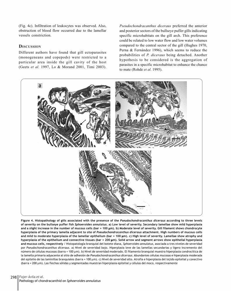
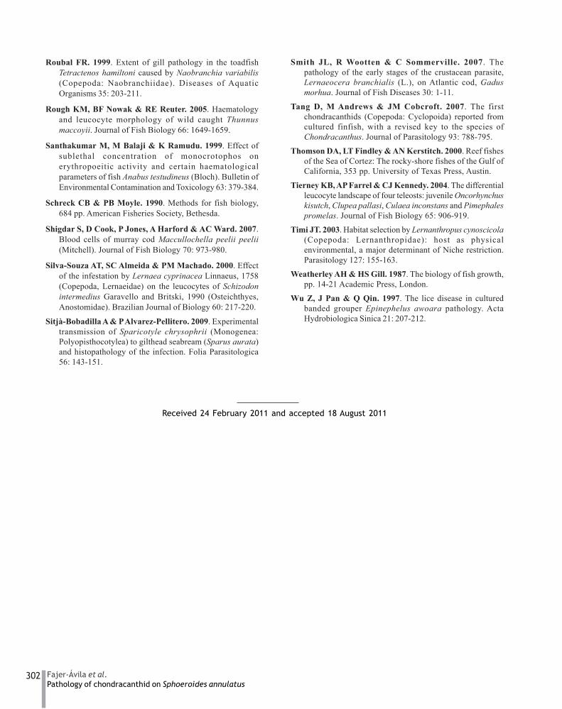
Lamellar cell disruption was characterised according tothree levels of severity and extent of the pathology towhere the parasite was attached. The lowest degree ofseverity was a mild focal hyperplasia of epithelium and afusion of the secondary lamellae with a slight increase inthe number of mucous cells associated with the presenceof the copepod (Fig. 4a).
There was chondrocyte hyperplasia of the primarylamellae adjacent to site of Pseudochondracanthusdiceraus attachment along with high numbers of mucouscells and mild to moderate hyperplasia of the lamellarepithelium (Fig. 4b). At this stage the severity and theextent of the pathology was considered moderate. Thehighest degree of severity was characterized by atrophyand hyperplasia of the epithelium and connective tissues
Table 1. Blood parameters of the bullseyepuffer fish, Sphoeroides annulatus, at twodifferent intensities of infection byPseudochondracanthus diceraus /Parámetros sanguíneos del botete diana,Sphoeroides annulatus, asociados a dosdiferentes intensidades de infestación conadultos de Pseudochondracanthus diceraus
Figure 3. Micrograph of blood smearsshowing: Erythrocytes (E), Lymphocytes(L), Granulocytes (G), Monocytes (M) andThrombocytes (T) of the bullseye pufferfish Sphoeroides annulatus (bar = 10 μμμμμm)/ Microfotografía de un frotis sanguíneo debotete diana, Sphoeroides annulatus (barra= 10 μm) mostrando: Eritrocitos (E), Linfocitos(L), Granulocitos (G), Monocitos (M) yTrombocitos (T)
298 Fajer-Ávila et al.Pathology of chondracanthid on Sphoeroides annulatus
(Fig. 4c). Infiltration of leukocytes was observed. Also,obstruction of blood flow occurred due to the lamellarvessels constriction.
DISCUSSION
Different authors have found that gill ectoparasites(monogeneans and copepods) were restricted to aparticular area inside the gill cavity of the host(Geets et al. 1997, Lo & Morand 2001, Timi 2003).
Pseudochondracanthus diceraus preferred the anteriorand posterior sectors of the bullseye puffer gills indicatingspecific microhabitats on the gill arch. This preferencecould be related to low water flow and low water volumescompared to the central sector of the gill (Hughes 1970,Perna & Fernández 1996), which seems to reduce theprobabilities of P. diceraus being detached. Anotherhypothesis to be considered is the aggregation ofparasites in a specific microhabitat to enhance the chanceto mate (Rohde et al. 1995).
Figure 4. Histopathology of gills associated with the presence of the Pseudochondracanthus diceraus according to three levelsof severity on the bullseye puffer fish Sphoeroides annulatus. a) Low level of severity. Secondary lamellae show mild hyperplasiaand a slight increase in the number of mucous cells (bar = 100 μμμμμm). b) Moderate level of severity. Gill filament shows chondrocytehyperplasia of the primary lamella adjacent to site of Pseudochondracanthus diceraus attachment. High numbers of mucous cellsand mild to moderate hyperplasia of the lamellar epithelium (bar = 100 μμμμμm). c) High level of severity. Lamellae show atrophy andhyperplasia of the epithelium and connective tissues (bar = 200 μμμμμm). Solid arrow and segment arrows show epithelial hyperplasiaand mucous cells, respectively / Histopatología branquial del botete diana, Sphoeroides annulatus, asociada a tres niveles de severidadpor Pseudochondracanthus diceraus. a) Nivel de severidad bajo. Hiperplasia leve de las lamellas secundarias y ligero incremento delnúmero de células mucosas (barra = 100 μm). b) Nivel de severidad moderado. El filamento branquial muestra hiperplasia condrocítica dela lamella primaria adyacente al sitio de adhesión de Pseudochondracanthus diceraus. Abundantes células mucosas e hiperplasia moderadadel epitelio de las laminillas branquiales (barra = 100 μm). c) Nivel de severidad alto. Atrofia e hiperplasia del tejido epitelial y conectivo(barra = 200 μm). Las flechas sólidas y segmentadas muestran hiperplasia epitelial y células del moco, respectivamente
299Vol. 46, Nº3, 2011Revista de Biología Marina y Oceanografía
The blood cells of the bullseye puffer fish wereconsistent with the typical characteristics of marine fish(Palíková et al. 1999, Rough et al. 2005, Pavlidis et al.2007), and freshwater fish (Conroy 1972, Ellis et al. 1989,Hrubec et al. 2000) except that the erythrocytes are smallerthan those of the other marine fish species reported byRough et al. (2005) and Pavlidis et al. (2007). Thedifference in the erythrocyte size may be linked to asedentary life style of the bullseye puffer fish. This is inagreement with Larsson et al. (1976) who hypothesisedthat fish with more active life styles had largererythrocytes than those with a sedentary life style.However, differences in sizes of blood cells between fishspecies reinforce the necessity to thoroughly characteriseeach species separately (Shigdar et al. 2007). Theclassification of certain types of leukocytes can also behampered by inconsistencies in nomenclature and theform of some cells (Cannon et al. 1980, Barber et al. 1981).Tierney et al. (2004) mentioned that small lymphocytesand thrombocytes can be nearly identical, largelymphocytes can appear in a variety of hues, texturesand shapes, so that they can be confused with monocytesand granulocytes, which sometimes are referred asheterophils or neutrophils.
In the present study the values of HCT, HGB andHCMC, regardless of the intensity of P. diceraus (for upto a maximum of 17) indicate that this parasite did notcause blood loss or anaemia. In contrast, Bennett &Bennett (2001) found that fish had severe focalhaemorrhage due to the penetration of the maxillipeds ofthe parasite copepod Dissonus manteri into the primarylamella of the leopard coral grouper Plectropomusleopardus, and anaemia was shown to be caused byLernaeocera branchialis in haddock Melanogrammusaeglefinus (Lester & Roubal 1995), cod Gadus morhua(Khan et al. 1990) and hake Merluccius merluccius(Guillaume et al. 1985).
On the other hand, a significantly smaller percentageof leukocytes in the blood were found in bullseye pufferfish with an infestation of 7-17 parasites per fish. This isimportant because fish leukocytes are involved inregulation of immunological function (Santhakumar et al.1999) and the reduced number of leukocytes may havebeen due to stress caused by the parasite. This suggestsa connection between parasitic infestations and impairedimmunological responses. A reduced number ofleukocytes also have been found in the snakehead murrelChanna striatus naturally infected with the isopodAlitropus typus (Achuthan-Nair & Balakrishnan-Nair
1983); moreover in Schizodon intermedius infected bythe copepod Lernaea cyprinacea (Silva-Souza et al. 2000)and in black scorpionfish Scorpaena porcus naturallyinfected by the leech Trachelobdella lubrica (Celik &Aydin 2006).
The evaluation of blood parameters can be the fastestway to detect stress symptoms. Infectious diseases andstress can elevate the number of neutrophils andmonocytes in blood (Ellsaesser & Miller 1985) anddecrease the number of lymphocytes as a protectiveresponse against the stress (Del Rio-Zaragoza et al. 2010).The numbers of lymphocytes, monocytes and neutrophilsare considered as secondary responses in different kindsof stresses (Martins et al. 2004). However, contrary toexpectation, the percentage of granulocytes did notincrease at the highest level of infection due probably tothe number of parasites, despite the increase of leukocytesobserved in the gill tissue. These results are similar to theresponses in some infected fish by some superficiallyattached ergasilid copepods (Paperna & Zwerner 1982,Roubal 1989a). Chronic infection by copepod gill parasitescan result in low to moderate infiltration of leukocytes tochronic inflammation seen in the surf breamAcanthopagrus australis on gill filaments infected withthe lenaepodid copepod Alella macrotrachelus (Roubal1989b).
The condition factor has been used as an indicator ofthe health status of a fish, and is useful to compare fishfrom different stocks (Weatherley & Gill 1987). The resultsof this study indicate that the numbers of parasites perfish were not high enough to cause a reduction in thecondition factor despite the gill injuries observed.
The localised hyperplasia at the site of attachment ofPseudochondracanthus diceraus was spread to the entiregill filament, losing its lamellar structure, and extendedonto adjacent uninfected filaments. This can partly beattributable to the action of the strong and well-developedmouthparts of this copepod during their feeding (Ho etal. 2000) as well as for other sessile copepods, and themovement of the thorax and appendages to ensure watermovement over the egg sacs so to avoid the settling ofmucus debris, as similarly reported by Andrew et al. (2010)for Chondracanthus goldsmidi. In addition to the lamellarvessels constriction, deprivation of blood supply canoccur by P. diceraus. As a result, copious mucous isproduced, forming a thick coating in the gill area thatdiminishes the respiratory efficiency. Andrews et al. (2010)reported extensive epithelial hyperplasia and necrosis ofgill tissue and ‘papilloma-like’ growths surrounding the
300 Fajer-Ávila et al.Pathology of chondracanthid on Sphoeroides annulatus
entire parasite resulting in deformation of the filament instriped trumpeter, Latris lineata infested byChondracanthus goldsmidi. Similar epithelial hyperplasiawas found in the gills of bullseye puffer associated withthe presence of P. diceraus, in contrast with the nodulesformed by connective tissue described by Roubal (1999)and Bennett & Bennett (2001) for the puffer Tetractenoshamiltoni (Richardson) and Plectropomus leopardus(Lacépède) parasitised by the copepods Naobranchiavariabilis and Dissonus manteri, respectively. This tissueresponse improves the possibility of the parasitesbecoming attached to the host. Chronic infestations byparasites can lead to a high number of inflammatory cellsthrough the entire gill filament; including neutrophils,thrombocytes, lymphocytes, and macrophages (Ferguson2006). In contrast, we only found a mild number ofleukocytes corresponding to the pathology describedassociated to P. diceraus on the bullseye puffer.
The genus Pseudochondracanthus was reported forParalichthyidae and Tetraodontidae fish families (Boxshall& Halsey 2004). However, P. diceraus has shown a highspecificity to their host and site of infection favouringtetraodontids fish, in particular the Sphoeroides genus.Prevalence of up to 67% of P. diceraus on wild S.annulatus from Santa María, La Reforma, Sinaloa, Mexicowas reported by Morales-Serna (2010). Female of P.diceraus have large bodies with a pair of strong clawantennae capable to attach to the gill filaments, whoselocking mechanism and feeding activities damage the gilltissue of the bullseye puffer fish. Thus, a high prevalenceand intensity of this copepod could become a potentialhealth risk to the bullseye puffer fish culture.
ACKNOWLEDGEMENTS
Thanks are given to R. M. Medina-Guerrero and S. Abad-Rosales for their technical assistance and to F. N. Morales-Serna for the specific identification of P. diceraus. Also, wethank the fishermen from Bahia de la Reforma, Navolato,Sinaloa, Mexico for their help in collecting the fish.
LITERATURE CITED
Achuthan-Nair G & N Balakrishnan-Nair. 1983. Effect ofinfestation with the isopod, Alitropus typus M. Edwards(Crustacea: Flabellifera: Aegidae) on the haematologicalparameters of the host fish, Channa striatus (Bloch).Aquaculture 30: 11-19.
Alvarez-Lajonchere L, MA Reina-Cañez, MA Camacho-Hernández & S Kraul. 2007. Design of a pilot-scaletropical marine finfish hatchery for a research centre atMazatlan, Mexico. Aquacultural Engineering 36: 81-96.
Andrews M, S Battaglene, J Cobcroft, M Adams, E Noga &B Nowak. 2010. Host response to the Chondracanthusgoldsmidi, a gill parasite of the striped trumpeter, Latrislineata (Forster), in Tasmania. Journal of Fish Diseases 33:211-220.
Barber DL, JE Mills-Westermann & MG White. 1981. Theblood cells of the Antarctic icefish Chaenocephalus aceratusLönnberg: light and electron microscopic observations.Journal of Fish Biology 19: 11-28.
Bennett SM & MB Bennett. 2001. Gill pathology caused byinfestation of adult and preadult Dissonus manteri Kabata(Copepoda: Dissonidae) on coral trout, Plectropomusleopardus (Lacépède) (Serranidae). Journal of Fish Diseases24: 523-533.
Bernet D, H Schmidt, W Meier, P Burkhardt-Holm & TWahli. 1999. Histopathology in fish: proposal for aprotocol to assess aquatic pollution. Journal of FishDiseases 22: 25-34.
Boxshall GA & SH Halsey. 2004. An introduction to copepoddiversity. Part 1, 421 pp. The Ray Society, London.
Bush A, KD Lafferty, JM Lotz & AW Shostak. 1997.Parasitology meets ecology on its own terms: Margolis etal. revisited. Journal of Parasitology 83: 575-583.
Cannon MS, HH Mollenhauer, TE Eurell, DH Lewis, AMCannon & C Tompkins. 1980. An ultrastructural studyof the leukocytes of the Channel Catfish, Ictaluruspunctatus. Journal of Morphology 164: 1-23.
Celik ES & S Aydin. 2006. Effect of Trachelobdella lubrica(Hirudinea: Piscicolidae) on biochemical and haematologicalcharacteristics of black scorpion fish (Scorpaena porcus,Linnaeus 1758). Fish Physiology and Biochemistry 32:255-260.
Chavez IS, R Luvizzotto-Santos, LAN Sampaio, A Bianchini& PE Martínez. 2006. Immune adaptive response inducedby Bicotylophora trachinoti (Monogenea: Diclidophoridae)infestation in pompano Trachinotus marginatus(Perciformes: Carangidae). Fish and Shellfish Immunology21: 242-250.
Conroy DA. 1972. Studies on the haematology of the Atlanticsalmon Salmo salar. Symposia of the Zoological Society ofLondon 30: 101-127.
Del Rio-Zaragoza OB, M Hernández-Rodríguez & LFBückle-Ramirez. 2008. Thermal stress effect on tilapiaOreochromis mossambicus (Pisces: Cichlidae) bloodparameters. Marine and Freshwater Behaviour andPhysiology 41: 135-145.
Del Rio-Zaragoza OB, EJ Fajer-Ávila & P Almazán-Rueda.2010. Haematological and gill responses to an experimentalinfection of dactylogyrid monogeneans on the spotted rosesnapper Lutjanus guttatus (Steindachner, 1869).Aquaculture Research 41: 1592-1601.
301Vol. 46, Nº3, 2011Revista de Biología Marina y Oceanografía
Ellis AE, RJ Roberts & P Tyler. 1989. The anatomy andphysiology of teleosts. In: Roberts RJ (ed). Fish pathology,pp. 13-52. Baillere and Tindall, Sydney.
Ellsaesser CF & NW Miller. 1985. Monocytes as accessorycells in fish immune responses. Development &Comparative Immunology 9: 803-809.
Fajer-Ávila EJ, A Roque, G Aguilar & N Duncan. 2004.Patterns of occurrence of the platyhelminth parasites ofthe wild bullseye puffer (Sphoeroides annulatus) in Sinaloa,Mexico. Journal of Parasitology 90: 415-418.
Fajer-Ávila EJ, I Martínez-Rodríguez, MI Abdo de la Parra,L Alvarez-Lajonchere & M Betancourt-Lozano. 2008.Effectiveness of freshwater treatment againstLepeophtheirus simplex (Copepoda: Caligidae) andNeobenedenia sp. (Monogenea: Capsalidae), skin parasitesof bullseye puffer fish, Sphoeroides annulatus reared intanks. Aquaculture 284: 277-280.
Ferguson HW. 2006. Systemic pathology of fish. A text andatlas of normal tissues in teleosts and their responses indisease, 366 pp. Scotian Press, London.
Geets A, H Coene & F Ollevier. 1997. Ectoparasites of thewhitespotted rabbitfish, Siganus sutor (Valenciennes, 1835)off the Kenyan Coast: distribution within the hostpopulation and site selection on the gills. Parasitology 115:69-79.
Guillaume C, L Douellou, B Romestand & J Trilles. 1985.Influence of a haematophageous parasite: Lernaeocerabranchialis (L., 1767) (Crustacea, Copepoda, Pennellidae),on some erythrocytic constants of the host fish: Merlucciusmerluccius. Revue des Travaux de I’Institut des PechesMaritimes 47: 55-61.
Ho JS, CL Lin & SN Chen. 2000. Species of Caligus Müller,1785 (Copepoda: Caligidae) parasitic on marine fishes ofTaiwan. Systematic Parasitology 46: 159-179.
Hrubec TC, JL Cardinale & SA Smith. 2000. Hematologyand plasma chemistry reference intervals for cultured tilapia(Oreochromis hybrid). Veterinary Clinical Pathology 29:7-12.
Hughes GM. 1970. Morphological measurements on the gillsof fishes in relation to their respiratory function. FoliaMorphologica 18: 78-95.
Jenyns L. 1842. Fish. In: Darwin C (ed). The zoology of thevoyage of H. M. S. Beagle, under the command of CaptainFitzroy, R. N. during the years 1832 to 1836. Part IV: 97-72. Smith, Elder and Co., London.
Khan RA, EM Lee & D Barker. 1990. Lernaeocerabranchialis: a potential pathogen to cod ranching. Journalof Parasitology 76: 913-917.
Larsson A, M Johansson-Sjöbeck & R Fänge. 1976.Comparative study of some hematological and biochemicalblood parameters in fishes from the Skagerrak. Journal ofFish Biology 9: 425-440.
Lester RJG & FR Roubal. 1995. Phylum Arthropoda. In:Woo PTK (ed). Fish diseases and disorders, Vol.1.Protozoan and metazoan infections, pp. 475-598. CABInternational, London.
Lin CL, JS Ho & SN Chen. 1994. Two species of Caligus(Copepoda: Caligidae) parasitic on black sea bream(Acanthopagrus schlegeli) cultured in Taiwan. FishPathology 29: 253-264.
Lo CM & S Morand. 2001. Gill parasites of Cephalopholisargus (Teleostei: Serranidae) from Moorea (FrenchPolynesia): site selection and coexistence. FoliaParasitologica 48: 30-36.
Martins ML, M Tavares-Diaz, RY Fujimoto, EM Onaka &DT Nomura. 2004. Haematological alterations of Leporinusmacrocephalus (Osteichtyes: Anostomidae) naturallyinfected by Goezia leporini (Nematoda: Anisakidae) in fishpond. Arquivo Brasileiro de Medicina Veterinária eZootecnia 56(5): 640-646.
Morales-Serna N. 2010. Ocurrencia de copépodos parásitosen el botete diana Sphoeroides annulatus (Jenyns, 1842)en el sistema lagunar Santa María La Reforma, Sinaloa,México. Tesis Doctoral, Posgrado en Ciencias del Mar yLimnología, Unidad Académica Mazatlán, UNAM,Mazatlán, 127 pp.
Palíková M, J Mares & J Jirásek. 1999. Characteristics ofleukocytes and thrombocytes of selected sturgeon species fromintensive breeding. Acta Veterinary Brunensis 68: 259-264.
Paperna I & DE Zwerner. 1982. Host- parasite relationshipof Ergasilus labracis, Kroyer (Cyclopidae, Ergasilidae) andthe striped bass Morone saxatilis (Walbaum) from the lowerChesapeake Bay. Annales de Parasitologie Humaine etComparée 57: 393-405.
Pavlidis M, WC Futter, P Katharios & P Divanach. 2007.Blood cell profile of six mediterranean mariculture fishspecies. Journal of Applied Ichthyology 23: 70-73.
Perna SA & MN Fernández. 1996. Gill morphometry offacultative air-breathing loricariid fish, Hypostomusplecostomus (Walbaurm) with special emphasis on aquaticrespiration. Fish Physiology and Biochemistry 15: 213-220.
Ricker WE. 1975. Computation and interpretation of biologicstatistics of fish populations, pp. 209-210. Department ofthe Environment, Fisheries and Marine Service, Ottawa.
Rohde K, C Hayward & M Heap. 1995. Aspects of the ecologyof metazoan ectoparasites of fishes. International Journalfor Parasitology 28: 263-273.
Roubal FR. 1989a. Comparative pathology of somemonogenean and copepod ectoparasites on the gills ofAcanthopagrus australis (family Sparidae). Journal of FishBiology 34: 503-514.
Roubal FR. 1989b. Pathological changes in the gill filaments ofAcanthopagrus australis (family Sparidae) associated withpost-settlement growth of a lernaepodid copepod Alellamacrotrachelus. Journal of Fish Biology 34: 333-342.
302 Fajer-Ávila et al.Pathology of chondracanthid on Sphoeroides annulatus
Roubal FR. 1999. Extent of gill pathology in the toadfishTetractenos hamiltoni caused by Naobranchia variabilis(Copepoda: Naobranchiidae). Diseases of AquaticOrganisms 35: 203-211.
Rough KM, BF Nowak & RE Reuter. 2005. Haematologyand leucocyte morphology of wild caught Thunnusmaccoyii. Journal of Fish Biology 66: 1649-1659.
Santhakumar M, M Balaji & K Ramudu. 1999. Effect ofsublethal concentration of monocrotophos onerythropoeitic activity and certain haematologicalparameters of fish Anabus testudineus (Bloch). Bulletin ofEnvironmental Contamination and Toxicology 63: 379-384.
Schreck CB & PB Moyle. 1990. Methods for fish biology,684 pp. American Fisheries Society, Bethesda.
Shigdar S, D Cook, P Jones, A Harford & AC Ward. 2007.Blood cells of murray cod Maccullochella peelii peelii(Mitchell). Journal of Fish Biology 70: 973-980.
Silva-Souza AT, SC Almeida & PM Machado. 2000. Effectof the infestation by Lernaea cyprinacea Linnaeus, 1758(Copepoda, Lernaeidae) on the leucocytes of Schizodonintermedius Garavello and Britski, 1990 (Osteichthyes,Anostomidae). Brazilian Journal of Biology 60: 217-220.
Sitjà-Bobadilla A & P Alvarez-Pellitero. 2009. Experimentaltransmission of Sparicotyle chrysophrii (Monogenea:Polyopisthocotylea) to gilthead seabream (Sparus aurata)and histopathology of the infection. Folia Parasitologica56: 143-151.
Smith JL, R Wootten & C Sommerville. 2007. Thepathology of the early stages of the crustacean parasite,Lernaeocera branchialis (L.), on Atlantic cod, Gadusmorhua. Journal of Fish Diseases 30: 1-11.
Tang D, M Andrews & JM Cobcroft. 2007. The firstchondracanthids (Copepoda: Cyclopoida) reported fromcultured finfish, with a revised key to the species ofChondracanthus. Journal of Parasitology 93: 788-795.
Thomson DA, LT Findley & AN Kerstitch. 2000. Reef fishesof the Sea of Cortez: The rocky-shore fishes of the Gulf ofCalifornia, 353 pp. University of Texas Press, Austin.
Tierney KB, AP Farrel & CJ Kennedy. 2004. The differentialleucocyte landscape of four teleosts: juvenile Oncorhynchuskisutch, Clupea pallasi, Culaea inconstans and Pimephalespromelas. Journal of Fish Biology 65: 906-919.
Timi JT. 2003. Habitat selection by Lernanthropus cynoscicola(Copepoda: Lernanthropidae): host as physicalenvironmental, a major determinant of Niche restriction.Parasitology 127: 155-163.
Weatherley AH & HS Gill. 1987. The biology of fish growth,pp. 14-21 Academic Press, London.
Wu Z, J Pan & Q Qin. 1997. The lice disease in culturedbanded grouper Epinephelus awoara pathology. ActaHydrobiologica Sinica 21: 207-212.
Received 24 February 2011 and accepted 18 August 2011