Embed Size (px)
Citation preview
Association of polyalanine and polyglutaminecoiled coils mediates expansion disease-relatedprotein aggregation and dysfunction
Ilaria Pelassa1, Davide Cora3, Federico Cesano2, Francisco J. Monje4, Pier Giorgio Montarolo1,5
and Ferdinando Fiumara1,∗
1Department of Neuroscience and 2Department of Chemistry, University of Torino, Torino 10125, Italy, 3Center for
Molecular Systems Biology, University of Torino, Torino 10123, Italy, 4Department of Neurophysiology and
Neuropharmacology, Medical University of Vienna, Vienna 1090, Austria and 5National Institute of Neuroscience (INN),
Torino 10125, Italy
Received October 3, 2013; Revised and Accepted January 30, 2014
The expansion of homopolymeric glutamine (polyQ) or alanine (polyA) repeats in certain proteins owing to gen-etic mutations induces protein aggregation and toxicity, causing at least 18 human diseases. PolyQ and polyArepeats can also associate in the same proteins, but the general extent of their association in proteomes is un-known. Furthermore, the structural mechanisms by which their expansion causes disease are not well under-stood, and these repeats are generally thought to misfold upon expansion into aggregation-prone b-sheetstructures like amyloids. However, recent evidence indicates a critical role for coiled-coil (CC) structures in trig-gering aggregation and toxicity of polyQ-expanded proteins, raising the possibility that polyA repeats may aswell form these structures, by themselvesor in associationwithpolyQ. Wefound through bioinformaticsscreen-ings that polyA, polyQ and polyQA repeats have a phylogenetically graded association in human and non-human proteomes and associate/overlap with CC domains. Circular dichroism and cross-linking experimentsrevealed that polyA repeats can form—alone or with polyQ and polyQA—CC structures that increase in stabilitywith polyA length, forming higher-order multimers and polymers in vitro. Using structure-guided mutagenesis,we studied the relevance of polyA CCs to the in vivo aggregation and toxicity of RUNX2—a polyQ/polyA proteinassociated with cleidocranial dysplasia upon polyA expansion—and found that the stability of its polyQ/polyACC controls its aggregation, localization and toxicity. These findings indicate that, like polyQ, polyA repeatsform CC structures that can trigger protein aggregation and toxicity upon expansion in human genetic diseases.
INTRODUCTION
Proteins often contain amino acid homopolymeric stretches ofvariable length, from a few up to tens of units (1). Thoseformed by glutamine (Q) and alanine (A) have been more exten-sively studied owing to their involvement in at least 18 humangenetic diseases, including Huntington’s disease and cleidocra-nial dysplasia (2,3). In these disorders, as a consequence oftriplet-expansion genetic mutations, polyQ and polyA stretchesin certain proteins expand beyond a critical length, assuming anaggregation-prone conformation that leads to the buildup ofintracellular protein aggregates and to cellular toxicity (4).
Interestingly, polyQ and polyA stretches can co-occur in thesame proteins, sometimes in contiguous or quasi-contiguousposition, even in proteins associated with polyQ- or polyA-expansion diseases such as Ataxin-7 (ATX7) and Runt-relatedtranscription factor 2 (RUNX2) (5,6). Moreover, alternateglutamine-alanine (QA) repeats of variable length (polyQA)have also been occasionally observed, either alone or in associ-ation with pure polyQ and/or polyA stretches (7,8). The co-occurrence of repeats of different amino acids has beennoticed in eukaryotic proteins of different species (9–12).However, a systematic phylogenetic analysis of the occurrenceas well as of the structural and functional meaning of the
∗ To whom correspondence should be addressed at: Department of Neuroscience, University of Torino, Corso Raffaello 30, Torino 10125, Italy. Tel: +39-0116708486; Email: [email protected]
# The Author 2014. Published by Oxford University Press.This is an Open Access article distributed under the terms of the Creative Commons Attribution License (http://creativecommons.org/licenses/by/3.0/),which permits unrestricted reuse, distribution, and reproduction in any medium, provided the original work is properly cited.
Human Molecular Genetics, 2014, Vol. 23, No. 13 3402–3420doi:10.1093/hmg/ddu049Advance Access published on February 4, 2014
Dow
nloaded from https://academ
ic.oup.com/hm
g/article-abstract/23/13/3402/659131 by guest on 04 April 2019
association between polyQ, polyA and polyQA repeats in pro-teomes is still lacking.
The structure of polyQ and polyA stretches has been thesubject of extensive research, because it is considered the key de-terminant of the molecular pathogenesis of polyQ- andpolyA-expansion diseases (4), in association with emergingRNA-mediated mechanisms (13). However, owing to their in-solubility and aggregation proneness, the atomic-level structureof full-length polyQ- or polyA-expanded proteins has not beendefined yet. PolyQ and polyA stretches are generally thoughtto have a tendency to misfold into b-sheets when they exceeda critical length, a transition held as underlying their aggregationand toxicity, in analogy with some well-characterized amyloidsknown to form b-sheet-based aggregates (3,14). However,recent studies have revealed different structural propensitiesand a greater complexity in the structural dynamics of polyQ pro-teins (4). In particular, we have recently found that polyQstretches associate and overlap with coiled-coil (CC) super-secondary structures, which are also enriched in the interactomesof polyQ proteins (15). Among other functions, CC structuresmediate protein–protein interactions, and protein oligo-/poly-merization (16). Indeed, we have found that CCs can triggeraggregation and mediate the toxicity of polyQ-expanded pro-teins such as huntingtin (15). These findings revealed a criticalrole for CC structures in the molecular pathogenesis ofpolyQ-expansion diseases and have been further confirmedand extended to other polyQ and Q-rich proteins (17–22). Theassociation, and at times even contiguity, of CC-prone polyQrepeats with polyA and polyQA repeats raises the questionwhether also these latter repeats can be involved in the assemblyof CC structures, thus suggesting the possibility that polyQ andpolyA stretches might share a common CC structural propensityand, upon expansion, cause toxicity and disease through aunitary CC-based mechanism. Indeed, experimental evidenceindicates that polyA stretches have a marked helical propensity(23,24), and early studies have even used the polyA helix struc-ture in idealized CC models (25). However, the actual potentialof polyA stretches to form CC-structures—either alone or in as-sociation with polyQ repeats—as well as the possible role of CCsin the molecular pathogenesis of polyA-expansion diseases is yetto be determined.
To address these issues, we have first undertaken a systematicbioinformatics screening of human and non-human proteomes inorder to define the degree of co-occurrence in proteins of polyQ,polyA and polyQA stretches, and their degree of association/overlap with CC domains. The results of our analysis indicateda close, phylogenetically graded association between polyQ,polyA and polyQA stretches, as well as a significant associ-ation/overlap of these repeats with CC structures. Based onthese findings, through a combination of biophysical, biochem-ical and cell biological approaches, we have studied both in vitroand in vivo the actual CC propensity of polyA stretches of vari-able length, their oligomeric states and their role in intracellularprotein aggregation and toxicity. Our results indicate that polyAstretches, either alone or in association with polyQ and polyQArepeats, can form CC structures and that these structures have acritical role for the aggregation and toxicity of mutant forms ofRUNX2, the molecular mediator of cleidocranial dysplasiaupon polyA expansion.
RESULTS
Phylogenetically graded association of polyQand polyA repeats in eukaryotic proteomes
To study the occurrence of polyQ and polyA repeats throughoutphylogenesis, and their co-occurrence as found in someexpansion-disease-related proteins (e.g. ATX7 and RUNX2,Fig. 1A), we have undertaken a bioinformatics screening ofthe human and other five eukaryotic proteomes searching forproteins containing repeats of at least four Qs (‘Q4’), four As(‘A4’), or both. We found that the human proteome contains2.29 and 7.73% of Q4 and A4 proteins, respectively (Fig. 1Band C). The percentages of polyQ and polyA proteins in theother proteomes seem to vary irrespective of phylogenetic rela-tionships (Fig. 1C). Strikingly, however, the ratio between thepercentage of polyA and polyQ proteins gradually increasesfrom 0.58 in Saccharomyces to 3.37 in Homo (Fig. 1D and E),with a strong correlation with evolutionary distances, estimatedas the time of evolutionary divergence of each species fromHomo sapiens (26) (Pearson’s r ¼ 20.94, P , 0.01, R2 ¼0.90). This indicates a phylogenetically graded, negative correl-ation in the occurrence of polyQ and polyA repeats in eukaryoticproteomes. We also noticed that QA tandem repeats (‘QA4’) arefrequently found inQ4andA4proteins and that theiroccurrence isalso negatively correlated with that of Q4 repeats throughoutphylogenesis (Fig. 1B and C; Supplementary Material, Fig. S1A).
Next, we studied how the association of polyA and polyQrepeats varies throughout evolution (Fig. 1D and F). In all ofthe proteomes, we found a significant over-representation of pro-teins containing both Q and A repeats with respect to what isexpected by chance (‘Q4 + A4’; P , 0.01, x2 test; Supplemen-tary Material, Figure S1B). QA4 repeats also significantlyco-occur in the same proteins with Q or A repeats (P , 0.01 inboth cases, x2 test; Fig. 1B and C; Supplementary Material,Fig. S1B, Table S1). Again, the percentage of these proteins inproteomes changes in apparent independence of phylogeneticdistances (Fig. 1C; Supplementary Material, Fig. S1C).However, there is a strong correlation between phylogenetic dis-tances and the overlap between the polyA and polyQ proteingroups, when values are normalized to the percentage of A4 pro-teins (Fig. 1F). Thus, proteomes with a higher percentage ofpolyA proteins have a higher proportion of these proteins con-taining also a polyQ stretch, and the more so the greater thephylogenetic distance from Homo.
Taken together, these observations show that polyQ, polyAand polyQA repeats occur and co-occur in eukaryotic proteomesin a phylogenetically graded manner from yeast to humans.
Association and overlap of polyA and polyQA repeatswith CC domains
PolyQ repeats frequently associate with CC domains in pro-teins and can participate themselves in the formation of CCsuper-secondary structures regulating the aggregation and tox-icity of polyQ proteins (15,17). Considering the frequent co-occurrence of polyQ, polyA and polyQA repeats that wasobserved, we assessed whether polyA and polyQA repeatsalso associate with, and can form, CC structures either aloneor in association with polyQ repeats. To this aim, we first
Human Molecular Genetics, 2014, Vol. 23, No. 13 3403
Dow
nloaded from https://academ
ic.oup.com/hm
g/article-abstract/23/13/3402/659131 by guest on 04 April 2019
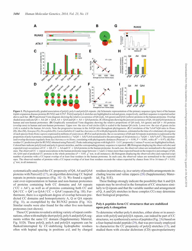
systematically analyzed the CC propensity of Q4, A4 and QA4proteins with Paircoil2 (27), an algorithm detecting CC heptadrepeats in protein sequences (Fig. 1G–I). We found a signifi-cant over-representation in human and non-human proteomesof proteins containing both CC domains and A4 repeats(‘CC + A4’), as well as of proteins containing both CC andQ4 (‘CC + Q4’) or QA4 (‘CC + QA4’) repeats (Fig. 1H). Fur-thermore, there is a significant degree of overlap between pre-dicted CC domains and homopolymeric A, Q or QA repeats(Fig. 1I), as exemplified by the RUNX2 protein (Fig. 1G).Similar results were also found for the other five non-humanproteomes (not shown).
These CC proteins revealed a striking variety of repeat combi-nations, often with multiple short polyQ, polyA and polyQA seg-ments within the same CC domain (Supplementary Material,Fig. S1D). These polyQ, polyA and polyQA segments can beflanked/interrupted by CC-stabilizing hydrophobic residuesoften with heptad spacing in positions a/d, and by charged
residues in positions e/g, in a variety of possible arrangements in-cluding leucine and valine zippers (28) (Supplementary Mater-ial, Fig. S1E).
These findings strongly indicate the possibility that A and QArepeats may be involved in the formation of CC structures simi-larly to Q repeats and that the variable number and arrangementof Q, A and QA stretches in these complex CCs may determinetheir biological properties.
PolyA peptides form CC structures that are stabilizedupon polyA elongation
To determine whether polyA stretches, either alone or in associ-ation with polyQ and polyQA repeats, can indeed be part of CCstructures, we synthesized a series of peptides (Fig. 2A) based ona four-heptad model peptide (29), that was previously employedto characterize the CC propensity of polyQ stretches (15), andstudied them with circular dichroism (CD) spectropolarimetry
Figure 1. Phylogenetically graded association of polyQ, polyA and polyQA repeats. (A) Schematic representation of the primary sequence (gray bars) of the humantriplet-expansion disease proteins RUNX2 and ATX7. PolyQ and polyA stretches are highlighted in red and green, respectively, and their sequence is reported earlierabove each bar. (B) Proportional Venn diagram showing the relative occurrence of Q4 (red), A4 (green) and QA4 (yellow) proteins in the human proteome. Overlapshaded areas indicate Q4 + A4, Q4 + QA4, A4 + QA4 and Q4 + A4 + QA4 proteins. (C) Histogram showing the percent occurrence of Q4, A4 and QA4 proteins inhuman and non-human proteomes. (D) Graphically normalized Venn diagrams showing the relative proportions of Q4 (red), A4 (green) and Q4 + A4 proteins(overlap area) in human and non-human proteomes. Upper row: the size of red circles (Q4) is scaled to the human Q4 circle. Lower row: the size of green circles(A4) is scaled to the human A4 circle. Note the progressive increase in the A4/Q4 ratio throughout phylogenesis. (E) Correlation of the %A4/%Q4 ratio in Homo(H), Mus (M), Xenopus (X), Drosophila (D), Caenorhabditis (C) and Saccharomyces (S) with phylogenetic distances, estimated as the time of evolutionary divergenceof each species from Homo sapiens expressed in millions of years (mya). (F) For each proteome, the co-occurrence of Q4 and A4 repeats in proteins is expressed as theproportion of polyA proteins containing a polyQ stretch (i.e. %(Q4 + A4)/%A4) normalized to the percentage of A4 proteins (i.e. %(Q4 + A4)/%A42). This normal-ized overlap decreases regularly from Saccharomyces to Homo, correlating with phylogenetic distances (Pearson’s r ¼ 0.98, P , 0.001, with a R2 ¼ 0.97). (G) Per-residue CC prediction plot for RUNX2 obtained using Paircoil2. Peaks indicating regions with high (0.8–1) CC propensity (1 minus P-score) are highlighted in black.Colored bars indicate polyQ (red) and polyA (green) stretches, and the corresponding primary sequence is reported. (H) Histogram displaying the observed (obs) andexpected (exp) occurrence of CC + Q4, CC + A4 and CC + QA4 proteins in the human proteome. In each case, the observed values are normalized to the expectedones. The observed CC + repeat associations in the human proteome range between 1.3 and 2.2 times more than expected based on the respective percentages of Q4,A4, QA4 and of predicted CC proteins in the whole proteome (P , 0.05, x2 test, in all instances). (I) Histogram displaying the observed (obs) and expected (exp)number of proteins with a CC/repeat overlap of at least four residues in the human proteome. In each case, the observed values are normalized to the expectedones. The observed number of proteins with a CC/repeat overlap of at least four residues exceeds the values expected by chance from 10 to 16 times (P , 0.01,x2 test, in all instances).
3404 Human Molecular Genetics, 2014, Vol. 23, No. 13
Dow
nloaded from https://academ
ic.oup.com/hm
g/article-abstract/23/13/3402/659131 by guest on 04 April 2019
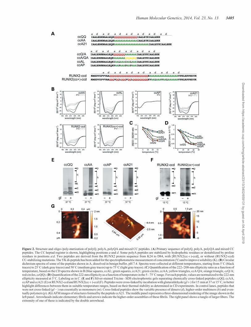
Figure 2. Structure and oligo-/poly-merization of polyQ, polyA, polyQA and mixed CC peptides. (A) Primary sequence of polyQ, polyA, polyQA and mixed CCpeptides. The CC heptad register is shown, highlighting positions a and d. Some polyA peptides are stabilized by hydrophobic residues or destabilized by prolineresidues in positions a/d. Two peptides are derived from the RUNX2 protein sequence from K24 to D84, with [RUNX2(cc+)-ccd], or without (RUNX2-ccd)CC-stabilizing mutations. The YK di-peptide has been added for the spectrophotometric measurement of concentration (Y) and to improve solubility (K). (B) Circulardichroism spectra of some of the peptides shown in A, dissolved in benign buffer, pH 7.4. Spectra were collected at different temperatures, starting from 58C (blacktraces) to 258C (dark gray traces) and 508C (medium gray traces) up to 758C (light gray traces). (C) Quantification of the 222-/208-nm ellipticity ratio as a function oftemperature, based on the CD spectra shown in B (blue squares, ccAL; green squares, ccA21; green circles, ccAA; yellow triangles, ccA/QA; orange triangle, ccQ/A;red circles, ccQQ). (D) Quantification of the 222-nmellipticity as a function of temperature in the 5–758C range. For each peptide, values are normalized to the 222-nmellipticity measured at 58C. Labeling as in C. (E and F) Silver-stained Tricine–SDS electrophoretic gels separating chemically cross-linked peptides ccQQ, ccAA,ccAP and ccA21 (E) or RUNX2-ccd and RUNX2(cc+)-ccd (F). Peptides were cross-linked by incubation with glutaraldehyde (gl+) for 15 min at 37 or 158C, to betterhighlight differences between them in suitable temperature ranges, based on their thermal stability as determined in CD experiments. In control lanes, peptides thatwere not cross-linked (gl2) run essentially as monomers (m). Cross-linked peptides show the variable presence of dimers (d), higher-order multimers (h) and even-tually polymers (p). (G) AFM images of structures formed by the peptide ccA21. The middle panel represents a three-dimensional rendering of the image shown in theleft panel. Arrowheads indicate elementary fibrils and arrows indicate the higher-order assemblies of these fibrils. The right panel shows a tangle of larger fibers. Theextremity of one of these is indicated by the double arrowhead.
Human Molecular Genetics, 2014, Vol. 23, No. 13 3405
Dow
nloaded from https://academ
ic.oup.com/hm
g/article-abstract/23/13/3402/659131 by guest on 04 April 2019
to define their secondary structure and CC propensity. In particu-lar, we inserted at the core of this model peptide polyA stretchesof different length (i.e. 14 alanines in peptide ccAA and 21 ala-nines in peptide A21) or polyA stretches in combination withpolyQ and polyQA repeats (peptides ccQ/A and ccA/QA).We also generated variants of peptide ccAA in which the CCpropensity was enhanced by the insertion of CC-stabilizinghydrophobic residues (peptide ccAL) or diminished by CC-disrupting prolines (peptide ccAP) in a/d heptad positions. Asa reference control, we also synthesized the ccQQ peptide, con-taining 14 Qs, whose CC propensity and stability were previous-ly characterized (15). Furthermore, we synthesized two peptidesencompassing the polyQ/polyA coiled-coil domain (ccd) ofRUNX2. One peptide (RUNX2-ccd) contains the wild-type(wt) sequence, whereas the other peptide, RUNX2(cc+)-ccd,contains the same ccd with CC-stabilizing mutations in a/dheptad positions similar to those used for the ccAL peptide.In fact, some As and Qs were replaced by hydrophobic residuessuch as leucine and valine. Valine was chosen because thisamino acid is already present in a/d heptad positions adjacentto the polyQ/polyA region of RUNX2 (SupplementaryMaterial, Fig. S1E). These peptides also contain a few RUNX2residues adjacent to the polyA/polyQ CC that were included tohelp solubilize the peptides, given the presence of charged resi-dues. The presence also of some scattered prolines in these flank-ing regions can limit their secondary structure stability, thusreducing their interference with the structural analysis of thepolyQ/polyA core of these peptides.
Circular dichroism is widely used for studying the folding andstability of CCs. Distinctive signatures allow to discriminatebetween single and coiled helices, based on the ellipticity ratioat 222 and 208 nm (≥1 for CCs), the inversion of this ratioinduced by trifluoroethanol (TFE), and the thermal stability ofthe folding (15,30–36).
The polyA peptide ccAA displayed CD spectra with minima at208 and 222 nm, and a 222-/208-nm ratio ≥1 (1.10), indicatinga-helical CC formation (Fig. 2B and C). As predictable for CCstructures, the CC helical folding was considerably stabilized bythe addition of canonical hydrophobic residues in position a/d ofthe polyA stretch (peptide ccAL, Fig. 2B and C) and completelydisrupted by prolines at the same positions (peptide ccAP), asshown by a single minimum at �200 nm indicating a randomcoil conformation (Fig. 2B).
The presence of a longer polyA stretch (peptide ccA21)induced a further increase in the 222-nm ellipticity with a222-/208-nm ratio ≥1, thus showing a greater degree ofhelical CC folding (Fig. 2B). Furthermore, the elongation ofthe polyA stretch conferred greater thermal stability to thehelical CC folding of ccA21 with respect to ccAA, as observedby analyzing both the 222-nm ellipticity and the 222-/208-nmratio at increasing temperatures (Fig. 2C and D). The helicalCC signatures and thermal stability of both ccAA and ccA21were more pronounced than those of ccQQ (Fig. 2B–D; Supple-mentary Material, Fig. S2A). As typical of CC structures, thepresence of TFE brought the 222-/208-nm ratio of ,1 withoutreducing the helical folding, indicating dissociation of thehelices (Supplementary Material, Fig. S2B). Taken together,these findings indicate that polyA stretches can form a-helicalCC structures that are even more stable that those formed bypolyQ stretches and that the CC folding of polyA stretches
can be modulated in opposite directions by appropriate CC-stabilizing or -destabilizing substitutions. Furthermore, theseresults also indicate that the elongation of polyA repeats leadsto an increased CC stability, as shown by the similar behaviorof ccA21 and ccAL peptides.
Next, we studied the two peptides combining polyA withpolyQ or polyQA stretches (peptides ccQ/A and ccA/QA, re-spectively). Interestingly, both displayed CC features, with adegree of stability that was intermediate between peptides withpure polyA or polyQ stretches (ccAA.ccA/QA.ccQ/A.ccQQ; Fig. 2C–D; Supplementary Material, Fig. S2A), inapparent correlation with the relative proportion of A and Q resi-dues in a/d (4:0 . 3:1 . 2:2 . 0:4). These observations indi-cate that the variable combination of polyQ, polyA andpolyQA stretches within CCs can finely tune their stability andtherefore their biological properties.
Finally, we analyzed the RUNX2 peptides. In agreement withthe predictions, RUNX2-ccd displayed clear a-helical features,with minima at 222 and 208 nm, and a 222-/208-nm elipticityratio of ≥1 (1.01) indicative of CC formation (Fig. 2B).RUNX2(cc+)-ccd displayed similar features and was morestable in thermal denaturation experiments (Fig. 2B; Sup-plementary Material, Fig. S2C), consistent with the presenceof CC-stabilizing substitutions in its polyQ/polyA stretch.Notably, about two-thirds of the amino acid sequence of thesetwo peptides are made of polyQ and polyA stretches (23Q +17A) separated only by a single glutamate residue. Thesequences flanking these stretches contain scattered proline resi-dues that would disrupt the CC structure in these flankingsequences (see Figs 1G and 2A). The fact that this peptide stillexhibits CC features strongly indicates that the polyQ andpolyA stretches at the core of this peptide are primarily respon-sible for the formation of the CC structure. Furthermore, astypical of CCs, the structure of both peptides was sensitive toTFE (e.g. Supplementary Material, Fig. S2D), which loweredthe 222-/208-nm elipticity ratio and enhanced at the same timethe alpha-helical folding. These findings strongly corroboratethe prediction that the polyQ/polyA stretch of RUNX2 canform CCs, and, together with the results obtained with modelpeptides, indicate that mutations within polyQ/polyA stretchessuch as those we employed can effectively enhance or disruptthe CC structure.
PolyA CC peptides form higher-order multimersand polymers in vitro
CC helical assemblies can range from dimers to polymers.To assess the oligomeric state of the peptides studied with CD,we used chemical cross-linking with glutaraldehyde (Fig. 2Eand F; Supplementary Material, Fig. S2E) and found that polyA-containing peptides can variably populate dimeric and higher-order multimeric states, up to polymers. Peptide ccAA formedin fact dimers, higher-order oligomers/multimers and even poly-mers, whereas ccQQ under the same conditions formed onlylower-order oligomers and ccAP was essentially monomeric,in very good agreement with the CC propensity and stability ofeach peptide as shown by the CD experiments (Fig. 2E). Thiscorrelation was further confirmed by studying peptides with alonger polyA tract or with mixed polyA/polyQ or /polyQAtracts. In fact, as for the CD experiments, ccQ/A and ccA/QA
3406 Human Molecular Genetics, 2014, Vol. 23, No. 13
Dow
nloaded from https://academ
ic.oup.com/hm
g/article-abstract/23/13/3402/659131 by guest on 04 April 2019
displayed intermediate features between ccQQ and ccAA(Supplementary Material, Fig. S2E), thus indicating that themultimerization tendency of polyA CCs can be finely modulatedby their combination with polyQ and polyQA segments. TheccA21 peptide showed an even higher tendency than ccAA toform multimers and polymers (Fig. 2E) that may drive aggrega-tion in vivo. Next, we analyzed the oligomeric state of theRUNX2 peptides (Fig. 2F). Chemical cross-linking showedthat RUNX2-ccd forms dimers and some lower-order oligomers,whereas RUNX2(cc+)-ccd forms, besides dimers, also higher-order multimers and polymers. These observations are in goodcorrelation with the differential CC stability of the two peptidesthat was shown by the CD experiments, similar to what wasobserved for the CC model peptides.
Finally, we characterized the ultrastructural morphologyof the polymeric assemblies formed by polyA CC peptides thatwere apparent in the chemical cross-linking experiments.To this aim, we performed an atomic force microscopy (AFM)analysis (37) of the ultrastructural organization of ccA21 poly-mers (Fig. 2G). CcA21 was chosen because it has a high poly-merization tendency, based on the cross-linking experiments.AFM showed that this peptide forms fibrillar structures witha hierarchical organization by which elongated elementaryfibrils (height ,1 nm; Fig. 2G, left and middle panels), oftenbranched, bundle to form progressively larger fibrils, reachingheights of .5 nm, that branch and intertwine forming complextangles (Fig. 2G, right panel). Interestingly, similar profiles havebeen observed in previous AFM studies of both CC (38) andpolyQ proteins (37). These findings show how polyA CC pep-tides can form fibrillar structures that organize into progressivelylarger bundles and tangles, which may represent the ultrastruc-tural substrate for aggregate formation in vivo.
Structure-guided mutagenesis of RUNX2, a polyQ/polyAprotein associated with cleidocranial dysplasia uponpolyA expansion
To define the relevance of polyA CCs to the in vivo aggregationand toxicity of proteins involved in human polyA-expansion dis-eases, we focused on the transcription factor RUNX2 (Fig. 3;Supplementary Material, Fig. S3A), the molecular mediator ofcleidocranial dysplasia upon polyA expansion. PolyA-expandedRUNX2 mislocalizes from the nucleus to the cytoplasm formingaggregates, causing ultimately cellular dysfunction. RUNX2contains both polyQ and polyA stretches separated by a singleglutamate residue, and this polyQ/polyA region is predicted toform a CC (Fig. 1G) that expands with polyA elongation up to10–12 additional alanines (Fig. 3B) as observed in cleidocranialdysplasia patients (39,40). RUNX2 thus offers a particularlysuitable model to explore the role of polyA CC structures, andtheir interplay with polyQ CCs, in the aggregation and dysfunc-tion of a protein involved in human polyA-expansion disease.
To directly test in vivo the role of CC structures in the aggre-gation and dysfunction of RUNX2, we generated differentmutant forms of polyA-expanded and non-expanded RUNX2(Fig. 3A) with variably enhanced or reduced CC stability, asdetermined with Paircoil2 (Fig. 3B; Supplementary Material,Fig. S3A). We first generated polyA length variants ofRUNX2 with +6A and +12A expansions, and 26A or 212Adeletions. Then, we designed mutants of RUNX2 with enhanced
(cc+ mutants) or reduced (cc2 mutants) CC propensity byusing the same type of mutations that were able to effectivelyenhance or impair CC formation in RUNX2-ccd and in otherCC peptides in the CD experiments (Fig. 2). We generated aset of mutants of non-expanded RUNX2 with enhanced CC pro-pensity by inserting canonical CC-stabilizing hydrophobic resi-dues (V and L) in positions a/d of two heptads within the CCheptad register established by the valine residues that immedi-ately precede and follow the polyQ/polyA region (Fig. 3A; Sup-plementary Material, Fig. S1E). These stabilizing residues wereintroduced in the polyQ region (RUNX2/cc+/#1), in the polyAregion (RUNX2/cc+/#2), or in both (RUNX2/cc+/#3) (Fig. 3Aand B). We also generated a set of polyA-expanded mutantswhose CC-propensity was disrupted by the insertion ofproline residues in heptad positions a/d in the polyQ region[RUNX2(+12A/cc2/#1)], or in the polyA region [RUNX2(+12A/cc2/#2)], or in both [RUNX2(+12A/cc2/#3)](Fig. 3A and B). A cc2 mutant of non-expanded, wt RUNX2was also generated [i.e. RUNX2(cc2); Fig. 3A and B].
In vivo subcellular distribution and aggregation of RUNX2are regulated by length and stability of its polyQ/polyA CC
To study the role of CC structures in the aggregation and local-ization of RUNX2 in vivo, we overexpressed GFP-tagged wtRUNX2 and its CC structural mutants in HEK293 cells and ana-lyzed their subcellular distribution and aggregation state throughconfocal fluorescence microscopy and biochemical approaches(Figs 4 and 5). One-way ANOVA indicated overall significantdifferences in the occurrence of both aggregation and mislocali-zation of RUNX2 related to the expression of the differentstructural mutants [F(7,80) ¼ 35.5, P , 0.001 and F(7,80) ¼ 30.2,P , 0.001, respectively].
Wild-type RUNX2 had an essentially nuclear localization in nu-meroussubnuclearsmall foci (Fig.4A, leftpanels), asobservedpre-viously (41). On the other hand, polyA-expanded RUNX2(+12A)(Fig.4A, right panels, EandG)was often found to have a prevailingcytoplasmic localization (32.4+0.2% of the cells, n ¼ 14, 350 ×350 mm microscopy fields; P , 0.001 versus wt, Tukey HSDpost hoc test), with one or more cytoplasmic or nuclear aggregates(38.6+2.3% of the cells, P , 0.001 versus wt), consistent withprevious studies of polyA-expanded forms of RUNX2 (42).Some cells displayed cytoplasmic aggregates but still had a pre-dominantly diffuse nuclear localization, whereas others had adiffuse cytoplasmic distribution without aggregates. Conversely,a mutant with partial polyA deletion had the same nuclear localiza-tion of the wt form (Fig. 4B).
All the RUNX2 CC-enhancing mutants displayed somedegrees of cytoplasmic mislocalization and aggregation, whichwere not significant when the CC-stabilizing mutations wererestricted to the polyQ region [RUNX2(cc+/#1); Supplemen-tary Material, Figs S3B; Fig. 4E and G] but were instead dramaticwhen the CC-stabilizing mutations were in the polyA stretch[RUNX2(cc+/#2) and /#3, Fig. 4C, E and G]. In fact, bothRUNX2(cc+/#2) and /#3 were more mislocalized and aggre-gated in cells than wt RUNX2 (P , 0.001 versus wt, TukeyHSD post hoc test, in all instances). Indeed, RUNX2(cc+/#2)and /#3 did not differ from RUNX2(+12A), both in terms of dis-tribution and aggregation (P . 0.65 versus RUNX2(+12A),Tukey HSD post hoc test, in all instances; Fig. 4E and G).
Human Molecular Genetics, 2014, Vol. 23, No. 13 3407
Dow
nloaded from https://academ
ic.oup.com/hm
g/article-abstract/23/13/3402/659131 by guest on 04 April 2019

These findings show how the effects of CC stabilization inthe polyQ/polyA region of RUNX2 on protein localization/aggregation closely mimic those of polyA expansion, supportingthe notion that polyA expansion triggers protein aggregationin vivo through CC stabilization. Furthermore, these resultsindicate a more prominent role of the polyA CC segment, withrespect to the polyQ section, in the regulation of RUNX2 local-ization/aggregation. Interestingly, we also found throughco-expression experiments that non-expanded wt RUNX2 isrecruited into aggregates formed either by polyA-expandedRUNX2(+12A) or by the CC-stabilized RUNX2(cc+/#2)mutant (Fig. 5A). These findings indicate that the excessiveelongation/stabilization of the RUNX2 polyQ/polyA CCdomain induces not only protein aggregation but also sequestra-tion of wt RUNX2, which may obviously play a role in the mo-lecular pathogenesis of cleidocranial dysplasia.
Next, we tested the effect on the localization/aggregation ofpolyA-expanded RUNX2 of CC-disrupting mutations in the
polyQ stretch, in the polyA stretch, or in both. We found thatpolyQ CC-disruption [mutant RUNX2(+12A/cc2/#1), Fig. 4D,left panels] moderately, but significantly, reduces mislocalizationand aggregation with respect to RUNX2(+12A) [P , 0.01 versusRUNX2(+12A) for aggregation, P , 0.04 for mislocalization,Tukey HSD post hoc test; Fig. 4F and H]. CC disruption in theexpanded polyA stretch [mutants RUNX2(+12A/cc2/#2) and/#3; Fig. 4D, middle and right panels] strongly reduces both mis-localization and aggregation with respect to the polyA-expandedform of RUNX2 [P , 0.001 versus RUNX2(+12A), TukeyHSD post hoc test, in all instances; Fig. 4F and H]. These resultssomewhat mirror those obtained with the cc+ mutants inshowing how the stability of the expanded polyQ/polyA CC ofRUNX2 critically regulates the localization and aggregation ofthe mutant protein, highlighting again a more prominent roleof the polyA with respect to the polyQ segment in RUNX2.
Finally, the aggregation propensity in cells of wt and mutantforms of RUNX2 was also assayed at the biochemical level, by
Figure 3. Structure-guided mutagenesis of wt and polyA-expanded RUNX2. (A) Design of polyQ and polyA mutants of RUNX2. For simplicity, only part of theprimary sequence, corresponding to the fragment between V31 and V76 of wt RUNX2, and between V31 and V88 of RUNX2(+12A), is shown. PolyA length variants(del, deletions and exp, expansions) are also shown. Substitutions with CC-stabilizing (cc+) and CC-destabilizing (cc2) amino acids are highlighted (arrowheads andletters) above each sequence. (B) CC propensity of RUNX2 mutants shown in A as determined by Paircoil2, expressed as 1 minus the P-score assigned to each aminoacid in the primary sequence. Protein segments whose CC propensity is 0.8–1 are highlighted in black. For simplicity, only the prediction for the N-terminal part ofRUNX2 is shown. The site and effect on CC propensity of the different polyA length variations (del, partial deletion and exp, expansion) and of the CC-stabilizing(cc+) and CC-destabilizing (cc2) mutations are highlighted by gray arrowheads. Vertical arrowheads indicate the site and effect of CC-stabilizing andCC-destabilizing mutations. Horizontal arrowheads indicate deletion or expansions of the predicted CC domain related to corresponding variations in polyA length.
3408 Human Molecular Genetics, 2014, Vol. 23, No. 13
Dow
nloaded from https://academ
ic.oup.com/hm
g/article-abstract/23/13/3402/659131 by guest on 04 April 2019
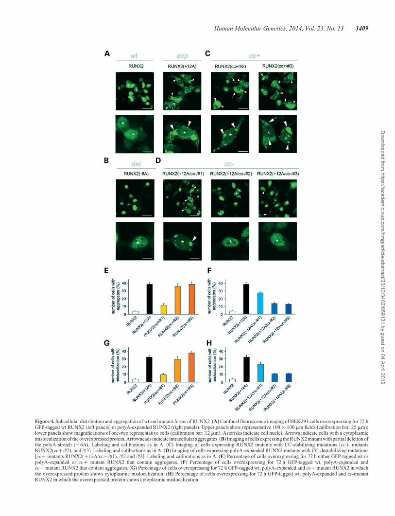
Figure 4. Subcellular distribution and aggregation of wt and mutant forms of RUNX2. (A) Confocal fluorescence imaging of HEK293 cells overexpressing for 72 hGFP-tagged wt RUNX2 (left panels) or polyA-expanded RUNX2 (right panels). Upper panels show representative 100 × 100 mm fields (calibration bar: 25 mm);lower panels show magnifications of one-two representative cells (calibration bar: 12 mm). Asterisks indicate cell nuclei. Arrows indicate cells with a cytoplasmicmislocalization of the overexpressed protein. Arrowheads indicate intracellular aggregates. (B) Imaging of cells expressing the RUNX2 mutant with partial deletion ofthe polyA stretch (26A). Labeling and calibrations as in A. (C) Imaging of cells expressing RUNX2 mutants with CC-stabilizing mutations [cc+ mutantsRUNX2(cc+/#2), and /#3]. Labeling and calibrations as in A. (D) Imaging of cells expressing polyA-expanded RUNX2 mutants with CC-destabilizing mutations[cc2 mutants RUNX2(+12A/cc2/#1), /#2 and /#3]. Labeling and calibrations as in A. (E) Percentage of cells overexpressing for 72 h either GFP-tagged wt orpolyA-expanded or cc+ mutant RUNX2 that contain aggregates. (F) Percentage of cells overexpressing for 72 h GFP-tagged wt, polyA-expanded andcc2 mutant RUNX2 that contain aggregates. (G) Percentage of cells overexpressing for 72 h GFP-tagged wt, polyA-expanded and cc+ mutant RUNX2 in whichthe overexpressed protein shows cytoplasmic mislocalization. (H) Percentage of cells overexpressing for 72 h GFP-tagged wt, polyA-expanded and cc-mutantRUNX2 in which the overexpressed protein shows cytoplasmic mislocalization.
Human Molecular Genetics, 2014, Vol. 23, No. 13 3409
Dow
nloaded from https://academ
ic.oup.com/hm
g/article-abstract/23/13/3402/659131 by guest on 04 April 2019
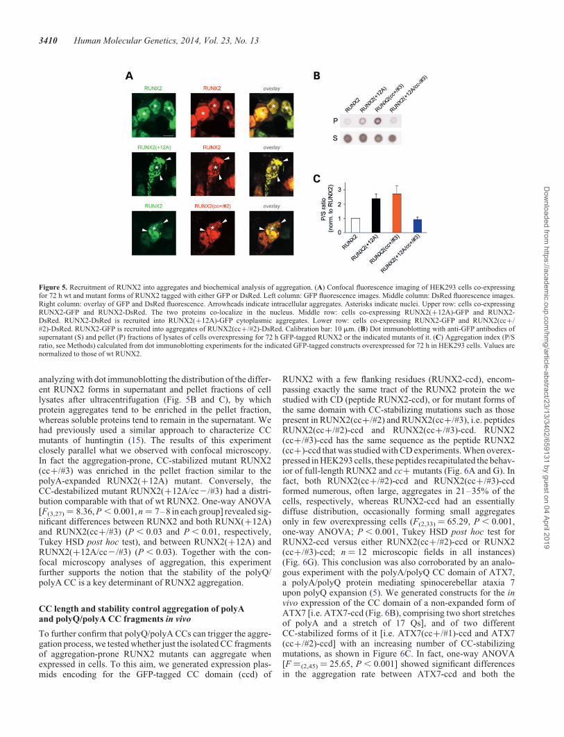
analyzing with dot immunoblotting the distribution of the differ-ent RUNX2 forms in supernatant and pellet fractions of celllysates after ultracentrifugation (Fig. 5B and C), by whichprotein aggregates tend to be enriched in the pellet fraction,whereas soluble proteins tend to remain in the supernatant. Wehad previously used a similar approach to characterize CCmutants of huntingtin (15). The results of this experimentclosely parallel what we observed with confocal microscopy.In fact the aggregation-prone, CC-stabilized mutant RUNX2(cc+/#3) was enriched in the pellet fraction similar to thepolyA-expanded RUNX2(+12A) mutant. Conversely, theCC-destabilized mutant RUNX2(+12A/cc2/#3) had a distri-bution comparable with that of wt RUNX2. One-way ANOVA[F(3,27) ¼ 8.36, P , 0.001, n ¼ 7–8 in each group] revealed sig-nificant differences between RUNX2 and both RUNX(+12A)and RUNX2(cc+/#3) (P , 0.03 and P , 0.01, respectively,Tukey HSD post hoc test), and between RUNX2(+12A) andRUNX2(+12A/cc2/#3) (P , 0.03). Together with the con-focal microscopy analyses of aggregation, this experimentfurther supports the notion that the stability of the polyQ/polyA CC is a key determinant of RUNX2 aggregation.
CC length and stability control aggregation of polyAand polyQ/polyA CC fragments in vivo
To further confirm that polyQ/polyA CCs can trigger the aggre-gation process, we tested whether just the isolated CC fragmentsof aggregation-prone RUNX2 mutants can aggregate whenexpressed in cells. To this aim, we generated expression plas-mids encoding for the GFP-tagged CC domain (ccd) of
RUNX2 with a few flanking residues (RUNX2-ccd), encom-passing exactly the same tract of the RUNX2 protein the westudied with CD (peptide RUNX2-ccd), or for mutant forms ofthe same domain with CC-stabilizing mutations such as thosepresent in RUNX2(cc+/#2) and RUNX2(cc+/#3), i.e. peptidesRUNX2(cc+/#2)-ccd and RUNX2(cc+/#3)-ccd. RUNX2(cc+/#3)-ccd has the same sequence as the peptide RUNX2(cc+)-ccd that was studied with CD experiments. When overex-pressed in HEK293 cells, these peptides recapitulated the behav-ior of full-length RUNX2 and cc+ mutants (Fig. 6A and G). Infact, both RUNX2(cc+/#2)-ccd and RUNX2(cc+/#3)-ccdformed numerous, often large, aggregates in 21–35% of thecells, respectively, whereas RUNX2-ccd had an essentiallydiffuse distribution, occasionally forming small aggregatesonly in few overexpressing cells (F(2,33) ¼ 65.29, P , 0.001,one-way ANOVA; P , 0.001, Tukey HSD post hoc test forRUNX2-ccd versus either RUNX2(cc+/#2)-ccd or RUNX2(cc+/#3)-ccd; n ¼ 12 microscopic fields in all instances)(Fig. 6G). This conclusion was also corroborated by an analo-gous experiment with the polyA/polyQ CC domain of ATX7,a polyA/polyQ protein mediating spinocerebellar ataxia 7upon polyQ expansion (5). We generated constructs for the invivo expression of the CC domain of a non-expanded form ofATX7 [i.e. ATX7-ccd (Fig. 6B), comprising two short stretchesof polyA and a stretch of 17 Qs], and of two differentCC-stabilized forms of it [i.e. ATX7(cc+/#1)-ccd and ATX7(cc+/#2)-ccd] with an increasing number of CC-stabilizingmutations, as shown in Figure 6C. In fact, one-way ANOVA[F ¼(2,45) ¼ 25.65, P , 0.001] showed significant differencesin the aggregation rate between ATX7-ccd and both the
Figure 5. Recruitment of RUNX2 into aggregates and biochemical analysis of aggregation. (A) Confocal fluorescence imaging of HEK293 cells co-expressingfor 72 h wt and mutant forms of RUNX2 tagged with either GFP or DsRed. Left column: GFP fluorescence images. Middle column: DsRed fluorescence images.Right column: overlay of GFP and DsRed fluorescence. Arrowheads indicate intracellular aggregates. Asterisks indicate nuclei. Upper row: cells co-expressingRUNX2-GFP and RUNX2-DsRed. The two proteins co-localize in the nucleus. Middle row: cells co-expressing RUNX2(+12A)-GFP and RUNX2-DsRed. RUNX2-DsRed is recruited into RUNX2(+12A)-GFP cytoplasmic aggregates. Lower row: cells co-expressing RUNX2-GFP and RUNX2(cc+/#2)-DsRed. RUNX2-GFP is recruited into aggregates of RUNX2(cc+/#2)-DsRed. Calibration bar: 10 mm. (B) Dot immunoblotting with anti-GFP antibodies ofsupernatant (S) and pellet (P) fractions of lysates of cells overexpressing for 72 h GFP-tagged RUNX2 or the indicated mutants of it. (C) Aggregation index (P/Sratio, see Methods) calculated from dot immunoblotting experiments for the indicated GFP-tagged constructs overexpressed for 72 h in HEK293 cells. Values arenormalized to those of wt RUNX2.
3410 Human Molecular Genetics, 2014, Vol. 23, No. 13
Dow
nloaded from https://academ
ic.oup.com/hm
g/article-abstract/23/13/3402/659131 by guest on 04 April 2019
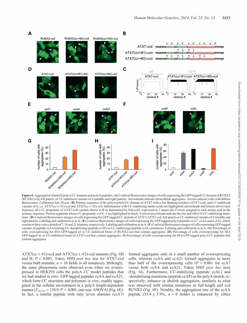
ATX7(cc+/#1)-ccd and ATX7(cc+/#2)-ccd mutants (Fig. 6Dand H; P , 0.001, Tukey HSD post hoc test for ATX7-ccdversus both mutants, n ¼ 16 fields in all instances). Strikingly,the same phenomena were observed even when we overex-pressed in HEK293 cells the polyA CC model peptides thatwe had studied in vitro. GFP-tagged peptides ccAA and ccA21,which form CC structures and polymers in vitro, readily aggre-gated in the cellular environment in a polyA length-dependentmanner (F(4,35)¼ 138.9, P , 0.001, one-way ANOVA) (Fig. 6E).In fact, a similar peptide with only seven alanines (ccA7)
formed aggregates only in a small number of overexpressingcells, whereas ccAA and ccA21 formed aggregates in morethan half of the overexpressing cells (P , 0.001 for ccA7versus both ccAA and ccA21, Tukey HSD post hoc test)(Fig. 6I). Furthermore, CC-stabilizing (peptide ccAL) and-destabilizing mutations (peptide ccAP) in the polyA stretch, re-spectively, enhance or abolish aggregation, similarly to whatwas observed with similar mutations in full-length and ccdRUNX2 (Fig. 6F). Notably, the aggregation rate of the ccAApeptide (53.4+ 3.9%, n ¼ 8 fields) is enhanced by either
Figure 6. Aggregation of polyQ/polyA CC domains and polyA peptides. (A) Confocal fluorescence images of cells expressing the GFP-tagged CC domain of RUNX2(RUNX2-ccd, left panel), or CC-stabilized variants of it (middle and right panels). Arrowheads indicate intracellular aggregates. Arrows indicate cells with diffusefluorescence. Calibration bar: 20 mm. (B) Primary sequence of the polyA/polyQ CC domain of ATX7 with a few flanking residues (ATX7-ccd), and CC-stabilizedvariants of it, i.e. ATX7(cc+/#1)-ccd and ATX7(cc+/#2)-ccd. Substitutions with CC-stabilizing amino acids are highlighted (arrowheads and letters) above eachsequence. (C) CC propensity of ATX7-ccd variants shown in B as determined by Paircoil2, expressed as 1 minus the P-score assigned to each amino acid in theprimary sequence. Protein segments whose CC propensity is 0.8–1 are highlighted in black. Vertical arrowheads indicate the site and effect of CC-stabilizing muta-tions. (D) Confocal fluorescence images of cells expressing the GFP-tagged CC domain of ATX7 (ATX7-ccd, left panel) or CC-stabilized variants of it (middle andright panels). Labeling and calibration as in A. (E) Confocal fluorescence images of cells expressing the GFP-tagged polyA peptides ccA7, ccAA and ccA21, whichcontain at their core a stretch of 7, 14 and 21 alanines, respectively. Labeling and calibration as in A. (F) Confocal fluorescence images of cells expressing GFP-taggedvariants of peptide ccAA bearing CC-destabilizing (peptide ccAP) or CC-stabilizing (peptide ccAL) mutations. Labeling and calibration as in A. (G) Percentage ofcells overexpressing for 48 h GFP-tagged wt or CC-stabilized forms of RUNX2-ccd that contain aggregates. (H) Percentage of cells overexpressing for 48 hGFP-tagged wt or CC-stabilized forms of ATX7-ccd that contain aggregates. (I) Percentage of cells overexpressing for 48 h GFP-tagged polyA CC peptides thatcontain aggregates.
Human Molecular Genetics, 2014, Vol. 23, No. 13 3411
Dow
nloaded from https://academ
ic.oup.com/hm
g/article-abstract/23/13/3402/659131 by guest on 04 April 2019
elongation of the polyA tract (peptide ccA21, 66.0+ 2.4%, n ¼8, P , 0.02, Tukey HSD post hoc test) or by its stabilization(peptide ccAL, 68.0+ 3.4%, n ¼ 8, P , 0.01, Tukey HSDpost hoc test) (Fig. 6I). Taken together, these findings indicatethat—in two different disease-related proteins as well as inmodel peptides—the stability/length of polyQ/polyA andpolyA CCs is a key determinant of the aggregation proneness,thus indicating the generality of the CC-triggered polyQ andpolyA aggregation mechanism.
Modulation of RUNX2 activity by length and stabilityof its polyQ/polyA CC
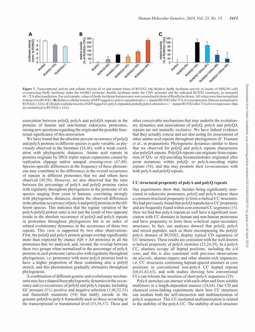
We also analyzed whether the length and stability of the polyQ/polyA CC also regulates the physiological transcriptional activ-ity of RUNX2. Evidence exists that either partial deletions of thepolyA stretch or short expansions of the polyQ stretch determinea decreased transcriptional activity of RUNX2 (43). To deter-mine the relevance of the CC structure of the polyQ/polyArepeats to the physiological function of RUNX2, we have inves-tigated whether—besides the length of the polyA stretch—alsothe stability of its CC structure has relevance to the transcription-al activity of RUNX2. To this aim, we have used a luciferasereporter assay to compare the transcriptional activity of wtRUNX2 with that of a polyA deletion mutant [i.e. RUNX2(212A)] and of a mutant in which the stability of the non-expanded polyA CC structure is disrupted by alanine-to-prolinemutations, i.e. RUNX2(cc2) (Fig. 3A and B). This experimentrevealed that CC disruption in the polyA stretch of RUNX2negatively modulates its transcriptional activity, parallelingthe effect of polyA deletion [F ¼ (2,61) ¼ 8.25, P , 0.01,one-way ANOVA] (Fig. 7A). In fact, the transcriptional activityof RUNX2(cc2) was 0.73+ 0.04 times that of wt RUNX2(P , 0.01, Tukey HSD post hoc test) Similarly, the RUNX2(212A) deletion mutant had a reduced level of activity(P , 0.03, Tukey HSD post hoc test versus wt RUNX2).These findings indicate that, besides the overall length, alsothe structure of the polyQ/polyA CC domain of RUNX2 is rele-vant to its physiological function.
Increased length and stability of the RUNX2 polyQ/polyACC induce cellular toxicity
PolyA-expansion in genetic diseases is associated not only withprotein aggregation/dysfunction but also with cellular toxicity(3). To assess the role of CCs in the toxicity of polyA-expandedRUNX2, we overexpressed in HEK293 cells GFP-taggedRUNX2, or RUNX2(+12A), or their cc+ and cc2 mutants,and used a colorimetric MTT-formazan assay to determinetheir relative toxicity (44) (Fig. 7B and C).
Overall, one-way ANOVA revealed significant differencesamong groups related to the overexpression of the differentRUNX2 mutants [F(9,462) ¼ 8.63, P , 0.001]. We observedsome significant toxicity after 72 h of RUNX2(+12A) overex-pression with respect to wt RUNX2 used as a control (i.e. forma-zan production was, respectively, 89.3+ 1.8% in n ¼ 94 culturewells versus 100.0+ 1.4% in n ¼ 119 wells, P , 0.01, TukeyHSD post hoc test; values normalized to wt). Conversely, morelimited expansions or partial deletions of the polyA stretch inRUNX2(+6A) and RUNX2(26A) did not cause evident
toxicity (n ¼ 39 and n ¼ 36, respectively, P . 0.90 versus wtin both cases). To compare the relative toxicity of the CCmutants, we normalized the toxicity of each mutant to that ofpolyA-expanded RUNX2(+12A) (Fig. 7B). Strikingly, theRUNX2(cc+/#2) in which the polyA CC was stabilized wasas toxic as the polyA-expanded form RUNX2(+12A) (n ¼ 76,P , 0.001 versus wt, Tukey HSD post hoc test). TheRUNX2(cc+/#1) mutant, in which the polyQ part of the CCwas stabilized, showed only a modest, non-significant degreeof toxicity. However, when both the polyQ and polyA sectionsof the CC were stabilized as in RUNX2(cc+/#3), the toxicitywas even higher than that of RUNX2(cc+/#2), more than2-fold as compared with RUNX2(+12A) (n ¼ 23, P , 0.001versus wt, Tukey HSD post hoc test), indicating that theoverall stability of the polyQ/polyA CC is relevant to toxicity.
Mirroring these results, polyA-expanded mutants withCC-destabilizing mutations (Fig. 7C) showed instead a verymarked loss of toxicity when the CC-destabilizing residueswere positioned within the polyA stretch [i.e. mutantRUNX2(+12A/cc2/#2) and /#3; P ¼ 0.95 versus wt and P ¼0.99 versus wt, respectively, Tukey HSD post hoc test],whereas the destabilization of the RUNX2(+12A) polyQportion by itself reduced toxicity partially but not significantly[mutant RUNX2(+12A/cc2/#1)].
Taken together, these findings indicate a critical role of theoverall length and stability of the polyQ/polyA CC of RUNX2for the induction of cellular toxicity. The greater relevance totoxicity of the polyA section of the RUNX2 CC with respect tothe polyQ section is also evident, paralleling what was observedfor subcellular distribution and aggregation.
DISCUSSION
The results of our study identify in numerous proteins of humanand non-human eukaryotic proteomes a systematic associationof polyQ, polyA and polyQA repeats with each other, and withCC domains, of which they can be a part. These associationsappear to be dynamically evolving throughout phylogenesisand have implications for human genetic diseases in whichpolyQ or polyA repeats, associated or not, are pathologicallyexpanded.
Philogenetically graded occurrence and associationof polyQ and polyA repeats
PolyQ and polyA repeats are widespread in proteomes (12).Their co-occurrence in the same proteins has also been occasion-ally noticed in different proteomes (11,10), but the general sig-nificance of their association is not clear. PolyQA repeats havealso been observed in some proteins (7,8), but little is knownabout their general association with polyQ and polyA repeats.
We have systematically screened six complete eukaryoticproteomes for the occurrence and co-occurrence in proteins ofpolyQ, polyA and polyQA repeats of four or more residues.This length threshold allowed us to detect both long repeats(e.g. RUNX2), as well as cryptic low-complexity regions (45)composed of short, often intermixed stretches of these aminoacids (e.g. PQN-41) that would go unnoticed using higher thresh-olds. Our analyses reveal a novel phylogenetically graded
3412 Human Molecular Genetics, 2014, Vol. 23, No. 13
Dow
nloaded from https://academ
ic.oup.com/hm
g/article-abstract/23/13/3402/659131 by guest on 04 April 2019
association between polyQ, polyA and polyQA repeats in theproteins of human and non-human eukaryotic proteomes,raising new questions regarding the origin and the possible func-tional significance of this association.
We have found that the absolute percent occurrence of polyQand polyA proteins in different species is quite variable, as pre-viously observed in the literature (18,46), with a weak correl-ation with phylogenetic distances. Amino acid repeats inproteins originate by DNA triplet repeat expansions caused byreplication slippage and/or unequal crossing-over (47,48).Species-specific differences in the frequency of these phenom-ena may contribute to the differences in the overall occurrenceof repeats in different proteomes that we and others haveobserved (49,50). However, we also observed that the ratiobetween the percentage of polyA and polyQ proteins varieswith regularity throughout phylogenesis in the proteome of sixspecies ranging from yeast to humans, correlating stronglywith phylogenetic distances, despite the observed differencesin the absolute occurrence of polyA and polyQ proteins in the dif-ferent species. This indicates that the regular variation of thepolyA/polyQ protein ratio is not just the result of two oppositetrends in the absolute occurrence of polyQ and polyA repeatsin proteomes throughout phylogenesis but is an index ofrelated evolutionary dynamics in the occurrence of these tworepeats. This view is supported by two other observations.First, the polyQ and polyA protein groups overlap significantlymore than expected by chance (Q4 + A4 proteins) in all theproteomes that we analyzed, and, second, the overlap betweenthese two groups when normalized to the percentage of polyAproteins in each proteome varies also with regularity throughoutphylogenesis, i.e. proteomes with more polyA proteins tend tohave a higher proportion of these containing also a polyQstretch, and this phenomenon gradually attenuates throughoutphylogenesis.
A combination of different genetic and evolutionary mechan-isms may have shaped these phylogenetic dynamics in the occur-rence and co-occurrence of polyQ and polyA repeats, includingGC pressure (51), positive and negative selection (1,46,52,53)and frameshift mutations (54) that stably encode in thegenome polyQ-to-polyA frameshifts such as those occurring atthe transcriptional or translational level (55,56,57). These and
other conceivable mechanisms that may underlie the evolution-ary dynamics and associations of polyQ, polyA and polyQArepeats are not mutually exclusive. We have indeed evidencethat they actually concur and are also acting for associations ofother amino acid repeats throughout phylogenesis (F. Fiumaraet al., in preparation). Phylogenetic dynamics similar to thosethat we observed for polyQ and polyA repeats characterizealso polyQA repeats. PolyQA repeats can originate from expan-sion of QA- or AQ-encoding hexanucleotides originated afterpoint mutations within polyQ- or polyA-encoding tripletrepeats (58), and this may promote their co-occurrence withboth polyA and polyQ repeats.
CC structural propensity of polyA and polyQ repeats
Our experiments show that, besides being significantly asso-ciated in eukaryotic proteomes, polyQ and polyA repeat sharea common structural propensity to forma-helical CC structures.We had previously found that polyQ repeats have CC propensityand are frequently found within conventional CC segments (15).Here we find that polyA repeats as well have a significant asso-ciation with CC domains in human and non-human proteomesand have propensity to form these a-helical super-secondarystructures. In fact, our analyses showed that polyQ, polyAand mixed peptides, such as those encompassing the polyQ/polyA domain of RUNX2, display typical CD signatures ofCC structures. These results are consistent with the well-knowna-helical propensity of polyA stretches (23,24,59). In a polyACC, alanines occupy all heptad positions, including the a/dcore, and this is also consistent with previous observationson alacoils, alanine-zippers and other alanine-rich sequences,i.e. CC structures containing heptad-spaced alanines at corepositions in conventional non-polyA CC heptad repeats(60,61,62,63), and with studies showing how conventionalCCs can tolerate the insertion of short polyA sequences (29).
PolyA stretches can interact with each other and form solublemultimers in a length-dependent manner (24,64). Our CD andchemical cross-linking experiments show how CC structurescan mediate both the self-interaction and multimerization ofpolyA sequences. This CC-mediated multimerization is relatedto the stability of the polyA CC. The stability of such structure
Figure 7. Transcriptional activity and cellular toxicity of wt and mutant forms of RUNX2. (A) Relative firefly luciferase activity in lysates of HEK293 cellsco-expressing firefly luciferase under the 6xOSE2 promoter, Renilla luciferase under the CMV promoter and the indicated RUNX2 constructs, as measured48–72 h after transfection. For each sample, values of firefly luciferase luminescence were normalized to those of Renilla luciferase. All values were then normalizedto those of wt RUNX2. (B) Relative cellular toxicity of GFP-tagged wt, polyA-expanded and cc+ mutant RUNX2 after 72 h of overexpression. Data are normalized toRUNX2(+12A). (C) Relative cellular toxicity of GFP-tagged wt, polyA-expanded, partially polyA-deleted or cc2 mutant RUNX2 after 72 h of overexpression. Dataare normalized to RUNX2(+12A).
Human Molecular Genetics, 2014, Vol. 23, No. 13 3413
Dow
nloaded from https://academ
ic.oup.com/hm
g/article-abstract/23/13/3402/659131 by guest on 04 April 2019
is increased by polyA elongation, which mimics the effects ofstabilization induced by hydrophobic residues in a/d positions.Interestingly, the ccA21 peptide, which contains a polyAstretch within the range of those causing human disease (3)and displays marked polymerization tendency in vitro, formshierarchically organized fibrillar structures that intertwine incomplex tangles, as shown by AFM analyses. These structures,which bear resemblance to those formed by other CCs (38)and polyQ proteins (37) may well represent the substrate of ag-gregation in vivo.
We have also found that polyA repeats can often associatewith polyQ and polyQA repeats in the context of CC-proneprotein domains. To determine the possible structural/functionalsignificance of these associations, we studied CC peptides con-taining pure repeats as well as repeat combinations and foundthat polyA CCs are relatively more stable than polyQ CCs, andthat mixed polyA/polyQ or /polyQA peptides have intermediatedegrees of stability. A sequence/structure combinatorial codecould finely tune the relative stability of complex polyQ,polyA and polyQA CCs such as those actually found in pro-teomes, providing a possible explanation as to why this type ofsequences is maintained, and subtly varied, throughout phylo-genesis. For instance, the polyQ/polyA length polymorphismand the polyQ/polyA length ratio in RUNX2 regulates its tran-scriptional activity and craniofacial variation in dogs and othercarnivores (65,66), and RUNX2 polyQ/polyA length poly-morphism is related to differences in bone mineral density inhumans (43). Our findings indicate that these skeletal variationsmay be related to graded variations in the stability of the RUNX2polyQ/polyA CC caused by the observed polymorphisms in therelative length of these repeats. Moreover, the results of our luci-ferase assay experiments also support this view by indicatingthat, besides the length, also the stability of the polyQ/polyACC has a role in modulating the physiological transcriptional ac-tivity of RUNX2.
Furthermore, our observations have obvious relevance tohuman skeletal diseases such as cleidocranial dysplasia inwhich the polyQ/polyA CC structure of RUNX2 is extendedby polyA expansion.
Coiled-coil stability as a determinant of polyA-expandedprotein aggregation and toxicity
PolyQ- and polyA-expansion diseases are related to the exces-sive elongation of polyQ or polyA repeats beyond a criticalthreshold. These expansions lead to protein aggregation and dys-function/toxicity, which are variably related to gain- orloss-of-function phenomena, causing severe developmental orneurodegenerative diseases (2,3). We have used the polyQ/polyA transcription factor RUNX2 as a model for studying invivo the role of CC structures in pathological protein aggregationand dysfunction upon polyA expansion, as occurring in cleido-cranial dysplasia. By structure-guided mutagenesis, we havegenerated wt and polyA-expanded variants of RUNX2 withenhanced or reduced CC stability and found that the stabilityof the RUNX2 CC has a critical role in determining the subcel-lular localization, aggregation and toxicity of the protein.
Consistent with previous reports (41,42), wt RUNX2 had anessentially nuclear localization, whereas polyA-expandedRUNX2 was often prevalently cytoplasmic, forming multiple
aggregates. We found that mutants in which the polyA CC wasstabilized by hydrophobic residues (cc+) displayed distributionand aggregation patterns comparable with polyA-expandedRUNX2. Conversely, when the polyA-expanded CC was desta-bilized (cc2), mislocalization and aggregation were substantial-ly reduced. These findings closely parallel what was observed inCD and cross-linking experiments with model peptides in whichpolyA CC structures were stabilized and made more prone tomultimerize, either by polyA elongation (peptide ccA21versus ccAA) or by substitutions with CC stabilizers (ccALversus ccAA), but were instead disrupted by CC-destabilizingmutations (ccAP versus ccAA). Even more notably, there wasa clear correspondence between the differential CC stability ofthe RUNX2-ccd and RUNX2(cc+)-ccd peptides and theirrespective in vitro polymerization and in vivo aggregation be-havior. In fact, CC-stabilized RUNX2(cc+)-ccd displayed agreater thermal stability as compared with its wt counterpart,formed higher-order multimers/polymers in vitro and had amarked aggregation tendency when expressed in living cells[i.e. RUNX2(cc+/#3)-ccd], mimicking the aggregation behav-ior of full-length RUNX2(cc+/#3). Similar phenomena wereobserved by overexpressing the CC domain of ATX7, i.e. theaggregation of non-polyQ-expanded ATX7-ccd can be trig-gered by CC-stabilizing mutations, thus showing the generalityof these CC-triggered aggregation mechanisms. Furthermore,even model polyA peptides were able to form in vivo aggregatesin a polyA length-dependent manner. In particular, the aggrega-tion rate of a peptide with 14 alanines (peptide ccAA) wasenhanced by either CC-stabilizing mutations (peptide ccAL)or by polyA elongation to 21 residues (peptide ccA21). Again,the in vivo aggregation behavior of ccAA, ccA21 and ccALcorrelated strongly with their respective CC stability asrevealed by CD. Thus, based on the biophysical and cell bio-logical experiments that we performed, polyA expansions canbe seen as inducing a critical increase in CC stability that pro-motes excessive multimerization and ultimately pathologicalaggregation.
Besides polymerization and aggregation, CC structures canmediate also homotypic and heterotypic protein–protein inter-actions (16). The mislocalization of the protein may resultfrom aberrant protein–protein interactions mediated by patho-logically elongated CCs (15), retaining the protein in the cyto-plasm after synthesis and/or altering its physiologicalnucleo-cytoplasmic shuttling (67). Abnormal CC interactionsmay also cause entrapment of other proteins into aggregatescausing protein-loss-of-function and toxicity through this mech-anism (15). Cleidocranial dysplasia associated with polyA ex-pansion in RUNX2 is, like other polyA and polyQ diseases, adominant genetic disease, i.e. in heterozygosis, cells containalso non-polyA-expanded copies of RUNX2 produced fromthe normal allele. Our experiments show that aggregatesformed by polyA-expanded—or otherwise CC-stabilized—RUNX2 can sequester into cytoplasmic inclusions even wtRUNX2. The loss-of-function of RUNX2, associated withpolyA-expansion or with other mutations, is a fundamentalpathogenetic mechanism of cleidocranial dysplasia (39). Theobserved entrapment of wt RUNX2 into the aggregates caninduce RUNX2 loss-of-function by subtracting to the nuclearcompartment even the normal copies of the protein that arepresent in the cell.
3414 Human Molecular Genetics, 2014, Vol. 23, No. 13
Dow
nloaded from https://academ
ic.oup.com/hm
g/article-abstract/23/13/3402/659131 by guest on 04 April 2019
The composite nature of the RUNX2 polyQ/polyA CC allowedus to define the relative contribution of the polyQ and polyA sec-tions to protein localization and aggregation. CC-stabilizing and-destabilizing mutations restricted to the polyQ stretch inducedmuch weaker phenotypes than those determined by the samemutations in the polyA stretch. Together with the in vitro experi-ments, these observations indicate that polyA CCs have a greaterstability as compared with polyQ CCs of the same length. There isevidence for the evolutionary selection of shorter and less poly-morphic polyA repeats with respect to polyQ repeats (68). Atthe same time, the disease threshold for polyA expansions islower than that for polyQ expansions (2,69). The fact that polyACCsare morestable thanpolyQ CCs can rationalize both phenom-ena in structural terms, i.e. polyA CCs become aggregation-proneand harmful to cells when they reach comparatively shorterlengths than polyQ CCs and are therefore under tighter evolution-ary constraints.
We have also studied the role of CC structures in the toxicity ofpolyA expansions. We have found that polyA-expandedRUNX2 has some toxicity when overexpressed in cells,similar to what was observed with other polyA-expanded pro-teins (3), and that cc+ mutants of non-expanded RUNX2 areas toxic, or even more toxic, than polyA-expanded RUNX2.Moreover, the toxicity related to the polyA expansion could besignificantly reduced or abolished by destabilizing the CC(cc2 mutants). Again, stabilization of the polyA section of theCC had a substantially greater effect than that of the polyQsection, in good agreement with previous studies comparingthe relative toxicity of polyQ and polyA expansions inAtaxin-7 (70). The toxicity of polyQ- and polyA-expanded pro-teins is thought to rely on several mechanisms, including seques-tration of the wt non-expanded protein and other proteins intoaggregates, as well as aberrant protein–protein interactions.Our findings suggest that, as for polyQ expansions (15), polyAexpansions may trigger these mechanisms through non-physiological CC-mediated interactions of the mutant proteinthat lead to toxic gain-of-function effects. Interestingly, whilethe most CC-stabilized RUNX2(cc+/#2) and (cc+/#3)mutants induced aggregation at levels comparable with thoseof the polyA-expanded mutant RUNX2(+12A), their toxicitylevel could be even two-three times higher than that ofRUNX2(+12A). Aggregation and toxicity in polyQ andpolyA-expansion diseases do not necessarily go in parallel. Infact, aggregates may not represent the only toxic entities inthese diseases (71–75) and may even have protective roles(76,77), although they can impair several cellular functions(78,79). Rather, dysfunctional toxic oligomers/multimers arethought to have a fundamental role in the molecular pathogenesisof these disorders (80). Our experiments strongly suggest thatCC-based oligomers/multimers may represent the fundamentaldeterminants of protein dysfunction and toxicity in polyA-expansion diseases such as cleidocranial dysplasia.
The molecular pathogenesis of polyA disease is generallythought to rely on protein misfolding events by which expandedpolyA stretches would assume a b-sheet-rich conformation,closely paralleling the behavior of other well-studied conven-tional amyloids such as Ab(1–42) (3,81). The structure ofpolyA sequences has been extensively used as a helical modelsystem by both molecular modeling approaches and structuralstudies on peptides (24,82–85). Besides the a-helical structure
(23,24,83), some polyA-based peptides have been shown to beable to assume in vitro multiple conformations, includingb-sheets (86,87) and PP-II structures (88). This fact has sug-gested that misfolding to aggregation-prone b-sheets could rep-resent the essential mechanism of polyA-expansion diseases,causing protein aggregation and toxicity in analogy with otherknown amyloid diseases (14). However, as noted by Bernackiand Murphy (24), in those studies in which the conversion tob-sheets was observed, the peptides were under substantiallynon-physiological conditions of temperature, pH and concentra-tion (86,87). Studies in benign buffers under more physiologicalconditions, similar to those we employed, consistently observeda-helical secondary structures even in very long polyA stretchesand did not show conversion to b-sheets (23,24,83). Rather, acritical observation in these and other studies is that the expan-sion of polyA stretches leads to an increase in helical structure(24,59,89). Our experiments extend these observations andshow that polyA expansion is related to a substantial stabiliza-tion of the CC super-secondary structure, with a parallel increasein multimerization propensity. These lines of evidence supportthe notion that the assembly of CC structures rather thanb-sheets primarily triggers the aggregation and toxicity ofpolyA-expanded proteins, as previously observed for polyQ pro-teins (15). This CC model does not exclude at all the possibilitythat CCs are intermediate structures in the aggregation process,and polyA b-sheets are subsequently formed at the level of mul-timers or aggregates/fibers (15,90,91). Furthermore, our findingsare also compatible with the notion that polyA stretches maypromote aggregation and/or toxicity in association with otherprotein domains (15,92,93).
In this view, the expansion of polyA and polyQ stretches leadsprimarily, rather than to misfolding, to an excessive enhance-ment of their native fold, which may ultimately explain thepathological enhancement of their native functions, an emerginggain-of-function mechanism observed in triplet-expansion dis-eases (94–96), as well as the triggering of protein aggregationand mislocalization causing protein loss-of-function (2,3).In conclusion, this work identifies a novel phylogenetic andstructural association between polyQ, polyA and polyQArepeats, which has implications for both the physiological func-tion of the proteins in which these repeats are normally found,and for the molecular pathogenesis of polyQ- and polyA-expan-sion diseases, providing for these a unified structural frameworkin which CC assembly and dynamics play a critical role.
MATERIALS AND METHODS
Bioinformatics
The FASTA protein sequences of the complete reference pro-teomes of Homo sapiens, Mus musculus, Xenopus tropicalis,Drosophila melanogaster, Caenorhabditis elegans and Sac-charomyces cerevisiae were downloaded from the Uniprotonline database of complete reference proteomes (available athttp://www.uniprot.org). Manually reviewed entries were con-sidered for Homo, Mus, and Saccharomyces proteomes, and allentries were considered for the other three species whosemanual review process is at early stages (see http://www.uniprot.org). The Paircoil2 CC-prediction software (27) was down-loaded from http://groups.csail.mit.edu/cb/paircoil2/. Ad hoc
Human Molecular Genetics, 2014, Vol. 23, No. 13 3415
Dow
nloaded from https://academ
ic.oup.com/hm
g/article-abstract/23/13/3402/659131 by guest on 04 April 2019
Perl scripts (http://www.perl.org) were generated to identify, ineach proteome, the proteins containing stretches of glutamine(Q), alanines (A) or glutamine-alanine (QA) of at least four resi-dues, as well as proteins containing predicted CCdomains accord-ing to Paircoil2, using a P-score ,0.05 as a detection threshold.Finally, the overlap between predicted CC segments and Q, Aand QA repeats was measured for each protein, and the numberof proteins with an observed overlap of ,4 or ≥4 residues wasdetermined. The overlap expected by chance was calculated con-sidering the relativepercent lengths of theCCdomain(s)andof therepeat region(s) in each protein, and the number of proteins withan expected overlap of ,4 or ≥4 residues was then determined.Observed and expected values were then compared statistically(see the section Results). Data were analyzed quantitativelyusing Excel (Microsoft). Phylogenetic distances between the sixspecies were estimated based on the time of evolutionary diver-gence of each species from Homo sapiens as derived from http://www.timetree.org (26). Venn diagrams were generated usingthe BioVenn software (97).
Plasmids
The RUNX2 DNA sequence was PCR-amplified using a proof-reading polymerase (Agilent) from a RUNX2-encodingplasmid (clone HsCD00295185; obtained from the PlasmId re-pository, Harvard University) and cloned into a pENTR/Dentry vector (Invitrogen). The RUNX2 sequence was thensubcloned in frame with Em-GFP into the pcDNA6.2/C-EmGFP-DEST destination vector (Invitrogen) by LR recom-bination (Gateway system, Invitrogen), to generate an ex-pression vector for the RUNX2-EmGFP fusion protein. Forsimplicity, we refer to EmGFP as GFP. The RUNX2 sequencewas also subcloned into another destination vector (pcDNA6.2/V5-DEST) in frame with a V5 tag by LR recombination. Wild-type and mutant forms of RUNX2 were also PCR-amplifiedand cloned into the pDsRed-Monomer-N In-Fusion Readyplasmid (Clontech) using the In-fusion cloning system (Clon-tech), following the manufacturer’s protocol, to generate DsRed-tagged constructs. The constructs encoding for GFP-tagged ccdfragments of RUNX2 and ATX7, and for GFP-tagged CC modelpeptides (ccA7, ccAA, ccA21, ccAP and ccAL) were generatedby cloning of PCR products, amplified using Taq polymerase(Sigma), into the pcDNA3.1/CT-GFP expression vector (Invitro-gen), following the manufacturer’s procedure. The PCR productsencoding for RUNX2-ccd, RUNX2(cc+/#2)-ccd and RUNX2(cc+/#3)-ccd were generated using as a template the pcDNA6.2/C-RUNX2-GFP, pcDNA6.2/C-RUNX2(cc+/#2)-GFP andpcDNA6.2/C-RUNX2(cc+/#3)-GFP expression vectors, re-spectively. The PCR products encoding for ATX7-ccd, ATX7(cc+/#1)-ccd, ATX7(cc+/#2)-ccd, ccA7, ccAA, ccA21, ccAPand ccAL were generated from partially overlapping syntheticDNA primers (Sigma). In all cloning procedures, PCR productswere gel-purified using the QIAquick Gel Extraction kit(Qiagen). Plasmids were amplified and purified using theQIAprep Spin Miniprep kit and the HiSpeed Plasmid Maxi kit(Qiagen).
Peptides
The synthetic peptides used in this study were obtained fromGenway Biotech (San Diego, California, USA) with .95%
purity, N-terminal acetylation and C-terminal amidation. Lyo-philized peptides were dissolved in a benign buffer (100 mM
NaCl and 10 mM phosphate buffer, pH 7.4) to obtain concen-trated stock solutions (2–5 mg/ml) that were used immediatelyfor CD experiments and then aliquoted and stored at 2808C forsubsequent experiments.
Circular dichroism spectropolarimetry
The structure of peptides was studied trough CD using a J-815spectropolarimeter (Jasco). Peptides were dissolved in 100 mM
NaCl and 10 mM phosphate buffer, pH 7.4 at a concentrationof 0.1–0.3 mg/ml, and spectra (190–260 nm) were acquired atthe desired temperatures measuring the ellipticity of peptidesamples in 0.1-mm quartz cuvettes every 0.5–1 nm (from 195to 260 nm). The mean residue molar ellipticity ([u]) was calcu-lated from the formula [u] ¼ u × mw/10 × (n–1) × c × pl,where u is the measured ellipticity, mw is the molecularweight of the peptide, n is the number of amino acids in thepeptide, c is the concentration of the peptide (mg/ml), and pl isthe cuvette pathlength (cm) (36). To test the thermal stabilityof peptides, the samples were heated from 5 to 758C (108C/min) while recording ellipticity at 222 nm. Circular dichroismdata and graphs were elaborated and smoothed using SpectraManager (Jasco) and Excel (Microsoft) software.
Chemical cross-linking
The oligomeric state of peptides was studied using chemicalcross-linking with glutaraldehyde, as previously described(15). Briefly, peptides stocks (diluted to 0.6–1.2 mg/ml inbenign buffer) were incubated for 15 min with 0.01% glutaralde-hyde (Sigma) at 378C, or with 0.05% glutaraldehyde at 158C.These two conditions were alternatively chosen to better high-light subtle differences between members of the differentpeptide sets that were analyzed. The reaction was stopped byadding ethanolamine (Sigma) to a final concentration of 0.1 M.Cross-linked and non-cross-linked peptides (usually �2 mgand �4–5 mg for RUNX2-ccd and RUNX2(cc+)-ccd peptides,as these two peptides were less efficiently silver-stained than theother peptides) were electrophoretically separated by Tricine–SDS–PAGE (98) and silver-stained using the SilverQuest kit(Invitrogen) following the manufacturer’s procedure. The elec-trophoretic mobility of peptides containing long polyQ stretcheswas slightly reduced with respect to what was expected based ontheir molecular weight, as previously observed for otherpolyQ-containing proteins.
Atomic force microscopy
AFM images were collected under ambient conditions in tappingmode by means of an Easyscan2 AFM (Nanosurf) equipped witha 10-mm scan head. Peptide stock solutions were incubated at48C for at least 72 h, a time frame during which their CDspectra retained their a-helical CC features. Stock solutionsbefore analysis were diluted to 0.05–0.25 mg/ml in benignbuffer and dropped onto freshly cleaved mica (Ted Pella, Inc.)for 1–2 min. The mica surface was then gently washed withultrapure water and dried under a moderate N2 stream.
3416 Human Molecular Genetics, 2014, Vol. 23, No. 13
Dow
nloaded from https://academ
ic.oup.com/hm
g/article-abstract/23/13/3402/659131 by guest on 04 April 2019
Site-directed mutagenesis
Site-directed mutagenesis of RUNX2-encoding plasmids wasperformed using the QuikChange Lightning Multi Site-DirectedMutagenesis Kit (Agilent), following the manufacturer’s proto-col. Mutagenic primers were designed using the QuikChangePrimer Design program, available at http://www.genomics.agilent.com. The presence of the desired mutations was verifiedthrough DNA sequencing.
Cell culture, transfection and imaging
HEK293 cells (293F strain; Gibco-Invitrogen) were maintainedfollowing standard procedures in Dulbecco’s modified Eagle’smedium supplemented with 10% fetal bovine serum. Accordingto the experimental requirements, the cells were plated either in96-well plastic plates or in 24-well plates containing glasspoly-L-lysine-coated glass coverslips. In protein overexpressionexperiments, cells were transfected with expression plasmids24 h after plating using Fugene 6 (Promega), following themanufacturer’s protocol, with a 3:1 of Fugene/DNA ratio. Forimaging analyses, transfected cells growing on glass coverslipswere fixed for 15–30 min with 4% formaldehyde (Thermo Sci-entific) in phosphate-buffered saline (PBS) 48–72 h after trans-fection, rinsed with PBS and mounted on microscopy slidesusing the Vectashield (Vector Laboratories) mountingmedium. Cells overexpressing GFP-tagged proteins were thenimaged using and Olympus FV300 laser confocal microscope.Fluoview (Olympus) and Photoshop (Adobe) software wereused for image display and analysis.
Analysis of protein localization and aggregation
Protein localization and aggregation were quantified on confocalfluorescence images of HEK293 cells overexpressing GFP-tagged wt and mutant forms of full-length RUNX2, 48–72 hafter transfection. The localization of the protein was assessedas being prevalently nuclear or cytoplasmic depending on therelative intensity of the diffuse fluorescence of the nucleus andof the cytoplasm in single confocal sections encompassingboth cell compartments. In some cases, cells overexpressingcertain RUNX2 mutants still displayed a substantially nucleardiffuse fluorescence but also some cytoplasmic and/or nuclearaggregates. These cells were assessed as having a prevalentlynuclear localization of the protein. The presence of aggregateswas determined by analyzing both single confocal sections andz-stack projections. Protein localization and aggregation werequantified for wt and mutant forms of RUNX2 by analyzing mul-tiple 350 × 350 mm microscopy fields (n ≥ 10) from at leastthree different coverslips of cells transfected in distinct experi-ments. At least 500 cells were counted for each overexpressedconstruct. The occurrence of protein aggregation in cells wasquantified in confocal images of HEK293 cell overexpressingGFP-tagged RUNX2-ccd, ATX7-ccd, their CC-stabilizedmutants, and ccA7, ccAA, ccA21, ccAP and ccAL, 48 h aftertransfection owing to the faster aggregation kinetics of thesepeptides. Protein aggregation was quantified by analyzing mul-tiple 175 × 175 mm microscopy fields (n ¼ 8–16) from at leastthree different coverslips of cells transfected in distinct
experiments. For each overexpressed construct, 150–350 cellswere counted.
Ultracentrifugation and dot immunoblotting assay
To assay the aggregation state in cells of wt and mutant forms ofRUNX2, HEK293 cells overexpressing the different GFP-tagged RUNX2 forms were lysed 72 h after transfection inlysis buffer (10 mM Tris, pH 7.5, 100 mM NaCl, 0.5% sodiumdeoxycholate and 0.5% Nonidet P-40, containing the proteaseinhibitors pepstatin and leupeptin (1 mg/ml), 0.5 mM phenyl-methylsulfonyl fluoride and 2 mM EDTA), and the lysateswere centrifuged at 20 000g for 30 min, as previously described(15). After centrifugation, supernatant and pellet fractionswere separated, and pellets were resuspended in lysis buffer.Equal volumes of supernatant and pellet fractions were thendot-immunoblotted on nitrocellulose membranes. Membraneswere probed with a primary monoclonal anti-GFP antibody(Novus Biologicals) and with an anti-mouse HRP-conjugatedsecondary antibody (Calbiochem). Antibody binding to themembranes was then revealed using the Pierce ECL WesternBlotting Substrate (Thermo Scientific), and chemiluminescencewas detected on film (GE Healthcare). Developed films wereimaged using a digital scanner (Canon), and blot intensitieswere quantitated by densitometry using the ImageJ software(NIH). In each experiment, an aggregation index was calculatedas the ratio between the blot intensities of pellet and supernatantfractions for each lysate, and values were then normalized to theratio of the wt RUNX2-GFP sample. The results of 7–8 experi-ments were then averaged and analyzed statistically.
Luciferase reporter assay
To assess the relative transcriptional activity of RUNX2 mutantswith respect to wt RUNX2, we performed a transcription report-er assay (41). The 6xOSE2 promoter sequence, which containsmultiple consensus sites for RUNX2 binding (41), was synthe-sized (GeneArt, Invitrogen) and subcloned into the HindIIIrestriction site of the pGL3-Basic expression vector (e1751;Promega) directly upstream of the firefly luciferase codingsequence. This pGL3-6xOSE2-Luciferase plasmid was thenco-transfected in HEK293 cells together with pcDNA6.2/C-EmGFP-DEST expression vectors encoding wt or mutantRUNX2 forms, and with a pRL-CMV vector (e2261; Promega) en-coding for Renilla luciferase under a CMV promoter, which wasused as a transfection control. The assessment of firefly luciferaseexpression driven by the 6xOSE2 promoter was performed 48–72 h after transfection by measuring sequentially the fireflyand Renilla luciferase luminescence of cell lysates using theDual-Glo luciferase assay system (e2920; Promega) followingthe manufacturer’s procedure. Firefly luciferase luminescencevalues for each lysate were normalized against the correspond-ing Renilla luciferase luminescence values. We also performedcontrol experiments in which the RUNX2-encoding plasmidsand pRL-CMV were co-transfected with pGL3-Basic withoutthe 6xOSE2 promoter, to measure background transcription.The normalized luminescence values of these promoter-lessexperiments were then subtracted from the luminescencevalues of the corresponding experiments that were performedwith the 6xOSE2 promoter. In some luciferase experiments,
Human Molecular Genetics, 2014, Vol. 23, No. 13 3417
Dow
nloaded from https://academ
ic.oup.com/hm
g/article-abstract/23/13/3402/659131 by guest on 04 April 2019
the pcDNA6.2/V5-DEST plasmid was used to drive the expres-sion of wt and mutant RUNX2 forms instead of the pcDNA6.2/C-EmGFP-DEST vector. Similar results were obtained with thetwo expression plasmids, and the data obtained with these con-structs were pooled and analyzed statistically.
MTT-formazan toxicity assay
Cell viability in HEK293 cultures was assessed after 72–96 h ofoverexpression of RUNX2-GFP or of its mutants through a col-orimetric reaction, by measuring the cellular reduction of3-(4,5-Dimethylthiazol-2-yl)-2,5-DiphenyltetrazoliumBromide (MTT; Molecular Probes-Invitrogen) to formazan(44). To this aim, cells grown in 96-well plates were incubatedfor 2–4 h at 378C with 0.4 mg/ml MTT. After removal of theculture medium, the formazan precipitate that had formed wassolubilized by adding 0.04 N HCl in isopropanol. The solubilizedformazan generated a purple coloration that was quantified usinga Glomax-Multi detection system (Promega) measuring absorb-ance at 560 nm.
Statistics
Data are expressed as mean+ standard error of mean (SEM).Statistical analyses were performed using SPSS (IBM) andExcel (Microsoft) software. Chi-squared (x2) with Yates’ cor-rection, Pearson’s r correlation test, Student’s t-test andone-way ANOVA, followed by post hoc tests, were usedwhere appropriate as indicated in the section Results. In allinstances, differences were considered as statistically significantwhen P , 0.05.
SUPPLEMENTARY MATERIAL
Supplementary Material is available at HMG online.
ACKNOWLEDGEMENTS
We thank Marica Cibelli for technical assistance with the cross-linking experiments, and Francesca Orso for help and sugges-tions on the spectrophotometric measurements. We are alsograteful to Antonino Natalello and Silvia Maria Doglia fortheir kind help with the CD experiments, Maurizio Giustettofor his advice on ultrastructural analyses and Mirella Ghirardifor helpful discussions.
Conflict of Interest statement. None declared.
FUNDING
This research was supported by Telethon Italia (grantGGP11223). Funding to pay the Open Access publicationcharges for this article was provided by Telethon Italia.
REFERENCES
1. Faux, N. (2012) Single amino acid and trinucleotide repeats, function andevolution. Adv. Exp. Med. Biol., 769, 26–40.
2. Orr, H.T. and Zoghbi, H.Y. (2007) Trinucleotide repeat disorders. Annu.
Rev. Neurosci., 30, 575–621.
3. Messaed, C. and Rouleau, G.A. (2009) Molecular mechanisms underlyingpolyalanine diseases. Neurobiol. Dis., 34, 397–405.
4. Almeida, B., Fernandes, S., Abreu, I.A. and Macedo-Ribeiro, S. (2013)Trinucleotide repeats, a structural perspective. Front. Neurol., 4, 76.
5. La Spada, A.R. and Taylor, J.P. (2010) Repeat expansion disease: progressand puzzles in disease pathogenesis. Nat. Rev. Genet., 11, 247–258.
6. Ducy, P., Zhang, R., Geoffroy, V., Ridall, A.L. and Karsenty, G. (1997)Osf2/Cbfa1, a transcriptional activator of osteoblast differentiation. Cell, 89,747–754.
7. Trumbly, R.J. (1988) Cloning and characterization of the CYC8 genemediating glucose repression in yeast. Gene, 73, 97–111.
8. Margolis, R.L., Abraham, M.R., Gatchell, S.B., Li, S.H., Kidwai, A.S.,Breschel, T.S., Stine, O.C., Callahan, C., McInnis, M.G. and Ross, C.A.(1997) cDNAs with long CAG trinucleotide repeats from human brain. Hum.
Genet., 100, 114–122.
9. Siwach, P., Pophaly, S.D. and Ganesh, S. (2006) Genomic and evolutionaryinsights into genes encoding proteins with single amino acid repeats. Mol.
Biol. Evol., 23, 1357–1369.
10. Alba, M.M., Santibanez-Koref, M.F. and Hancock, J.M. (1999) Amino acidreiterations in yeast are overrepresented in particular classes of proteinsand show evidence of a slippage-like mutational process. J. Mol. Evol., 49,789–797.
11. Karlin, S., Chen, C., Gentles, A.J. and Cleary, M. (2002) Associationsbetween human disease genes and overlapping gene groups and multipleamino acid runs. Proc. Natl. Acad. Sci. USA., 99, 17008–17013.
12. Faux, N.G., Bottomley, S.P., Lesk, A.M., Irving, J.A., Morrison, J.R., de laBanda, M.G. and Whisstock, J.C. (2005) Functional insights from thedistribution and role of homopeptide repeat-containing proteins. Genome
Res., 15, 537–551.
13. Krzyzosiak, W.J., Sobczak, K., Wojciechowska, M., Fiszer, A., Mykowska,A. and Kozlowski, P. (2012) Triplet repeat RNA structure and its role aspathogenic agent and therapeutic target. Nucleic Acids Res., 40, 11–26.
14. Eisenberg, D. and Jucker, M. (2012) The amyloid state of proteins in humandiseases. Cell, 148, 1188–1203.
15. Fiumara, F., Fioriti, L., Kandel, E.R. and Hendrickson, W.A. (2010)Essential role of coiled coils for aggregation and activity of Q/N-rich prionsand PolyQ proteins. Cell, 143, 1121–1135.
16. Parry, D.A., Fraser, R.D. and Squire, J.M. (2008) Fifty years of coiled-coilsand alpha-helical bundles, a close relationship between sequence andstructure. J. Struct. Biol., 163, 258–269.
17. Blum, E.S., Abraham, M.C., Yoshimura, S., Lu, Y. and Shaham, S. (2012)Control of nonapoptotic developmental cell death in Caenorhabditis elegans
by a polyglutamine-repeat protein. Science, 335, 970–973.
18. Schaefer, M.H., Wanker, E.E. and Andrade-Navarro, M.A. (2012) Evolutionand function of CAG/polyglutamine repeats in protein–protein interactionnetworks. Nucleic Acids Res., 40, 4273–4287.
19. Petrakis, S., Rasko, T., Russ, J., Friedrich, R.P., Stroedicke, M., Riechers,S.P., Muehlenberg, K., Moller, A., Reinhardt, A., Vinayagam, A. et al.
(2012) Identification of human proteins that modify misfolding andproteotoxicity of pathogenic ataxin-1. PLoS Genet., 8, e1002897.
20. Miettinen, M.S., Knecht, V., Monticelli, L. and Ignatova, Z. (2012)Assessing polyglutamine conformation in the nucleating event by moleculardynamics simulations. J. Phys. Chem. B., 116, 10259–10265.
21. Hey, F., Czyzewicz, N., Jones, P. and Sablitzky, F. (2012) DEF6, a novelsubstrate for the Tec kinase ITK, contains a glutamine-richaggregation-prone region and forms cytoplasmic granules that co-localizewith P-bodies. J. Biol. Chem., 287, 31073–31084.
22. Krzyzosiak, W.J. (2013) Coiled-coil motifs enhance the toxicity of polyQproteins. Bioessays, 35, 500.
23. Gratzer, W.B. and Doty, P. (1963) A conformational examination ofpoly(L-alanine) and poly(D.L-alanine) in aqueous solution. J. Am. Chem.
Soc., 85, 1193–1197.
24. Bernacki, J.P. and Murphy, R.M. (2011) Length-dependent aggregation ofuninterrupted polyalanine peptides. Biochemistry, 50, 9200–9211.
25. Tarry, D.A.D. and Suzuki, E. (1969) Intrachain potential energy of thea-helix and of a coiled coil strand. Biopolymers, 7, 189–197.
26. Hedges, S.B., Dudley, J. and Kumar, S. (2006) TimeTree, a publicknowledge-base of divergence times among organisms. Bioinformatics, 22,2971–2972.
3418 Human Molecular Genetics, 2014, Vol. 23, No. 13
Dow
nloaded from https://academ
ic.oup.com/hm
g/article-abstract/23/13/3402/659131 by guest on 04 April 2019
27. McDonnell, A.V., Jiang, T., Keating, A.E. and Berger, B. (2006) Paircoil2,improved prediction of coiled coils from sequence. Bioinformatics, 22,356–358.
28. Peters, C.S., Liang, X., Li, S., Kannan, S., Peng, Y., Taub, R. and Diamond,R.H. (2001) ATF-7, a novel bZIP protein, interacts with the PRL-1protein-tyrosine phosphatase. J. Biol. Chem., 276, 13718–13726.
29. Hicks, M.R., Walshaw, J. and Woolfson, D.N. (2002) Investigating thetolerance of coiled-coil peptides to nonheptad sequence inserts. J. Struct.Biol., 137, 73–81.
30. Hodges, R.S., Saund, A.K., Chong, P.C., St-Pierre, S.A. and Reid, R.E.(1981) Synthetic model for two-stranded alpha-helical coiled-coils. Design,synthesis, and characterization of an 86-residue analog of tropomyosin.J. Biol. Chem., 256, 1214–1224.
31. Graddis, T.J., Myszka, D.G. and Chaiken, I.M. (1993) Controlled formationof model homo- and heterodimer coiled coil polypeptides. Biochemistry, 32,12664–12671.
32. Myszka, D.G. and Chaiken, I.M. (1994) Design and characterization ofan intramolecular antiparallel coiled coil peptide. Biochemistry, 33,2363–2372.
33. Kohn, W.D., Kay, C.M. and Hodges, R.S. (1995) Protein destabilization byelectrostatic repulsions in the two-stranded alpha-helical coiled-coil/leucinezipper. Protein Sci., 4, 237–250.
34. Kwok, S.C. and Hodges, R.S. (2003) Clustering of large hydrophobes in thehydrophobic core of two-stranded alpha helical coiled-coils controls proteinfolding and stability. J. Biol. Chem., 278, 35248–35254.
35. McNamara, C., Zinkernagel, A.S., Macheboeuf, P., Cunningham, M.W.,Nizet, V. and Ghosh, P. (2008) Coiled-coil irregularities and instabilitiesin group A Streptococcus M1 are required for virulence. Science, 319,1405–1408.
36. Hodges, R.S., Mills, J., McReynolds, S., Kirwan, J.P., Tripet, B. andOsguthorpe, D. (2009) Identification of a unique stability control region thatcontrols protein stability of tropomyosin, A two-stranded alpha-helicalcoiled-coil. J. Mol. Biol., 392, 747–762.
37. Burke,K.A., Godbey, J. and Legleiter, J. (2011) Assessingmutant huntingtinfragment and polyglutamine aggregation by atomic force microscopy.Methods, 53, 275–284.
38. Kirmse, R., Qin, Z., Weinert, C.M., Hoenger, A., Buehler, M.J. and Kreplak,L. (2010) Plasticity of intermediate filament subunits. PLoS One, 5, e12115.
39. Mundlos, S., Otto, F., Mundlos, C., Mulliken, J.B., Aylsworth, A.S.,Albright, S., Lindhout, D., Cole, W.G., Henn, W., Knoll, J.H. et al. (1997)Mutations involving the transcription factor CBFA1 cause cleidocranialdysplasia. Cell, 89, 773–779.
40. Michelson-Kerman, M., Leshinsky-Silver, E., Lev, D., Singer, A. andVinkler, C. (2012) A long polyalanine repeats expansion in the RUNX2 genein a patient with Cleidocranial dysplasia. Presented at the 63rd AnnualMeeting of The American Society of Human Genetics, November 610, SanFrancisco, CA (Abstract #3174F).
41. Baniwal, S.K., Khalid, O., Sir, D., Buchanan, G., Coetzee, G.A. and Frenkel,B. (2009) Repression of Runx2 by androgen receptor (AR) in osteoblasts andprostate cancer cells, AR binds Runx2 and abrogates its recruitment to DNA.Mol. Endocrinol., 23, 1203–1214.
42. Albrecht, A.N., Kornak, U., Boddrich, A., Suring, K., Robinson, P.N.,Stiege, A.C., Lurz, R., Stricker, S., Wanker, E.E. and Mundlos, S. (2004) Amolecular pathogenesis for transcription factor associated poly-alanine tractexpansions. Hum. Mol. Genet., 13, 2351–2359.
43. Morrison, N.A., Stephens, A.A., Osato, M., Polly, P., Tan, T.C., Yamashita,N., Doecke, J.D., Pasco, J., Fozzard, N., Jones, G. et al. (2012) Glutaminerepeat variants in human RUNX2 associated with decreased femoral neckBMD, broadband ultrasound attenuation and target gene transactivation.PLoS One, 7, e42617.
44. Mosmann, T. (1983) Rapid colorimetric assay for cellular growth andsurvival, application to proliferation and cytotoxicity assays. J. Immunol.
Methods, 65, 55–63.45. Hancock, J.M. (1993) Evolution of sequence repetition and gene
duplications in the TATA-binding protein TBP (TFIID). Nucleic Acids Res.,21, 2823–2830.
46. Lavoie, H., Debeane, F., Trinh, Q.D., Turcotte, J.F., Corbeil-Girard, L.P.,Dicaire, M.J., Saint-Denis, A., Page, M., Rouleau, G.A. and Brais, B. (2003)Polymorphism, shared functions and convergent evolution of genes withsequences coding for polyalanine domains. Hum. Mol. Genet., 12,2967–2979.
47. Warren, S.T. (1997) Polyalanine expansion in synpolydactyly might resultfrom unequal crossing-over of HOXD13. Science, 275, 408–409.
48. Owens, K.M., Quinonez, S.C., Thomas, P.E., Keegan, C.E., Lefebvre, N.,Roulston, D., Larsen, C.A., Stadler, H.S. and Innis, J.W. (2013) Analysis ofde novo HOXA13 polyalanine expansions supports replication slippagewithout repair in their generation. Am. J. Med. Genet A., 161A, 1019–1027.
49. Kruglyak, S., Durrett, R.T., Schug, M.D. and Aquadro, C.F. (1998)Equilibrium distributions of microsatellite repeat length resulting from abalance between slippage events and point mutations. Proc. Natl. Acad. Sci.
USA., 95, 10774–10778.
50. Laidlaw, J., Gelfand, Y., Ng, K.W., Garner, H.R., Ranganathan, R., Benson,G. and Fondon, J.W. III. (2007) Elevated basal slippage mutation ratesamong the Canidae. J. Hered., 98, 452–460.
51. Cocquet, J., De Baere, E., Caburet, S. and Veitia, R.A. (2003) Compositionalbiases and polyalanine runs in humans. Genetics, 165, 1613–1617.
52. Mularoni, L., Ledda, A., Toll-Riera, M. and Alba, M.M. (2010) Naturalselection drives the accumulation of amino acid tandem repeats in humanproteins. Genome Res., 20, 745–754.
53. Wren, J.D., Forgacs, E., Fondon, J.W. III, Pertsemlidis, A., Cheng, S.Y.,Gallardo, T., Williams, R.S., Shohet, R.V., Minna, J.D. and Garner, H.R.(2000) Repeat polymorphisms within gene regions, phenotypic andevolutionary implications. Am. J. Hum. Genet., 67, 345–356.
54. Tucker, P.K. and Lundrigan, B.L. (1993) Rapid evolution of the sexdetermining locus in Old World mice and rats. Nature, 364, 715–717.
55. Gaspar, C., Jannatipour, M., Dion, P., Laganiere, J., Sequeiros, J., Brais, B.and Rouleau, G.A. (2000) CAG tract of MJD-1 may be prone to frameshiftscausing polyalanine accumulation. Hum. Mol. Genet., 9, 1957–1966.
56. Davies, J.E. and Rubinsztein, D.C. (2006) Polyalanine and polyserineframeshift products in Huntington’s disease. J. Med. Genet., 43, 893–896.
57. Girstmair, H., Saffert, P., Rode, S., Czech, A., Holland, G., Bannert, N. andIgnatova, Z. (2013) Depletion of cognate charged transfer RNA causestranslational frameshifting within the expanded CAG stretch in huntingtin.Cell Rep., 3, 148–159.
58. Green, H. and Wang, N. (1994) Codon reiteration and the evolution ofproteins. Proc. Natl. Acad. Sci. USA., 91, 4298–4302.
59. Lin, J.C., Barua, B. and Andersen, N.H. (2004) The helical alaninecontroversy, an (Ala)6 insertion dramatically increases helicity. J. Am.
Chem. Soc., 126, 13679–13684.
60. Licht, J.D., Grossel, M.J., Figge, J. and Hansen, U.M. (1990) DrosophilaKruppel protein is a transcriptional repressor. Nature, 346, 76–79.
61. Gernert, K.M., Surles, M.C., Labean, T.H., Richardson, J.S. and Richardson,D.C. (1995) The Alacoil, a very tight, antiparallel coiled-coil of helices.Protein Sci., 4, 2252–2260.
62. Liang, W., Warrick, H.M. and Spudich, J.A. (1999) A structural model forphosphorylation control of Dictyostelium myosin II thickfilament assembly.J. Cell. Biol., 147, 1039–1048.
63. Wanke, D., Hohenstatt, M.L., Dynowski, M., Bloss, U., Hecker, A., Elgass,K., Hummel, S., Hahn, A., Caesar, K., Schleifenbaum, F. et al. (2011)Alanine zipper-like coiled-coil domains are necessary for homotypicdimerization of plant GAGA-factors in the nucleus and nucleolus. PLoS
One, 6, e16070.
64. Oma, Y., Kino, Y., Toriumi, K., Sasagawa, N. and Ishiura, S. (2007)Interactions between homopolymeric amino acids (HPAAs). Protein Sci.,16, 2195–2204.
65. Fondon, J.W. III and Garner, H.R. (2004) Molecular origins of rapid andcontinuous morphological evolution. Proc. Natl. Acad. Sci. USA., 101,18058–18063.
66. Sears, K.E., Goswami, A., Flynn, J.J. and Niswander, L.A. (2007) Thecorrelated evolution of Runx2 tandem repeats, transcriptional activity, andfacial length in carnivora. Evol. Dev., 9, 555–565.
67. Pockwinse, S.M., Rajgopal, A., Young, D.W., Mujeeb, K.A., Nickerson, J.,Javed, A., Redick, S., Lian, J.B., van Wijnen, A.J., Stein, J.L. et al. (2006)Microtubule-dependent nuclear-cytoplasmic shuttling of Runx2. J. Cell.
Physiol., 206, 354–362.
68. Gojobori, J. and Ueda, S. (2011) Elevated evolutionary rate in genes withhomopolymeric amino acid repeats constituting nondisordered structure.Mol. Biol. Evol., 28, 543–550.
69. Brown, L.Y. and Brown, S.A. (2004) Alanine tracts, the expanding story ofhuman illness and trinucleotide repeats. Trends Genet., 20, 51–58.
70. Latouche, M., Fragner, P., Martin, E., El Hachimi, K.H., Zander, C., Sittler,A., Ruberg, M., Brice, A. and Stevanin, G. (2006) Polyglutamine andpolyalanine expansions in ataxin7 result in different types of aggregation andlevels of toxicity. Mol. Cell. Neurosci., 31, 438–445.
Human Molecular Genetics, 2014, Vol. 23, No. 13 3419
Dow
nloaded from https://academ
ic.oup.com/hm
g/article-abstract/23/13/3402/659131 by guest on 04 April 2019
71. Saudou, F., Finkbeiner, S., Devys, D. and Greenberg, M.E. (1998)Huntingtin acts in the nucleus to induce apoptosis but death does notcorrelate with the formation of intranuclear inclusions. Cell, 95, 55–66.
72. Hackam, A.S., Hodgson, J.G., Singaraja, R., Zhang, T., Gan, L., Gutekunst,C.A., Hersch, S.M. and Hayden, M.R. (1999) Evidence for both the nucleusand cytoplasm as subcellular sites of pathogenesis in Huntington’s disease incell culture and in transgenic mice expressing mutant huntingtin. Philos.Trans. R. Soc. Lond. B Biol. Sci., 354, 1047–1055.
73. Rankin, J., Wyttenbach, A. and Rubinsztein, D.C. (2000) Intracellular greenfluorescent protein-polyalanine aggregates are associated with cell death.Biochem. J., 348, 15–19.
74. Fan, X., Dion, P., Laganiere, J., Brais, B. and Rouleau, G.A. (2001)Oligomerization of polyalanine expanded PABPN1 facilitates nuclearprotein aggregation that is associated with cell death. Hum. Mol. Genet., 10,2341–2351.
75. Bao, Y.P., Cook, L.J., O’Donovan, D., Uyama, E. and Rubinsztein, D.C.(2002) Mammalian, yeast, bacterial, and chemical chaperones reduceaggregate formation and death in a cell model of oculopharyngeal musculardystrophy. J. Biol. Chem., 277, 12263–12269.
76. Arrasate, M., Mitra, S., Schweitzer, E.S., Segal, M.R. and Finkbeiner, S.(2004) Inclusion body formation reduces levels of mutant huntingtin and therisk of neuronal death. Nature, 431, 805–810.
77. Berger, Z., Davies, J.E., Luo, S., Pasco, M.Y., Majoul, I., O’Kane, C.J. andRubinsztein, D.C. (2006) Deleterious and protective properties of anaggregate-prone protein with a polyalanine expansion. Hum. Mol. Genet.,15, 453–465.
78. Lee, W.C., Yoshihara, M. and Littleton, J.T. (2004) Cytoplasmic aggregatestrap polyglutamine-containing proteins and block axonal transport in aDrosophila model of Huntington’s disease. Proc. Natl. Acad. Sci. USA., 101,3224–3229.
79. Shao, J. and Diamond, M.I. (2007) Polyglutamine diseases, emergingconcepts in pathogenesis and therapy. Hum. Mol. Genet., 16, R115–R123.
80. Li, M., Chevalier-Larsen, E.S., Merry, D.E. and Diamond, M.I. (2007)Soluble androgen receptor oligomers underlie pathology in a mouse modelof spinobulbar muscular atrophy. J. Biol. Chem., 282, 3157–3164.
81. Fandrich, M., Schmidt, M. and Grigorieff, N. (2011) Recent progress inunderstanding Alzheimer’s I2-amyloid structures. Trends Biochem. Sci., 36,338–345.
82. Graf, J., Nguyen, P.H., Stock, G. and Schwalbe, H. (2007) Structure anddynamics of the homologous series of alanine peptides, a joint moleculardynamics/NMR study. J. Am. Chem. Soc., 129, 1179–1189.
83. Xiong, K., Asciutto, E.K., Madura, J.D. and Asher, S.A. (2009) Saltdependence of an alpha-helical peptide folding energy landscapes.Biochemistry, 48, 10818–10826.
84. Petukhov, M., Uegaki, K., Yumoto, N., Yoshikawa, S. and Serrano, L.(1999) Position dependence of amino acid intrinsic helical propensities II,non-charged polar residues, Ser, Thr, Asn, and Gln. Protein Sci., 8,2144–2150.
85. Palencar, P. and Bleha, T. (2011) Molecular dynamics simulationsof the folding of poly(alanine) peptides. J. Mol. Model., 17,2367–2374.
86. Blondelle, S.E., Forood, B., Houghten, R.A. and Perez-Paya, E. (1997)Polyalanine-based peptides as models for self-associated beta-pleated-sheetcomplexes. Biochemistry, 36, 8393–8400.
87. Shinchuk, L.M., Sharma, D., Blondelle, S.E., Reixach, N., Inouye, H. andKirschner, D.A. (2005) Poly-(L-alanine) expansions form core beta-sheetsthat nucleate amyloid assembly. Proteins, 61, 579–589.
88. Shi, Z., Olson, C.A., Rose, G.D., Baldwin, R.L. and Kallenbach, N.R. (2002)Polyproline II structure in a sequence of seven alanine residues. Proc. Natl.
Acad. Sci. USA., 99, 9190–9195.
89. Tavanez, J.P., Bengoechea, R., Berciano, M.T., Lafarga, M.,Carmo-Fonseca, M. and Enguita, F.J. (2009) Hsp70 chaperones and type IPRMTs are sequestered at intranuclear inclusions caused by polyalanineexpansions in PABPN1. PLoS One, 4, e6418.
90. Narayanan, S., Walter, S. and Reif, B. (2006) Yeast prion-protein, sup35,fibril formation proceeds by addition and substraction of oligomers.Chembiochem, 7, 757–765.
91. Abedini, A. and Raleigh, D.P. (2009) A role for helical intermediates inamyloid formation by natively unfolded polypeptides? Phys. Biol., 6,015005.
92. Tavanez, J.P., Calado, P., Braga, J., Lafarga, M. and Carmo-Fonseca, M.(2005) In vivo aggregation properties of the nuclear poly(A)-binding proteinPABPN1. RNA, 11, 752–762.
93. Winter, R., Kuhn, U., Hause, G. and Schwarz, E. (2012)Polyalanine-independent conformational conversion of nuclearpoly(A)-binding protein 1 (PABPN1). J. Biol. Chem., 287, 22662–22671.
94. Kratter, I.H. and Finkbeiner,S. (2010) PolyQ disease, too manyQs, too muchfunction? Neuron, 67, 897–899.
95. Orr, H.T. (2012) Polyglutamine neurodegeneration, expanded glutaminesenhance native functions. Curr. Opin. Genet. Dev., 22, 251–255.
96. Blum, E.S., Schwendeman, A.R. and Shaham, S. (2013) PolyQ disease,misfiring of a developmental cell death program? Trends Cell. Biol., 23,168–174.
97. Hulsen, T., de Vlieg, J. and Alkema, W. (2008) BioVenn—a webapplication for the comparison and visualization of biological lists usingarea-proportional Venn diagrams. BMC Genomics., 9, 488.
98. Schaegger, H. (2006) Tricine-SDS-PAGE. Nat. Protoc., 1, 16–22.
3420 Human Molecular Genetics, 2014, Vol. 23, No. 13
Dow
nloaded from https://academ
ic.oup.com/hm
g/article-abstract/23/13/3402/659131 by guest on 04 April 2019