Embed Size (px)
Citation preview

Computed Tomography
Asst.Suchart Kiatwattanacharoen
Fculty of Associated Medical Science
CMU.

เอกซเรยคอมพวเตอร
Gantry
Couch

Computed Tomography • Computed Tomography (CT. Scan)
• Computed Axial Tomography (CAT. Scan) เครองเอกซเรยทหมนรอบตวผปวย โดยม Detector รบรงสเอกซทผานตว
ผปวยในองศาตางๆ แปลงขอมลสญญาณไฟฟาทไดในแตละองศา มาสรางภาพ (Image processing) ตดขวาง (Axial) ในแตละสวนของรางกาย เพอตรวจสอบความผดปกตภายในรางกาย

A/D and DAS Computer


Generation of CT
• Generation 1 (Translate:Rotate)
• Generation 2 (Translate:Rotate)
• Generation 3 (Rotate:Rotate)
• Generation 4 (Rotate:Rotate)
• Generation 5 (Electron Beam CT; EBCT)
• Generation 6 (Spiral or Helical CT.)
• Generation 7 (Multi detector CT. ; MDCT)
http://www.youtube.com/watch?v=3DnU_esTr8M

Type of Computed Tomography
• Single slice CT
=> Slice by slice CT
=> Helical CT
• Multislice CT
=> Dual slice
=> 4 -256 Slices CT.

Sequence scan or Cluster scan
Helical scan or Spiral scan

สวนประกอบเครองเอกซเรยคอมพวเตอร (CT-Scan) • Gantry
– X-Ray Tube – Detector – DAS (Data Acquisition Syatem) – A/D converter
• Couch – Motor drive – Moving control
• Console (Computer) – Operating controller computer – Image management and application computer – 3D work station
• Display monitor – Operating monitor – Diagnostic monitor
• Software and PACS system – Operating and viewing software – 3D and post processing software – PACS management software
DAS
Computer / Image
processing
Display monitor
X-Ray tube and Detector

Gantry • X-Ray Tube
– High heat unit • kV x mA x Sec. ===> up to
10MHU.
• Control grid x-ray tube
• Generator
– High output current
• Detector
– Gas filled ==> Xenon gas
– Semiconductor == > GdWO4
– CaF2 ,Bi4Ge3O12 ,BGO

• Slip ring (for MDCT)
– Bushless for continuous radiation
• DAS (Data Acquisition Data)
– Transform signal to data
• A/D converter => Analog to Digitals
Bit depth => 14 bits

Couch
• Motor drive moving
• Support up to 300 kgs.
Console • Scanning operation
computer
• Display monitor
• 3D working

Display
• Operating Monitor
• Diagnostic monitor => 1-2 M

Principle of CT
• ใชหลกการสรางภาพโดยเคลอนหลอดเอกซเรยรอบตวผปวย ไปยงหววดรงส จากนนค านวณหาคาความแตกตางของเนอเยอสวนตางๆ
• แบงสวนขอมลของเนอเยอเปนชองเลกๆ เรยกวา Pixel เมอสรางภาพโดยมความหนา เรยกวา Voxel

CT number
• เปนคาทใชเปรยบเทยบความแตกตางคาสมประสทธการดดกลนรงสเอกซของเนอเยอตางๆ กบน า ในบรเวณตางๆ ของภาพ (คา K เปนคาคงท =1000 และ CT.number มหนวยเปน Hounsfield unit)

CT.Number เนอเยอ คาเฉลย
CT.Number (120kV)
อากาศ ไขมน น า CSF กลามเนอ WM GM Blood Coagulate
-1000 -90 + 10
0 10 + 5 45 + 5
30-35 + 5 35-40 + 5
55 + 5 70 + 5
เนอเยอ คาเฉลย CT.Number
(120kV) Blood Kidney Pancrease Lymphoma Liver Bone(Spongy) โลหะ
30 + 10 45 + 5
45 + 10 65 + 5 70 + 5
130+ 100 > 2000

Pixel • Pixels are the smallest individual element in an
image
• Quantized values that represent the brightness of a given color at any specific point (pixel).

Contrast resolution : The ability of gray scale which separated minimal contrast level
2 bits (4 level)
3 bits (8 level)
Tissue

Contrast resolution & bit per pixel • High bit depth (bit per pixel) give long scale
contrast
• Low bit depth become short scale contrast

Bit depth and contrast resolution (Represent on Gradation level)
1 0 1 bit =>
00 01 2 bit => 10 11
3 bit =>
8 bit =>
000 001 010 100 011 101 110 111
10011010 11111111 00000000

Parameter ทส ำคญ
• Slice width
• Slice interval (Increment)
• Slice reconstruction
• Scanning time
• Rotation time
• Exposure time
• PITCH
• Table speed
• Reconstruction time
• Image Reconstruction
• Image Reformation
• Retrospective
• Prospective
• 3D Volume rendering
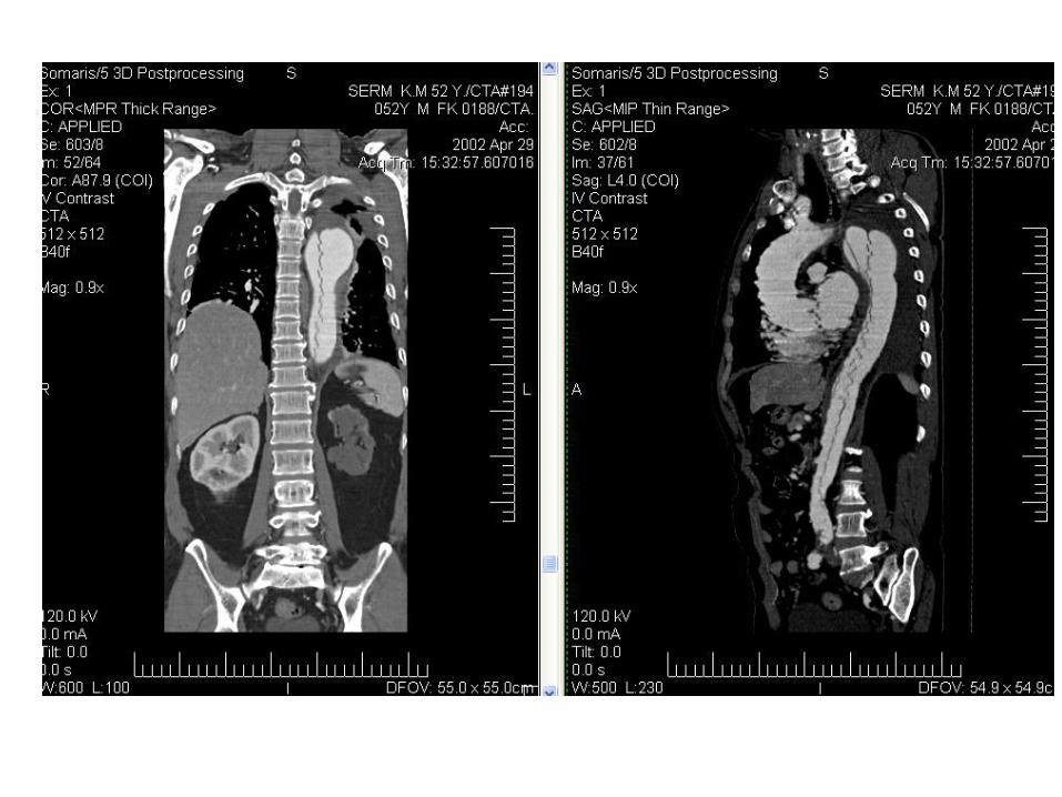
• CTA
• CT.perfusion

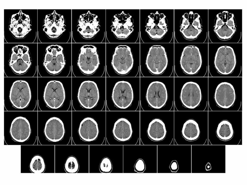
ตวอยำงภำพจำกเครอง CT. scan
Normal brain Epidural Hemorrhage
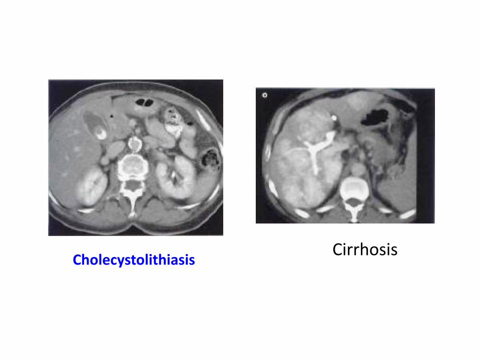
Cholecystolithiasis Cirrhosis


Software and PACS system
• Image Processing software
– Image reconstruction => Signal to image
– Reconstruction reformation => Axial to Sagittal or Axial to Coronal
– 3D volume rendering => Advanced reconstruction
– Virtual endoscopy

DICOM remote viewer
PACS svr.
Client (Remote)
Viewer
CT. (Modality)
CT (Local) Viewer
3D-Work Station

SET ZERO