Embed Size (px)
Citation preview
Correction
MEDICAL SCIENCESCorrection for “Atrial natriuretic peptide prevents cancer me-tastasis through vascular endothelial cells,” by Takashi Nojiri,Hiroshi Hosoda, Takeshi Tokudome, Koichi Miura, Shin Ishikane,Kentaro Otani, Ichiro Kishimoto, Yasushi Shintani, MasayoshiInoue, Toru Kimura, Noriyoshi Sawabata, Masato Minami,Tomoyuki Nakagiri, Soichiro Funaki, Yukiyasu Takeuchi, HajimeMaeda, Hiroyasu Kidoya, Hiroshi Kiyonari, Go Shioi, Yuji Arai,Takeshi Hasegawa, Nobuyuki Takakura, Megumi Hori, YukoOhno, Mikiya Miyazato, Naoki Mochizuki, Meinoshin Okumura,and Kenji Kangawa, which was first published March 16, 2015;10.1073/pnas.1417273112 (Proc Natl Acad Sci USA 112:4086–4091).The authors wish to note the following: “During the process of
data checking, we detected mistakes in the expression of samplenumbers, error bar expressions, and figure preparation. Threemembers of our team were involved in these errors and despitechecking the data ahead of publication, we did not notice thecalculation and expression errors in the figure preparation. Thesenior author apologizes for the inconvenience for these honesterrors, which, importantly, do not affect the clinical outcomes ormain results of the study.“The error bars in Figs. 2F, 3B, and 4E, and Fig. S5Bwere presented
incorrectly. The legends for Figs. 2 and 3 and Fig. S5 noted incorrectsample sizes. There was an error in the preparation of Table S1.“For Fig. 3, we showed representative images in Fig. 3 A and D
and apologize for use of the same image in the two panels andfor the confusion it might have caused. We have updated thefigure to include an image taken from the same experiment.“We showed the representative organ in Fig. 2 A and C, and
showed all image data in Fig. S1 A and C. Considering that thedata show the number of metastases, the size ratio change isacceptable because the number of metastases has not changed.We apologize for not making this clear in our figure legend.“Due to an error in data compilation, Fig. S8 data are different
and the statistical outcome is correspondingly changed andamended in the main text (italics), on page 4090, right column,first paragraph, lines 13–16:‘To ascertain the efficiency of ANP administration, we mea-
sured the blood levels of cGMP. However, the data did not reachstatistical significance when ANP (0.5 μg·kg·min) was infusedsubcutaneously in the mice (SI Appendix, Fig. S8), although the bloodcGMP levels showed a tendency of increase after ANP administration.’”The editor has read and approved these changes. The cor-
rected Figs. 2–4 appear below with their respective correctedlegends. The online version of the article has been corrected.The SI Appendix has also been corrected online, with correctionsmade to Fig. S5 and its legend, Fig. S8 and its legend, andTable S1.
www.pnas.org PNAS | August 14, 2018 | vol. 115 | no. 33 | E7883–E7886
CORR
ECTION

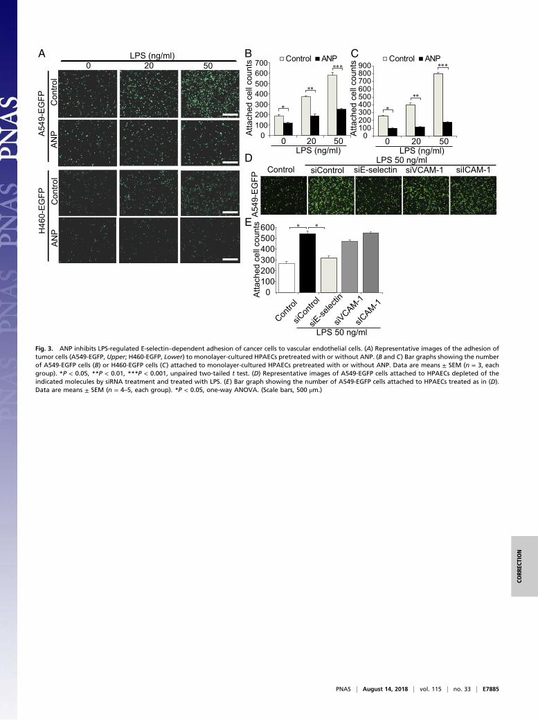
Fig. 2. ANP inhibits the LPS-augmented metastasis of A549-EGFP lung cancer cells and B16/F10 mice melanoma cells to the lung. (A) Representative EGFPimages of the lungs of mice that were pretreated with or without LPS and then injected with A549-EGFP cells (1 × 10⁶ cells per mouse) and continuouslytreated with or without ANP for 4 wk. The mice were killed 6 wk after the injection of tumor cells. (B) Bar graph showing the number of nodules representingpulmonary metastasis of A549-EGFP cells in mice grouped as in A. Data are means ± SD (n = 6, each group). ***P < 0.001, unpaired two-tailed t test. (C)Representative images of the lungs of mice that were pretreated with or without LPS and then injected with B16/F10 cells (2 × 10⁵ cells per mouse) andcontinuously treated with or without ANP for 2 wk. The mice were killed 2 wk after the injection of the tumor cells. (D) Bar graph showing the number ofnodules representing pulmonary metastasis of B16/F10 cells in mice grouped as in C. Data are means ± SD (n = 6, each group). ***P < 0.001, unpaired two-tailed t test. (E) Representative images of the lungs and hearts (Top and Middle, respectively) and histological cross-sections of the hearts (H&E staining,Bottom) of the GC-Aflox/flox mice and EC GC-A-KO mice after injection of B16/F10 cells (2 × 10⁵ cells per mouse). The mice were killed 2 wk after the injection ofthe tumor cells. (Scale bars, 500 μm.) Red arrows indicate metastasis in the heart. (F) Bar graph showing the number of nodules representing pulmonarymetastasis of B16/F10 cells in mice grouped as in E. Data are means ± SD (n = 9, 7, each group). **P < 0.01, unpaired two-tailed t test. (G) Kaplan–Meier curvescomparing survival times between GC-Aflox/flox and EC GC-A-KO mice after injection of B16/F10 cells (2 × 10⁵ cells per mouse). n = 12, 11 (each group), *P <0.05, log-rank test. (H) Representative images of the lungs of WT and EC GC-A-Tg mice after injection of B16/F10 cells (5 × 10⁵ cells per mouse). The mice werekilled 2 wk after the injection of tumor cells. (I) Bar graph showing the number of nodules representing pulmonary metastasis of B16/F10 cells in mice groupedas in H. Data are means ± SD (n = 10, 8, each group). **P < 0.01, unpaired two-tailed t test. (J) Kaplan–Meier curves comparing survival times betweenWT andEC GC-A-Tg mice after injection of B16/F10 (5 × 10⁵ cells per mouse). n = 15 (each group), *P < 0.05, log-rank test. Whole images of lungs were shown in SIAppendix, Fig. S1.
E7884 | www.pnas.org
A0 20 50
LPS (ng/ml)C
ontro
lC
ontro
lA
NP
AN
PA54
9-E
GFP
H46
0-E
GFP
0100200300400500600700
0 20 50 0 20 50LPS (ng/ml) LPS (ng/ml)
Atta
ched
cel
l cou
nts
Atta
ched
cel
l cou
nts
0100200300400500600700800900
Control ANP
∗ ∗
B C
A54
9-E
GFP
siControl siE-selectin siVCAM-1 siICAM-1LPS 50 ng/ml
ControlD
∗∗
∗∗∗
∗∗
∗∗∗
E
Atta
ched
cel
l cou
nts
LPS 50 ng/ml
∗
0100200300400500600 ∗
Contro
l
siCon
trol
siE-se
lectin
siVCAM-1
ANPControl
sICAM-1
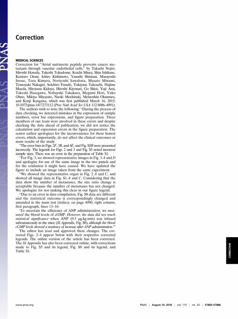
Fig. 3. ANP inhibits LPS-regulated E-selectin–dependent adhesion of cancer cells to vascular endothelial cells. (A) Representative images of the adhesion oftumor cells (A549-EGFP, Upper; H460-EGFP, Lower) to monolayer-cultured HPAECs pretreated with or without ANP. (B and C) Bar graphs showing the numberof A549-EGFP cells (B) or H460-EGFP cells (C) attached to monolayer-cultured HPAECs pretreated with or without ANP. Data are means ± SEM (n = 3, eachgroup). *P < 0.05, **P < 0.01, ***P < 0.001, unpaired two-tailed t test. (D) Representative images of A549-EGFP cells attached to HPAECs depleted of theindicated molecules by siRNA treatment and treated with LPS. (E) Bar graph showing the number of A549-EGFP cells attached to HPAECs treated as in (D).Data are means ± SEM (n = 4–5, each group). *P < 0.05, one-way ANOVA. (Scale bars, 500 μm.)
PNAS | August 14, 2018 | vol. 115 | no. 33 | E7885
CORR
ECTION

Published under the PNAS license.
Published online August 6, 2018.
www.pnas.org/cgi/doi/10.1073/pnas.1811802115
0 5 20 50LPS (ng/ml) ANP + LPS (ng/ml)
E-selectin0 5 20 50
VCAM-1
ICAM-1
GAPDH
A
100
kDa
100100
Con
trol
LPS
A
NP
+LP
S
NF-NF-κBκBC
Con
trol
LPS
A
NP
+LP
S
GE-selectinE-selectin CD31CD31 Merge/Merge/DAPIDAPI
05
1015202530
Contro
l
Vehicle
ANP
∗∗
Nuc
lear
NF-
κB p
ositi
ve c
ells
(%)
D
LPS
E-selectin
GAPDH
LPSANP
+- +- - +
E-s
elec
tin m
RN
A E F
0
5
10
15
20
0 1 2
Control ANP
rela
tive
expr
essi
on
LPS (mg/kg)
∗∗
B
E-selectin
GAPDH
LPSANP
siRNA Control GC-A+- +- - + - - +
- ++
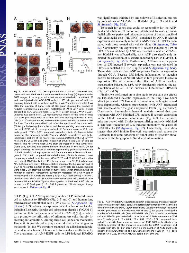
Fig. 4. ANP–GC-A signaling attenuates LPS-induced E-selectin expression. (A) Immunoblot analysis of the lysates of HPAECs pretreated with or without ANPfollowed by LPS stimulation; antibodies used are indicated on the left. Each blot is representative of six independent experiments. (B) E-selectin expressionassessed by immunoblot analysis of the lysates of HPAECs transfected with the indicated siRNAs and stimulated with LPS. The result shown is representative ofsix independent experiments. (C) Bright field images (Left) and NF-κB immunofluorescence images (Right) of HPAECs that were unstimulated (control, Top),stimulated with LPS alone (Middle), or pretreated with ANP followed by LPS stimulation (Bottom). Each image is representative of five independent ex-periments. (Scale bars, 100 μm.) (D) Quantitative analyses of C. Each column shows the percentage of HPAECs with nuclear NF-κB expression in the indicatedgroup. Data are means ± SEM (n = 5, each group); **P < 0.01, unpaired two-tailed t test. (E) Quantitative reverse transcriptase PCR analysis of E-selectin mRNAlevels in the lungs of mice pretreated with ANP or vehicle (control) and treated with LPS. Data are normalized relative to 36B4 mRNA levels. Data are means ±SEM (n = 6, each group); *P < 0.05, unpaired two-tailed t test. (F) Immunoblot analysis of E-selectin levels in lung lysates of mice pretreated with or withoutANP followed by LPS stimulation (1.0 mg/kg) for 5 h. Each blot is representative of six independent experiments. (G) E-selectin images (Left), CD31 images(Center), and merged images with DAPI staining (Right) of the lungs of mice pretreated with or without ANP followed by LPS stimulation (1.0 mg/kg) for 5 h.Each image is representative of six independent experiments. Nuclei are stained with DAPI (blue). (Scale bars, 100 μm.)
E7886 | www.pnas.org

Atrial natriuretic peptide prevents cancer metastasisthrough vascular endothelial cellsTakashi Nojiria,b, Hiroshi Hosodac, Takeshi Tokudomea, Koichi Miurac, Shin Ishikanea, Kentaro Otanic, Ichiro Kishimotod,Yasushi Shintanib, Masayoshi Inoueb, Toru Kimuraa,b, Noriyoshi Sawabatab, Masato Minamib, Tomoyuki Nakagirib,Soichiro Funakib, Yukiyasu Takeuchie, Hajime Maedae, Hiroyasu Kidoyaf, Hiroshi Kiyonarig, Go Shioig, Yuji Araih,Takeshi Hasegawai, Nobuyuki Takakuraf, Megumi Horij, Yuko Ohnoj, Mikiya Miyazatoa, Naoki Mochizukik,Meinoshin Okumurab, and Kenji Kangawaa,1
aDepartment of Biochemistry, cDepartment of Regenerative Medicine and Tissue Engineering, hDepartment of Molecular Biology, and kJST-CREST/Department of Cell Biology, National Cerebral and Cardiovascular Center Research Institute, Suita-city, Osaka 565-8565, Japan; bDepartment of GeneralThoracic Surgery and jDepartment of Mathematical Health Science, Osaka University Graduate School of Medicine, Suita-city, Osaka, 565-0871, Japan;dDepartment of Endocrinology and Metabolism, National Cerebral and Cardiovascular Center Hospital, Suita-city, Osaka 565-8565, Japan; eDepartment ofGeneral Thoracic Surgery, National Hospital Organization Toneyama Hospital, Toyonaka-city, Osaka, 560-8552, Japan; fDepartment of Signal Transduction,Research Institute for Microbial Diseases, Suita-city, Osaka 565-0871, Japan; gLaboratory for Animal Resources and Genetic Engineering, RIKEN Centerfor Developmental Biology, Kobe-city, Hyogo 650-0047, Japan; and iDrug Research Section II, Fukushima Research Laboratories, TOA EIYO Ltd.,Fukushima-city, Fukushima 960-0280, Japan
Edited* by Masashi Yanagisawa, University of Texas Southwestern Medical Center, Dallas, TX, and approved February 18, 2015 (received for reviewSeptember 7, 2014)
Most patients suffering from cancer die of metastatic disease.Surgical removal of solid tumors is performed as an initial attemptto cure patients; however, surgery is often accompanied withtrauma, which can promote early recurrence by provoking de-tachment of tumor cells into the blood stream or inducing systemicinflammation or both. We have previously reported that adminis-tration of atrial natriuretic peptide (ANP) during the perioperativeperiod reduces inflammatory response and has a prophylactic effecton postoperative cardiopulmonary complications in lung cancersurgery. Here we demonstrate that cancer recurrence after curativesurgery was significantly lower in ANP-treated patients than incontrol patients (surgery alone). ANP is known to bind specificallyto NPR1 [also called guanylyl cyclase-A (GC-A) receptor]. In mousemodels, we found that metastasis of GC-A–nonexpressing tumorcells (i.e., B16 mouse melanoma cells) to the lung was increased invascular endothelium-specific GC-A knockout mice and decreased invascular endothelium-specific GC-A transgenic mice compared withcontrol mice. We examined the effect of ANP on tumor metastasis inmice treated with lipopolysaccharide, which mimics systemic inflam-mation induced by surgical stress. ANP inhibited the adhesion ofcancer cells to pulmonary arterial and micro-vascular endothelialcells by suppressing the E-selectin expression that is promoted byinflammation. These results suggest that ANP prevents cancer me-tastasis by inhibiting the adhesion of tumor cells to inflamedendothelial cells.
cardiac peptide | cancer metastasis | vascular endothelial cell |inflammation | surgery
The majority of cancer patients die from tumor metastasis.Despite substantial advances in our understanding of the
mechanisms of tumor metastasis, effective prevention of metas-tasis has not been well established. Surgical removal of solidtumors is performed to cure patients if the primary tumor meetssurgical indications; however, postoperative cancer recurrence isa major problem. Surgical trauma itself influences the de-velopment of early recurrence (1, 2). First, the procedure duringtumor removal might provoke detachment of tumor cells; con-sistently, the number of circulating tumor cells is increasedduring primary tumor resection (3, 4). We previously reportedthat the presence of circulating tumor cells in pulmonary veinsduring lung cancer surgery could be a prognostic indicator forearly cancer recurrence (4). Second, surgical trauma provokesa severe systemic inflammatory reaction. Emerging evidencesuggests that systemic inflammation can accelerate the adhesionof circulating tumor cells to the vascular endothelium of distant
organs, which is the first step of extravasation in hematogenousmetastasis (5, 6).We identified human atrial natriuretic peptide (ANP) as a di-
uretic, natriuretic, and vasodilating hormone from the humanheart in 1984 (7). ANP binds specifically to the guanylyl cyclase-A(GC-A) receptor to exhibit biological functions, including pro-motion of diuresis, antifibrotic action, and inhibition of renin-angiotensin-aldosterone (8, 9). Thus, ANP has been used clinicallyfor the treatment of heart failure since 1995 in Japan. Wepreviously reported that administration of human ANP duringthe perioperative period reduces inflammatory responses andhas a prophylactic effect on postoperative cardiopulmonarycomplications in lung cancer surgery (10–12). In those studies,
Significance
Postoperative cancer recurrence is a major problem followingcurative cancer surgery. Perioperative systemic inflammationinduces the adhesion of circulating tumor cells released fromthe primary tumor to the vascular endothelium of distantorgans, which is the first step in hematogenous metastasis. Wehave previously reported that administration of atrial natri-uretic peptide (ANP) during the perioperative period reducesinflammatory response and has a prophylactic effect on post-operative cardiopulmonary complications in lung cancer sur-gery. Here, we demonstrate that cancer recurrence after lungcancer surgery was significantly lower in ANP-treated patientsthan in control patients (surgery alone). We show that ANPprevents cancer metastasis by suppressing the inflammatoryreaction of endothelial cells, thereby inhibiting cancer cell ad-hesion to vascular endothelial cells.
Author contributions: T. Nojiri, H.H., T.T., N.T., M. Miyazato, N.M., M.O., and K.K. de-signed research; T. Nojiri, H.H., T.T., K.M., S.I., K.O., I.K., Y.S., M.I., T.K., N.S., M. Minami,T. Nakagiri, S.F., Y.T., H.M., H. Kidoya, and N.M. performed research; T. Nojiri, H. Kiyonari,G.S., and Y.A. contributed new reagents/analytic tools; T. Nojiri, T.H., M.H., and Y.O.analyzed data; and T. Nojiri, N.M., and K.K. wrote the paper.
Conflict of interest statement: K.K., T. Nojiri, H.H., and M.O. have filed the patent relatedto atrial natriuretic peptide for the treatment of cancer metastasis with Daiichi-SankyoPharmaceutical Inc. (PCT/JP2012/054841).
*This Direct Submission article had a prearranged editor.
Freely available online through the PNAS open access option.
Data deposition: The data reported in this paper have been deposited in the Gene Ex-pression Omnibus (GEO) database, www.ncbi.nlm.nih.gov/geo (accession no. GSE56976).1To whom correspondence should be addressed. Email: [email protected].
This article contains supporting information online at www.pnas.org/lookup/suppl/doi:10.1073/pnas.1417273112/-/DCSupplemental.
4086–4091 | PNAS | March 31, 2015 | vol. 112 | no. 13 www.pnas.org/cgi/doi/10.1073/pnas.1417273112

ANP was used to promote diuresis during perioperative right-side heart failure caused by lung damage. Here, we further an-alyzed the effect of ANP on prevention of cancer recurrenceafter surgery and found that ANP might have antitumor meta-static activity. We explored the antimetastatic action of ANP byusing tissue-specific GC-A transgenic and knockout mice of tu-mor metastasis models. Our results suggest that ANP could beuseful as an antimetastasis peptide to prevent cancer recurrenceafter surgery.
ResultsClinical Impacts of ANP Therapy on Cancer Recurrence After LungCancer Surgery. We performed a retrospective study of the in-cidence of cancer recurrence in lung cancer patients after curativesurgery, comparing patients who underwent perioperative ANPtreatment with those who were subjected to surgery alone (controlpatients). The 2-y relapse-free survival (RFS) after surgery wassignificantly greater in ANP-treated patients than in controlpatients (91% vs. 75%, P = 0.018) (Fig. 1A). To eliminate bias, wereanalyzed the data by using propensity score matching. The 2-yRFS in the propensity score-matched analysis was also signifi-cantly greater in ANP-treated patients than in control patients(91% vs. 67%, P = 0.0013) (Fig. 1B and SI Appendix, Table S1).We hypothesized from these retrospective observations that ANPmay prevent recurrence of lung cancer.
Antimetastatic Effects of ANP in Hematogenous Pulmonary MetastaticModels. Vascular inflammation is considered to render the endo-thelium adhesive to circulating tumor cells, thereby allowing themetastasis of tumor cells (5, 6). We previously reported that post-operative complications induced by inflammation are reduced byANP (10–12). Therefore, to investigate whether ANP inhibits themetastasis of cancer cells to inflamed organs, we examined the ef-fect of ANP on tumor metastases in mice injected with LPS, whichmimics systemic inflammation induced by surgical stress (6, 13).The LPS-treated mice showed numerous hematogenous pulmonarymetastases of intravenously injected A549 lung cancer cellsexpressing EGFP (A549-EGFP) cells (Fig. 2 A and B and SI Ap-pendix, Fig. S1A). In contrast, the mice pretreated with ANPexhibited a large and significant reduction of LPS-induced pul-monary metastasis of A549-EGFP cells (Fig. 2 A and B and SIAppendix, Fig. S1A). ANP also significantly inhibited the pulmonaryhematogenous metastasis of B16/F10 melanoma cells, which do notexpress GC-A (Fig. 2 C and D and SI Appendix, Fig. S1 B and C).Furthermore, we confirmed that ANP significantly inhibited thepulmonary hematogenous metastasis of A549-EGFP (SI Appendix,Fig. S2 A and B) and B16/F10 (SI Appendix, Fig. S2 C and D) cellseven without LPS, suggesting that ANP inhibits tumor metastasisboth under LPS-induced general inflammation and under non–
LPS-induced massive inflammation. More importantly, thesedata indicate that ANP acts through GC-A expressed in nontumormouse cells.To eliminate the direct effect of ANP on tumor cell proliferation,
we first examined the direct effects of ANP on the growth of cancercells and found that GC-A was expressed in A549 and H460 humanlung cancer cells (SI Appendix, Fig. S1B). Even though GC-A wasexpressed on A549 and H460 cells, ANP did not induce the pro-liferation of these tumor cells (SI Appendix, Fig. S3 A–C). Natri-uretic peptide receptor-C, which is also known as a receptor ofANP, was expressed in A549, H460, and B16/F10 cells (SI Ap-pendix, Fig. S3A); however, there were no significant effects of ANPon the growth of A549, H460, or B16/F10 cells (SI Appendix, Fig. S3B–D). These results suggest that the inhibitory effect of ANP ontumor metastasis is dependent upon GC-A expressed on cells otherthan tumor cells.We considered that GC-A expressed on endothelial cells
might be responsible for the antimetastatic effect of ANP becausecancer cell attachment to endothelial cells is the initial step inmetastasis (5, 6). Vascular endothelial cells abundantly expressGC-A, which exhibits a protective role in the cardiovascular system(8, 9). Therefore, to clearly show that the antimetastatic effect ofANP does not depend on GC-A expression in tumor cells, weexamined the hematogenous pulmonary metastasis of B16/F10cells in both endothelium-specific GC-A knockout mice (termedEC GC-A-KO mice) and GC-A transgenic mice (termed EC GC-A-Tg mice) (SI Appendix, Fig. S4). EC GC-A-KO mice exhibiteda significant elevation of blood pressure and cardiac hypertrophycompared with GC-Aflox/flox mice. These phenotypic data wereconsistent with the previous report (14). The number of pulmonarymetastases was significantly higher in EC GC-A-KO mice than inGC-Aflox/flox mice (Fig. 2 E and F). Furthermore, cardiac metas-tases were found in one-third of EC GC-A-KO mice, whereas nocardiac metastasis was found inGC-Aflox/flox mice (Fig. 2E). Overallsurvival was significantly shorter in EC GC-A-KO mice com-pared with GC-Aflox/flox mice (Fig. 2G). In contrast, the number ofpulmonary metastases was significantly lower in ECGC-A-Tg micethan in WT mice (Fig. 2 H and I), and EC GC-A-Tg mice survivedsignificantly longer than WT mice (Fig. 2J). Collectively, these datasuggest that endothelial GC-A activated by ANP prevents hema-togenous pulmonary metastasis of cancer cells in mice.
Mechanism of the Effect of ANP on Cancer Cell Adhesion to VascularEndothelial Cells. We next attempted to uncover the molecularmechanism behind ANP-mediated inhibition of tumor metastasisthrough vascular endothelial cells. The attachment of A549-EGFP and H460 lung cancer cells expressing EGFP (H460-EGFP) cells to cultured human pulmonary artery endothelialcells (HPAECs) stimulated with LPS was dependent on the dose
ANP (+) ANP + Surgery (n = 77)
ANP (-) Surgery alone (n = 390)
All patients
0
60
40
80
100
20
ANP(+)Patients at risk
ANP(-)
0 12 24 36(Months)
77390
69327
47216
29128
Rel
apse
-free
sur
viva
l (%
)
ANP (+) ANP + Surgery (n = 77)
ANP (-) Surgery alone (n = 77)
Pair-matched patients
0
60
40
80
100
20
0
ANP(+)Patients at risk
ANP(-)
12 24 36 (Months)
77 77
6960
4733
2923
Rel
apse
-free
sur
viva
l (%
)P = 0.018 (log-rank) P = 0.0013 (log-rank)
A B
Fig. 1. Effect of ANP treatment on RFS in patients with surgically resected nonsmall cell lung cancer. (A) Kaplan–Meier curves of the ANP group and controlgroup (surgery alone) in all patients (P = 0.018, log-rank test). (B) Kaplan–Meier curves of the above groups in propensity score-matched patients (P = 0.0013,log-rank test). RFS was measured from the day of surgery to cancer recurrence.
Nojiri et al. PNAS | March 31, 2015 | vol. 112 | no. 13 | 4087
MED
ICALSC
IENCE
S
of LPS (Fig. 3A). ANP significantly inhibited LPS-induced tumorcell attachment to HPAECs (Fig. 3 B and C) and human lungmicrovascular endothelial cells (HMVEC-L) (SI Appendix, Fig.S5 A–C). LPS induces the expression of cell adhesion molecules,including E-selectin, vascular cell adhesion molecule-1 (VCAM-1),and intercellular adhesion molecule-1 (ICAM-1) (15), which inturn promote the infiltration of inflammatory cells, thereby in-creasing inflammation. Among these cell adhesion molecules,E-selectin is considered to play a central role in hematogenousmetastasis (16–18). We therefore examined the adhesion molecule-dependent attachment of tumor cells to vascular endothelial cells.The attachment of A549-EGFP cells to LPS-induced HPAECs
was significantly inhibited by knockdown of E-selectin, but notby knockdown of VCAM-1 or ICAM-1 (Fig. 3 D and E andSI Appendix, Fig. S6A).To search for genes that could be responsible for the ANP-
mediated inhibition of tumor cell attachment to vascular endo-thelial cells, we performed microarray analyses of human umbilicalvein endothelial cells (HUVECs) stimulated by ANP. E-selectinexpression was markedly reduced in ANP-treated HUVECs com-pared with those treated with vehicle alone (SI Appendix, TableS2). Consistently, the expression of E-selectin induced by LPS inHPAECs was inhibited by ANP, whereas that of neither VCAM-1nor ICAM-1 was affected (Fig. 4A). ANP also significantly in-hibited the expression of E-selectin induced by LPS in HMVEC-L(SI Appendix, Fig. S5D). Furthermore, ANP-mediated suppres-sion of LPS-induced E-selectin expression was not observed inHPAECs depleted of GC-A (Fig. 4B and SI Appendix, Fig. S6B).These data indicate that ANP suppresses E-selectin expressionthrough GC-A. Because LPS induces inflammation by inducingnuclear translocation of NF-κB, which in turn promotes E-selectinexpression (19), we examined the effect of ANP on nucleartranslocation induced by LPS. ANP significantly inhibited the ac-cumulation of NF-κB in the nucleus of LPS-induced HPAECs(Fig. 4 C and D).Finally, we performed an in vivo study to evaluate the effects
on LPS-induced E-selectin expression in the lung. Five hoursafter injection of LPS, E-selectin expression in the lung increaseddose-dependently, whereas pretreatment with ANP attenuatedthis increase at both the gene and protein levels (Fig. 4 E and F).Consistently, immunohistochemical analysis showed that pre-treatment with ANP inhibited LPS-induced E-selectin expressionin the CD31+ vascular endothelium (Fig. 4G). Furthermore,mice pretreated with E-selectin–neutralizing antibody exhibiteda significant reduction of LPS-induced pulmonary metastasis ofB16/F10 cells (SI Appendix, Fig. S7). Collectively, these datasuggest that ANP inhibits E-selectin expression and reduces theE-selectin–mediated adhesion of tumor cells to vascular endo-thelium of the lung upon LPS-induced inflammation.
Fig. 2. ANP inhibits the LPS-augmented metastasis of A549-EGFP lungcancer cells and B16/F10 mice melanoma cells to the lung. (A) RepresentativeEGFP images of the lungs of mice that were pretreated with or without LPSand then injected with A549-EGFP cells (1 × 10⁶ cells per mouse) and con-tinuously treated with or without ANP for 4 wk. The mice were killed 6 wkafter the injection of tumor cells. (B) Bar graph showing the number ofnodules representing pulmonary metastasis of A549-EGFP cells in micegrouped as in A. Data are means ± SD (n = 6, each group). ***P < 0.001,unpaired two-tailed t test. (C) Representative images of the lungs of micethat were pretreated with or without LPS and then injected with B16/F10cells (2 × 10⁵ cells per mouse) and continuously treated with or without ANPfor 2 wk. The mice were killed 2 wk after the injection of the tumor cells.(D) Bar graph showing the number of nodules representing pulmonary metas-tasis of B16/F10 cells in mice grouped as in C. Data are means ± SD (n = 6,each group). ***P < 0.001, unpaired two-tailed t test. (E) Representativeimages of the lungs and hearts (Top and Middle, respectively) and histo-logical cross-sections of the hearts (H&E staining, Bottom) of the GC-Aflox/flox
mice and EC GC-A-KO mice after injection of B16/F10 cells (2 × 10⁵ cells permouse). The mice were killed 2 wk after the injection of the tumor cells.(Scale bars, 500 μm.) Red arrows indicate metastasis in the heart. (F) Bargraph showing the number of nodules representing pulmonary metastasisof B16/F10 cells in mice grouped as in E. Data are means ± SD (n = 9, 7, eachgroup). **P < 0.01, unpaired two-tailed t test. (G) Kaplan–Meier curvescomparing survival times between GC-Aflox/flox and EC GC-A-KO mice afterinjection of B16/F10 cells (2 × 10⁵ cells per mouse). n = 12, 11 (each group),*P < 0.05, log-rank test. (H) Representative images of the lungs of WT and ECGC-A-Tgmice after injection of B16/F10 cells (5 × 10⁵ cells per mouse). The micewere killed 2 wk after the injection of tumor cells. (I) Bar graph showing thenumber of nodules representing pulmonary metastasis of B16/F10 cells inmice grouped as in H. Data are means ± SD (n = 10, 8, each group). **P < 0.01,unpaired two-tailed t test. (J) Kaplan–Meier curves comparing survival timesbetween WT and EC GC-A-Tg mice after injection of B16/F10 (5 × 10⁵ cells permouse). n = 15 (each group), *P < 0.05, log-rank test. Whole images of lungswere shown in SI Appendix, Fig. S1.
A0 20 50
LPS (ng/ml)
Con
trol
Con
trol
AN
PA
NPA
549-
EG
FPH
460-
EG
FP
0100200300400500600700
0 20 50 0 20 50LPS (ng/ml) LPS (ng/ml)
Atta
ched
cel
l cou
nts
Atta
ched
cel
l cou
nts
0100200300400500600700800900
Control ANP
∗ ∗
B C
A54
9-E
GFP
siControl siE-selectin siVCAM-1 siICAM-1LPS 50 ng/ml
ControlD
∗∗
∗∗∗
∗∗
∗∗∗
E
Atta
ched
cel
l cou
nts
LPS 50 ng/ml
∗
0100200300400500600 ∗
Contro
l
siCon
trol
siE-se
lectin
siVCAM-1
ANPControl
sICAM-1
Fig. 3. ANP inhibits LPS-regulated E-selectin–dependent adhesion of cancercells to vascular endothelial cells. (A) Representative images of the adhesionof tumor cells (A549-EGFP, Upper; H460-EGFP, Lower) to monolayer-culturedHPAECs pretreated with or without ANP. (B and C) Bar graphs showing thenumber of A549-EGFP cells (B) or H460-EGFP cells (C) attached to monolayer-cultured HPAECs pretreated with or without ANP. Data are means ± SEM(n = 3, each group). *P < 0.05, **P < 0.01, ***P < 0.001, unpaired two-tailed t test. (D) Representative images of A549-EGFP cells attached toHPAECs depleted of the indicated molecules by siRNA treatment andtreated with LPS. (E ) Bar graph showing the number of A549-EGFP cellsattached to HPAECs treated as in (D). Data are means ± SEM (n = 4–5, eachgroup). *P < 0.05, one-way ANOVA. (Scale bars, 500 μm.)
4088 | www.pnas.org/cgi/doi/10.1073/pnas.1417273112 Nojiri et al.

DiscussionAlthough many clinical trials aimed at preventing cancer re-currence during the perioperative period have been conducted,no prophylactic treatments have been established. The failure ofthese trials might be ascribed to the risk of surgery alone, and theside effects of the chemicals used in the trials (20, 21). We pre-viously showed that ANP prevents the incidence of postoperativecomplications after lung cancer surgery (10–12). Here we dem-onstrate that cancer recurrence after curative surgery was signifi-cantly lower in ANP-treated patients than in control patients,suggesting that ANP could potentially be used to prevent cancerrecurrence after surgery. We assumed two possibilities as to howANP inhibited tumor metastases; one was that ANP directlyinhibited the tumor cell proliferation and the other was that ANPindirectly inhibited tumor cell metastases by acting on nontumorcells. In previous studies of the direct effects of ANP on cancercells, both inhibitory and stimulatory effects of ANP on the growthof cancer cells have been reported (22, 23); therefore, the directeffects of ANP on cancer cells remain controversial. In the presentstudy, we focused on the possibility that ANP indirectly inhibitstumor cell metastases through effects on nontumor cells.Our discovery that mice pretreated with ANP exhibited a
dramatic reduction of LPS-induced pulmonary metastasis ofintroduced cancer cells provides direct evidence that ANP canprevent tumor metastasis in mice. This notion is supported by ourfinding that mice that specifically overexpress or lack expression
of the receptor of ANP (i.e., GC-A) in the vascular endotheliumhave reduced or enhanced numbers of metastases, respectively,compared with the appropriate control mice. These results sug-gest that ANP prevents early relapse in patients at least in part bypreventing metastasis through the vascular endothelium.Surgical procedures induce postoperative complications and
early recurrence after surgery by releasing inflammatory cyto-kines, such as IL-1β and TNF-α (1, 2). Recent studies indicatethat postoperative complications, including severe inflammatoryreaction and infection, after various types of cancer surgery areassociated with poor cancer-specific survival (24–26). Endothe-lial cells that become inflamed during surgery are considered tobe prone to adhering to circulating tumor cells, thereby allowingthe initiation of metastasis (5, 6). Although most circulating tu-mor cells undergo rapid cell death by apoptosis (27, 28), it ispossible that surgical inflammation promotes the adherence ofresidual cancer cells to inflamed endothelial cells (5, 6). ANP hasanti-inflammatory and anti-infectious activity on endothelialcells. ANP pretreatment reduces serum TNF-α levels and NF-κBactivation by inhibiting IκB-phosphorylation in mice injectedwith LPS (29) and has a protective role against LPS-induced lunginjury and endothelial barrier dysfunction (30). Our finding thatANP has anti-inflammatory action (i.e., suppression of LPS-induced E-selectin) in vascular endothelial cells in mice isconsistent with these studies. Taken together, our results sug-gest that ANP-mediated inhibition of metastasis occurs throughinhibition of the inflammatory response.Among the vascular adhesion molecules, E-selectin is essential
for recruitment of inflammatory cells to damaged tissues (31),and it enables circulating tumor cells to roll and tether on theendothelium. Recent studies have shown that cross-talk betweenE-selectin and integrins could facilitate the movement of notonly inflammatory cells but also tumor cells through the endo-thelium to inflammatory foci (16–18). In fact, tumor metastasis isincreased in the lungs of E-selectin–overexpressing mice andreduced in E-selectin knockout mice (18). Therefore, E-selectinis considered to play a central role in hematogenous metastasis(16–18). In a clinical study, Gogali et al. reported that serumlevels of soluble E-selectin in lung cancer patients were signifi-cantly elevated compared with those in control subjects (32).However, we assume that the antimetastasis activity of ANP doesnot solely depend upon the suppression of E-selectin, becauseextravasation of cancer cells in the metastatic process is regu-lated by many other steps. Recent experimental reports dem-onstrated that inflammatory chemokines including chemokineligand (CCL) 2 and CCL5 contributed to not only leukocyterecruitment but also tumor cell homing to activated endothelialcells (33, 34). Because we focused on only E-selectin expressionsin this study, further studies are necessary to elucidate thedetailed mechanism and role of the ANP–GC-A system incancer metastasis.Because most current chemotherapeutic agents are cytotoxic
and cause many side effects, chemotherapy cannot be usedduring surgical resection to prevent cancer recurrence. In con-trast, ANP is an endogenous and physiological peptide and hasbeen proved not to cause severe adverse effects when used inpatients with heart failure (35). Because the target of ANP isconsidered to be vascular endothelium in all organs that expressthe GC-A receptor, including lung, liver, and brain, ANP mightinhibit hematogenous cancer metastasis to all organs express-ing GC-A receptor and could be used for all kinds of malig-nant tumors.
Materials and MethodsClinical Study. We retrospectively evaluated 552 consecutive patients whounderwent curative surgery for nonsmall cell lung cancer at Osaka UniversityHospital and National Hospital Organization ToneyamaHospital fromAugust2007 to December 2011. Patientswith carcinoma in situ and those undergoing
0 5 20 50LPS (ng/ml) ANP + LPS (ng/ml)
E-selectin0 5 20 50
VCAM-1
ICAM-1
GAPDH
A100
kDa
100100
Con
trol
LPS
A
NP
+LP
S
NF-NF-κBκBC
Con
trol
LPS
A
NP
+LP
S
GE-selectinE-selectin CD31CD31 Merge/Merge/DAPIDAPI
05
1015202530
Contro
l
Vehicle
ANP
∗∗
Nuc
lear
NF-
κB p
ositi
ve c
ells
(%)
D
LPS
E-selectin
GAPDH
LPSANP
+- +- - +
E-s
elec
tin m
RN
A E F
0
5
10
15
20
0 1 2
Control ANP
rela
tive
expr
essi
on
LPS (mg/kg)
∗∗
BE-selectin
GAPDH
LPSANP
siRNA Control GC-A+- +- - + - - +
- ++
Fig. 4. ANP–GC-A signaling attenuates LPS-induced E-selectin expression.(A) Immunoblot analysis of the lysates of HPAECs pretreated with or withoutANP followed by LPS stimulation; antibodies used are indicated on the left.Each blot is representative of six independent experiments. (B) E-selectinexpression assessed by immunoblot analysis of the lysates of HPAECs trans-fected with the indicated siRNAs and stimulated with LPS. The result shownis representative of six independent experiments. (C ) Bright field images(Left) and NF-κB immunofluorescence images (Right) of HPAECs thatwere unstimulated (control, Top), stimulated with LPS alone (Middle),or pretreated with ANP followed by LPS stimulation (Bottom). Each imageis representative of five independent experiments. (Scale bars, 100 μm.)(D) Quantitative analyses of C. Each column shows the percentage of HPAECswith nuclear NF-κB expression in the indicated group. Data are means ± SEM(n = 5, each group); **P < 0.01, unpaired two-tailed t test. (E) Quantitativereverse transcriptase PCR analysis of E-selectin mRNA levels in the lungs ofmice pretreated with ANP or vehicle (control) and treated with LPS. Data arenormalized relative to 36B4 mRNA levels. Data are means ± SEM (n = 6, eachgroup); *P < 0.05, unpaired two-tailed t test. (F) Immunoblot analysis ofE-selectin levels in lung lysates of mice pretreated with or without ANP fol-lowed by LPS stimulation (1.0 mg/kg) for 5 h. Each blot is representative ofsix independent experiments. (G) E-selectin images (Left), CD31 images(Center), and merged images with DAPI staining (Right) of the lungs of micepretreated with or without ANP followed by LPS stimulation (1.0 mg/kg) for5 h. Each image is representative of six independent experiments. Nuclei arestained with DAPI (blue). (Scale bars, 100 μm.)
Nojiri et al. PNAS | March 31, 2015 | vol. 112 | no. 13 | 4089
MED
ICALSC
IENCE
S

a limited resection, including wedge resection, were excluded. Patients withincomplete postoperative follow up (n= 8) were also excluded. Segmentectomyfor curative surgery was not excluded. Finally, 467 patients who underwentcurative surgery were included in the present study.
RFS, defined as the time from the day of surgery to cancer recurrence, wascompared between patients who received ANP during the perioperativeperiod and those that received surgery only. In the ANP group, the subjectsreceived ANP intravenously at 0.025 μg·kg·min (Daiichi-Sankyo Pharmaceu-tical) without a bolus for 3 d continuously, starting just before the inductionof general anesthesia. We previously reported that ANP has a prophylacticeffect against postoperative cardiopulmonary complications for patientswith elevated preoperative brain natriuretic peptide levels (10, 12, 36).Therefore, we performed a propensity score-matched analysis to reduce thetreatment selection bias for each group. The propensity score was estimatedby using a logistic regression model adjusted for age, sex, pathologicalstaging [lung cancer tumor, node, metastases (TMN) staging seventh edi-tion], cancer histology, and preoperative brain natriuretic peptide levels.These variables were chosen for potential associations with the outcome ofinterest. An independent statistician selected the patients by matchingpropensity scores without access to clinical outcome information. Patientcharacteristics for the full and propensity score-matched cohorts are listed inSI Appendix, Table S1. In our matching algorithm, one patient who receivedANP was matched to one patient who did not receive ANP by using nearest-neighbor matching without replacement. To measure covariate balance, weused the standardized difference. Estimation of propensity scores andmatching were performed by using MATLAB r2011b software (Mathworks).Lung cancer-specific RFS was compared by using Kaplan–Meier estimatesand the log-rank test for equality of survival curves. Calculations wereconducted by using JMP statistical software (SAS Institute).
All patients received predefined treatment including chemotherapyaccording to the clinical guidelines for lung cancer in Japan. ANP treatmentwas performed just to prevent postoperative complications; therefore, therewere no differences in the treatments including chemotherapy betweencontrol patients and ANP patients. We obtained complete pathological andfollow-up data from all subjects. The study protocol was approved by theInstitutional Review Boards of both institutions, and all patients gave writ-ten informed consent to participate in the study (Trial registration ID: JPRN-UMIN4880). The median follow-up duration was 36 mo (18–60 mo). Allsubjects underwent follow-up examinations at 3-mo intervals postoperatively:each evaluation included a physical examination, chest X-ray and bloodtests including tumor markers. Thoraco-abdominal CT scans were gener-ally performed at 6-mo intervals and additional bone scintigraphy andMRI of the brain for the detection of cancer recurrence were performedevery year.
Cell Lines. The human lung cancer cell lines, A549-EGFP and H460-EGFP, wereobtained from Wako and maintained in RPMI-1640 medium supplementedwith 10% (vol/vol) FBS. The mice melanoma cell line, B16/F10, and the A549,H460, MCF-7, OVCAR3, CaCo2, GCIY, HepG2, ME-180, PANC1, and PC3 celllines were obtained from the American Type Culture Collection andmaintained in DMEM supplemented with 10% FBS at 37 °C under 5%CO2. HPAECs, HMVEC-L, and HUVECs were purchased from Lonza, main-tained in EGM-2 according to the manufacturer’s instructions, and usedwithin passages three to five.
Experimental Lung Metastasis Model. All animal experiments were performedaccording to the protocol approved by the Animal Care Ethics Committeeof the National Cerebral and Cardiovascular Center Research Institute. Six-week-old BALB/c nu/nu and C57BL/6 mice were purchased from Japan SLC.BALB/c nu/numice for A549-EGFP cells (1 × 10⁶ cells per mouse) or C57BL/6micefor B16/F10 cells (2–5 × 10⁵ cells per mouse) were used in this study. In the
experiments with LPS (Fig. 2 A–D), mice were divided into three groups:vehicle alone (control), vehicle/LPS, and ANP/LPS group with two kinds ofcancer cells (either A549-EGFP or B16/F10 cells). ANP in 0.9% saline or vehiclewas continuously injected by using osmotic pumps (Alzet Model 1002 or2004, DURECT) implanted subcutaneously in the upper back of the mice1 d before LPS injection. On the next day, the mice were intravenouslyinjected with or without 1.0 mg/kg LPS (Wako). Five hours after LPS in-jection, cancer cells (either A549-EGFP or B16/F10 cells) were injected intothe tail vein. In the experiments without LPS (SI Appendix, Fig. S2), micewere divided into two groups: vehicle and ANP groups with two kinds ofcancer cells (either A549-EGFP or B16/F10 cells). ANP or vehicle alone wasimplanted 1 d before cancer cells injection. On the next day, cancer cells(either A549-EGFP or B16/F10 cells) were injected into the tail vein. To as-certain the efficiency of ANP administration, we measured the blood levelsof cGMP. However, the data did not reach statistical significance when ANP(0.5 μg·kg·min) was infused subcutaneously in the mice (SI Appendix, Fig. S8),although the blood cGMP levels showed a tendency of increase after ANPadministration. ANP (0.5 μg·kg·min) or vehicle infusion was started 1 d beforethe injection of cancer cells. At this dose, ANP did not change the blood pressureor heart rate of the mice (SI Appendix, Table S3). ANP or vehicle infusion wascontinued until the mice were euthanized. Six or 8 wk (A549-EGFP) or 2 wk (B16/F10) after tumor cell injection, the mice were killed for evaluation of pulmonarymetastases. The number of nodules reflecting pulmonary metastasis of A549-EGFP or B16/F10 cells was counted by using images obtained with a fluorescentmicroscope (OV100, Olympus) or a camera (CX6, Ricoh).
Adhesion Assay. To quantify tumor cell adhesion to HPAECs or HMVEC-L,a standardized cell adhesion assay was performed by using a modification ofthe method of van Rossen et al. (37). Briefly, HPAEC or HMVEC-L monolayerswere established in 35-mm collagen-coated dishes (IWAKI). Before coculturewith tumor cells, HPAECs or HMVEC-L were either pretreated with 0.1 μMANP for 15 min or untreated and subsequently treated with LPS (0–50 pg/mL)for 30 min, then washed three times with fresh M199 medium (Gibco; Invi-trogen) containing 1% BSA (Sigma-Aldrich). A549-EGFP or H460-EGFP tumorcells (2 × 10⁵ cells per dish) were added to the confluent monolayer-culturedHPAECs or HMVEC-L and cocultured for 3 h. The dishes were then washedthree times with PBS to remove nonadherent tumor cells, and the cells werefixed with 4% (wt/vol) paraformaldehyde. The number of remaining EGFP+
cells in the fixed dishes was counted by using images obtained with a fluo-rescence microscope (FSX100, Olympus) and a computer-aided manipulatorprogram (Cell-sense, Olympus). In addition, the adhesion of the tumor cells toHPAECs depleted of either E-selectin, VCAM-1, or ICAM-1, and stimulated withLPS, was similarly analyzed.
Statistics. Data are presented as means ± SEM and were analyzed by usinga two-tailed Student’s t-test for paired samples or one-way ANOVA formultiple groups. P values less than 0.05 were considered statisticallysignificant.
ACKNOWLEDGMENTS. We thank T. N. Sato (Nara Institute of Science andTechnology) and M. Yanagisawa (University of Texas Southwestern MedicalCenter) for their gift of Tie2-Cremice; M. M. Taketo and M. Sonoshita (KyotoUniversity) for giving valuable advice; T. Mabuchi, H. Mondo, Y. Nakamura,and AntiCancer Japan Inc. for their technical assistance; and K. Shioya forhelping us to care for the mice. This work was supported by research grantsfrom the Ministry of Education, Culture, Sports, Science and Technology ofJapan; Osaka Cancer Society, Japan Research Foundation for Clinical Phar-macology, Kobayashi Foundation for Cancer Research, Takeda Science Foun-dation, and Mochida Memorial Foundation for Medical and PharmaceuticalResearch (to T. Nojiri); and grants from the Ministry of Education, Culture,Sports, Science and Technology of Japan and Takeda Science Foundation(to K.K.).
1. Ferri LE, Law S, Wong KH, Kwok KF, Wong J (2006) The influence of technical com-plications on postoperative outcome and survival after esophagectomy. Ann SurgOncol 13(4):557–564.
2. ten Kate M, et al. (2004) Influence of proinflammatory cytokines on the adhesion of hu-man colon carcinoma cells to lungmicrovascular endothelium. Int J Cancer 112(6):943–950.
3. Koch M, et al. (2005) Detection of hematogenous tumor cell dissemination predictstumor relapse in patients undergoing surgical resection of colorectal liver metastases.Ann Surg 241(2):199–205.
4. Funaki S, et al. (2011) Novel approach for detection of isolated tumor cells in pulmonaryvein using negative selection method: Morphological classification and clinical im-plications. Eur J Cardiothorac Surg 40(2):322–327.
5. Giavazzi R, Foppolo M, Dossi R, Remuzzi A (1993) Rolling and adhesion of humantumor cells on vascular endothelium under physiological flow conditions. J Clin Invest92(6):3038–3044.
6. McDonald B, et al. (2009) Systemic inflammation increases cancer cell adhesion tohepatic sinusoids by neutrophil mediated mechanisms. Int J Cancer 125(6):1298–1305.
7. Kangawa K, Matsuo H (1984) Purification and complete amino acid sequence ofα-human atrial natriuretic polypeptide (α-hANP). Biochem Biophys Res Commun118(1):131–139.
8. Li Y, et al. (2002) Guanylyl cyclase-A inhibits angiotensin II type 1A receptor-mediatedcardiac remodeling, an endogenous protective mechanism in the heart. Circulation106(13):1722–1728.
9. Kishimoto I, et al. (2009) Natriuretic peptide signaling via guanylyl cyclase (GC)-A: Anendogenous protective mechanism of the heart. Curr Cardiol Rev 5(1):45–51.
10. Nojiri T, et al. (2012) Effect of low-dose human atrial natriuretic peptide on post-operative atrial fibrillation in patients undergoing pulmonary resection for lungcancer: A double-blind, placebo-controlled study. J Thorac Cardiovasc Surg 143(2):488–494.
4090 | www.pnas.org/cgi/doi/10.1073/pnas.1417273112 Nojiri et al.

11. Nojiri T, et al. (2013) Low-dose human atrial natriuretic peptide for the prevention ofpostoperative cardiopulmonary complications in chronic obstructive pulmonary dis-ease patients undergoing lung cancer surgery. Eur J Cardiothorac Surg 44(1):98–103.
12. Nojiri T, et al. (2012) Effects of low-dose human atrial natriuretic peptide for preventingpost-operative cardiopulmonary complications in elderly patients undergoing pul-monary resection for lung cancer. Eur J Cardiothorac Surg 41(6):1330–1334.
13. Haupt W, et al. (1998) Monocyte function before and after surgical trauma. Dig Surg15(2):102–104.
14. Sabrane K, et al. (2005) Vascular endothelium is critically involved in the hypotensiveand hypovolemic actions of atrial natriuretic peptide. J Clin Invest 115(6):1666–1674.
15. Haraldsen G, Kvale D, Lien B, Farstad IN, Brandtzaeg P (1996) Cytokine-regulatedexpression of E-selectin, intercellular adhesion molecule-1 (ICAM-1), and vascular celladhesion molecule-1 (VCAM-1) in human microvascular endothelial cells. J Immunol156(7):2558–2565.
16. Barthel SR, Gavino JD, Descheny L, Dimitroff CJ (2007) Targeting selectins and selectinligands in inflammation and cancer. Expert Opin Ther Targets 11(11):1473–1491.
17. Tremblay PL, Huot J, Auger FA (2008) Mechanisms by which E-selectin regulates di-apedesis of colon cancer cells under flow conditions. Cancer Res 68(13):5167–5176.
18. Hiratsuka S, et al. (2011) Endothelial focal adhesion kinase mediates cancer cellhoming to discrete regions of the lungs via E-selectin up-regulation. Proc Natl AcadSci USA 108(9):3725–3730.
19. Jersmann HP, Hii CS, Ferrante JV, Ferrante A (2001) Bacterial lipopolysaccharide andtumor necrosis factor alpha synergistically increase expression of human endothelialadhesion molecules through activation of NF-kappaB and p38 mitogen-activatedprotein kinase signaling pathways. Infect Immun 69(3):1273–1279.
20. de Bree E, et al. (2003) Intraoperative hyperthermic intraperitoneal chemotherapywith docetaxel as second-line treatment for peritoneal carcinomatosis of gynaeco-logical origin. Anticancer Res 23(3C):3019–3027.
21. Glehen O, et al. (2004) Cytoreductive surgery combined with perioperative in-traperitoneal chemotherapy for the management of peritoneal carcinomatosis fromcolorectal cancer: A multi-institutional study. J Clin Oncol 22(16):3284–3292.
22. Serafino A, et al. (2012) Anti-proliferative effect of atrial natriuretic peptide on co-lorectal cancer cells: Evidence for an Akt-mediated cross-talk between NHE-1 activityand Wnt/β-catenin signaling. Biochim Biophys Acta 1822(6):1004–1018.
23. Kong X, et al. (2008) Natriuretic peptide receptor a as a novel anticancer target.Cancer Res 68(1):249–256.
24. Lerut T, et al. (2009) Postoperative complications after transthoracic esophagectomyfor cancer of the esophagus and gastroesophageal junction are correlated with earlycancer recurrence: Role of systematic grading of complications using the modifiedClavien classification. Ann Surg 250(5):798–807.
25. Andalib A, Ramana-Kumar AV, Bartlett G, Franco EL, Ferri LE (2013) Influence ofpostoperative infectious complications on long-term survival of lung cancer patients:A population-based cohort study. J Thorac Oncol 8(5):554–561.
26. Walker KG, et al. (2004) Anastomotic leakage is predictive of diminished survival afterpotentially curative resection for colorectal cancer. Ann Surg 240(2):255–259.
27. Stott SL, et al. (2010) Isolation and characterization of circulating tumor cells frompatients with localized and metastatic prostate cancer. Sci Transl Med 2(25):25ra23.
28. Méhes G, Witt A, Kubista E, Ambros PF (2001) Circulating breast cancer cells arefrequently apoptotic. Am J Pathol 159(1):17–20.
29. Ladetzki-Baehs K, et al. (2007) Atrial natriuretic peptide, a regulator of nuclear factor-kappaB activation in vivo. Endocrinology 148(1):332–336.
30. Xing J, Birukova AA (2010) ANP attenuates inflammatory signaling and Rho pathwayof lung endothelial permeability induced by LPS and TNFalpha. Microvasc Res 79(1):56–62.
31. Bevilacqua MP, Stengelin S, Gimbrone MA, Jr, Seed B (1989) Endothelial leukocyteadhesion molecule 1: An inducible receptor for neutrophils related to complementregulatory proteins and lectins. Science 243(4895):1160–1165.
32. Gogali A, et al. (2010) Soluble adhesion molecules E-cadherin, intercellular adhesionmolecule-1, and E-selectin as lung cancer biomarkers. Chest 138(5):1173–1179.
33. Borsig L, Wolf MJ, Roblek M, Lorentzen A, Heikenwalder M (2014) Inflammatorychemokines and metastasis—Tracing the accessory. Oncogene 33(25):3217–3224.
34. Läubli H, Spanaus KS, Borsig L (2009) Selectin-mediated activation of endothelial cellsinduces expression of CCL5 and promotes metastasis through recruitment of mono-cytes. Blood 114(20):4583–4591.
35. Kitakaze M, et al.; J-WIND investigators (2007) Human atrial natriuretic peptide andnicorandil as adjuncts to reperfusion treatment for acute myocardial infarction(J-WIND): Two randomised trials. Lancet 370(9597):1483–1493.
36. Nojiri T, et al. (2011) B-type natriuretic Peptide as a predictor of postoperative car-diopulmonary complications in elderly patients undergoing pulmonary resection forlung cancer. Ann Thorac Surg 92(3):1051–1055.
37. van Rossen ME, et al. (2001) Effect of inflammatory cytokines and growth factors ontumour cell adhesion to the peritoneum. J Pathol 193(4):530–537.
Nojiri et al. PNAS | March 31, 2015 | vol. 112 | no. 13 | 4091
MED
ICALSC
IENCE
S