Embed Size (px)
Citation preview
84 Copyrights © 2014 The Korean Society of Radiology
INTRODUCTION
Autosomal dominant polycystic kidney disease (ADPKD) is one of the most commonly inherited renal diseases, and it pri-marily manifests as multiple, bilateral renal cysts, leading to mas-sive enlargement of the kidneys and progressive renal failure (1). The localization of polycystins, encoded by PKD genes, outside of renal tubules results in various extrarenal manifestations of ADPKD (2). Extrarenal manifestations of ADPKD include non-renal cysts in liver, seminal vesicle, pancreas, and various cardiac manifestations such as valvular abnormalities, left ventricular hypertrophy (LVH), early onset hypertension (HTN), and peri-cardial effusions. Intracranial manifestations include intracrani-al aneurysms (ICA) and arachnoid cyst. Other less common ex-trarenal manifestations are diverticular disease, abdominal hernias, and bronchiectasis (2-4). However, to the best of our
knowledge, no case report of ADPKD accompanied with ICA and dilated cardiomyopathy (DCMP) is reported. Herein, we report the first case of ADPKD combined with ICA and DCMP diagnosed in a 26-year-old male patient.
CASE REPORT
A 26-year-old man was admitted to the emergency room for one day; he suffered from presumably ADPKD and also diag-nosed with hemoptysis and dyspnea. One month prior to this case, he was diagnosed with asthma and received medical treat-ment and periodic follow-up in our hospital. His blood pressure had been high for several years; however, he did not receive any anti-hypertensive treatment. His father had undergone kidney transplantation because of end-stage renal disease caused by ADPKD and had a history of ruptured ICA.
Case ReportpISSN 1738-2637 / eISSN 2288-2928J Korean Soc Radiol 2014;71(2):84-88http://dx.doi.org/10.3348/jksr.2014.71.2.84
Received February 6, 2014; Accepted May 29, 2014Corresponding author: Semin Chong, MDDepartment of Radiology, Chung-Ang University Hospital, Chung-Ang University College of Medicine, 102 Heukseok-ro, Dongjak-gu, Seoul 156-755, Korea.Tel. 82-2-6299-2646 Fax. 82-2-6918-6229E-mail: [email protected]
This is an Open Access article distributed under the terms of the Creative Commons Attribution Non-Commercial License (http://creativecommons.org/licenses/by-nc/3.0) which permits unrestricted non-commercial use, distri-bution, and reproduction in any medium, provided the original work is properly cited.
Extrarenal manifestations of autosomal dominant polycystic kidney disease (ADP-KD) include non-renal, various intracranial, and cardiac cysts. A 26-year-old man with presumed ADPKD was also diagnosed with hemoptysis and dyspnea. The chest radiograph and CT scans indicated bilateral consolidations, ground-glass opacities, and bilateral pleural effusions, indicating symptoms of pulmonary edema. Based on the systolic dysfunction, left atrial enlargement, and left ventricular enlargement by echocardiography, he was diagnosed with dilated cardiomyopathy (DCMP). On screening brain CT angiography, an intracranial aneurysm (ICA) was detected in the right middle cerebral artery. Although the reason of a strong connection between ADPKD and DCMP is still unclear apart from the connection between ADPKD and ICA, DCMP may be considered as another cardiovascular manifestation of ADPKD when idiopathic DCMP is diagnosed in a patient with ADPKD.
Index termsAutosomal Dominant Polycystic Kidney DiseaseDilated CardiomyopathyIntracranial Aneurysm
Autosomal Dominant Polycystic Kidney Disease Combined with Intracranial Aneurysm and Dilated Cardiomyopathy: A Case Report1
두개내동맥류와 확장심근병증을 동반한 상염색체우성 다낭신장병: 증례 보고1
Bo Mi Chung, MD1, Semin Chong, MD1, Wang-Soo Lee, MD2, Sung-Nam Hwang, MD3
1Department of Radiology, 2Division of Cardiology, 3Department of Neurosurgery, Chung-Ang University Hospital, Chung-Ang University College of Medicine, Seoul, Korea
Bo Mi Chung, et al
85jksronline.org J Korean Soc Radiol 2014;71(2):84-88
nosed with persistent hemoptysis and dyspnea, and was re-ad-mitted. Follow-up chest CT scans revealed interval progression of the cardiomegaly and pulmonary artery trunk enlargement (3.9 cm in diameter). The presence of focal GGO in the right middle lobe indicated aspirated blood. The BNP increased to 1398 pg/mL. Echocardiography showed aggravated systolic dys-function (EF, 14%), mild pulmonary HTN, and plethora of the inferior vena cava. He received conservative treatment and was discharged after relief. Two months later, he was admitted to the emergency because of unconsciousness. On follow-up brain CT angiography, the previous right MCA aneurysm was slightly in-creased in diameter (5 mm) and was accompanied by a sub-arachnoid hemorrhage. Aneurysmal rupture was suspected, and an emergency operation was performed. At surgery, severe brain swelling was observed; however, clipping of the aneurysm was performed successfully. He was recovered without neurologic deficit.
DISCUSSION
ADPKD is one of the most commonly inherited renal diseas-es and systemic disorders, characterized by multiple, bilateral re-nal cysts with progressive enlargement of kidneys (1). Mutations in two genes, PKD1 and PKD2, of polycystic kidney disease have been known to be associated with ADPKD. PKD1 and PKD2 code for polycystin1 and 2 proteins, respectively. The lo-calization of polycystins outside of renal tubules results in vari-ous extrarenal manifestations of ADPKD (2). Ultrasonography (US) is used for diagnosing ADPKD. US can detect cysts larger than 0.5 cm in diameter. In high-risk individuals with family history of ADPKD, at least three cysts (unilateral or bilateral) are needed for diagnosis in the age range 15−39 years. At least two and four cysts are the criteria for individuals in the age range 40−59 years and older than 60 years, respectively. CT and mag-netic resonance imaging (MRI) can detect smaller simple cysts less than 1 cm and can be used as screening modality if cyst size is limited to ≥ 1 cm. CT is especially useful in the evaluation of renal complications in ADPKD such as kidney stones, cyst hem-orrhage, infection, and suspicious masses. MRI is particularly helpful in evaluating complex renal cysts (5).
ICA is more common in patients with ADPKD than in the general population (4.0−11.7% vs. 1.0%) (6). Moreover, the risk
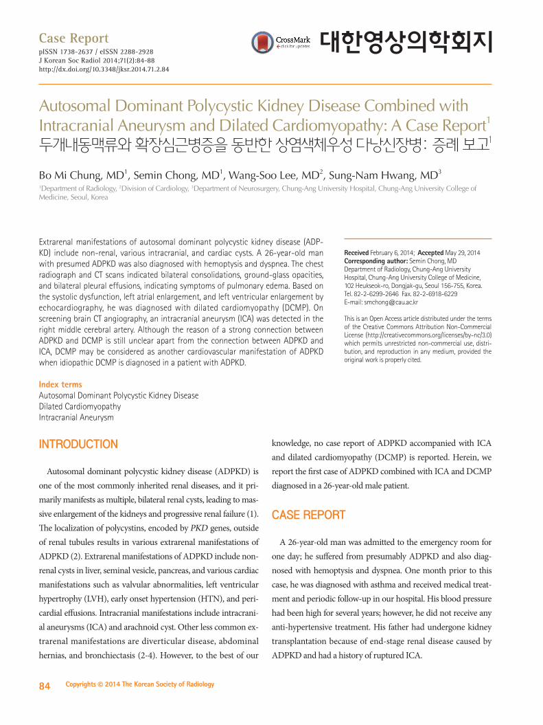
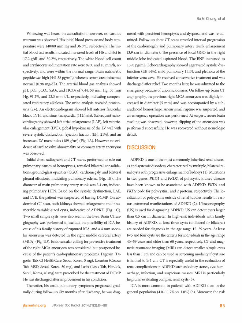
Wheezing was heard on auscultation; however, no cardiac murmur was observed. His initial blood pressure and body tem-perature were 140/80 mm Hg and 36.6°C, respectively. The ini-tial blood test results indicated increased levels of Hb and Hct to 17.2 g/dL and 50.2%, respectively. The white blood cell count and erythrocyte sedimentation rate were 8250 and 10 mm/h, re-spectively, and were within the normal range. Brain natriuretic peptide was high (441.38 pg/mL), whereas serum creatinine was normal (0.98 mg/dL). The arterial blood gas analysis showed pH, pO2, pCO2, SaO2, and HCO3 of 7.44, 58 mm Hg, 30 mm Hg, 91.2%, and 22.3 mmol/L, respectively, indicating compen-sated respiratory alkalosis. The urine analysis revealed protein-uria (2+). An electrocardiogram showed left anterior fascicular block, LVH, and sinus tachycardia (112/min). Subsequent echo-cardiography showed left atrial enlargement (LAE), left ventric-ular enlargement (LVE), global hypokinesia of the LV wall with severe systolic dysfunction [ejection fraction (EF), 21%], and an increased LV mass index (189 g/m2) (Fig. 1A). However, no evi-dence of cardiac valve abnormality or coronary artery aneurysm was observed.
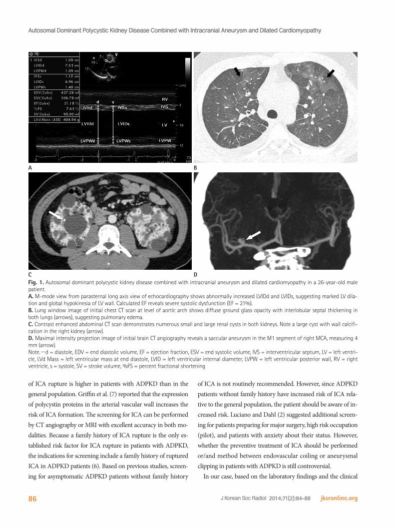
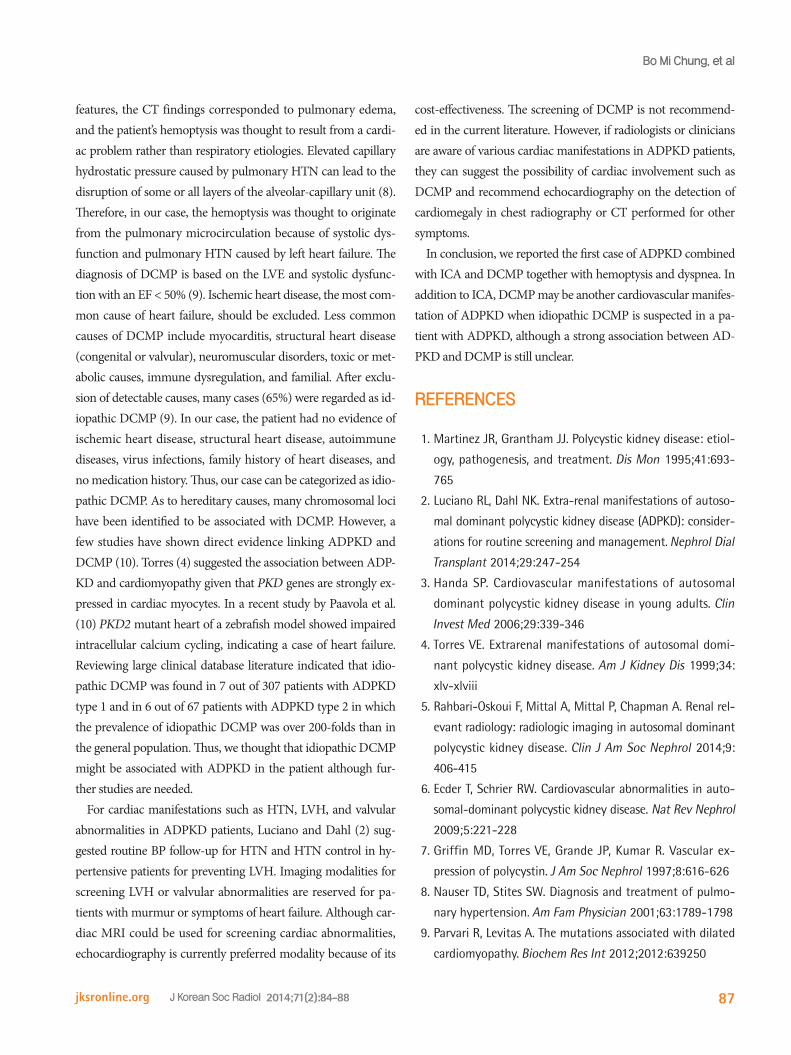
Initial chest radiograph and CT scans, performed to rule out pulmonary causes of hemoptysis, revealed bilateral consolida-tions, ground-glass opacities (GGO), cardiomegaly, and bilateral pleural effusions, indicating pulmonary edema (Fig. 1B). The diameter of main pulmonary artery trunk was 3.4 cm, indicat-ing pulmonary HTN. Based on the systolic dysfunction, LAE, and LVE, the patient was suspected of having DCMP. On ab-dominal CT scan, both kidneys showed enlargement and innu-merable variable-sized cysts, indicative of ADPKD (Fig. 1C). Two small simple cysts were also seen in the liver. Brain CT an-giography was performed to exclude the possibility of ICA be-cause of his family history of ruptured ICA, and a 4 mm saccu-lar aneurysm was detected in the right middle cerebral artery (MCA) (Fig. 1D). Endovascular coiling for preventive treatment of the right MCA aneurysm was considered but postponed be-cause of the patient’s cardiopulmonary problems. Digoxin (Di-gosin Tab, CJ HealthCare, Seoul, Korea, 5 mg), Losartan (Cozzar Tab, MSD, Seoul, Korea, 50 mg), and Lasix (Lasix Tab, Handok, Seoul, Korea, 40 mg) were prescribed for the treatment of DCMP. He was discharged after improvement in his condition.
Thereafter, his cardiopulmonary symptoms progressed grad-ually during follow-up. Six months after discharge, he was diag-
Autosomal Dominant Polycystic Kidney Disease Combined with Intracranial Aneurysm and Dilated Cardiomyopathy
86 jksronline.orgJ Korean Soc Radiol 2014;71(2):84-88
of ICA is not routinely recommended. However, since ADPKD patients without family history have increased risk of ICA rela-tive to the general population, the patient should be aware of in-creased risk. Luciano and Dahl (2) suggested additional screen-ing for patients preparing for major surgery, high risk occupation (pilot), and patients with anxiety about their status. However, whether the preventive treatment of ICA should be performed or/and method between endovascular coiling or aneurysmal clipping in patients with ADPKD is still controversial.
In our case, based on the laboratory findings and the clinical
of ICA rupture is higher in patients with ADPKD than in the general population. Griffin et al. (7) reported that the expression of polycystin proteins in the arterial vascular wall increases the risk of ICA formation. The screening for ICA can be performed by CT angiography or MRI with excellent accuracy in both mo-dalities. Because a family history of ICA rupture is the only es-tablished risk factor for ICA rupture in patients with ADPKD, the indications for screening include a family history of ruptured ICA in ADPKD patients (6). Based on previous studies, screen-ing for asymptomatic ADPKD patients without family history
Fig. 1. Autosomal dominant polycystic kidney disease combined with intracranial aneurysm and dilated cardiomyopathy in a 26-year-old male patient.A. M-mode view from parasternal long axis view of echocardiography shows abnormally increased LVIDd and LVIDs, suggesting marked LV dila-tion and global hypokinesia of LV wall. Calculated EF reveals severe systolic dysfunction (EF = 21%). B. Lung window image of initial chest CT scan at level of aortic arch shows diffuse ground glass opacity with interlobular septal thickening in both lungs (arrows), suggesting pulmonary edema. C. Contrast enhanced abdominal CT scan demonstrates numerous small and large renal cysts in both kidneys. Note a large cyst with wall calcifi-cation in the right kidney (arrow). D. Maximal intensity projection image of initial brain CT angiography reveals a saccular aneurysm in the M1 segment of right MCA, measuring 4 mm (arrow).Note.-d = diastole, EDV = end diastolic volume, EF = ejection fraction, ESV = end systolic volume, IVS = interventricular septum, LV = left ventri-cle, LVd Mass = left ventricular mass at end diastole, LVID = left ventricular internal diameter, LVPW = left ventricular posterior wall, RV = right ventricle, s = systole, SV = stroke volume, %FS = percent fractional shortening
C
A
D
B
Bo Mi Chung, et al
87jksronline.org J Korean Soc Radiol 2014;71(2):84-88
cost-effectiveness. The screening of DCMP is not recommend-ed in the current literature. However, if radiologists or clinicians are aware of various cardiac manifestations in ADPKD patients, they can suggest the possibility of cardiac involvement such as DCMP and recommend echocardiography on the detection of cardiomegaly in chest radiography or CT performed for other symptoms.
In conclusion, we reported the first case of ADPKD combined with ICA and DCMP together with hemoptysis and dyspnea. In addition to ICA, DCMP may be another cardiovascular manifes-tation of ADPKD when idiopathic DCMP is suspected in a pa-tient with ADPKD, although a strong association between AD-PKD and DCMP is still unclear.
REFERENCES
1.MartinezJR,GranthamJJ.Polycystickidneydisease:etiol-
ogy,pathogenesis,andtreatment.DisMon1995;41:693-
765
2.LucianoRL,DahlNK.Extra-renalmanifestationsofautoso-
maldominantpolycystickidneydisease(ADPKD):consider-
ationsforroutinescreeningandmanagement.NephrolDial
Transplant2014;29:247-254
3.HandaSP.Cardiovascularmanifestationsofautosomal
dominantpolycystickidneydiseaseinyoungadults.Clin
InvestMed2006;29:339-346
4.TorresVE.Extrarenalmanifestationsofautosomaldomi-
nantpolycystickidneydisease.AmJKidneyDis 1999;34:
xlv-xlviii
5.Rahbari-OskouiF,MittalA,MittalP,ChapmanA.Renalrel-
evantradiology:radiologicimaginginautosomaldominant
polycystickidneydisease.ClinJAmSocNephrol 2014;9:
406-415
6.EcderT,SchrierRW.Cardiovascularabnormalitiesinauto-
somal-dominantpolycystickidneydisease.NatRevNephrol
2009;5:221-228
7.GriffinMD,TorresVE,GrandeJP,KumarR.Vascularex-
pressionofpolycystin.JAmSocNephrol1997;8:616-626
8.NauserTD,StitesSW.Diagnosisandtreatmentofpulmo-
naryhypertension.AmFamPhysician2001;63:1789-1798
9.ParvariR,LevitasA.Themutationsassociatedwithdilated
cardiomyopathy.BiochemResInt2012;2012:639250
features, the CT findings corresponded to pulmonary edema, and the patient’s hemoptysis was thought to result from a cardi-ac problem rather than respiratory etiologies. Elevated capillary hydrostatic pressure caused by pulmonary HTN can lead to the disruption of some or all layers of the alveolar-capillary unit (8). Therefore, in our case, the hemoptysis was thought to originate from the pulmonary microcirculation because of systolic dys-function and pulmonary HTN caused by left heart failure. The diagnosis of DCMP is based on the LVE and systolic dysfunc-tion with an EF < 50% (9). Ischemic heart disease, the most com-mon cause of heart failure, should be excluded. Less common causes of DCMP include myocarditis, structural heart disease (congenital or valvular), neuromuscular disorders, toxic or met-abolic causes, immune dysregulation, and familial. After exclu-sion of detectable causes, many cases (65%) were regarded as id-iopathic DCMP (9). In our case, the patient had no evidence of ischemic heart disease, structural heart disease, autoimmune diseases, virus infections, family history of heart diseases, and no medication history. Thus, our case can be categorized as idio-pathic DCMP. As to hereditary causes, many chromosomal loci have been identified to be associated with DCMP. However, a few studies have shown direct evidence linking ADPKD and DCMP (10). Torres (4) suggested the association between ADP-KD and cardiomyopathy given that PKD genes are strongly ex-pressed in cardiac myocytes. In a recent study by Paavola et al. (10) PKD2 mutant heart of a zebrafish model showed impaired intracellular calcium cycling, indicating a case of heart failure. Reviewing large clinical database literature indicated that idio-pathic DCMP was found in 7 out of 307 patients with ADPKD type 1 and in 6 out of 67 patients with ADPKD type 2 in which the prevalence of idiopathic DCMP was over 200-folds than in the general population. Thus, we thought that idiopathic DCMP might be associated with ADPKD in the patient although fur-ther studies are needed.
For cardiac manifestations such as HTN, LVH, and valvular abnormalities in ADPKD patients, Luciano and Dahl (2) sug-gested routine BP follow-up for HTN and HTN control in hy-pertensive patients for preventing LVH. Imaging modalities for screening LVH or valvular abnormalities are reserved for pa-tients with murmur or symptoms of heart failure. Although car-diac MRI could be used for screening cardiac abnormalities, echocardiography is currently preferred modality because of its
Autosomal Dominant Polycystic Kidney Disease Combined with Intracranial Aneurysm and Dilated Cardiomyopathy
88 jksronline.orgJ Korean Soc Radiol 2014;71(2):84-88
clingintheheartandpredisposetodilatedcardiomyopa-
thy.JMolCellCardiol2013;58:199-208
10.PaavolaJ,SchliffkeS,RossettiS,KuoIY,YuanS,SunZ,et
al.Polycystin-2mutations leadto impairedcalciumcy-
두개내동맥류와 확장심근병증을 동반한 상염색체우성 다낭신장병: 증례 보고1
정보미1 · 정세민1 · 이왕수2 · 황성남3
상염색체우성 다낭신장병의 신장 외 소견에는 비신장성 낭종들과 다양한 두개 내 및 심장 소견들이 있다. 상염색체우성 다
낭신장병이 있는 26세 남성이 객혈과 호흡곤란으로 내원하였다. 단순흉부방사선사진과 흉부전산화단층촬영에서 양측성
경화와 간유리음영, 그리고 양측성 흉막 삼출 소견들이 보여 폐부종이 의심되었다. 심장초음파에서 수축기 기능이상, 좌심
방비대, 그리고 좌심실비대의 소견을 바탕으로 확장심근병증으로 진단되었다. 뇌전산화단층혈관조영 선별검사에서 우측
중대뇌동맥에 두개내동맥류가 발견되었다. 상염색체우성 다낭신장병과 확장심근병증의 연관성이 아직 불분명하지만 상
염색체우성 다낭신장병 환자에서 원인불명의 확장심근병증이 의심될 때 두개내동맥류뿐만 아니라 확장심근병증은 상염
색체우성 다낭신장병의 또 다른 심혈관계 소견일 수 있다.
중앙대학교 의과대학 중앙대학교병원 1영상의학과, 2순환기내과, 3신경외과