Embed Size (px)
Citation preview
Acta Neuropathol (1994) 87 : 337-342 �9 Springer-Verlag 1994
M. Sabatelli �9 T. Mignogna �9 G. Lippi �9 S. Servidei G. Manfredi �9 E. Ricci �9 E. Bertini - M. Lo Monaco E Tonali
Autosomal recessive hypermyelinating neuropathy
Received: 17 August 1993 / Revised, accepted: 20 October 1993
Abstract We studied three pat ients f rom two kinships, affected by early onset heredi tary mo to r and sensory neuropa thy with p robab le autosomal recessive inherit- ance ( H M S N type HI) . Morphological studies of sural nerve biopsies revealed an abnormal myel in pro- liferation. Two adult pat ients with long- term follow up, lost ability to walk at 28 and 22 years and showed severe involvement of the cranial nerves, Our observat ions suggest that "hypermyel ina t ion neuropa thy" with early onset is a progressive disease with poo r long- term prog- nosis. In one kinship the occurrence of the disease in two sibs of bo th sexes but not in parents , is consistent with an au tosomal recessive inheritance. Familial cases of hypermyel ina t ion neuropa thy have not been described in previous reports . Morphological aspects of this condit ion are compared with o ther forms of hyper- myel inat ion neuropathy.
Key words Polyneuropa thy �9 Hereditary- m o t o r and sensory neuropa thy �9 Demyel ina t ion �9 Myelin sheath foldings
Introduction
Hered i t a ry mo to r and sensory neuropathies (HMSN) include a he te rogeneous group of disorders. The mos t f requent forms are H M S N type I and I I which are in- her i ted as an au tosomal dominant trait.
Supported by Telethon-Italy for the project: "Chronic inflammatory polyradiculoneuropathy: electrophysiological and immunopathological studies"
M. Sabatelli (t~) �9 T. Mignogna. G. Lippi - S. Servidei G. Manfredi - E. Ricci - M. Lo Monaco �9 P. Tonali Neurological Institute, Catholic University, Pol. A. Gemelli, Largo Gemelli, 8, 1-00168 Rome, Italy
E. Bertini Department of Neuropediatrics, Hospital Bambino Gesu', Rome, Italy
H M S N type I I I is not a well-defined entity. Charac- teristic features are autosomal recessive inheri tance, cogenital or infantile onset, variable expression and markedly decreased nerve conduct ion velocity. Pathological changes in sural nerve biopsy f rom typical forms are severe loss of myet inated fibers with hypo- and demyel inat ion and f requent onion bulbs [4]. In recent years, however, some pat ients have been repor ted with a virtual absence of myelin sheath and absent or atypical onion bulb format ions (hypomyelina- t ion neuropathy) [6, 8, 9, 11, 17] or with an abnormal myelin overgrowth (hypermyel inat ion neuropathy) [5, 7, 15, 16, 19]. It is still unclear whether or not these conditions represent distinct clinico-pathological en- tities.
We describe three pat ients f rom two kinships, affected by early onset H M S N with p robab le autosomal recessive inheri tance, showing the features of "hyper- myel inat ion neuropa thy" .
Case reports
Patient 1
This 30-year-old man was born normally at term after a normal pregnancy. His 53-year-old mother was normal at clinical and elec- trophysiologieal examination. His father was referred to as being healthy but was not examined by us. Parents were not con- sanguineous; they were from a little town of 1000 inhabitants, near Isernia, Italy. One sister, aged 33 years, was normal at clinical exmination. Another sister was affected by the., same disease (patient 2).
The patient walked normally at 13 months. Around the age of 18 months the parents noticed that he walked with a waddling gait, on tip toes. When he was 11 years old a diagnosis of Charcot- Marie-Tooth was established in another neurological institute. At the time the patient showed bilateral foot drop, pes cavus with retraction of Achille's tendons, waddling gait, mild weakness of small hand muscles and areflexia; there was no sensory loss. Height was 123 cm (below 3rd centile). Routine hematological examinations and serum CK were normal. Electrophysiological examination revealed absent sensor 3 , action potentials and "severe" reduction of motor nerve conduction velocity.
338
During the following years progression of the disease was slow, but by the age of 26 the patient noticed progressive weakness of proximal limb muscles. At 29 he lost the ability to walk anaided. When first seen by us, the patient showed a severe and generalized weakness. Muscle power was F = 0 (Medical Research Council scale) in the tibio-peronei, Triceps sura, extensor carpi, extensor digitorum communis and small hand muscles, F = 1-2 in the iliop- soas and glutei, F = 2-3 in the biceps brachii, triceps brachii and quadriceps, F = 4 in the deltoids and neck flexors. There was a moderate weakness of masticatory and facial muscles: forced closure of lids and of jaw could easily be overcome by the exami- nator's fingers. The patient could walk a few steps only with major assistance. Tendon reflexes were absent. Sensor), examination showed mild loss of sensation distally in both legs and a marked acrocyanosis was also present. Intelligence was normal. Routine hematological exams were normal. Serum CK was 260 IU/1 [nor- mal value (n.y.) < 230], lactic acid was i mEq/1 (n.v. <2.2). Brain CT scan, brain stem evoked potentials and audiometric examina- tion were normal. Respiratory functional tests were low normal.
Electromyographic studies revealed fibrillation potentials and pseudomyotonic discharges in all nmscles. No voluntary activation was detected in the tibioperonei, small hand muscles, extensor carpi and digitorum communis, while in deltoid, biceps brahii and quadriceps few high frequency giant motor unit potentials were observed. Sensory action potentials were absent recording from radial, median and sural nerves. No muscle action potentials could be elicited from electrical stimulation of peroneal, median and ulnar nerves. Motor latency from Erb's point to deltoid and biceps brachii -were markedly prolonged: 13.5 ms (n.y. < 5.1) and 10 ms (n.y. < 5.7), respectively.
floor. )all deep tendon reflexes were absent. There was bilateral pes cavus with retraction of Achille's tendons. Intelligence was normal.
Electrophysiological examination demonstrated absence of sensory action potentials on testing the median, radial and 'sural nerves. Motor conduction velocities were 16 m/s and 14 m/s in the right ulnar and median nerves, respectively; no action potential was elicited from electrical stimulation of the right peroneal nerve. Electromyographic examination was performed only in the quadriceps femoris and was unremarkable. The following tests were normal: full blood count, fasting blood glucose, serum elec- trolytes~ creatine kinase, lactic acid, urinary organic acids, serum and urine carnitine, EEG, ECG and fiver echography.
Materials and methods
Nerve
A whole surat nerve biopsy specimen was fixed in 2.5 % glutar- aldehyde in phosphate buffer and post-fixed in 1% phosphate- buffered osmium tetroxide. The tissue was dehydrated, infiltrated and embedded in Spurt resin. Sections, 1 pm thick, were stained with totuidine blue. Thin sections were stained with uranyl acetate and lead citrate prior to examination in a Philips EM 400 electron microscope.
Muscle
Patient 2
This 28-year-old-woman was the sister of the patient 1. Early motor and intellectual milestones were normal: she could walk unaided at 13 months. Since the age of 14-15 month she developed progressive difficulty in walking. When she was 4-5 years old wasting and weakness of hands was observed. At around 15 years of age dysphonia, dysphagia and difficulty in chewing appeared. Progression was more severe than in her brother and at 22 years she lost the ability to walk.
Clinical examination revealed severe weakness and wasting of tibio-peronei (F=0), quadriceps (F=2) and iliopsoas (F=I ) . In upper limbs, motor power was F = 0 in flexor and extensor carpi, extensor digitorum communis and small hand muscles, F = 2-3 in deltoid, biceps and triceps brachii and neck flexors. Moreover, there was marked dyshonia due to vocal cord palsy, dysphagia, weakness of masticatory muscles and of facial muscles; the face was amimic and the patient was not able to close her eyes com- pleteb: She was not able to walk and could stand only with assist- ance. Respiratory muscles were spared. Tendon reflexes were absent. There was mild loss of sensation distally in both legs. Rou- tine blood examinations were normal. Brain CI" was normal. Serum CK was 266 IU/1 (n.y. < 230). Electromyographie changes were similar to those observed in the brother. Sensitive action potentials were all absent. Compound muscular action potentials were not evocable from peroneal, ulnar and median nerves stimu- lation.
Patient 3
A 4-year-old boy was admitted to our hospital because of clumsy gait and frequent falls since the age of 2 years. He was the only child of healthy and unrelated parents. Both parents were normal at clinical and electrophysiological examination. Pregnancy and delivery were normal. The patient had normal early motor mile- stones and he walked at 11 month.
Neurological examination showed mild bilateral facial weak- ness, moderate weakness and atrophy of distal limb muscles and neck flexors. He used the Gowers' manoeuvre to rise from the
For histochemical studies, the biopsies were frozen in liquid nitrogen-cooled isopentane, cross-sectioned at 7- to 8-~m thick- ness in a cryostat, and processed for routine histochemical analysis as described [3]. Assays of mitochondrial and 13-oxidation enzyme activities and of carnitine were performed as described [18].
Results
Sura l n e r v e b i o p s y
P a t h o l o g i c a l changes o n sural n e r v e b i o p s y we re s imi la r in al l t h r e e cases and t hey will be d e s c r i b e d toge ther .
Light microscopy
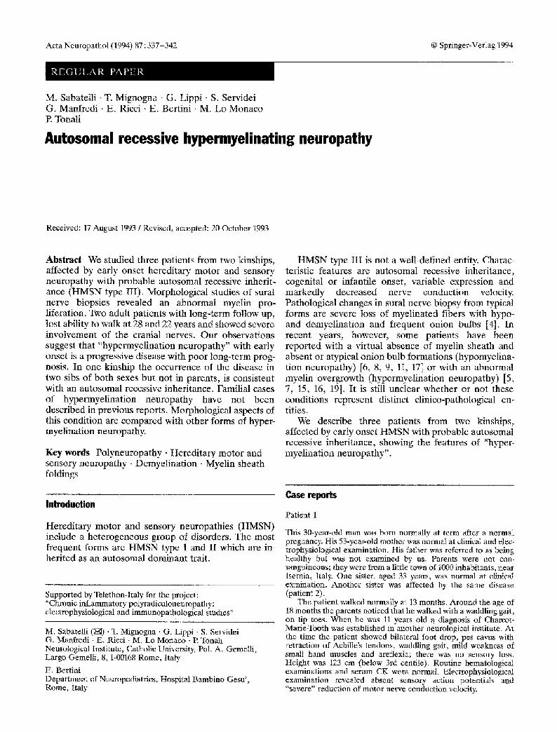
O n s e m i t h i n t r a n s v e r s e sec t ions t h e r e was seve re r educ - t i on in t he n u m b e r o f m y e l i n a t e d f ibe rs , m o r e r e m a r k - ab l e in p a t i e n t s 1 a n d 2 (Figs . 1, 2). T h e m o s t s t r ik ing f ind ing , in all cases , was the p r e s e n c e o f m a r k e d over- g rowth o f the m y e l i n shea th in t he m a j o r i t y o f mye l in - a t e d f ibers . S o m e f ibers s h o w e d a th in mye l in shea th wi th r e spec t to axona l d i ame te r . O n i o n bu lbs were f ie - q u e n t in p a t i e n t s 1 a n d 2, whi le t hey we re ra re in p a t i e n t 3.
Teased fiber analysis
O n e h u n d r e d f ibers f rom p a t i e n t 3 we re t e a s e d , whi le on ly few f ibers cou ld b e o b t a i n e d f rom t h e b iops i e s of p a t i e n t s 1 and 2. A l l f ibers s h o w e d a b n o r m a l mye l in
339
Fig. 1 a,b Semithin transverse section of sural nerve biopsy from patients 1 (a) and 2 (b). There is severe fiber loss, many fibers show abnormal myelin proliferation. Some onion bulbs are present. Toluidine blue, x 780
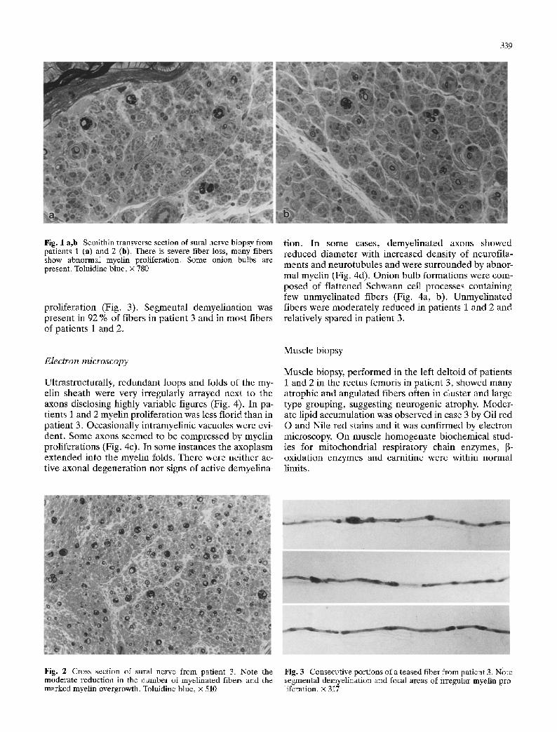
proliferation (Fig. 3). Segmental demyelination was present in 92 % of fibers in patient 3 and in most fibers of patients 1 and 2.
Electron microscopy
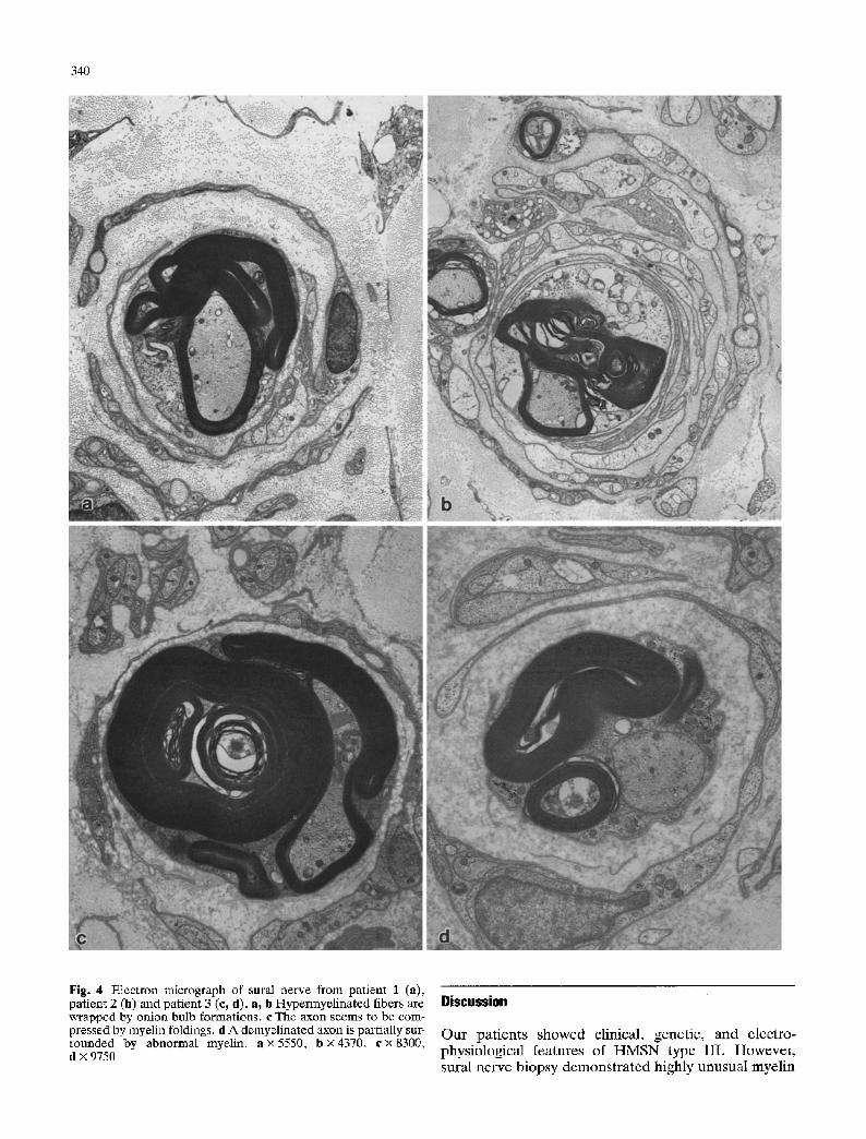
Ultrastructurally, redundant loops and folds of the my- elin sheath were very irregularly arrayed next to the axons disclosing highly variable figures (Fig. 4). In pa- tients 1 and 2 myelin proliferation was less florid than in patient 3. Occasionally intramyelinic vacuoles were evi- dent. Some axons seemed to be compressed by myelin proliferations (Fig. 4c). In some instances the axoplasm extended into the myelin folds. There were neither ac- tive axonal degeneration nor signs of active demyelina-
tion. In some cases, demyelinated axons showed reduced diameter with increased density of neurofila- ments and neurotubules and were surrounded by abnor- mal myelin (Fig. 4d). Onion bulb formations were com- posed of flattened Schwann cell processes containing few unmyelinated fibers (Fig. 4a, b). Unmyelinated fibers were moderately reduced in patients 1 and 2 and relatively spared in patient 3.
Muscle biopsy
Muscle biopsy, performed in the left deltoid of patients 1 and 2 in the rectus femoris in patient 3, slhowed many atrophic and angulated fibers often in cluster and large type grouping, suggesting neurogenic atrophy. Moder- ate lipid accumulation was observed in case 3 by Oil red O and Nile red stains and it was confirmed by electron microscopy. On muscle homogenate biochemical stud- ies for mitochondrial respiratory chain enzymes, ~- oxidation enzymes and carnitine were within normal limits.
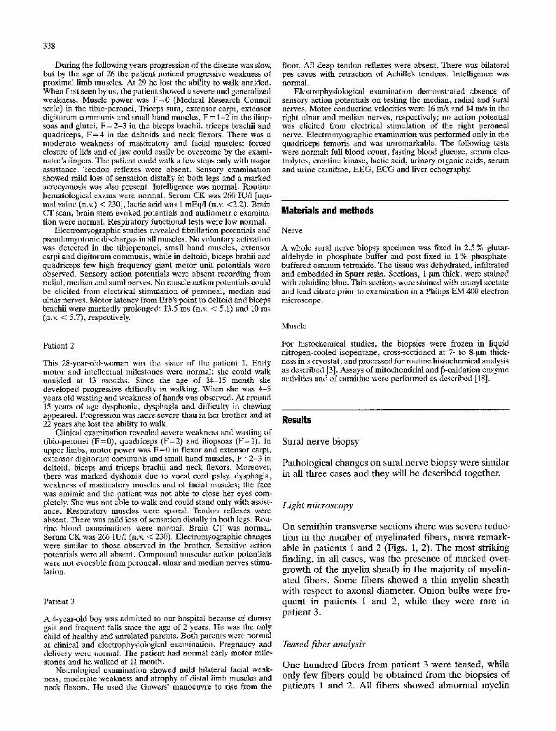
Fig. 2 Cross section of sural nerve from patient 3. Note the moderate reduction in the number of myelinated fibers and the marked myelin overgrowth. Toluidine blue, • 510
Fig. 3 Consecutive portions of a teased fiber from patient 3. Note segmental demyelination and focal areas of irregular myelin pro- liferation. • 317
340
Fig. 4 Electron micrograph of sural nerve from patient 1 (a), patient 2 (b) and patient 3 (e, d). a, b Hypermyelinated fibers are wrapped by onion bulb formations, c The axon seems to be com- pressed by myelin foldings, d A demyelinated axon is partially sur- rounded by abnormal myelin, a x 5550, b • 4370, c x 8300, d x 9750
Discussion
O u r p a t i e n t s s h o w e d cl inical , gene t ic , and e lec t ro - phys io log i ca l f ea tu re s o f H M S N t y p e I I I . H o w e v e r , sura l n e r v e b i o p s y d e m o n s t r a t e d h igh ly u n u s u a l mye l in
34t
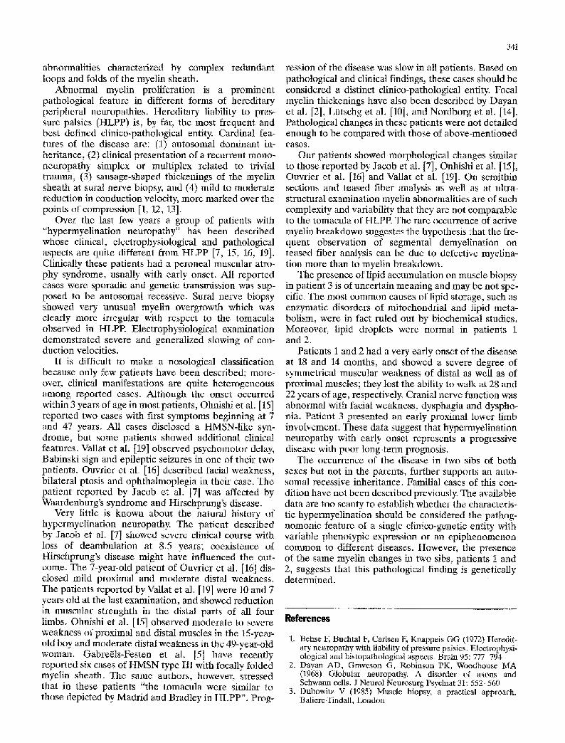
abnormalities characterized by complex redundant loops and folds of the myelin sheath.
Abnormal myelin proliferation is a prominent pathological feature in different forms of hereditary peripheral neuropathies. Hereditary liability to pres- sure palsies (HLPP) is, by far, the most frequent and best defined clinico-pathological entity. Cardinal fea- tures of the disease are: (1) autosomal dominant in- heritance, (2) clinical presentation of a recurrent mono- neuropathy simplex or multiplex related to trivial trauma, (3) sausage-shaped thickenings of the myelin sheath at sural nerve biopsy, and (4) mild to moderate reduction in conduction velocity, more marked over the points of compression [1, 12, 13].
Over the last few years a group of patients with "hypermyelination neuropathy" has been described whose clinical, electrophysiological and pathological aspects are quite different from HLPP [7, 15, 16, 19]. Clinically these patients had a peroneal muscular atro- phy syndrome, usually with early onset. All reported cases were sporadic and genetic transmission was sup- posed to be autosomal recessive. Sural nerve biopsy showed very unusual myelin overgrowth which was clearly more irregular with respect to the tomacula observed in HLPR Electrophysiological examination demonstrated severe and generalized slowing of con- duction velocities.
It is difficult to make a nosological classification because only few patients have been described; more- over, clinical manifestations are quite heterogeneous among reported cases. Although the onset occurred within 3 years of age in most patients, Ohnishi et al. [15] reported two cases with first symptoms beginning at 7 and 47 years. All cases disclosed a HMSN-like syn- drome, but some patients showed additional clinical features. Vallat et al. [19] observed psychomotor delay, Babinski sign and epileptic seizures in one of their two patients. Ouvrier et al. [16] described facial weakness, bilateral ptosis and ophthalmoplegia in their case. The patient reported by Jacob et al. [7] was affected by Waardenburg's syndrome and Hirschprung's disease.
Very little is known about the natural history of hypermyelination neuropathy. The patient described by Jacob et al. [7] showed severe clinical course with loss of deambulation at 8.5 years; coexistence of Hirschprung's disease might have influenced the out- come. The 7-year-old patient of Ouvrier et al. [16] dis- closed mild proximal and moderate distal weakness. The patients reported by Vallat et al. [19] were 10 and 7 years old at the last examination, and showed reduction in muscular strenghth in the distal parts of all four limbs. Ohnishi et al. [15] observed moderate to severe weakness of proximal and distal muscles in the 15-year- old boy and moderate distal weakness in the 49-year-old woman. Gabre~ls-Festen et al. [5] have recently reported six cases of HMSN type III with focally folded myelin sheath. The same authors, however, stressed that in these patients "the tomacula were similar to those depicted by Madrid and Bradley in H L P P ' . Prog-
ression of the disease was slow in all patients. Based on pathological and clinical findings, these cases should be considered a distinct clinico-pathological entity. Focal myelin thickenings have also been described by Dayan et al. [2], Ltitschg et al. [10], and Nordborg et al. [14], Pathological changes in these patients were not detailed enough to be compared with those of above-mentioned cases.
Our patients showed morphological changes similar to those reported by Jacob et al. [7], Onhishi et al. [i5], Ouvrier et al. [16] and Vallat et al. [19]. On semithin sections and teased fiber analysis as well as at ultra- structural examination myelin abnormalities are of such complexity and variability that they are not comparable to the tomacula of HLPE The rare occurrence of active myelin breakdown suggestes the hypothesis that the fre- quent observation of segmental demyelination on teased fiber analysis can be due to defective myelina- tion more than to myelin breakdown.
The presence of lipid accumulation on muscle biopsy in patient 3 is of uncertain meaning and may be not spe- cific. The most common causes of lipid storage, such as enzymatic disorders of mitochondrial and lipid meta- bolism, were in fact ruled out by biochemical studies. Moreover, lipid droplets were normal in patients 1 and 2.
Patients 1 and 2 had a very early onset of the disease at 18 and 14 months, and showed a severe degree of symmetrical muscular weakness of distal as well as of proximal muscles; they lost the ability to walk at 28 and 22 years of age, respectively. Cranial nerve fimction was abnormal with facial weakness, dysphagia and dyspho- nia. Patient 3 presented an early proximal lower limb involvement. These data suggest that hypermyelination neuropathy with early onset represents a progressive disease with poor long-term prognosis.
The occurrence of the disease in two sibs of both sexes but not in the parents, further supports an auto- somal recessive inheritance. Familial cases of this con- dition have not been described previously. The available data are too scanty to establish whether the characteris- tic hypermyelination should be considered the pathog- nomonic feature of a single clinico-genetic entity with variable phenotypic expression or an epiphenomenon common to different diseases. However, the presence of the same myelin changes in two sibs, patients 1 and 2, suggests that this pathological finding is genetically determined.
References
1. Behse F, Buchtal F, Carlsen E Knappeis GG (1972) Heredit- ary neuropathy with liability of pressure palsies. Electrophysi- ologicat and histopathological aspects. Brain 95:777-794
2. Dayan AD, Graveson G, Robinson PK, Woodhouse MA (1968) Globular neuropathy. A disorder of axons and Schwann cells. J Neurol Neurosurg Psychiat 31:552-560
3. Dubowitz V (1985) Muscle biopsy, a practical approach. Baliere-Tindail, London
342
4. Dyck PJ, Chance P, Lebo R, Carney JA (1993) Hereditary motor and sensory neuropathies. In: Dyck PJ, Thomas PK (eds) Peripheral Neuropathy, 3rd edn. WB Saunders, Phila- delphia, pp 1094-1136
5. Gabre61s-Festen AAWM, Joosten EMG, Gabre~ls FJM, Stegeman DF, Vos AJM, Busch HFM (1990) Congenital demyelinating motor and sensory neuropathy with focally folded myelin sheaths. Brain 113:1629-1643
6. Guzzetta G, Ferriere G, Lyon G (1982) Congenital hypomy- elination polyneuropathy. Pathological findings compared with polyneuropathies starting later in life. Brain 105: 395- 416
7. Jacobs JM, Wilson J (1992) An unusual demyelinating neuro- pathy in a patient with Waardenburg's syndrome. Acta Neuro- pathol 83:670-674
8. Karch SB, Urich H (1975) Infantile polyneuropathy with defective myelination: an autopsy study. Dev Med Child Neu- rol 17:504-511
9. Kennedy WR, Sung JH, Berry JF (1977) A case of congenital hypomyelination neuropahty: clinical, morphological and chemical studies. Arch Neurol 34:337-345
10. Lt~tschg J, Vassella F, Boltshauser E, Dias K, Meier C (1985) Heterogeneity of congenital motor and sensory neuropathies. Neuropediatrics 16:33-38
11. Lyon G (1969) Ultrastructural study of a nerve biopsy from a case early infantile chronic neuropathy. Acta Neuropathol (Berl) 13:131-142
12. Madrid R, Bradley WG (1975) The pathology of neuropathies with focal thickenings of the myelin sheath (tomaculous neu- ropathy). Studies on the formation of the abnormal myelin sheath. J Neurol Sci 25:415-448
13. Meier C, Moll C (1982) Hereditary neuropathy with liability to pressure palsies. Report of two families and review of the literature. J Neurol 228:73-95
14. Nordborg C, Conradi N, Sourander P, Hagberg B, Western- berg B (1984) Hereditary motor and sensory neuropathy of demyelinating and remyelinating type in children. Ultrastruc- tural and morphometric studies on sural nerve biopsy speci- mens from ten sporadic cases. Acta Neuropathol (Berl) 65: 1- 9
15. Ohnishi A, Murai Y, Ikeda M, Fujita T, Furuya H, Kuroiwa Y (1989) Autosomal recessive motor and sensory neuropathy with excessive myelin outfolding. Muscle Nerve 12:568-575
16. Ouvrier R, McLeod JG, Pollard J (1990) Congenital dismy- elinating neuropathy. In: Rapin I (ed) Peripheral neuropathy in childhood. Raven Press, New York, pp 210-214
17. Palix C, Coignet J (1978) Un cas de polyneuropathie p6riph6- rique n6onatale par amy61inisation. P6diatrie 33:201-207
18. Papadimitriou A, Servidei S (1991) Late onset lipid storage myopathy due to multiple acyl CoA dehydrogenase deficiency triggered by valproate. Neuromusc Dis 1:247-252
19. Vallat JM, Gil R, Leboutet MJ, Hugon J, Moulies D (1987) Congenital hypo- and hypermyelination neuropathy. Acta Neuropathol (Berl) 74:197-201


![Autosomal recessive ichthyosis with limb reduction defect ... · including autosomal dominant, autosomal recessive and X-linked inheritance [1,2]. Associated cutaneous and extracutaneous](https://img.pdfslide.net/doc/110x75/5ec8c9b91adfdf12ab3e663c/autosomal-recessive-ichthyosis-with-limb-reduction-defect-including-autosomal.jpg)


